295,00 € – 895,00 €
Product details
Synonyms = Coagulation Factor VIII, Factor VIII Related Antigen, F8VWF, von Willebrand Antigen 2,von Willebrand Disease (vWD)
Antibody type = Recombinant Rabbit monoclonal / IgG
Clone = MSVA-521R
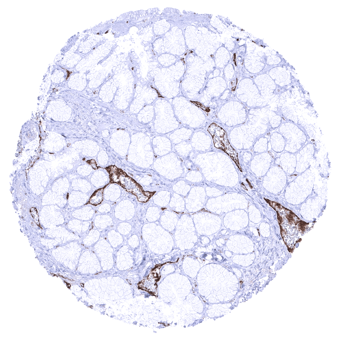
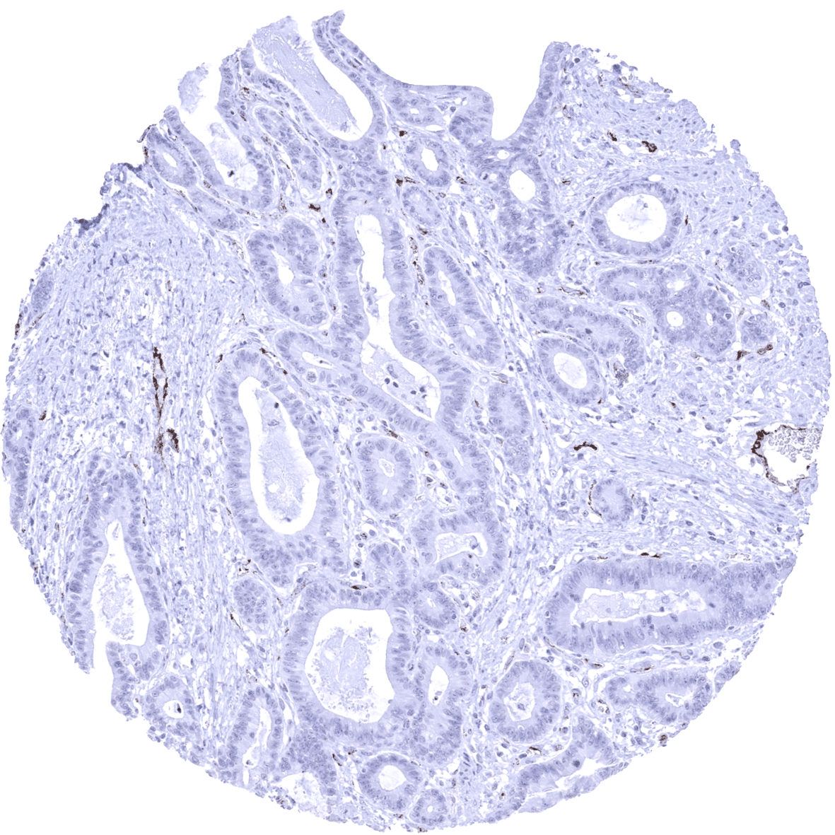
Positive control =Colon: A moderate to strong endothelial vWF immunostaining should be seen in a fraction of blood vessels.
Negative control =Colon: vWF immunostaining should be completely absent in epithelial and muscular cells.
Cellular localization = Cytoplasm.
Reactivity = Human
Application = Immunohistochemistry
Dilution = 1:50
Intended Use = Research Use Only
Relevance of Antibody
vWF is a marker for endothelium and megakaryocytes.
Biology Behind
Von Willebrand factor (vWF) is a glycoprotein coded by the vWF gene on 12p13.3. vWF circulates in the blood in two distinct compartments. Plasma vWF, is synthesized and secreted from endothelial cells while platelet vWF is synthesized by megakaryocytes. vWF represents a key protein in hemostasis. vWF is not an enzyme but contributes to hemostasis by binding to other proteins such as factor VIII, activated platelet receptors or collagen. vWF protects inactive factor VIII from degradation and releases it, if triggered by thrombin. vWF forms a bridge between collagen exposed under injured endothelium and platelets to support blood clot formation under high shear blood flow. Increased vWF plasma levels occur in many cardiovascular, neoplastic, and connective tissue diseases and are linked to an increased risk of thrombosis. VWF is deficient in von Willebrand disease which results in increased bleeding tendency mainly of the skin and mucous membranes.
Staining Pattern in Normal Tissues
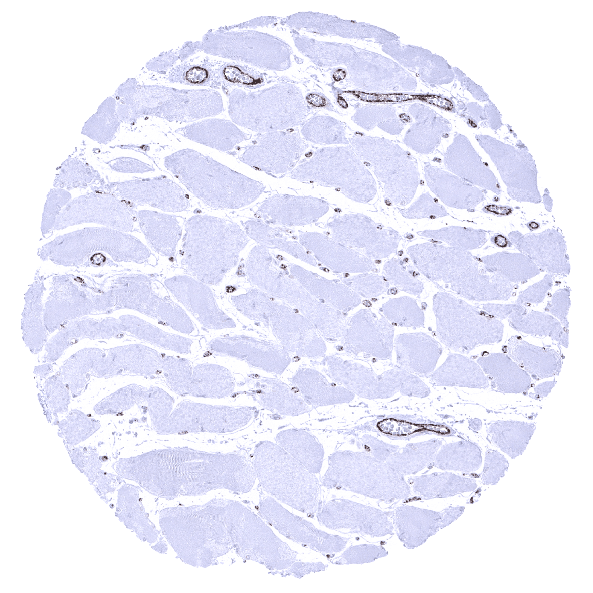
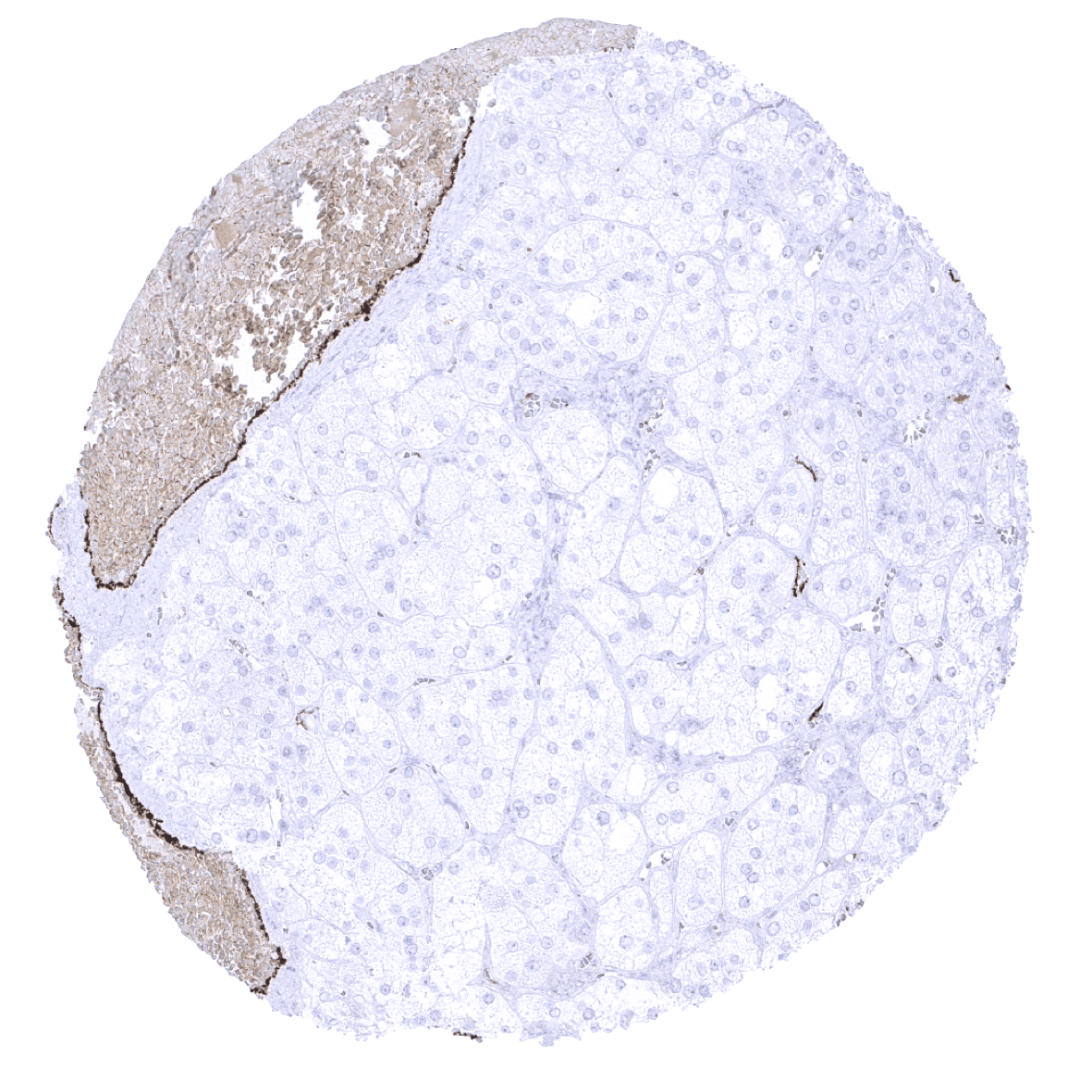
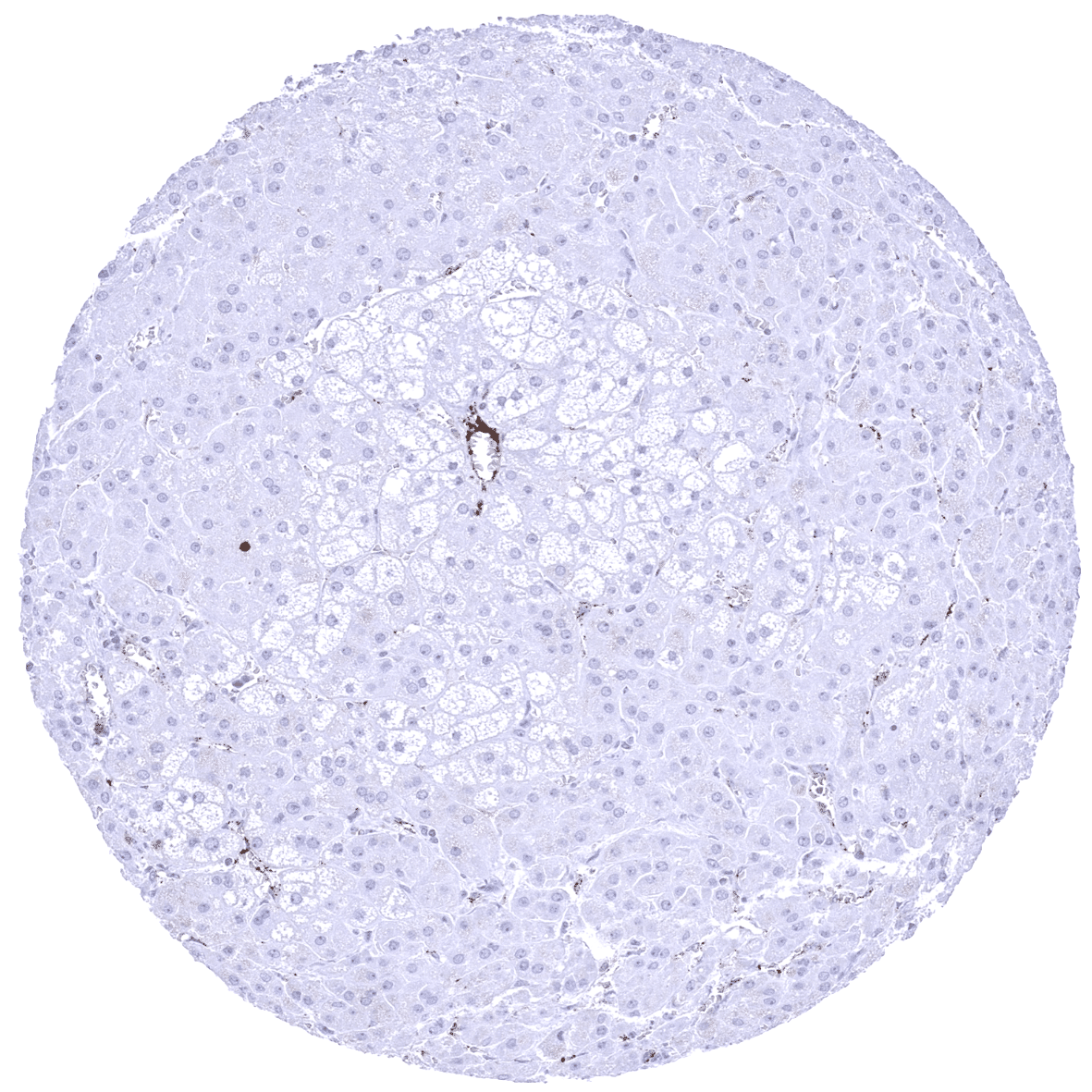
In normal tissues, vWF immunostaining is only seen in endothelial cells, megakaryocytes, and platelets. Within endothelial cells, the level of vWF expression varies between different locations. Endothelial vWF expression is generally higher in veins and venules than in arteries and arterioles and vWF expression in capillaries varies between organs and even within organs. A particularly low endothelial vWF expression is seen in kidney, liver, stomach and in various exocrine and endocrine glands (pancreas, salivary glands, adrenal gland, thyroid, parathyroid).
These findings are largely consistent with the RNA and protein data described in the Human Protein Atlas (Tissue expression vWF)
Positive control: Colon: A moderate to strong endothelial vWF immunostaining should be seen in a fraction of blood vessels.
Negative control: Colon: vWF immunostaining should be completely absent in epithelial and muscular cells.

Staining Pattern in Relevant Tumor Types
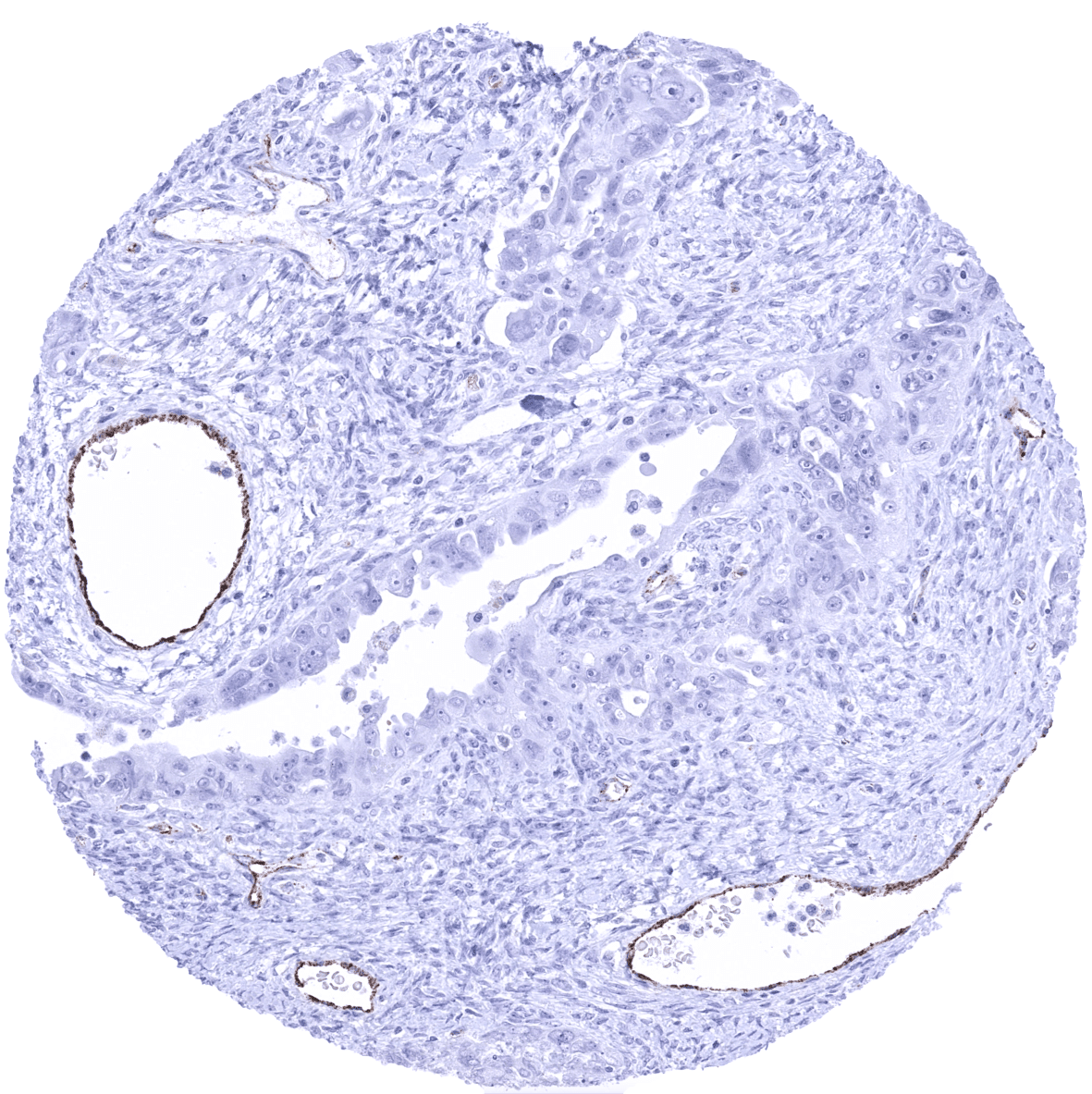
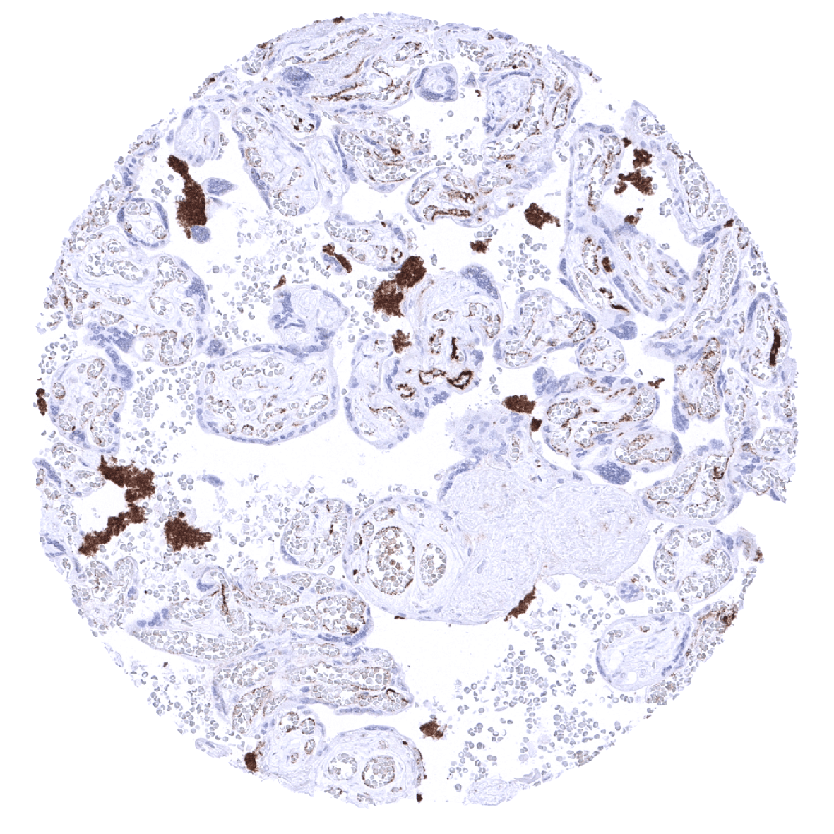
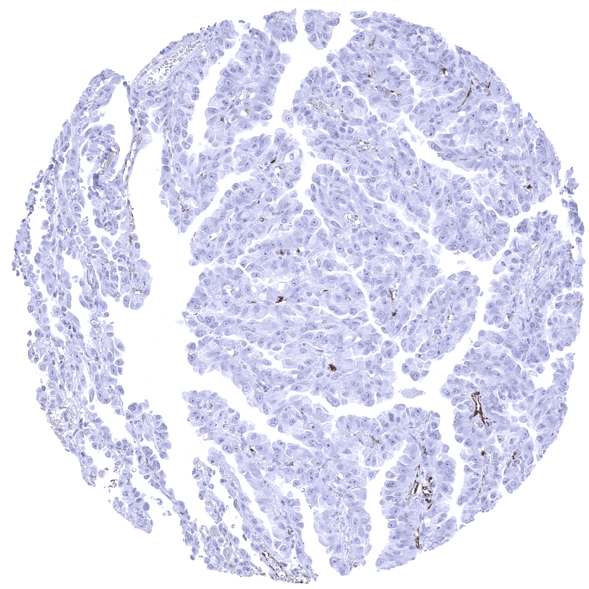
An endothelial vWF immunostaining of variable intensity is usually seen in a fraction of small intratumoral blood vessels. The quantity of stained vessels and the staining intensity appears to vary between tumors.
The TCGA findings on vWF RNA expression in different tumor categories have been summarized in the Human Protein Atlas.
Compatibility of Antibodies
No data available at the moment
Protocol Recommendations
IHC users have different preferences on how the stains should look like. Some prefer high staining intensity of the target stain and even accept some background. Others favor absolute specificity and lighter target stains. Factors that invariably lead to more intense staining include higher concentration of the antibody and visualization tools, longer incubation time, higher temperature during incubation, higher temperature and longer duration of the heat induced epitope retrieval (slide pretreatment). The impact of the pH during slide pretreatment has variable effects and depends on the antibody and the target protein.
All images and data shown here and in our image galleries are obtained by the manual protocol described below. Other protocols resulting in equivalent staining are described as well.
-Manual protocol
Freshly cut sections should be used (less than 10 days between cutting and staining). Heat-induced antigen retrieval for 5 minutes in an autoclave at 121°C in pH 7,8 Target Retrieval Solution buffer. Apply MSVA-572R at a dilution of 1:50 at 37°C for 60 minutes. Visualization of bound antibody by the EnVision Kit (Dako, Agilent) according to the manufacturer’s directions.
Potential Research Applications
- The function of capillary vWF remains unclear. It has been suggested that adequate vWF supply for the bloodstream requires a larger endothelial cell area than the large vessels alone can supply.
- Data have suggested that vWF may be upregulated in activated endothelial cells. vWF expression levels in vessels of neoplastic tissues could therefore represent a surrogate of the angiogenic potential of a tumor.
- It is currently unclear whether increased vWF levels in cancer patients might be caused by vWF expression of cancer cells.
- RNA expression data have shown that vWF expression levels are particularly high in kidney cancer. The role of vWF in renal cell carcinoma should be investigated.
Evidence for Antibody Specificity in IHC
There are two ways how the specificity of antibodies can be documented for immunohistochemistry on formalin fixed tissues. These are: 1. comparison with a second independent method for target expression measurement across a large number of different tissue types (orthogonal strategy), and 2. Comparison with one or several independent antibodies for the same target and showing that all positive staining results are also seen with other antibodies for the same target (independent antibody strategy).
Orthogonal validation is not well suited for validation of targets such as endothelial markers that are expressed in every organ but IHC data obtained by MSVA-521R were compared with data from three independent RNA screening studies, including the Human Protein Atlas (HPA) RNA-seq tissue dataset, the FANTOM5 project, and the Genotype-Tissue Expression (GTEx) project, which are all summarized in the Human Protein Atlas (Tissue expression vWF). The particularly low levels of vWF RNA in organs with numerous vWF negative capillaries such as the pancreas and the kidney fit well with IHC data obtained by MSVA-521R.
Comparison of antibodies: True expression of vWF in endothelial cells and in megakaryocytes is supported by comparable findings obtained by the polyclonal antibody CAB001694 for which numerous images are depicted the human protein atlas.
