Adrenal gland - A strong endothelial vWF immunostaining is seen in a venule while vWF staining is rather low or absent in adrenocortical capillaries.
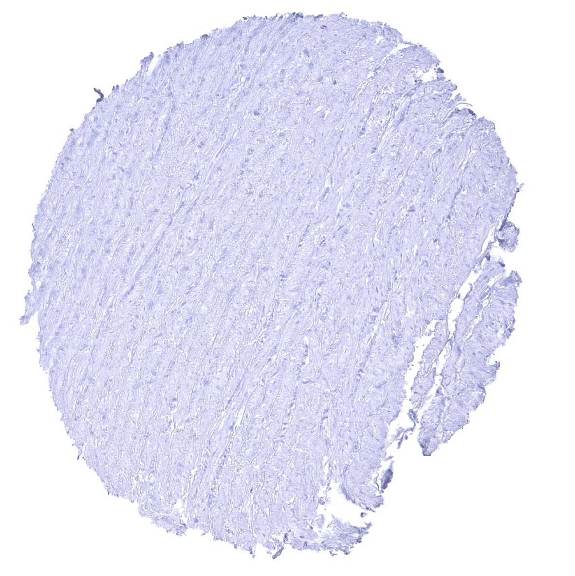
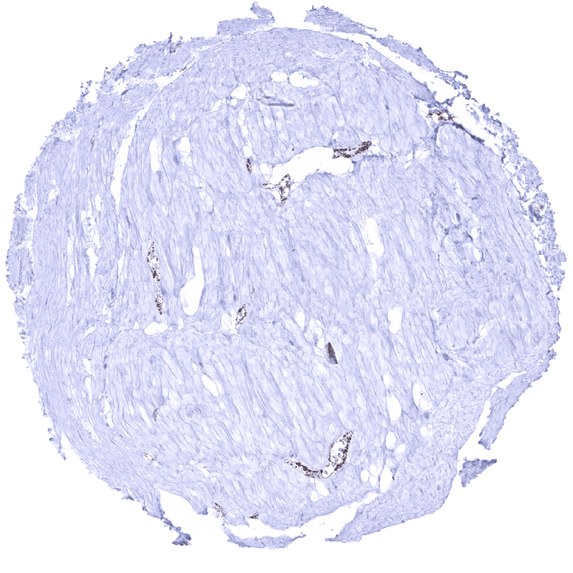
Aorta, media
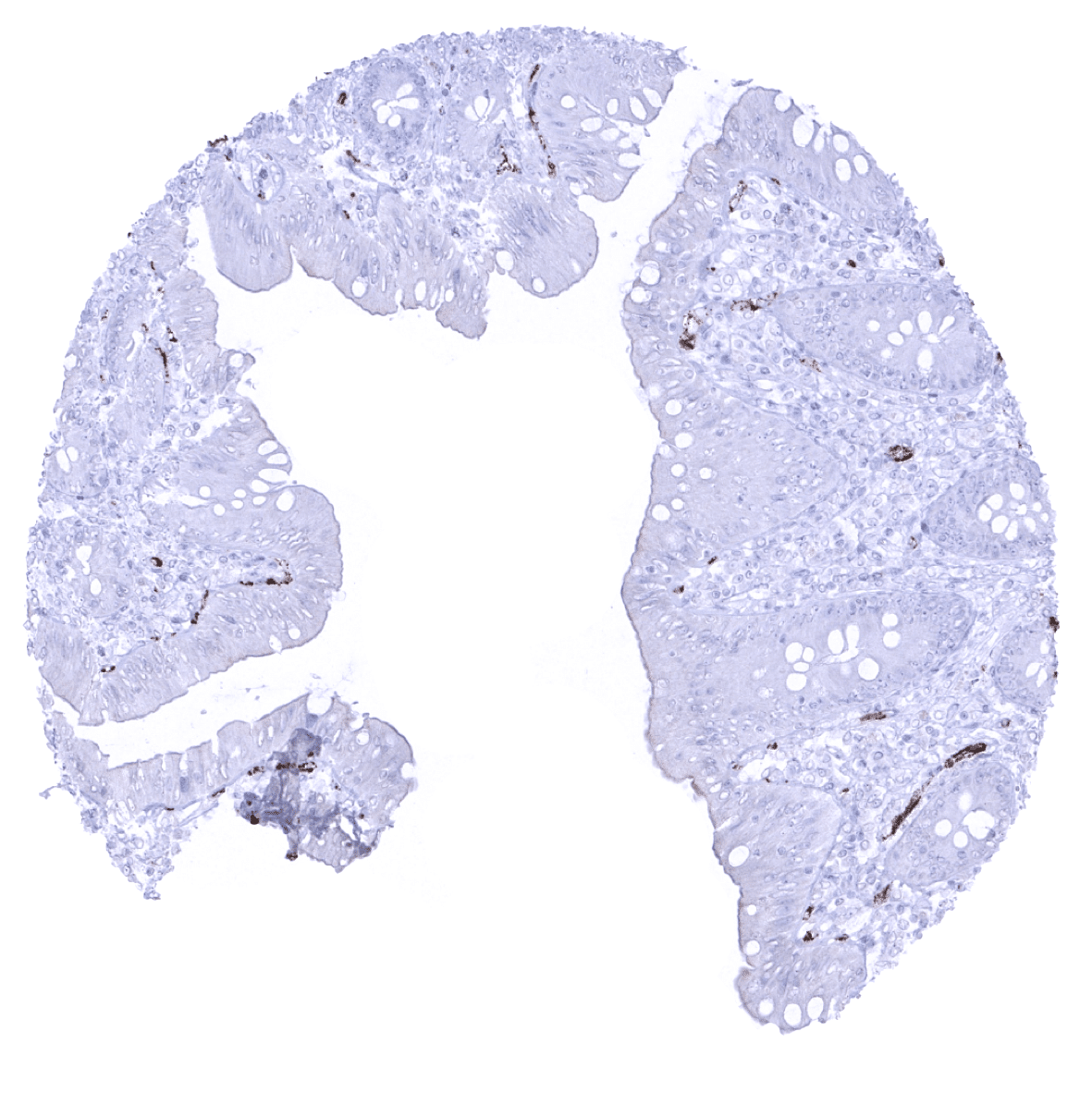
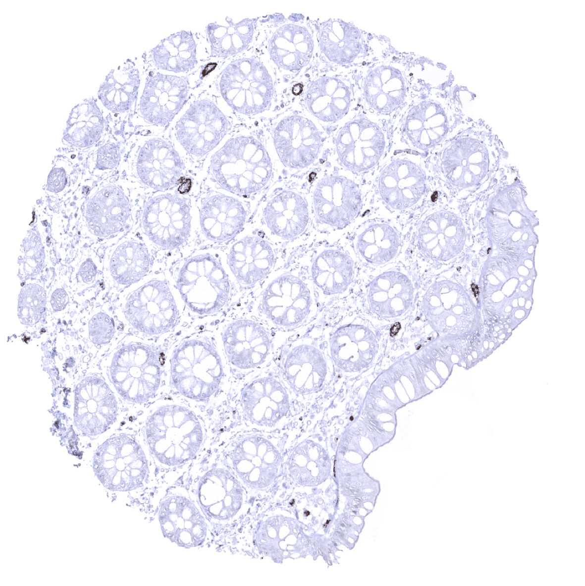
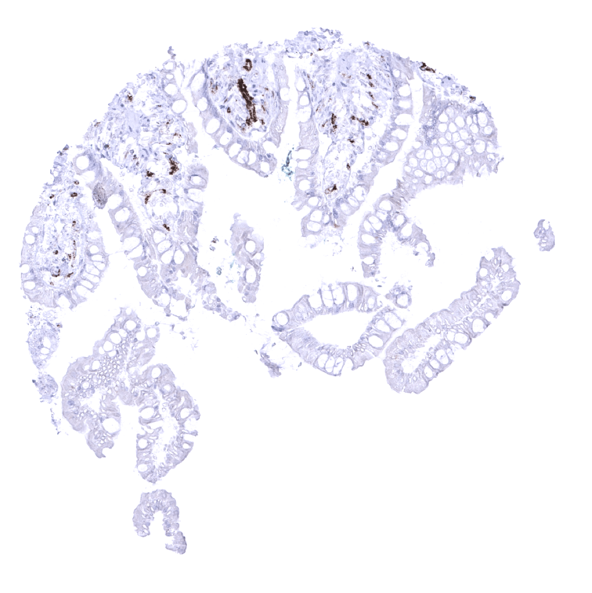
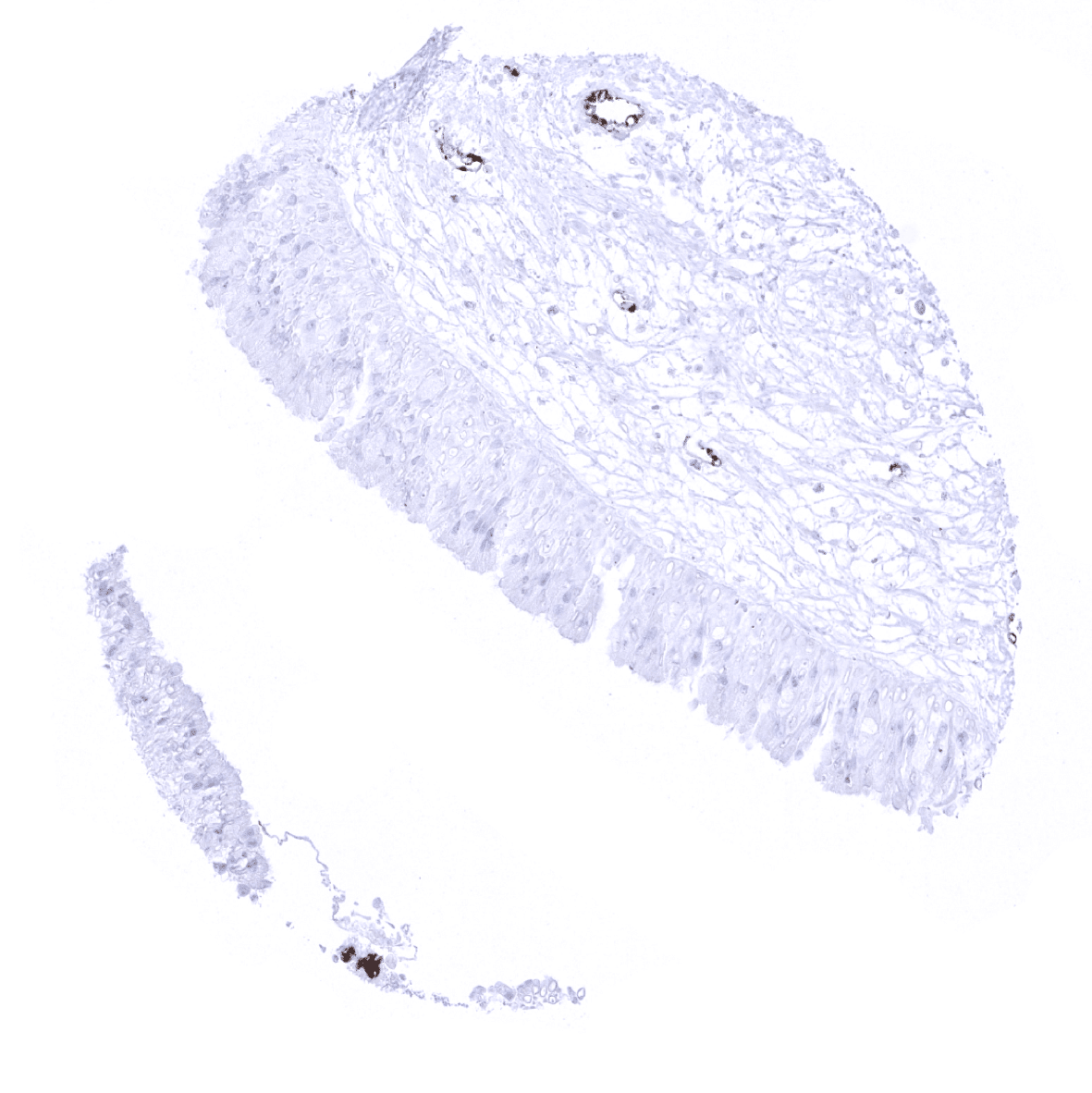
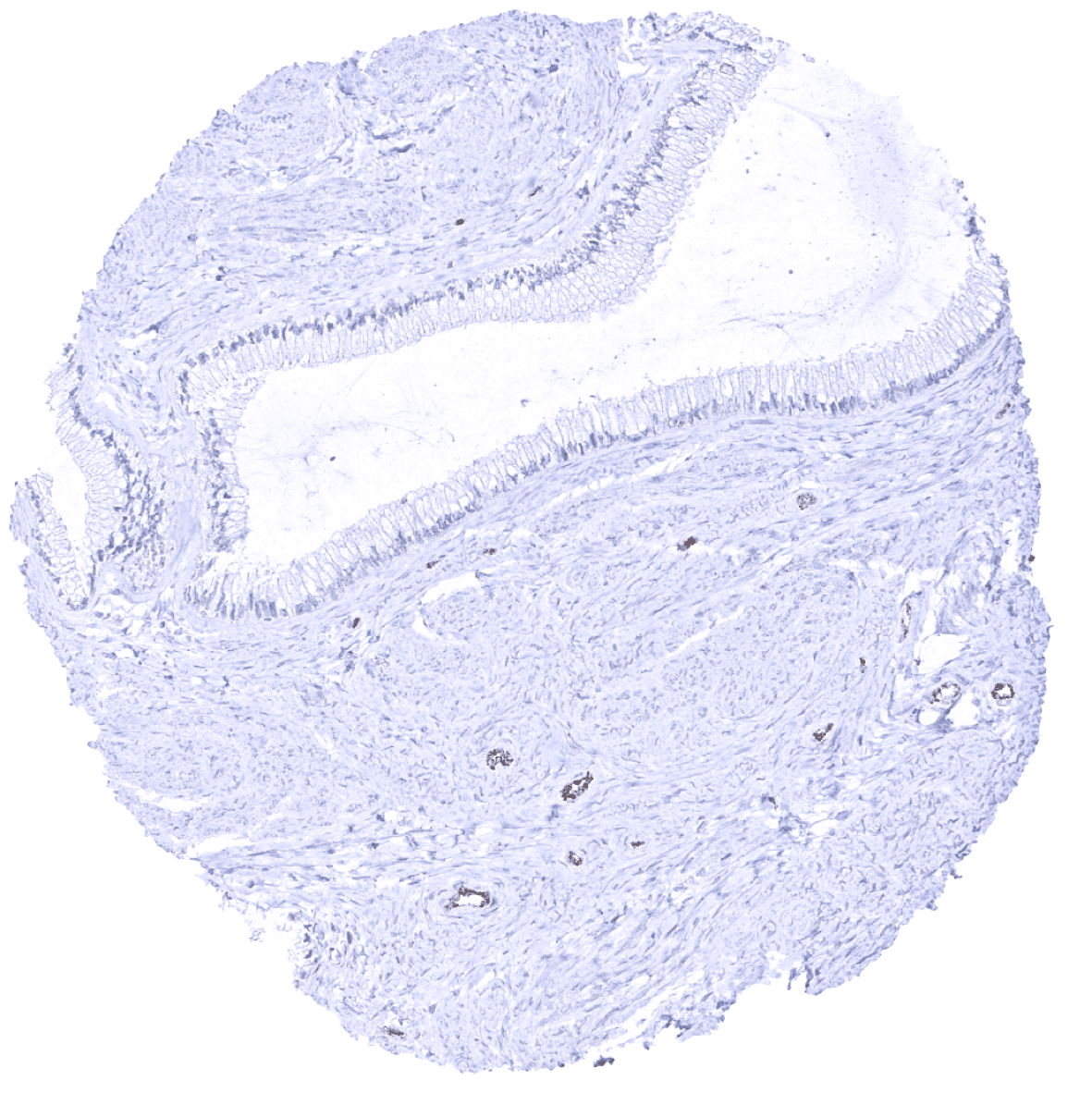
Appendix, mucosa
Appendix, muscular wall - Strong vWF immunostaining of endothelial cells.
Bone marrow - A strong vWF immunostaining occurs in megacaryocytes.
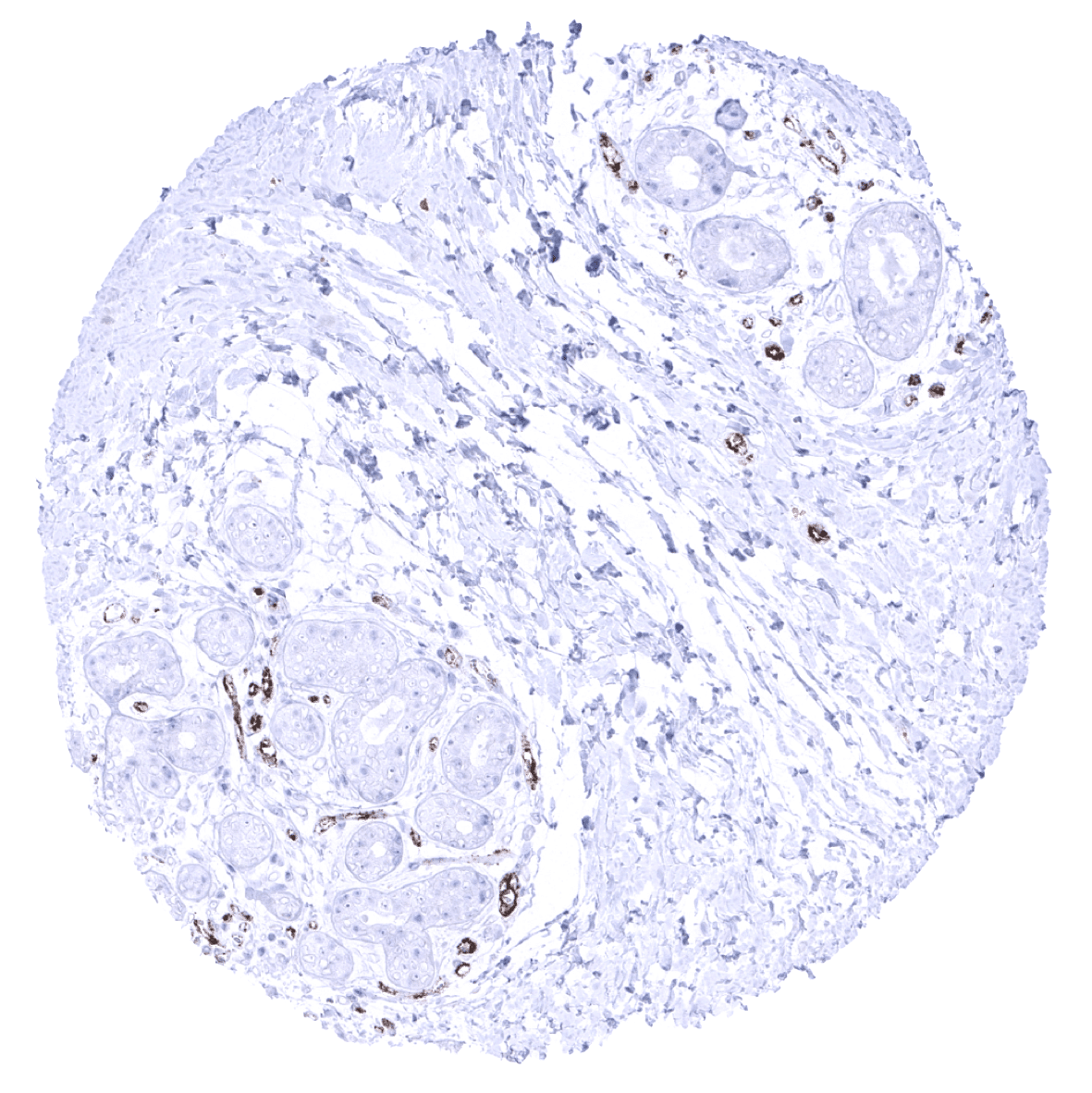
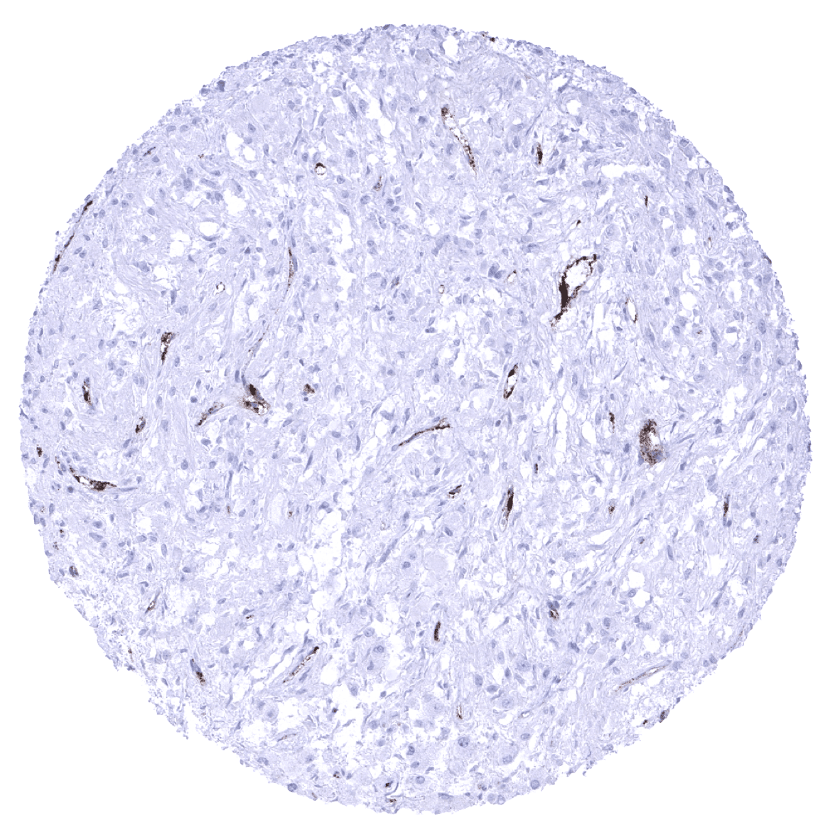
Breast
Bronchus, mucosa

Cerebellum (molecular layer, Purkinje cell layer, granule cell layer, white matter)
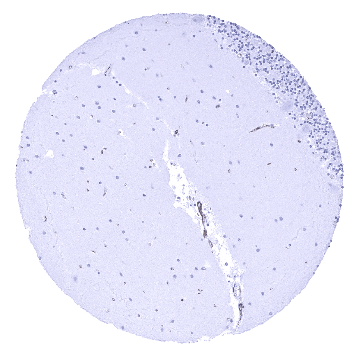
Cerebellum (molecular layer, Purkinje cell layer, granule cell layer)
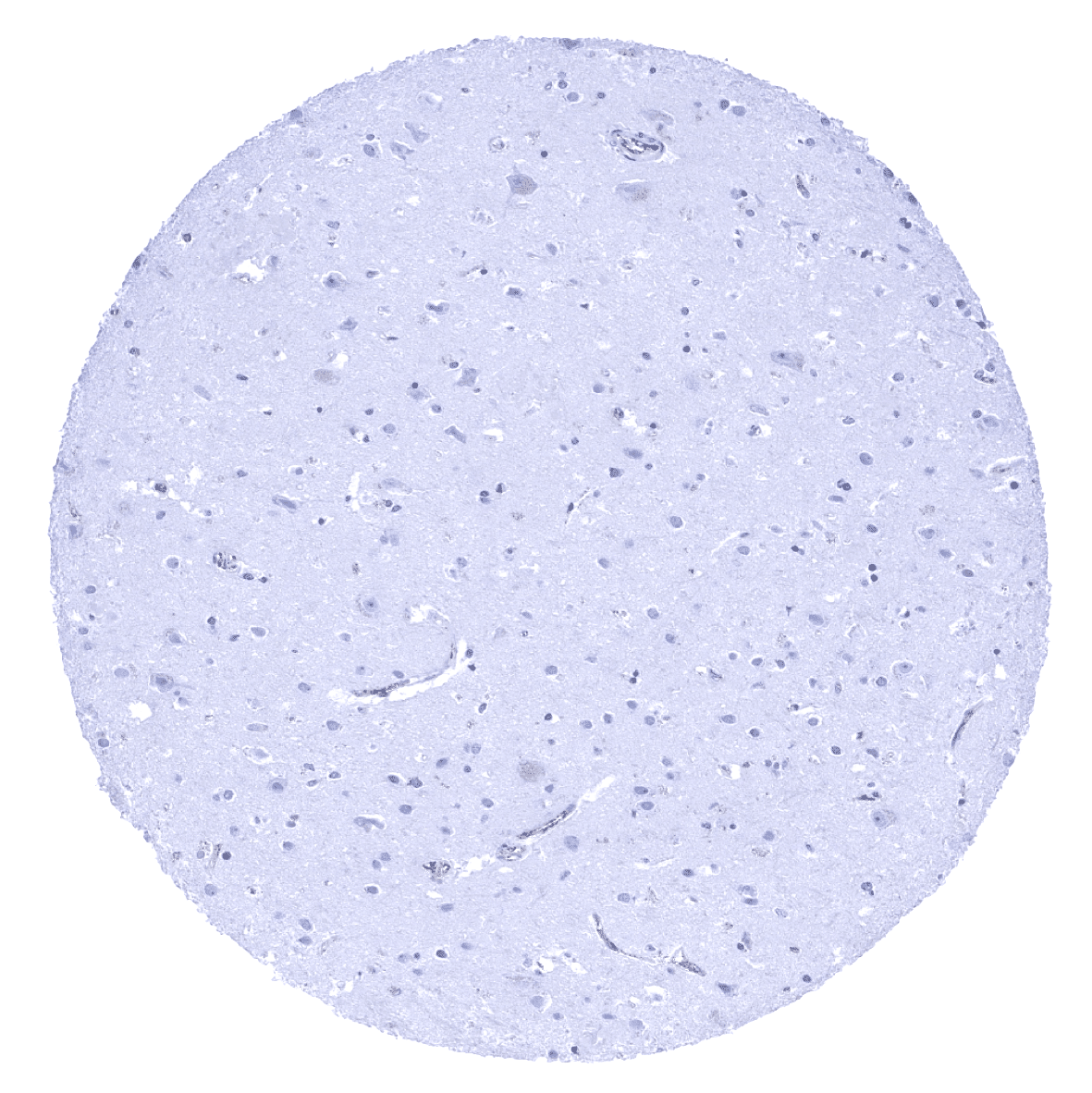
Cerebrum, grey matter

Cerebrum, white matter
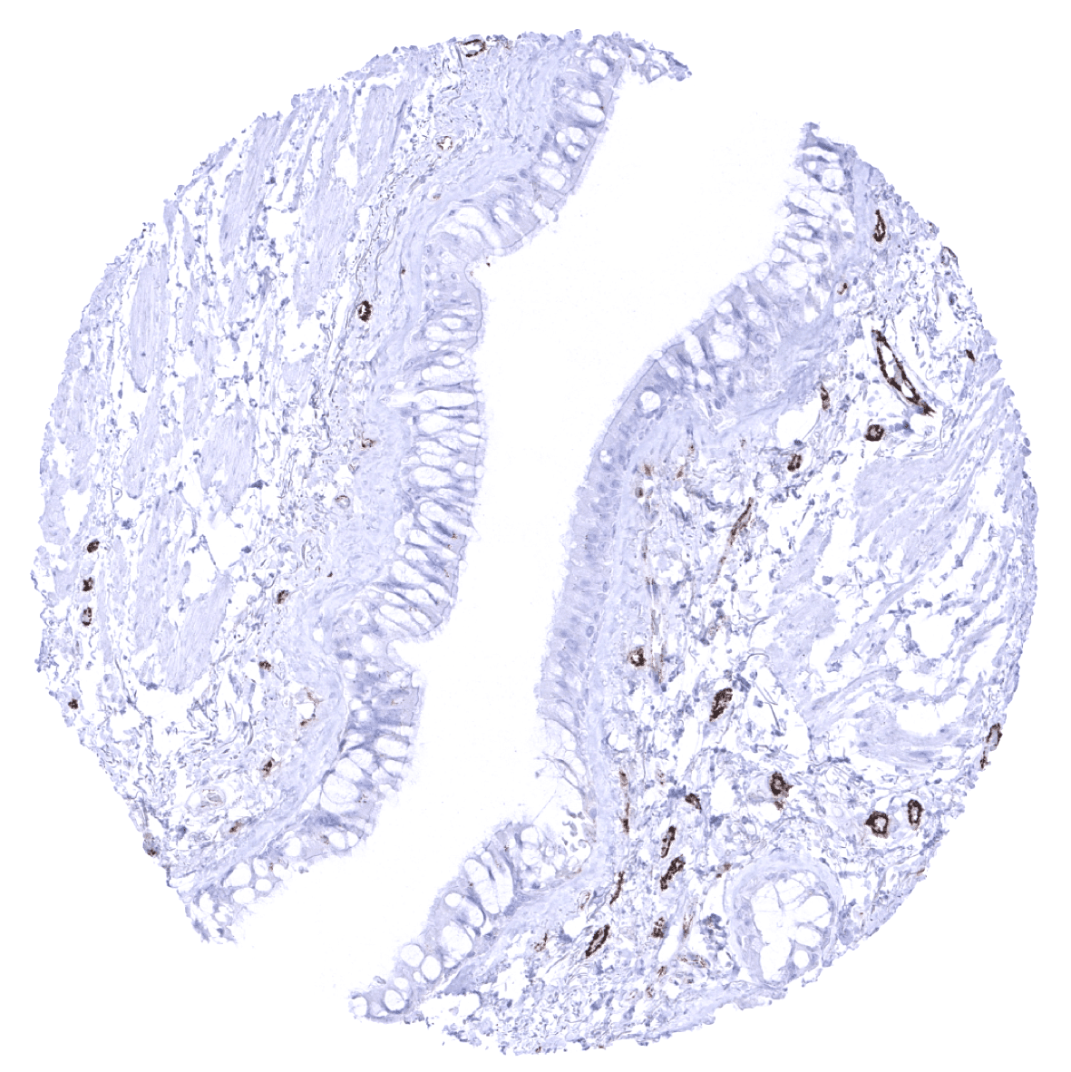
Colon descendens, mucosa
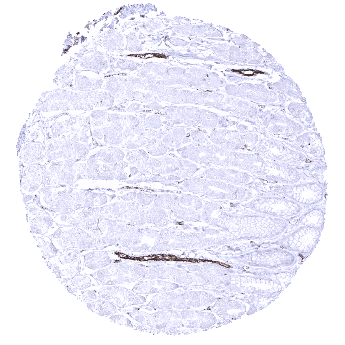
Colon descendens, muscular wall
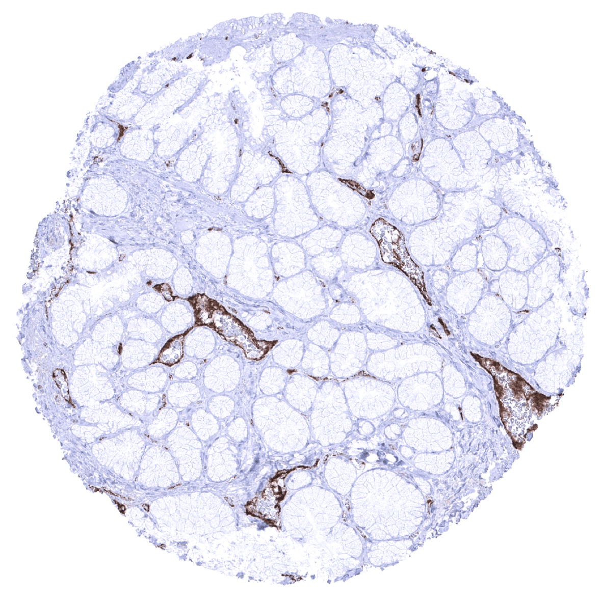
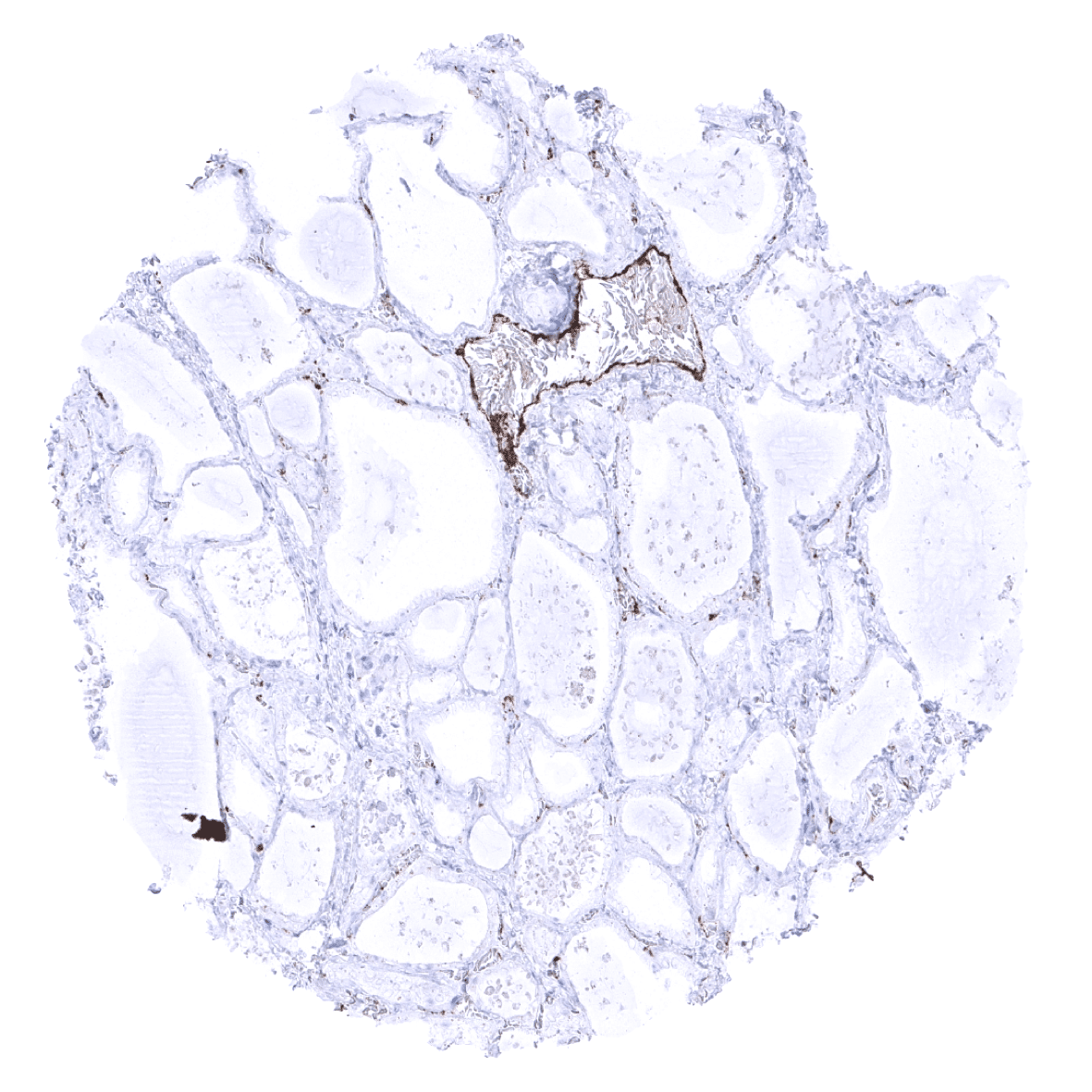
Duodenum, Brunner gland

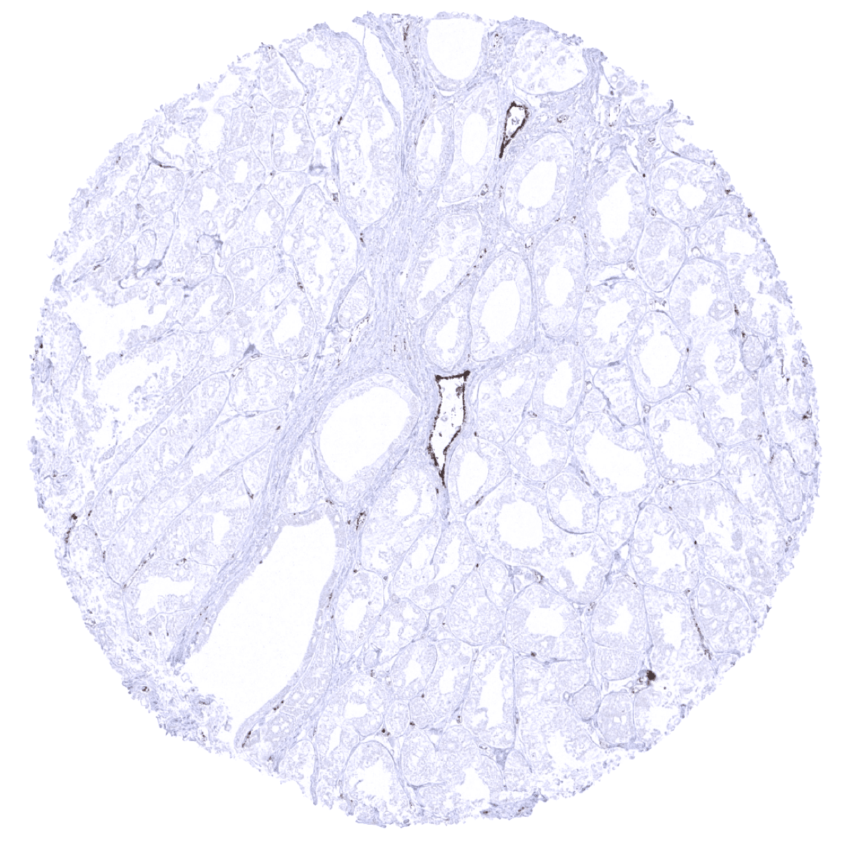
Duodenum, mucosa - A moderate endothelial vWF immunostaining is seen in postcapillary venules while staining intensity is low or absent in capillaries.

Epididymis
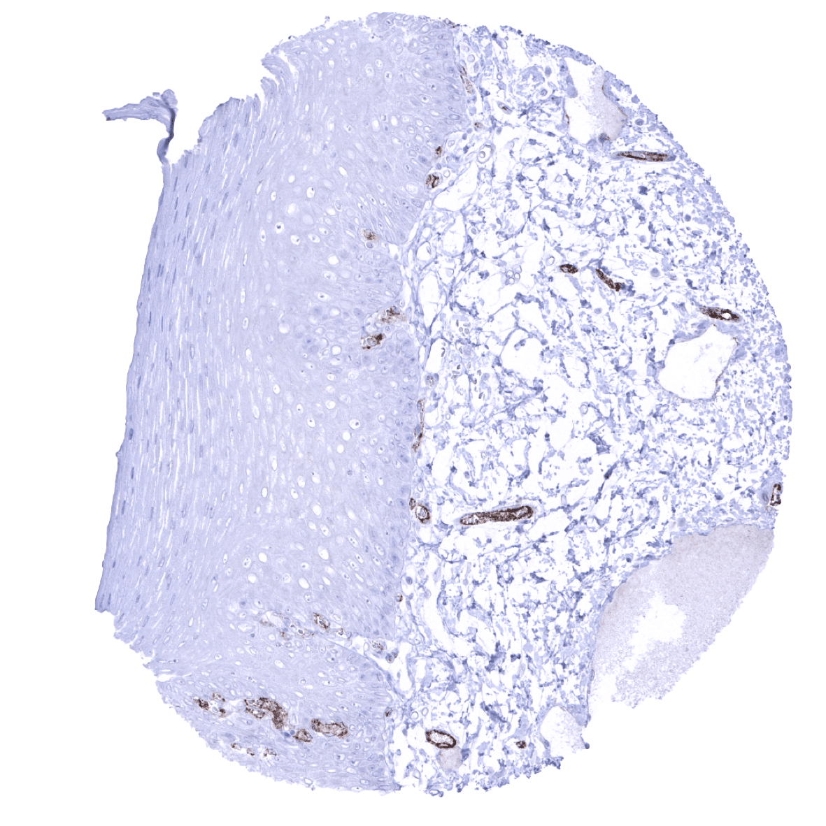
Esophagus, squamous epithelium
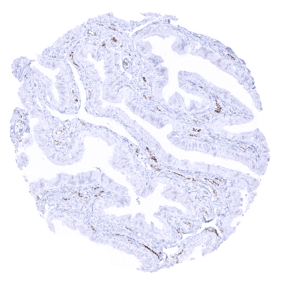
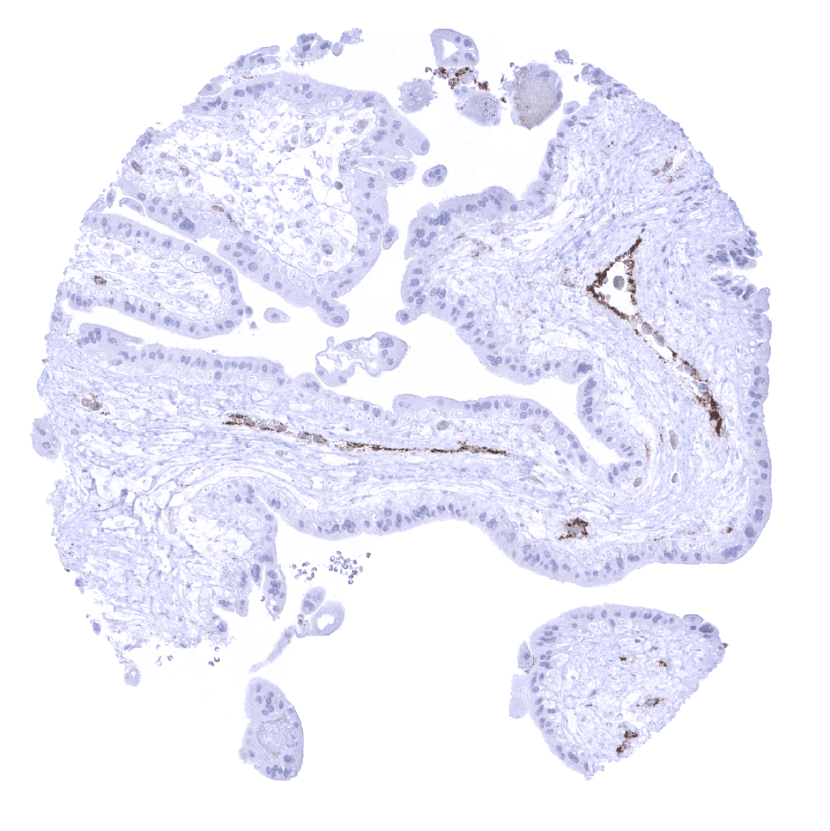
Fallopian tube, mucosa
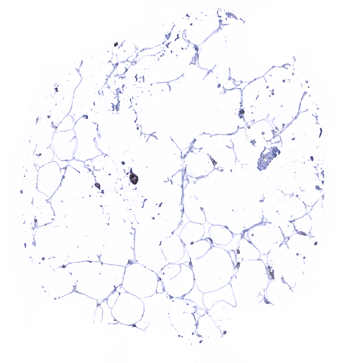
Fat

Gallbladder, epithelium
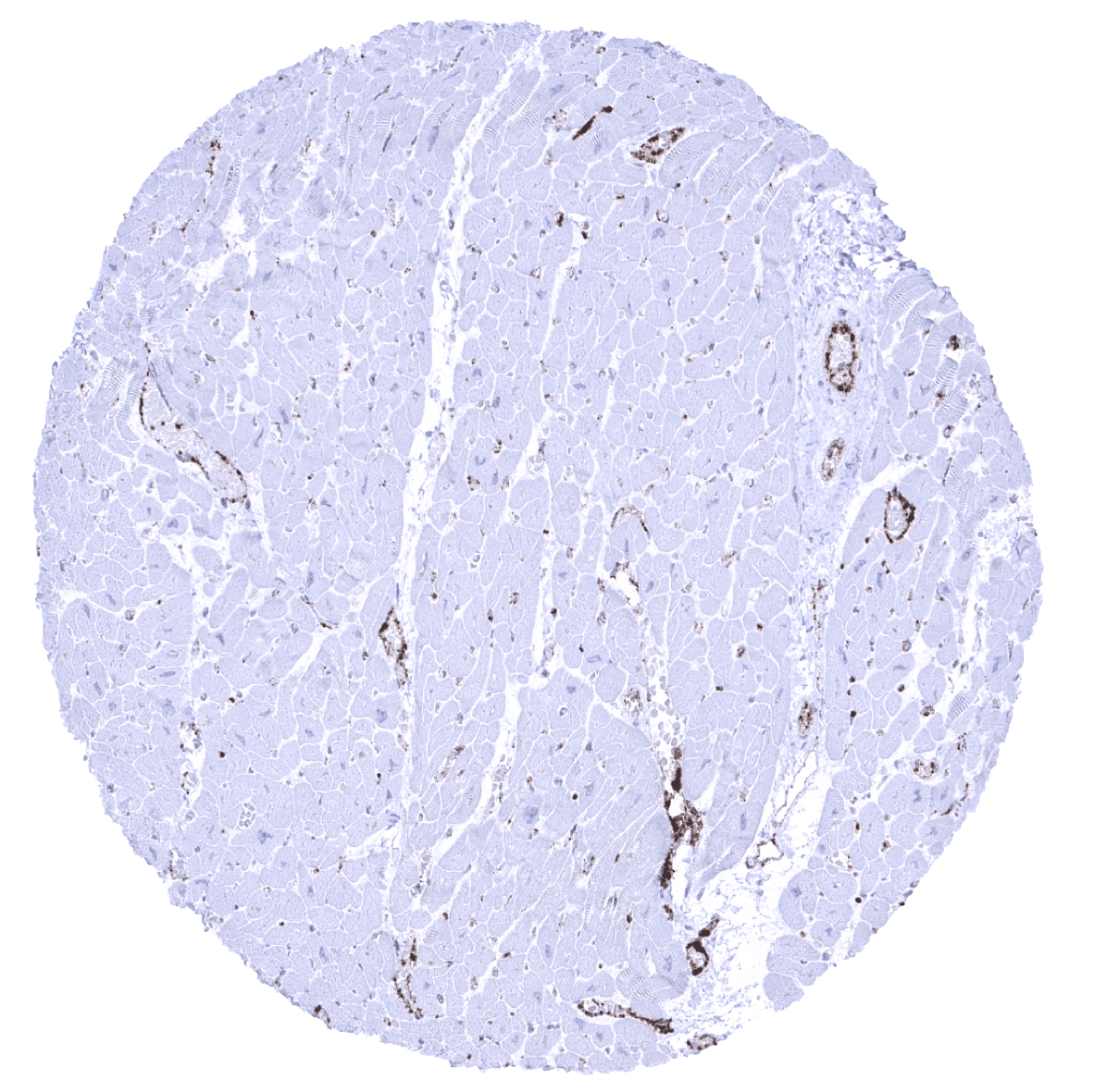
Heart muscle
Ileum, mucosa

Kidney, cortex - Endothelial vWF immunostaining is weak or absent in most small vessels of the kidney.

Kidney, medulla - Endothelial vWF immunostaining is weak or absent in most small vessels of the kidney.
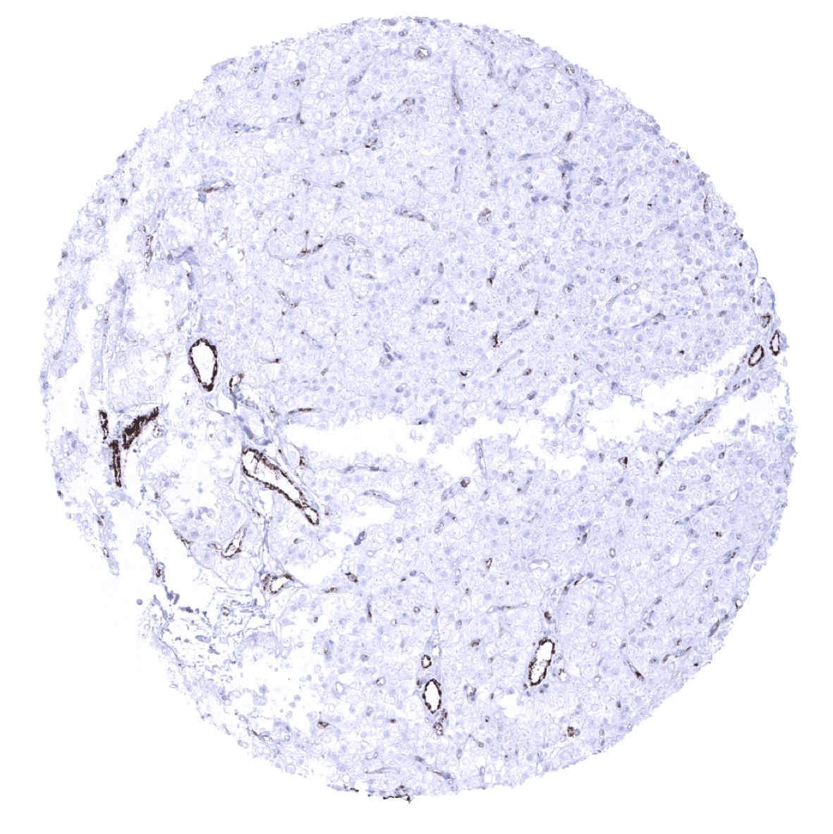
Liver - A weak endothelial vWF immunostaining is seen in a central vein while staining is absent in sinusoids.

Lung

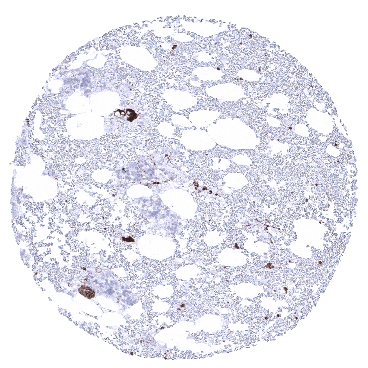
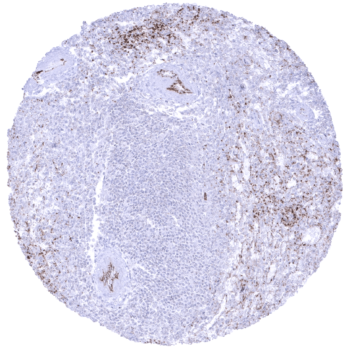
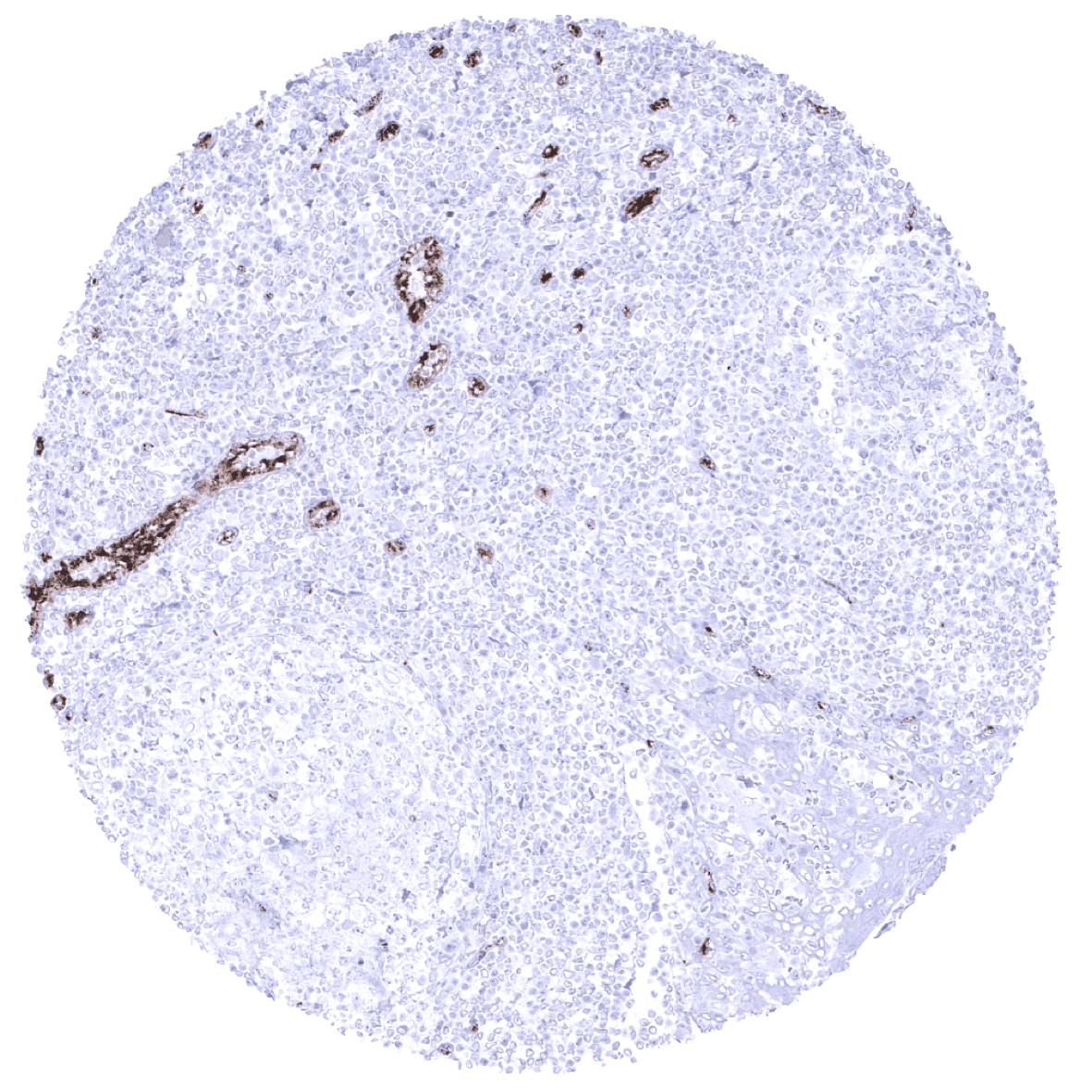
Lymph node - Variable endothelial cell vWF immunostaining in capillaries and other small vessels in a lymph node.
Ovary, stroma - Weak vWF immunostaining in capillaries.
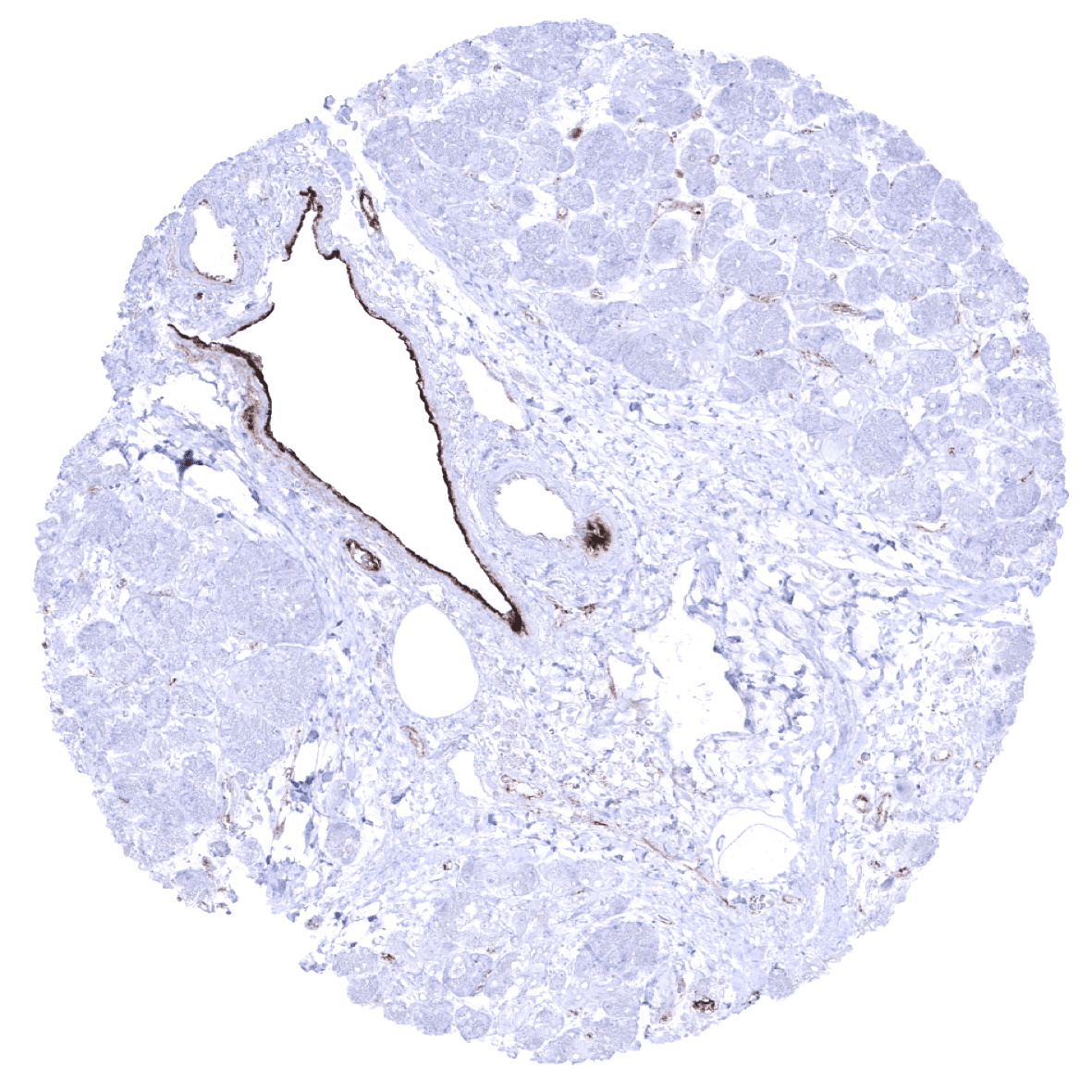
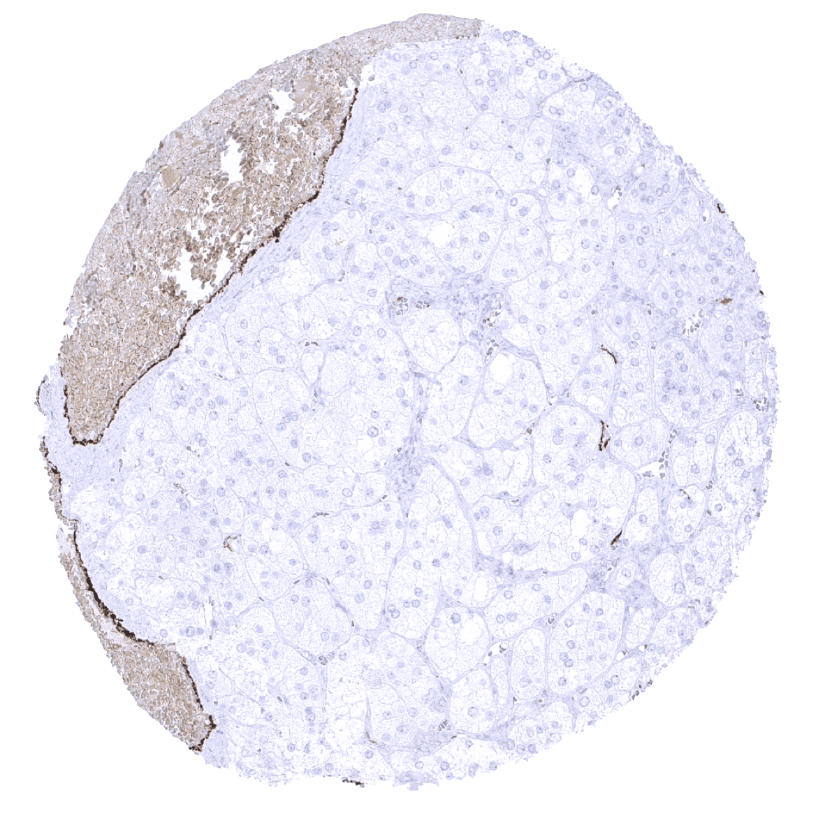
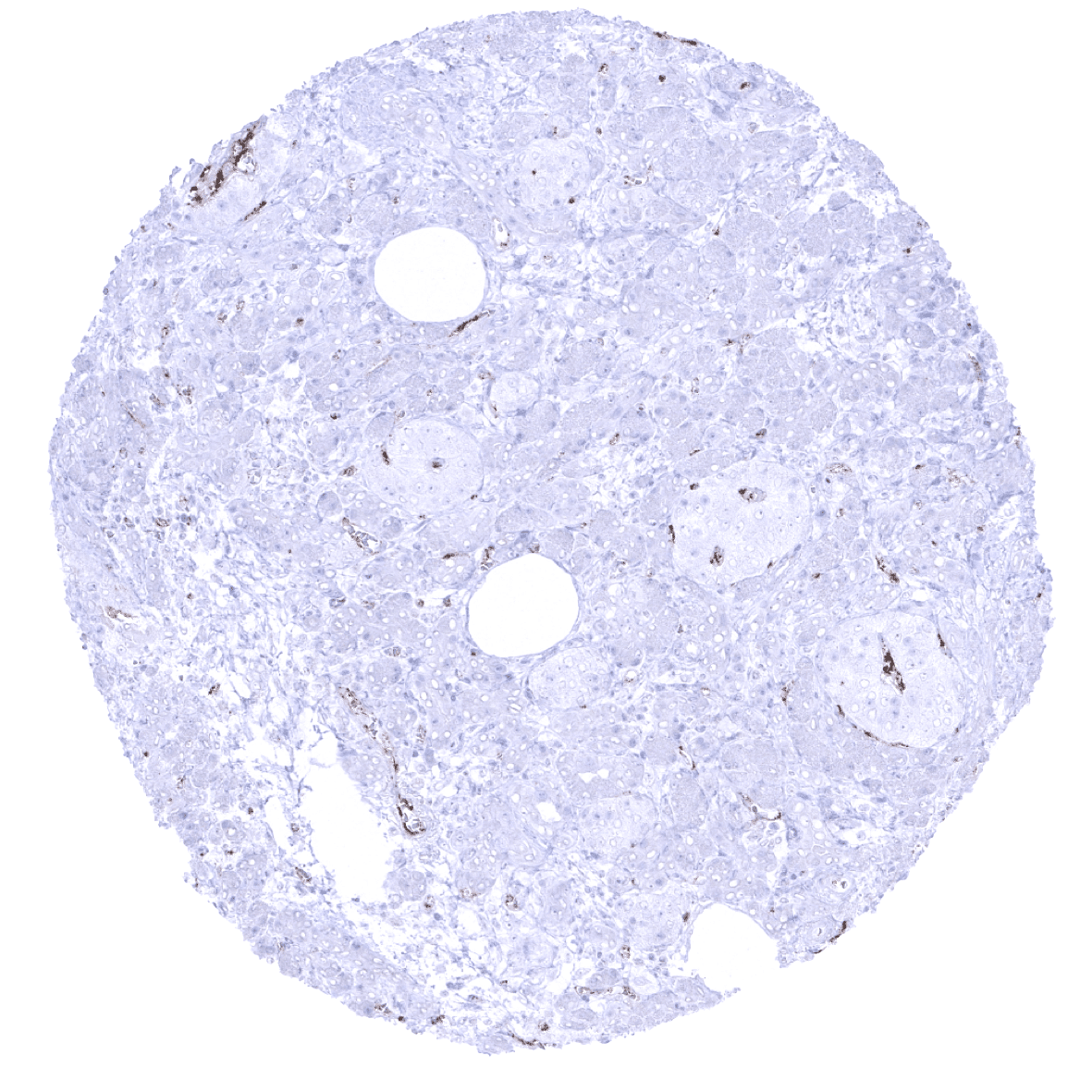
Pancreas - A strong endothelial vWF immunostaining is seen in a venule while vWF staining is rather low or absent in arterioles and most other small vessels of the pancreas.
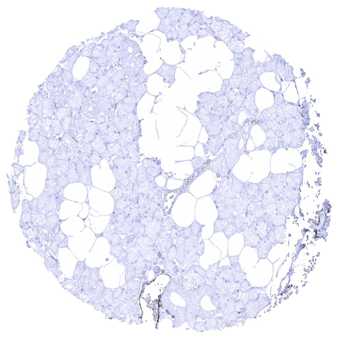
Pancreas - Endothelial vWF immunostaining is rather low or even absent in small vessels of the pancreas.
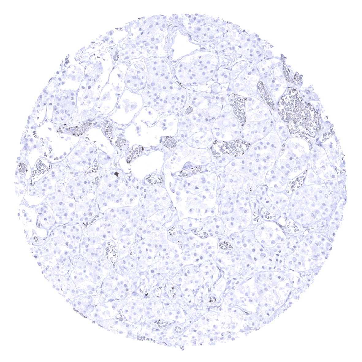
Parathyroid gland - A strong endothelial vWF immunostaining occurs in postcapillary venules while vWF staining is low in capillaries.
Parotid gland - Only a weak vWF staining is seen in most small vessels of the parotid gland.
Pituitary gland, anterior lobe - vWF immunostaining is very low or absent in small blood vessels in this adenohypophysis sample.
Pituitary gland, posterior lobe

Placenta (amnion)
Placenta (chorion)
Placenta, early
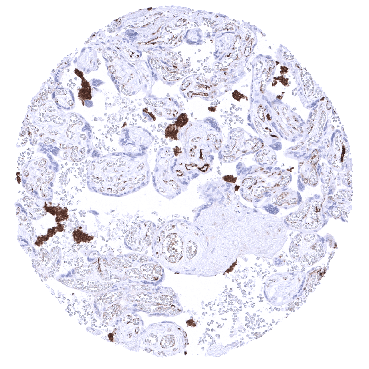
Placenta, mature - Endothelial vWF immunostaining of variable intensity occurs in the placenta. A strong vWF positivity is also seen in blood clots.

Prostate

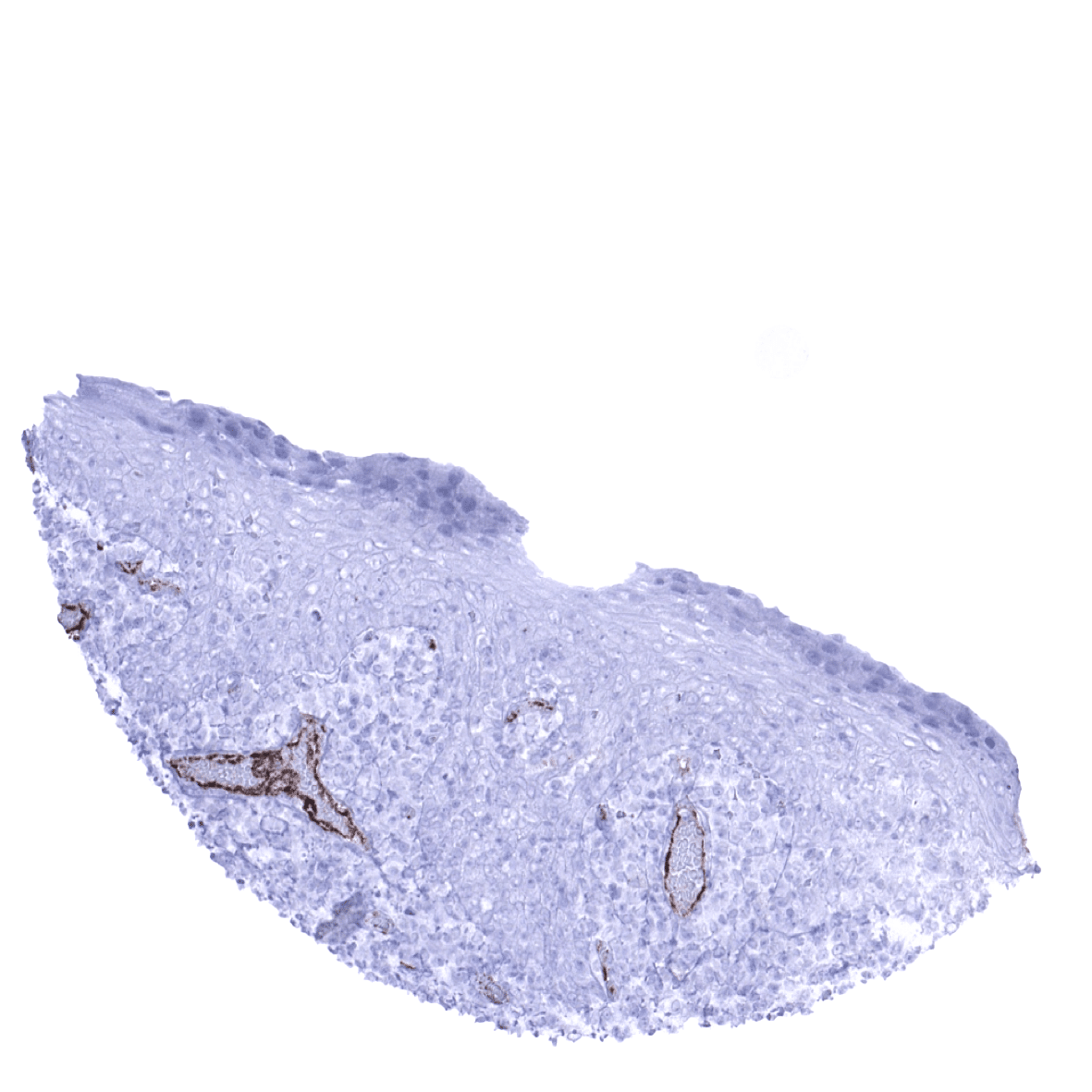
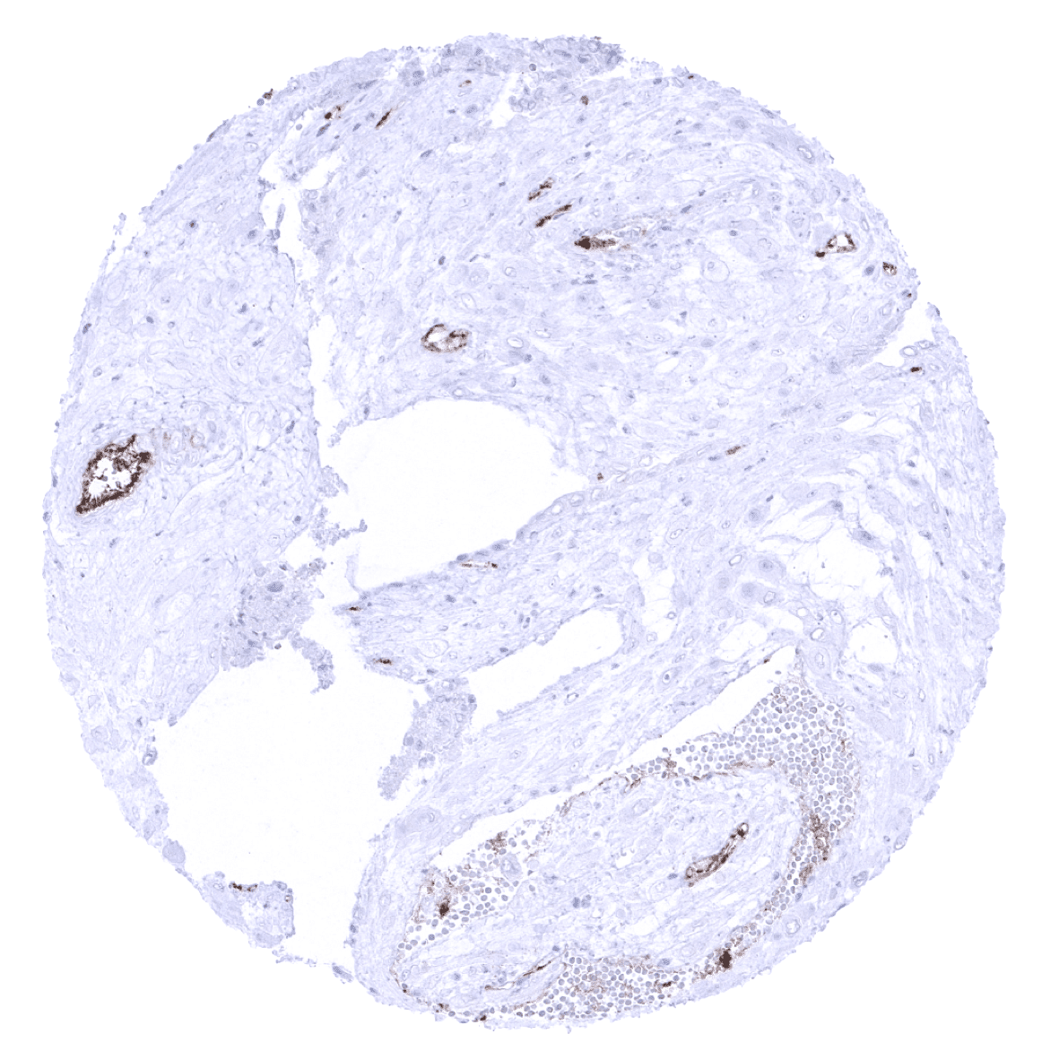
Rectum, mucosa - Only few small blood vessels show a strong endothelial vWF immunostaining while staining intensity is low or absent in most capillaries.

Seminal vesicle
Sinus paranasales
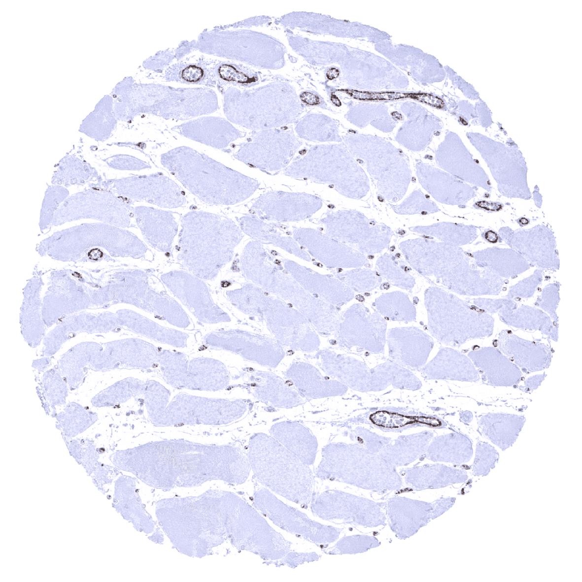
Skeletal muscle - Moderate vWF immunostaining in capillaries, strong staining in postcapillary venules.
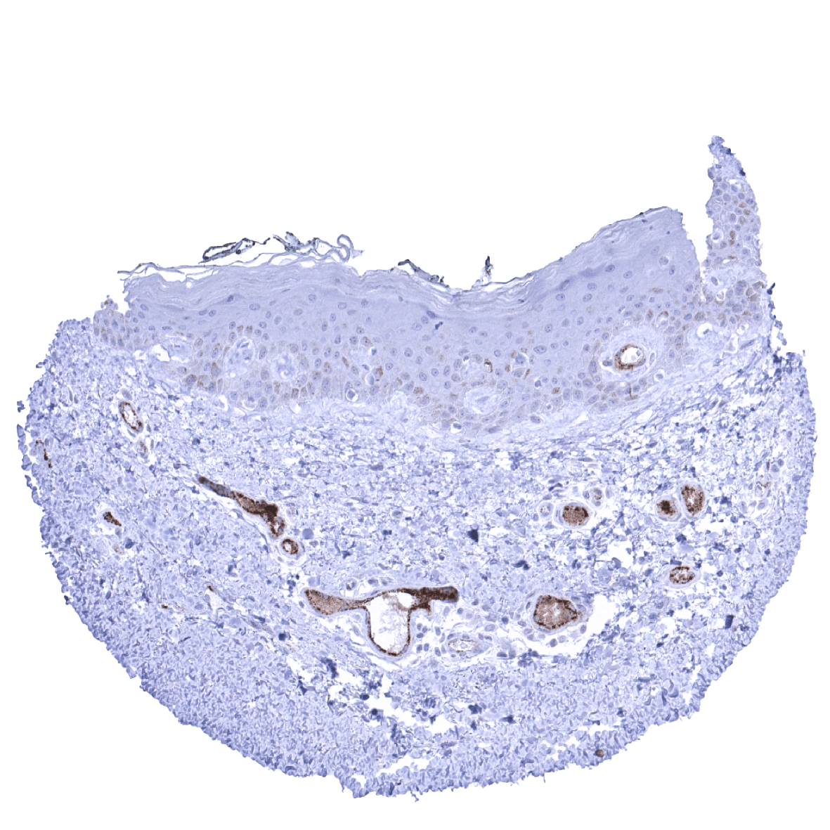
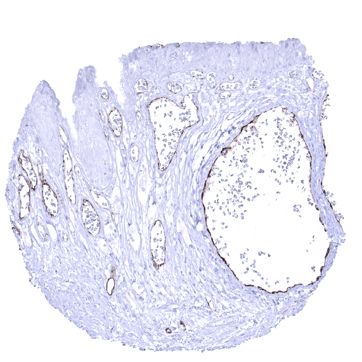
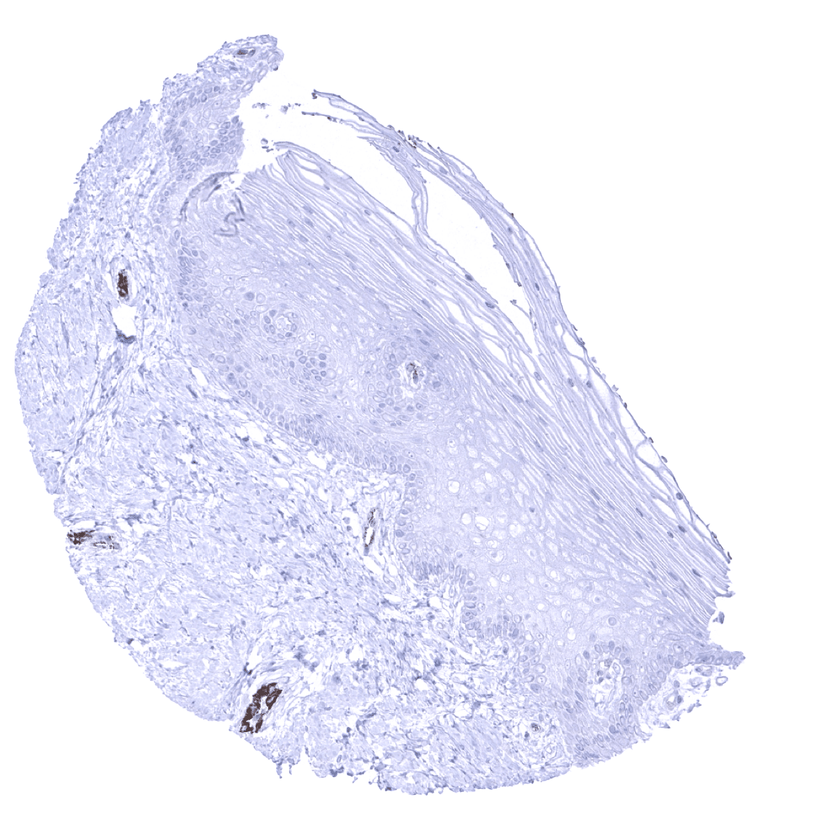
Skin
Spleen
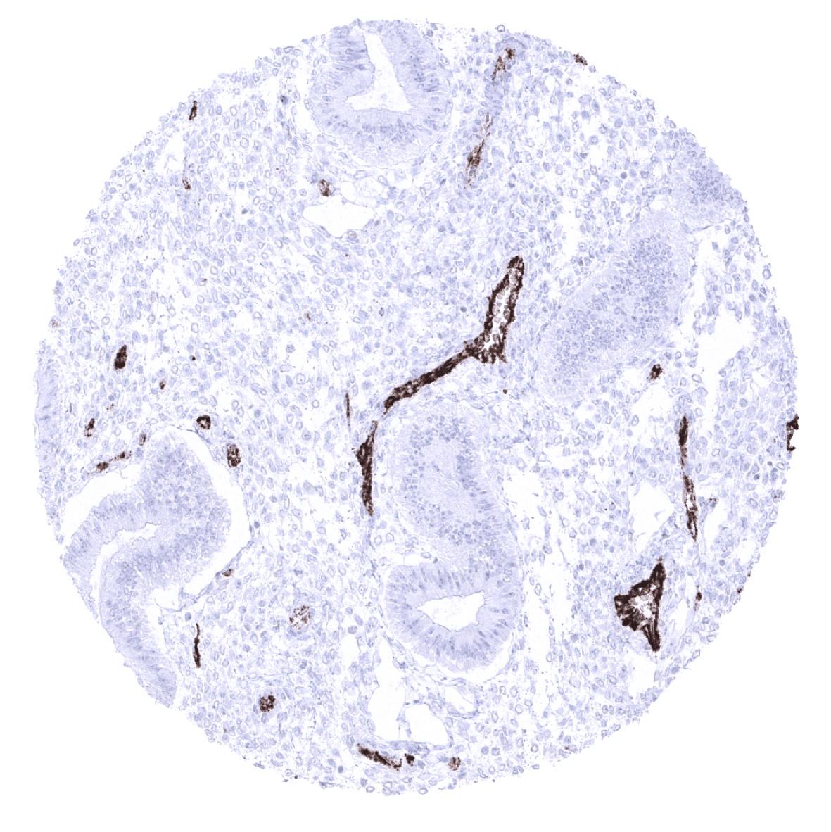
Stomach, antrum - A strong endothelial vWF immunostaining is seen in postcapillary venules while staining intensity is low or absent in capillaries.
Stomach, corpus - A strong endothelial vWF immunostaining is seen in postcapillary venules while staining intensity is low or absent in capillaries.

Testis
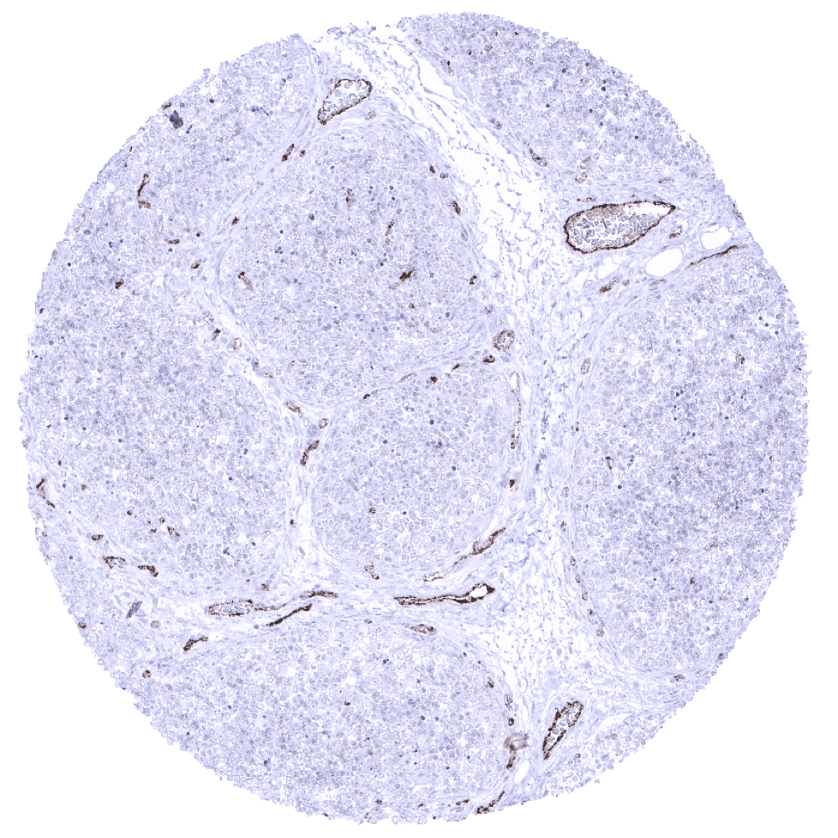
Thymus
Thyroid gland - A strong endothelial vWF immunostaining occurs in a venule while vWF staining is low in capillaries.
Tonsil
Tonsil, surface epithelium
Urinary bladder, urothelium - Variable endothelial vWF immunostaining in capillaries and venules with lowest staining intensity of epithelium adjacent capillaries.
Uterus, ectocervix
Uterus, endocervix
Uterus, endometrium (pregnancy)
Uterus, endometrium (proliferation)

Uterus, endometrium (secretion)

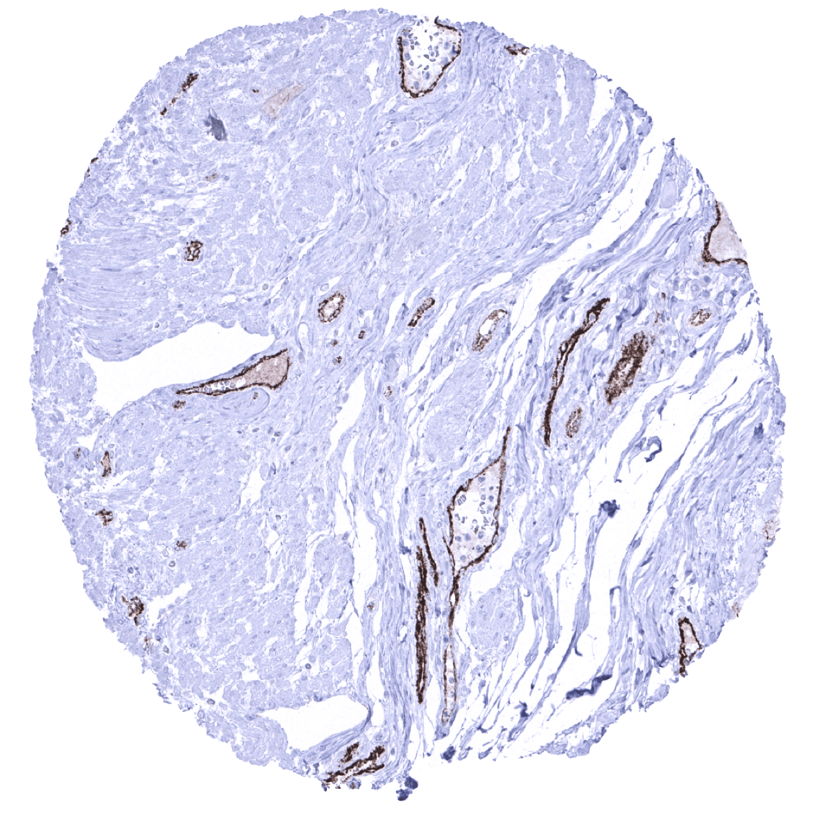
Uterus, myometrium - Moderate vWF immunostaining in capillaries, strong staining in postcapillary venules.