295,00 € – 895,00 €
Product details
Synonyms = Tetraspanin-20; Tspan-20; TSPAN20; UP1b; UPIb; UPK1B; Uroplakin-1b
Antibody type = Mouse monoclonal / IgG
Clone = MSVA-734M
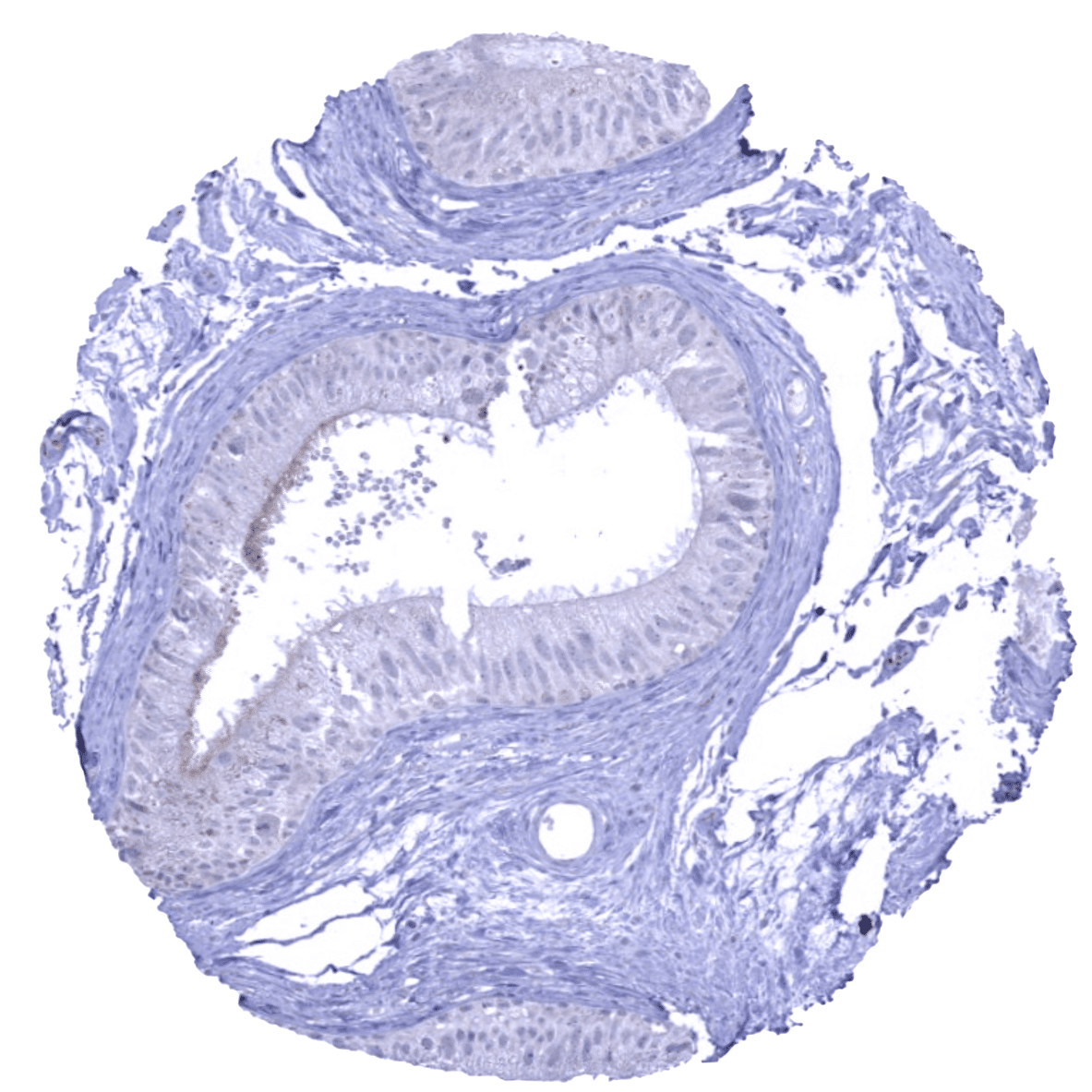
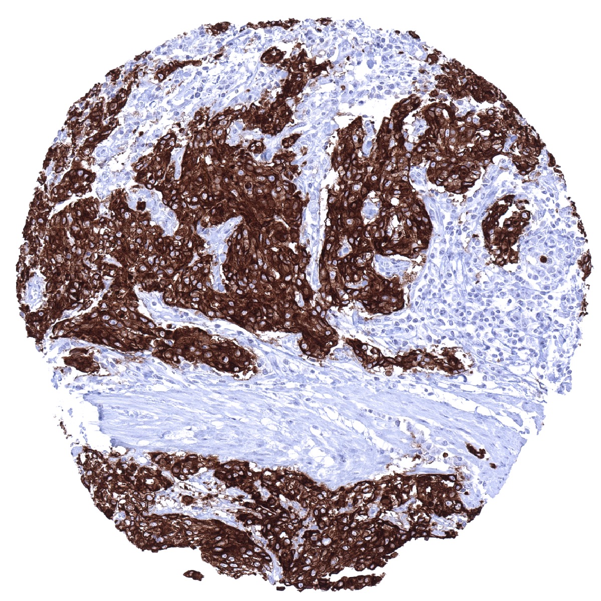
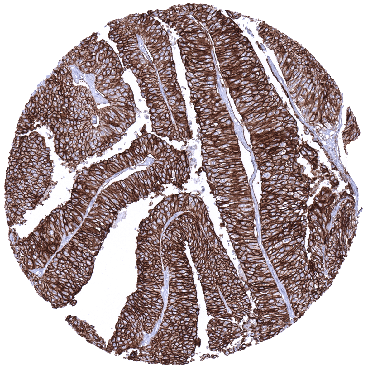
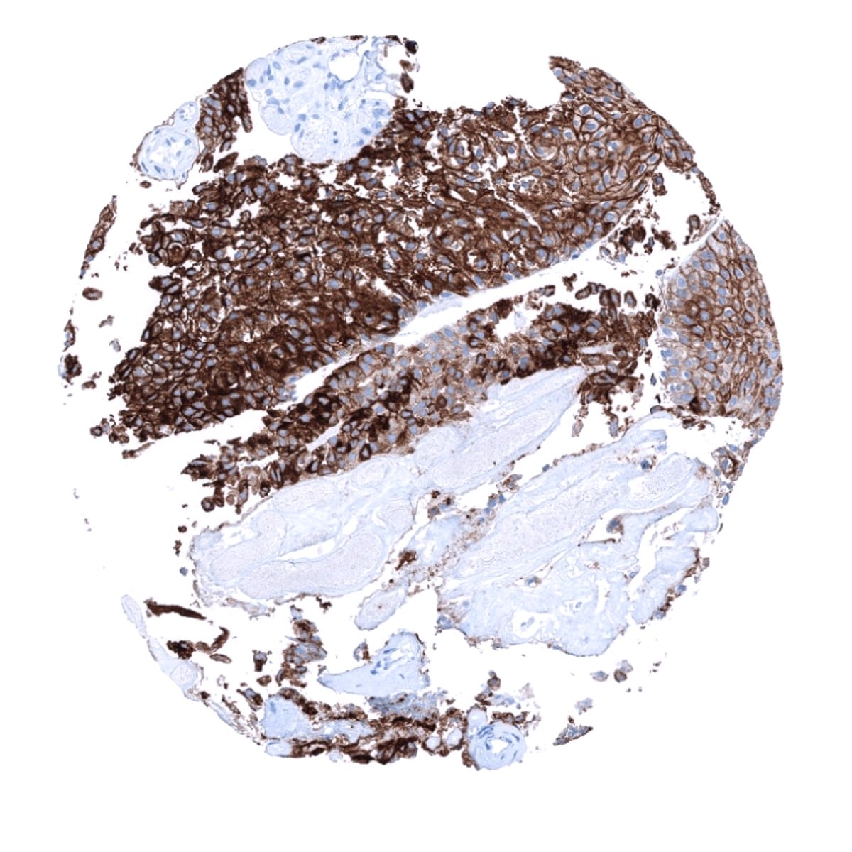
Positive control = Urinary bladder: A strong membranous and cytoplasmic UPK1b immunostaining should be seen in the urothelium (the staining can be limited to the top cell layers or equally involve all cell layers).
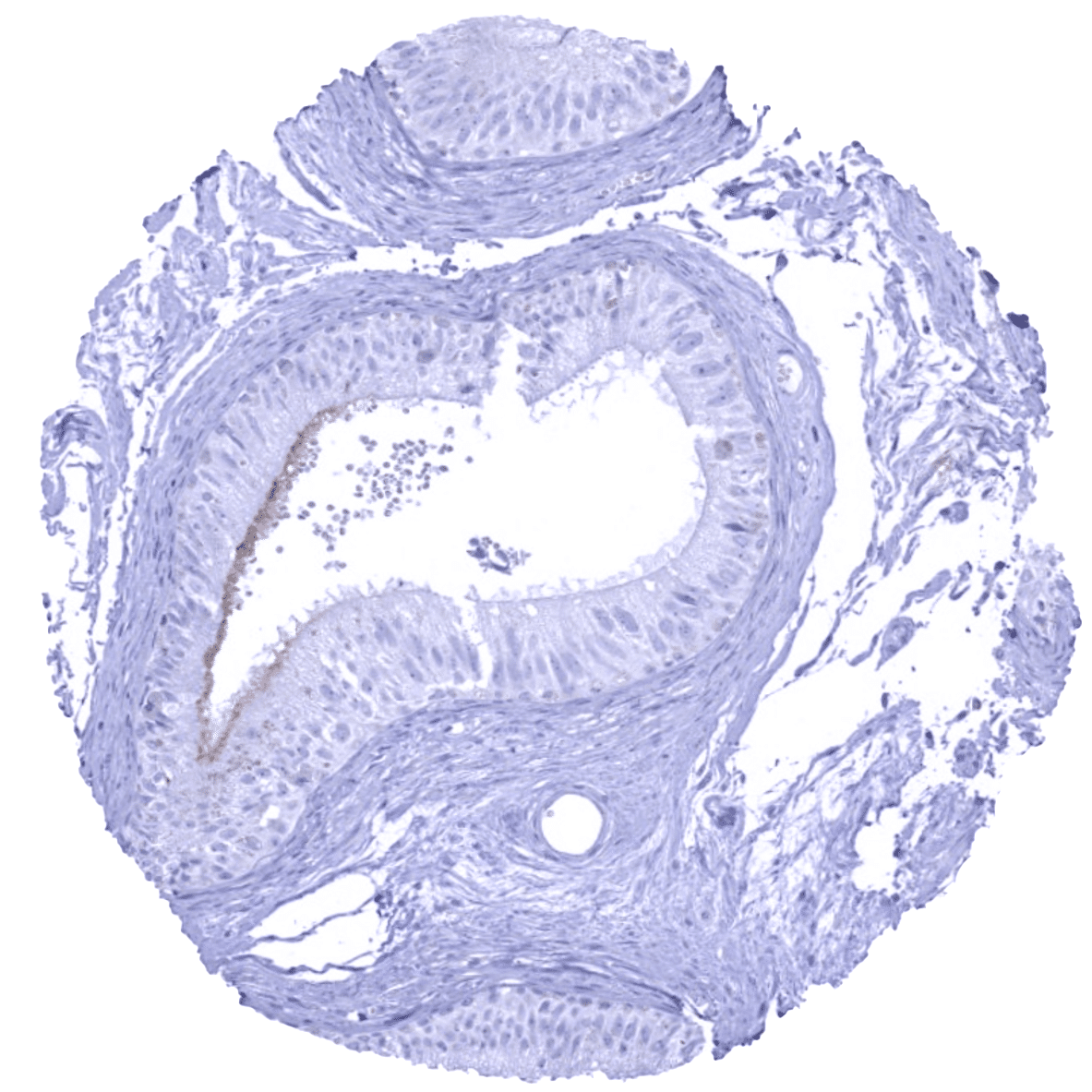
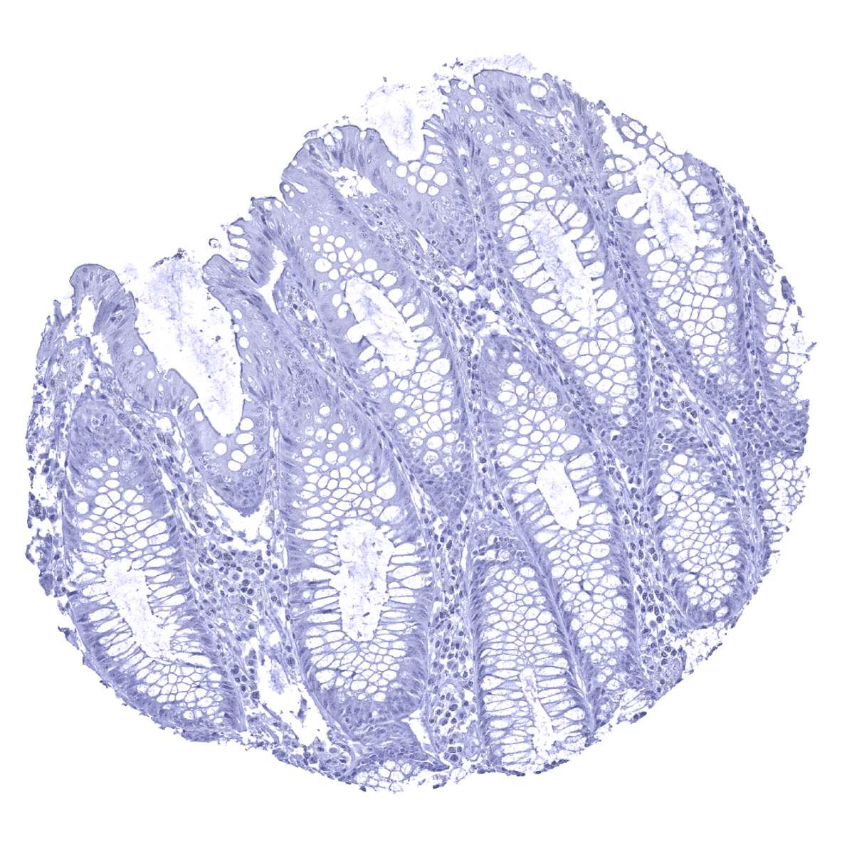
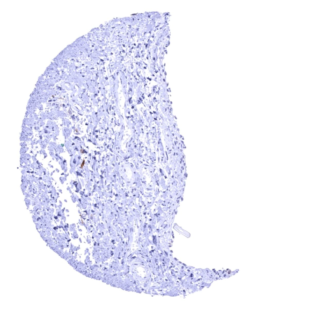
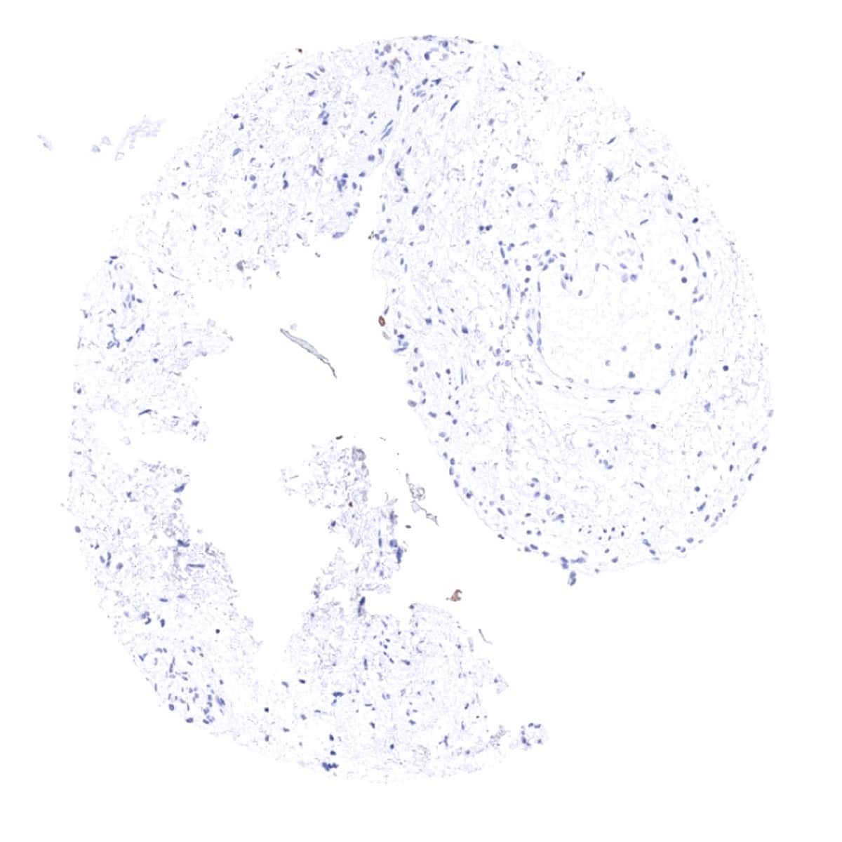
Negative control = Colon: UPK1b immunostaining should be absent in all cells of the colon mucosa.
Cellular localization = Cell Surface and cytoplasm
Reactivity = Human
Application = Immunohistochemistry
Dilution = 1:100 – 1:200
Intended Use = Research Use Only
Relevance of Antibody
Uroplakin 1B is a marker for urothelial carcinomas
Biology Behind
Uroplakin 1B (Upk1b) is a 29.6 kDa protein which is encoded by the UPK1b gene located at 3q13.3-q21. In humans, there are 4 other uroplakins (Upk 1a, -2, -3a, -3b), which assemble into dimers of Upk1a/Upk2 and Upk1b/Upk3 and eventually into heterotetramers that are deposited at the surface of the epithelial cells lining the urinary bladder, ureter, and renal pelvis. These membrane plaques are termed asymmetric unit membranes (AUMs) and serve as elastic stabilizers that prevent the bladder wall from mechanical stress and rupture during urinary bladder distension. In addition, AUMs may contribute to the regulation of membrane permeability and signal transduction, which may also impact aspects of tumorigenesis such as cellular growth and motility.
Staining Pattern in Normal Tissues
Uroplakin 1B staining pattern in Normal Tissues with antibody MSVA-734M (Images shown in our “Normal Tissue Gallery”)
| Brain | Cerebrum | Negative. |
| Cerebellum | Negative. | |
| Endocrine Tissues | Thyroid | Negative. |
| Parathyroid | Negative. | |
| Adrenal gland | Negative. | |
| Pituitary gland | Negative. | |
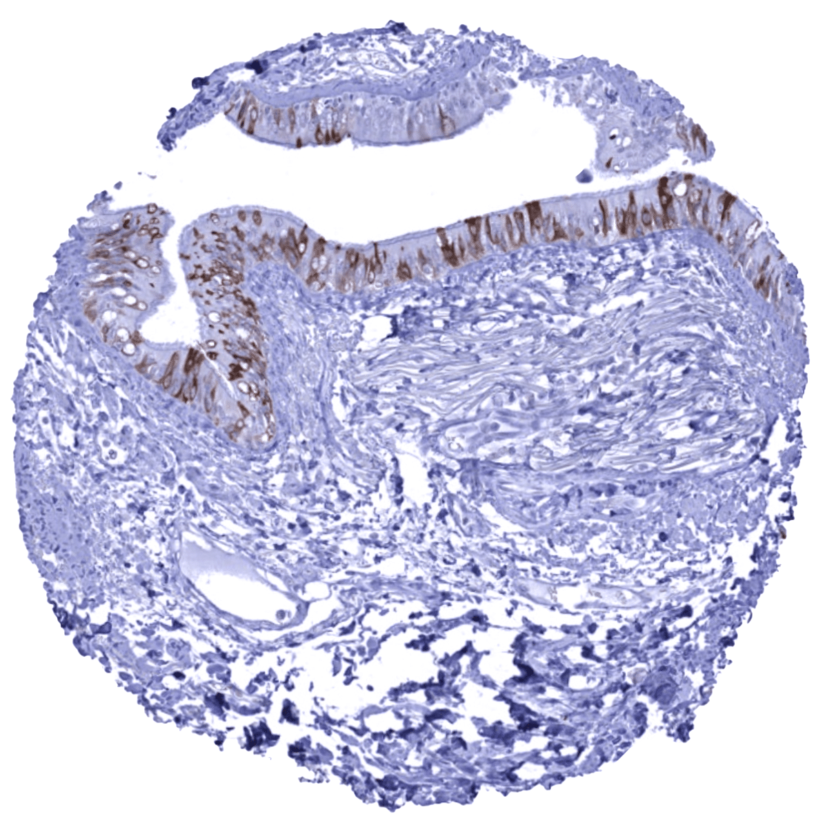
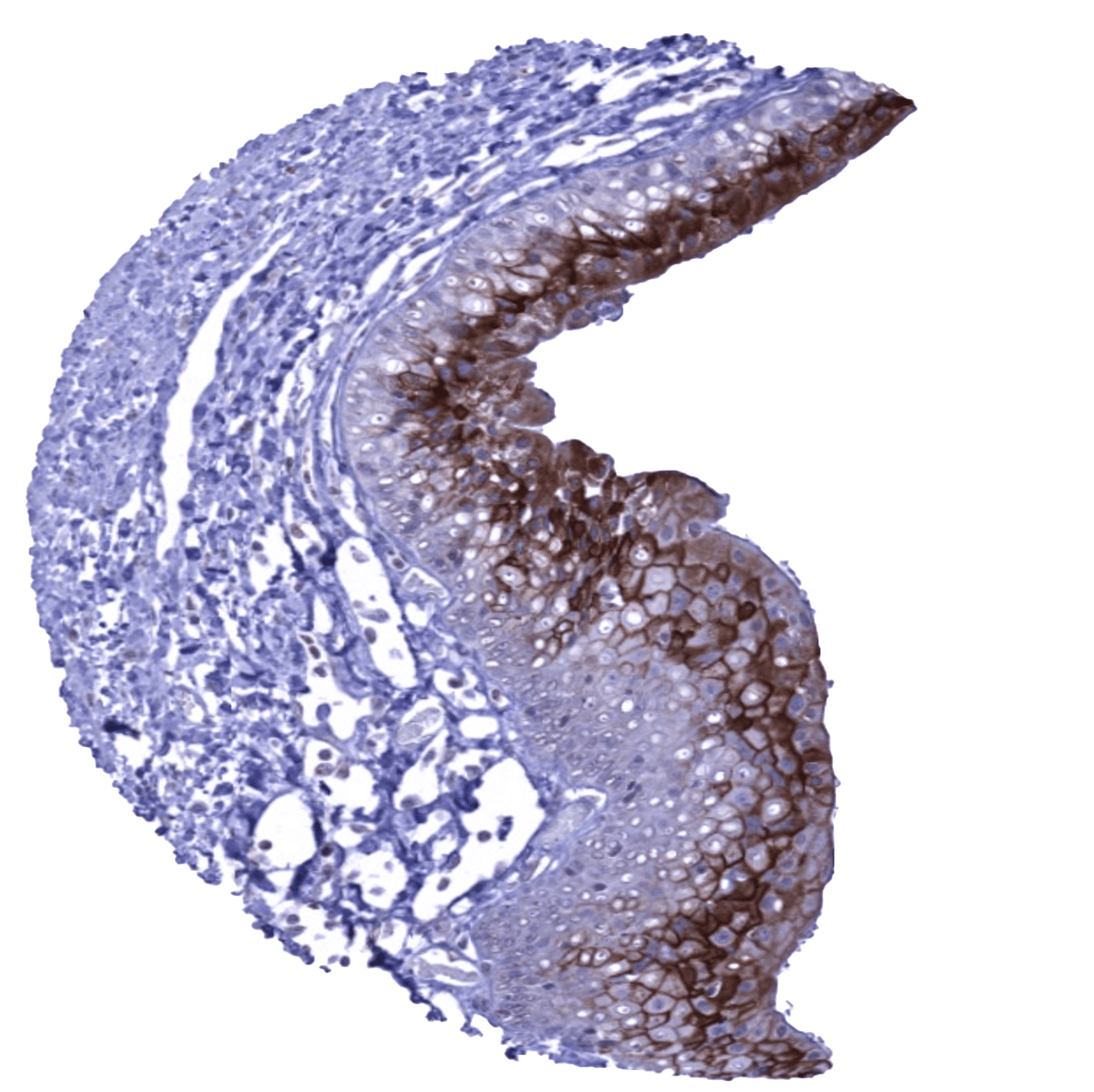
| Respiratory system | Respiratory epithelium | A moderate to strong Upk1b staining occurs in a fraction of cells. |
| Lung | ||
| Gastrointestinal Tract | Salivary glands | A variable (weak to strong) Upk1b positivity can sometimes occur in some serous cells in salivary glands. |
| Esophagus | Usually negative. Scattered Upk1b positive cells can occur. | |
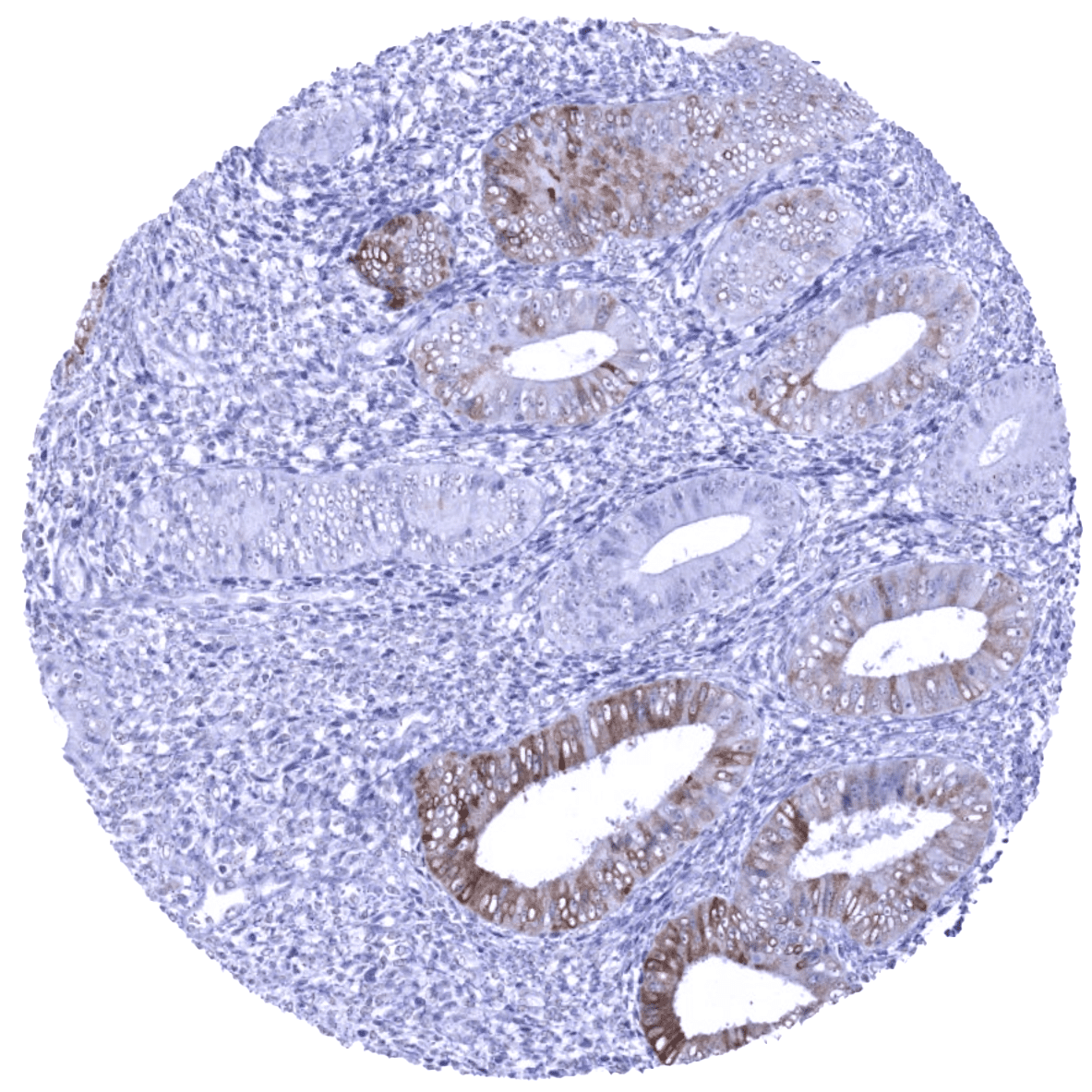
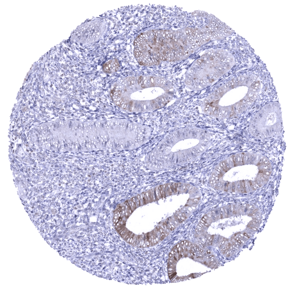
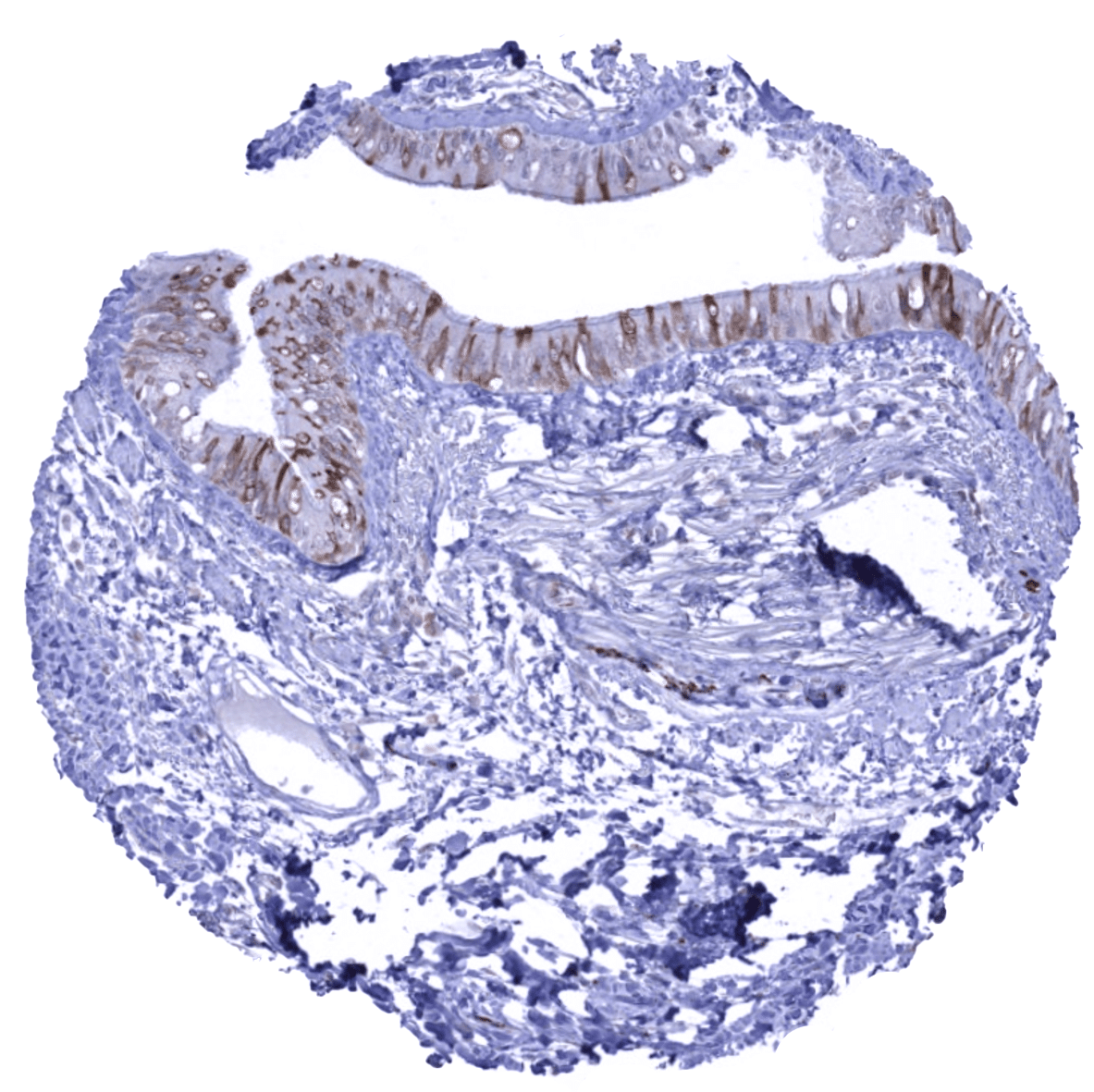
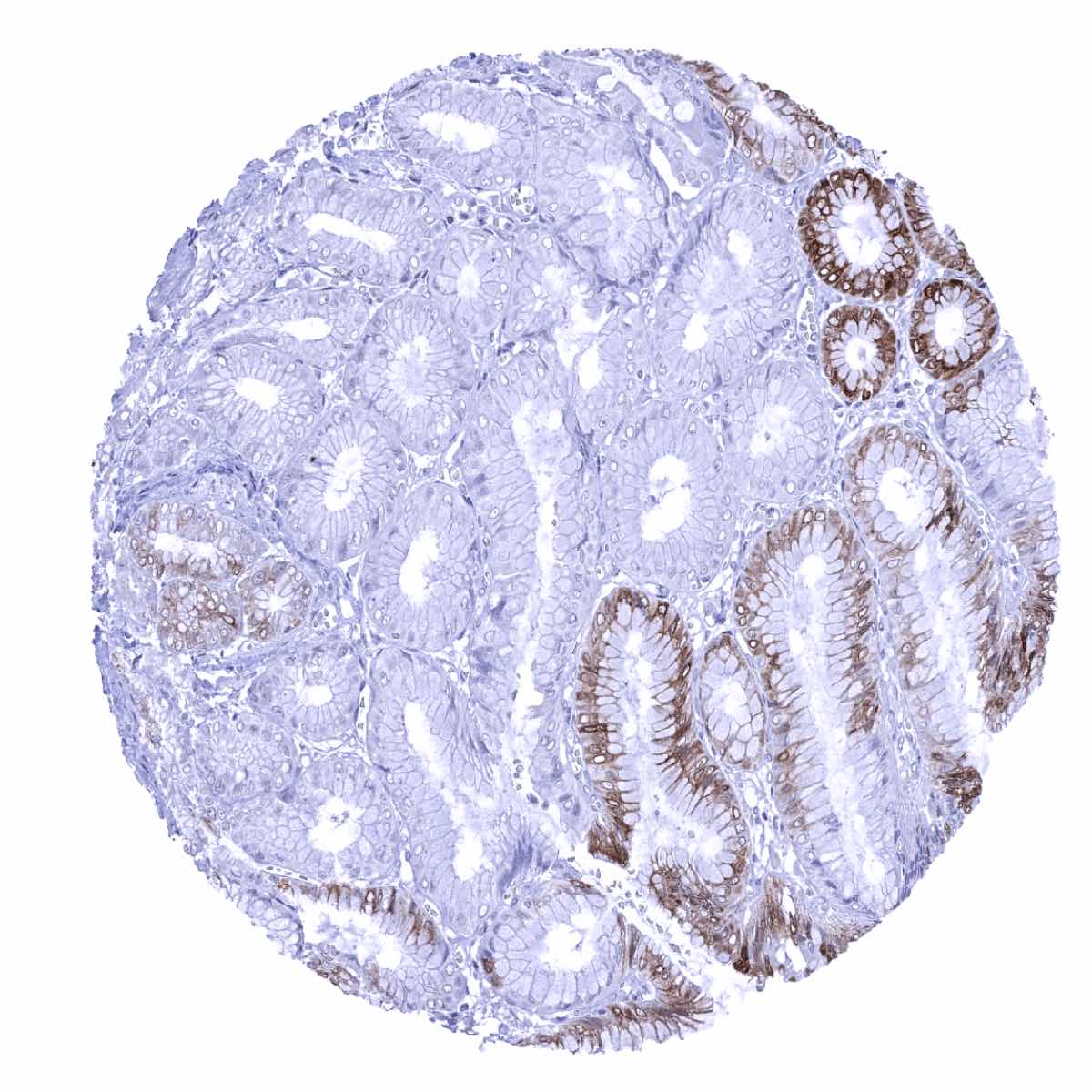
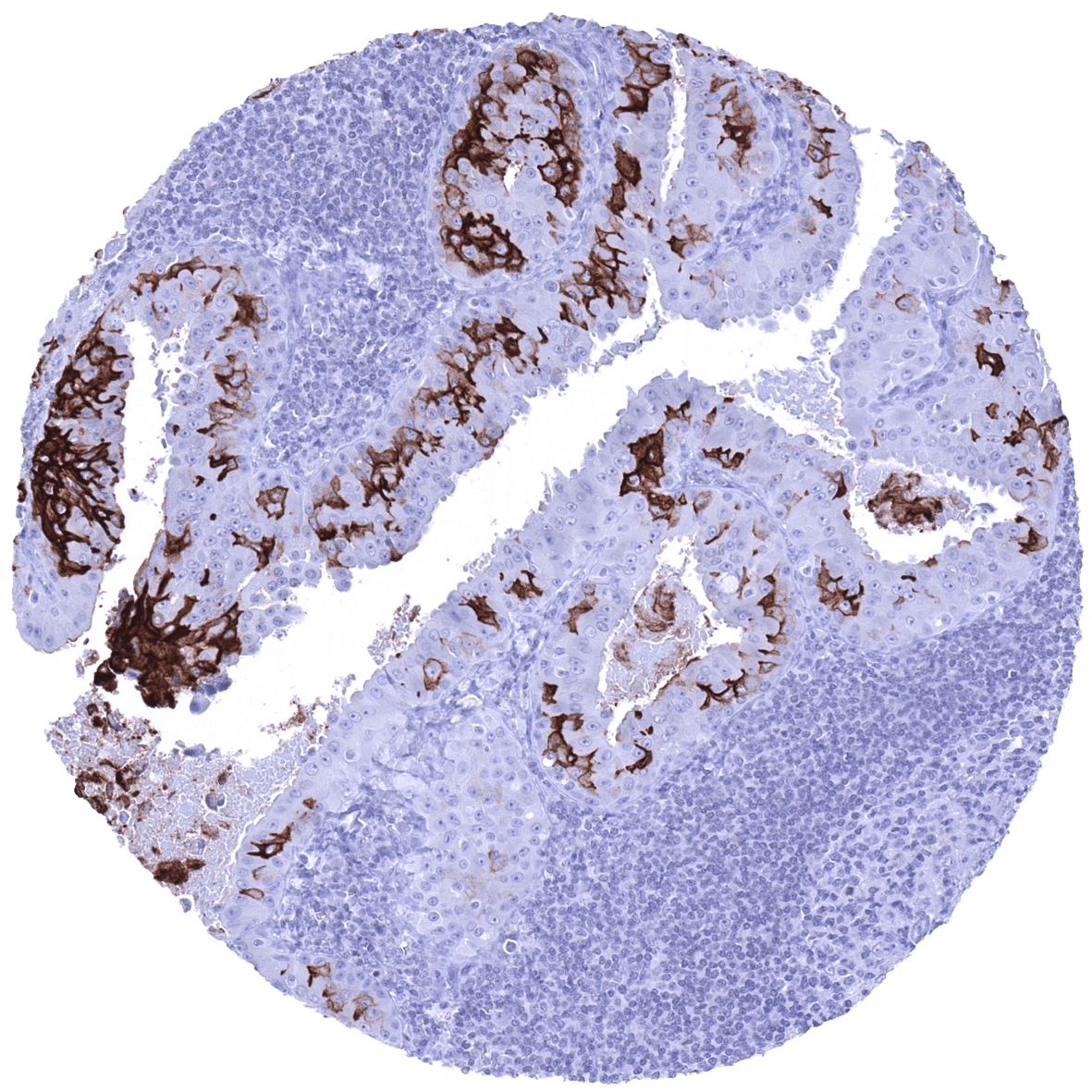
| Stomach | A moderate to strong Upk1b staining is seen in superficial epithelial cells and parietal cells. | |
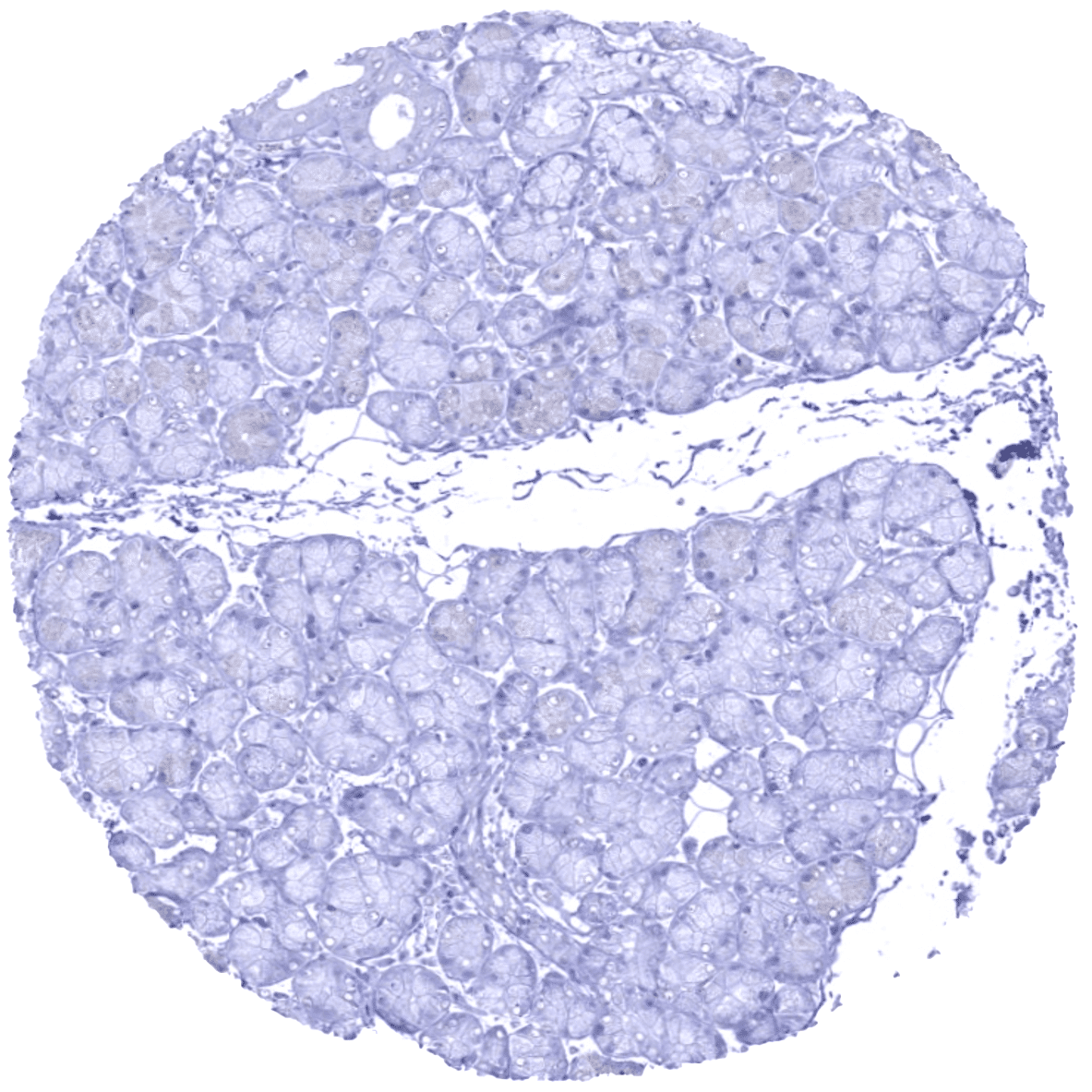
| Colon | Negative. | |
| Duodenum | Negative. | |
| Rectum | Negative. | |
| Small intestine | Negative. | |
| Liver | A variable (weak to strong) cytoplasmic and membranous Upk1b positivity can sometimes occur in intrahepatic bile ducts. Hepatocytes are negative. | |
| Gallbladder | A variable (weak to strong) cytoplasmic and membranous Upk1b positivity can sometimes occur in gallbladder epithelium | |
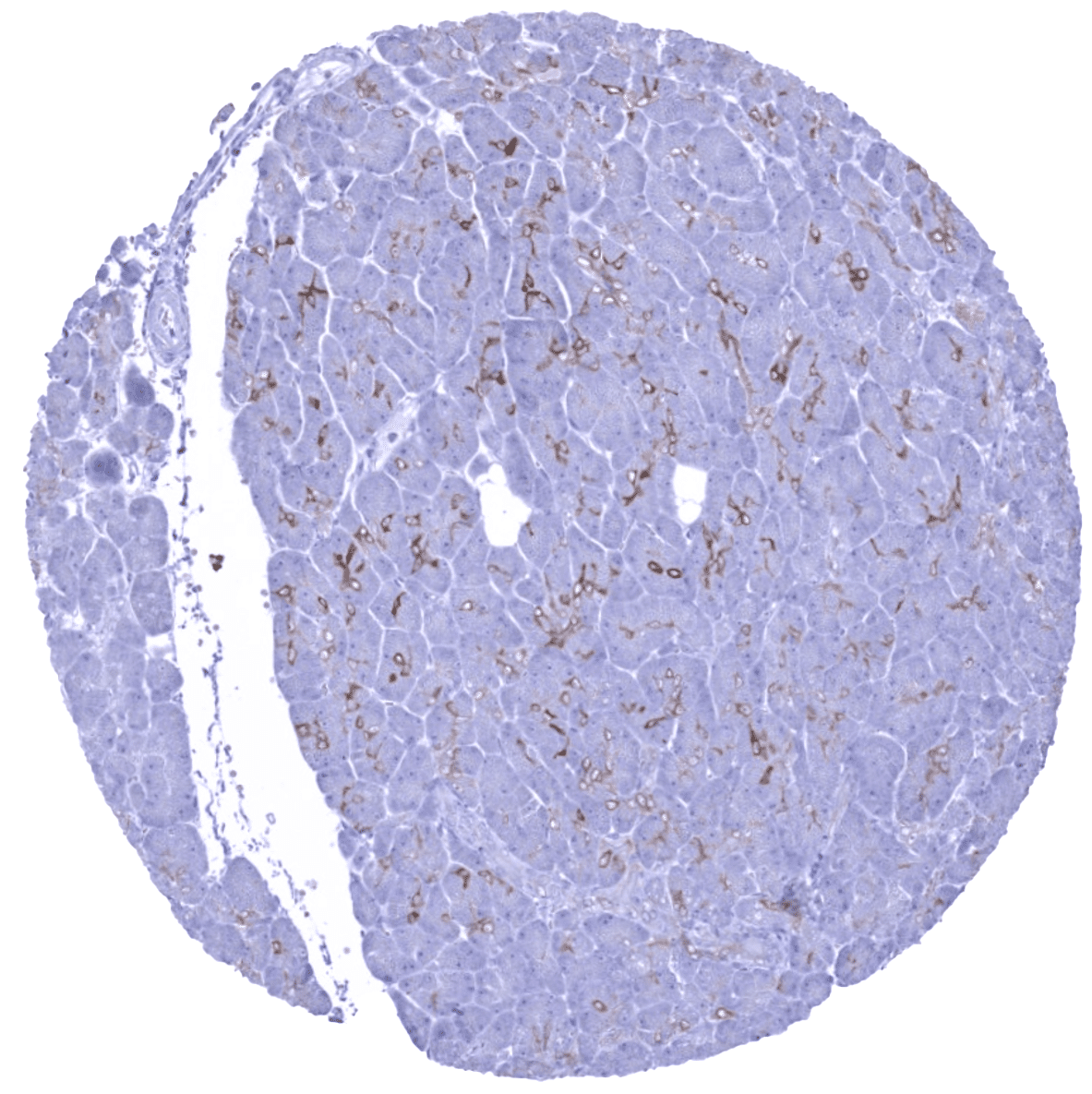
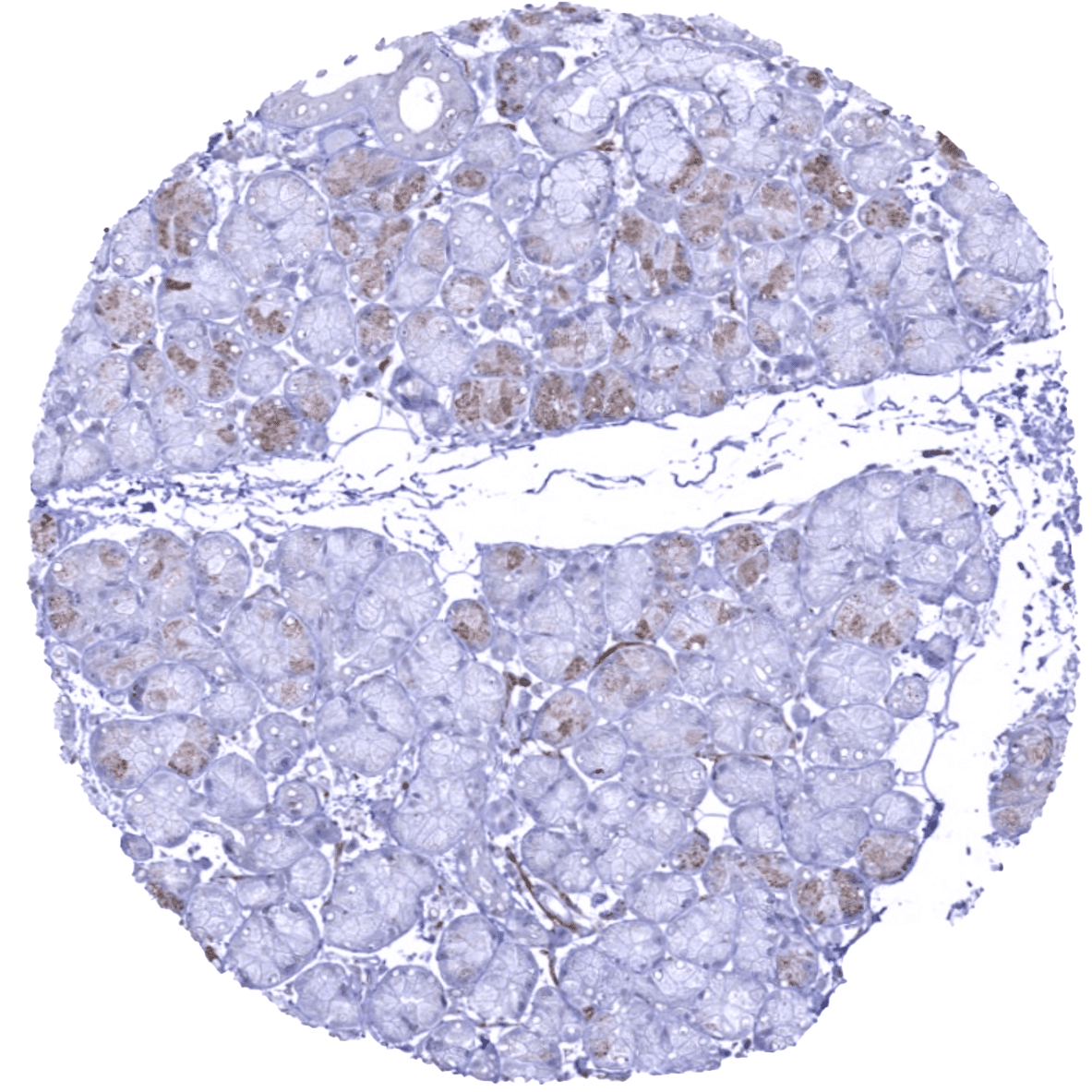
| Pancreas | Weak to moderate staining in intercalated ducts of the pancreas. Excretory ducts of the pancreas can also stain positive. | |
| Genitourinary | Kidney | A variable (weak to strong) cytoplasmic and membranous Upk1b positivity can sometimes occur in the parietal layer of Bowman capsule and in few tubuli in the kidney |
| Urothelium | Variable Upk1b staining. Equally involving all cell layers in the majority of samples but limited to the upper cell layers in some samples. | |
| Male genital | Prostate | Negative. |
| Seminal vesicles | Negative. | |
| Testis | Negative. | |
| Epididymis | Apical membrane of tall columnar cells of the epididymis may show a faint positive staining. | |
| Female genital | Breast | Negative. |
| Uterus, ectocervix | Usually negative. Scattered Upk1b positive cells can occur. | |
| Uterus endocervix | A variable (weak to strong) positivity can sometimes occur in some endocervical glands. | |
| Uterus, endometrium | A variable (weak to strong) Upk1b positivity can sometimes occur in some endometrial glands. | |
| Fallopian Tube | A variable (weak to strong) Upk1b positivity can sometimes occur in a fraction of cells in the fallopian tube. | |
| Ovary | Negative. | |
| Placenta early | A small fraction of the syncytiotrophoblast cells may stain positive. | |
| Placenta mature | A small fraction of the syncytiotrophoblast cells may stain positive. | |
| Amnion | Moderate to strong Upk1b positivity in amnion cells. | |
| Chorion | Moderate to strong Upk1b positivity in chorion cells. | |
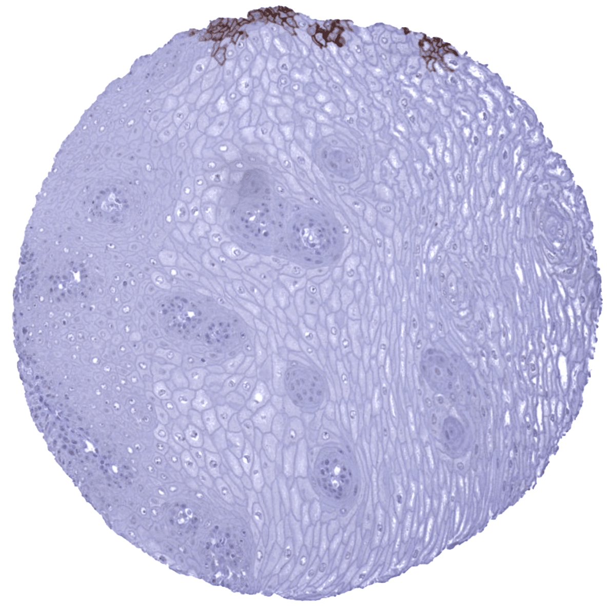
| Skin | Epidermis | Usually negative. Scattered Upk1b positive cells can occur. |
| Sebaceous glands | Negative. | |
| Muscle/connective | Heart | Negative. |
| Skeletal | Negative. | |
| Smooth muscle | Negative. | |
| Fat | Negative. | |
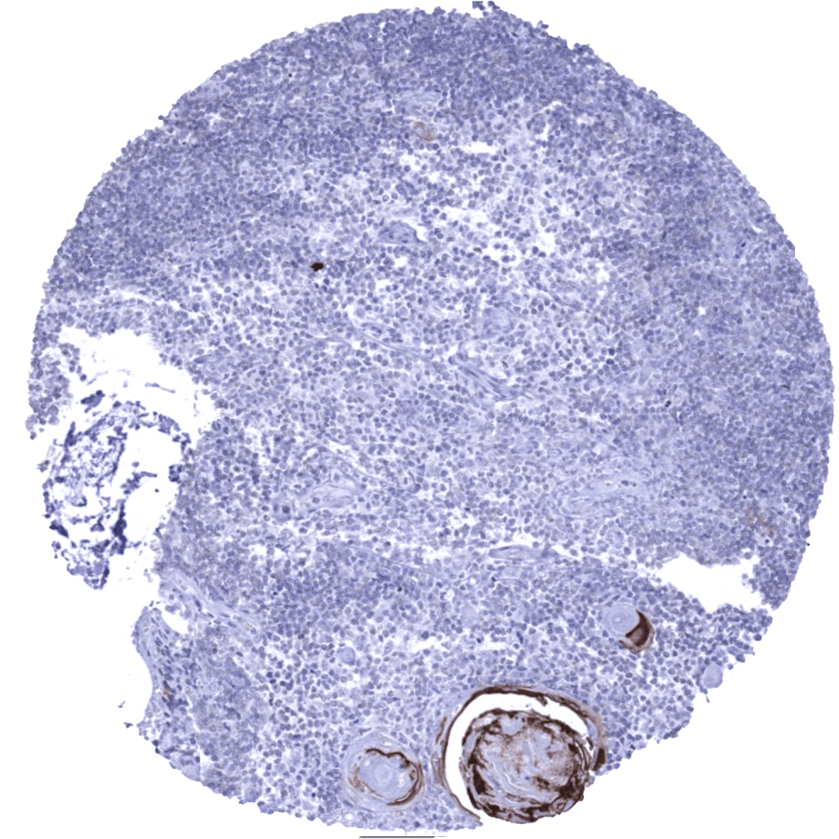
| Bone marrow/lymphoid | Bone marrow | Negative. |
| Lymph node | Negative. | |
| Spleen | Negative. | |
| Thymus | Upk1b is positive in a subset of cells in corpuscles of Hassall’s. No staining of inflammatory cells. | |
| Tonsil | Upk1b is regularly positive in the surface epithelium and in the intermediate cell layer of tonsil crypt epithelium. No staining of inflammatory cells. | |
| Remarks | Staining is typically cytoplasmic and membranous. |
These findings are largely consistent with the RNA and protein data described in the Human Protein Atlas (Tissue expression Uroplakin 1B) All organs with documented Upk1b RNA expression (urinary bladder, kidney, prostate, gallbladder, stomach, placenta, fallopian tube, uterine cervix, tonsil) with the only exception of smooth muscle are IHC positive for MSV-734M. Given the immediate vicinity of smooth muscle to multiple Upk1b positive tissues, smooth muscle RNA positivity may represent a contamination artifact.
Positive control: Urinary bladder: A strong membranous and cytoplasmic UPK1b immunostaining should be seen in the urothelium (the staining can be limited to the top cell layers or equally involve all cell layers).
Negative control: Colon: UPK1b immunostaining should be absent in all cells of the colon mucosa.


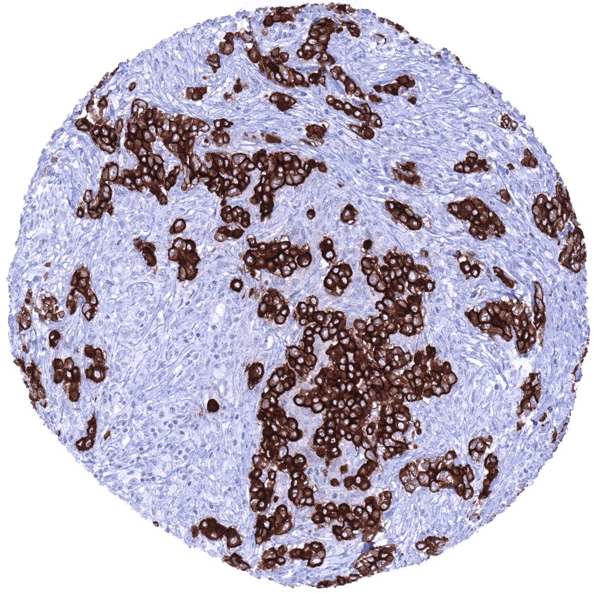
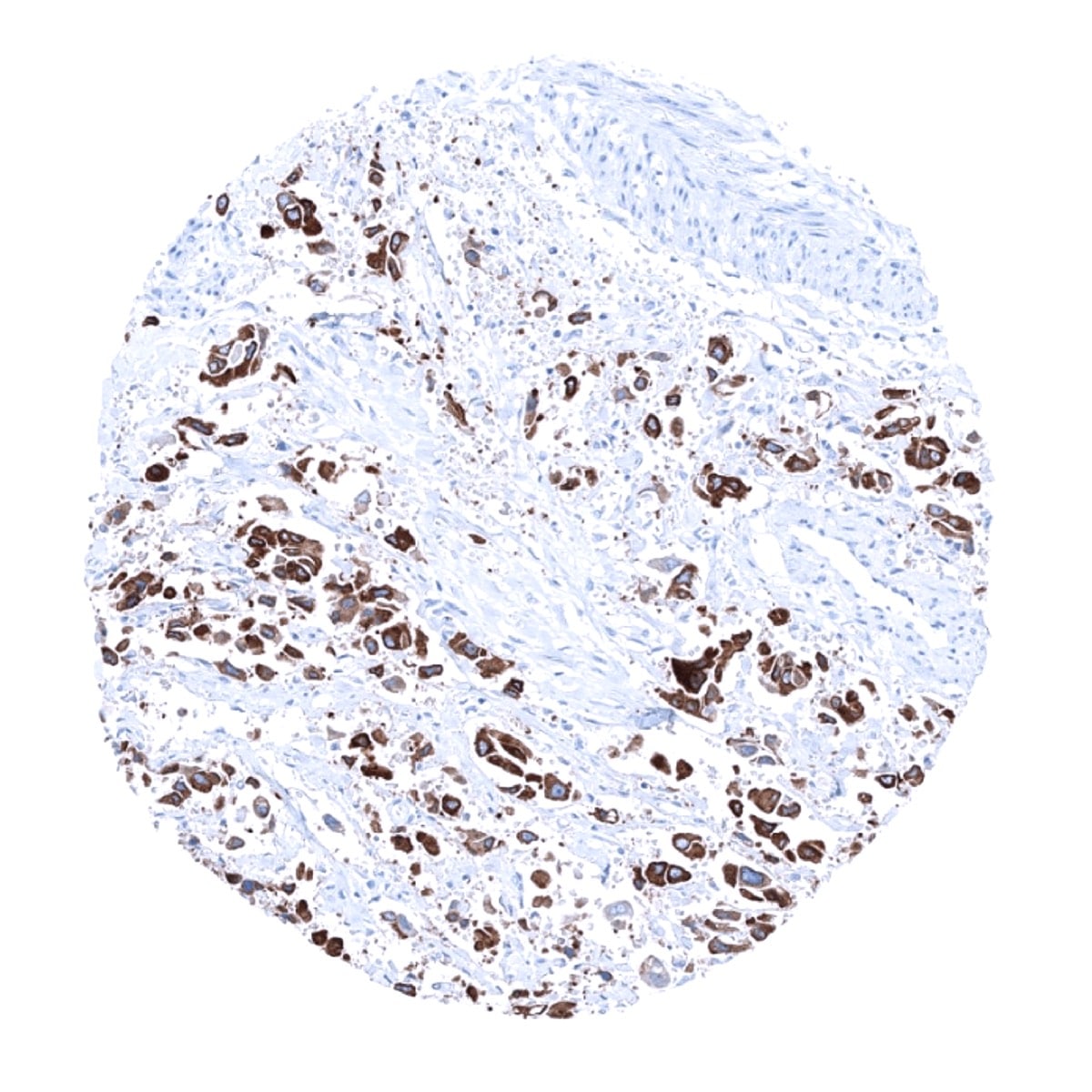
Staining Pattern in Relevant Tumor Types
The TCGA database on RNA expression in cancer has described upregulation of Upk1b in a fraction of urothelial, head and neck, lung, endometrial, cervical, ovarian and renal cancers.
The TCGA findings on Uroplakin 1B RNA expression in different tumor categories have been summarized in the Human Protein Atlas.
Compatibility of Antibodies
Uroplakin 1b (MSVA-734M) publication summary:
Relevant publication: Reiswich et al. “Large-scale human tissue analysis identifies Uroplakin 1b as a putative diagnostic marker in surgical pathology“ Published in Human Pathology 2022 May 9; S0046-8177(22)00116-2. doi: 10.1016/j.humpath.2022.05.002. PMID: 35550834
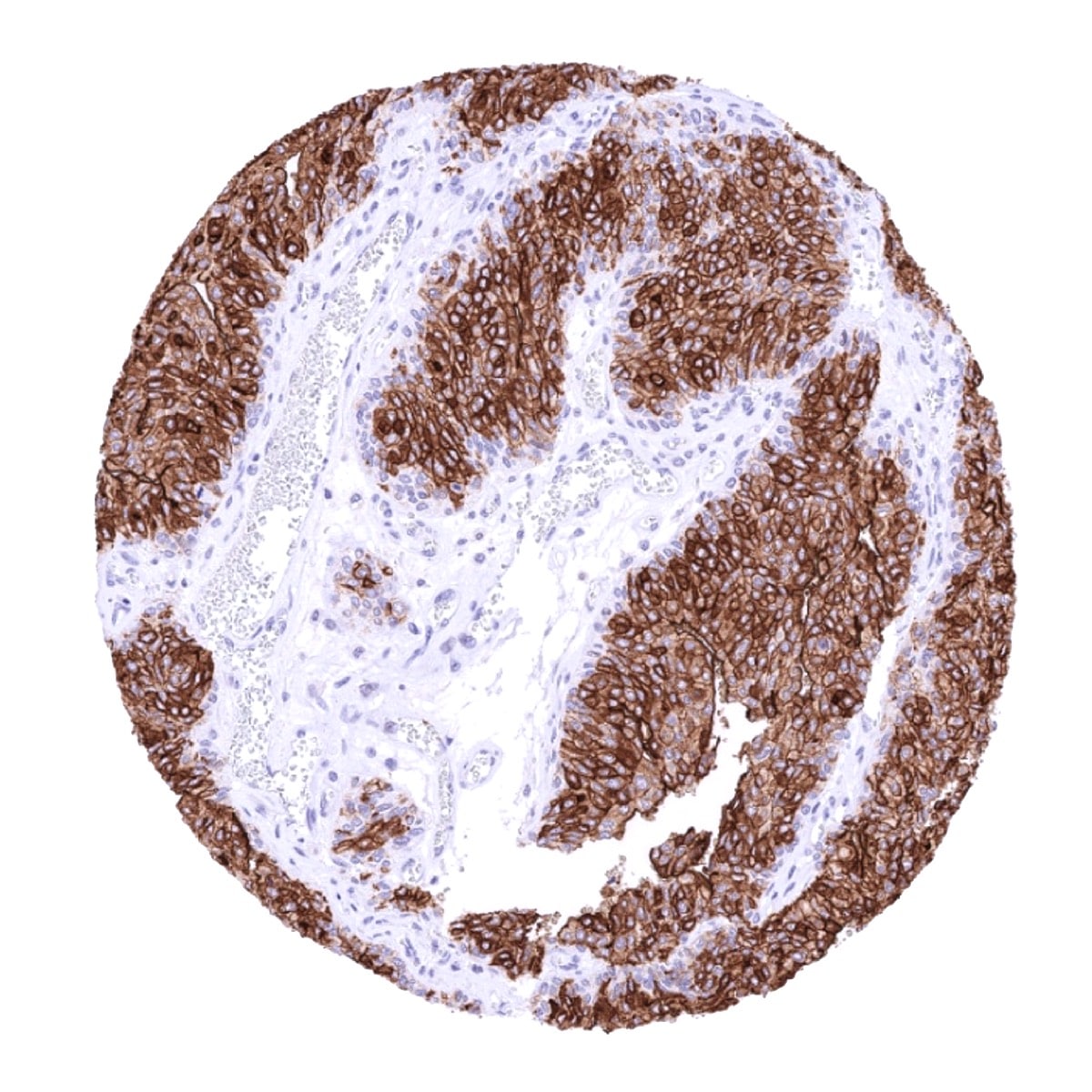
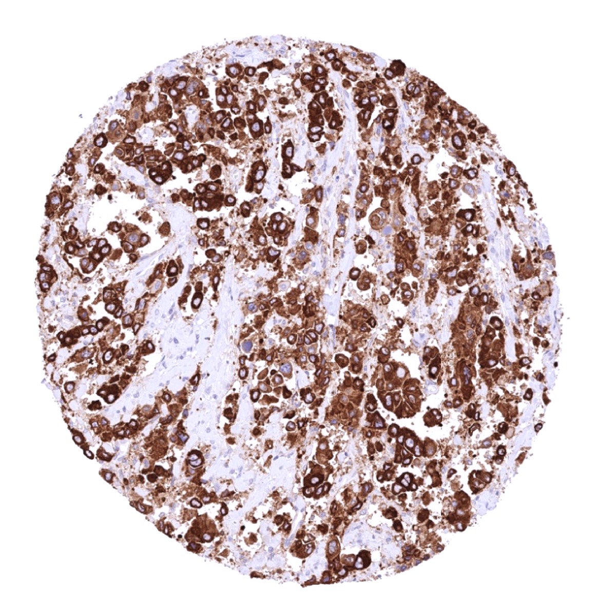
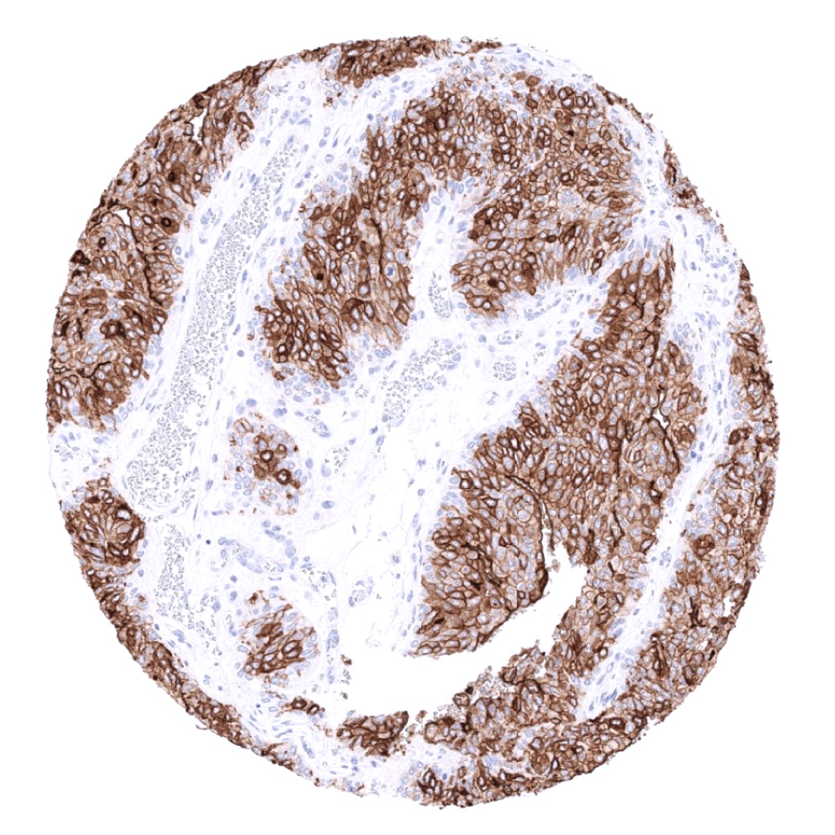
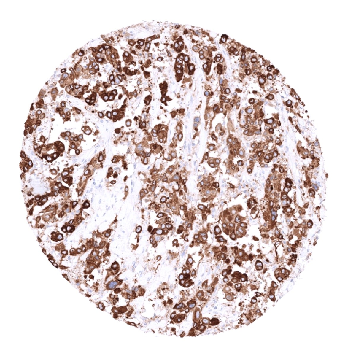
In this study, 11’868 tumors were analyzed from 127 different tumor categories by using the following protocol: Heat-induced antigen retrieval for 5 minutes in an autoclave at 121°C in pH 7.8 Target Retrieval Solution buffer. MSVA-734M at a dilution of 1:150 at 37°C for 60 minutes. Visualization of bound antibody by the EnVision Kit (Dako, Agilent). This protocol was also used for all stainings depicted in our tumor and normal tissue galleries.
At least one case with a positive Upk1b immunostaining was seen in 61 (48%) and at least one case with a strong Upk1b immunostaining was seen in 39 (31%) of 127 tumor categories. The distribution of positive staining results is shown in an “organ-systematic” (Figure 1) and in a “ranking order” (Figure 2) below (images based on a compilation of data from (Reiswich et al.). Results on possible associations with histopathological and clinical parameters of tumor aggressiveness are also summarized below (Table 1, based on data from Reiswich et al.).
Authors conclusions on diagnostic utility of Upk1b immunostaining with respect to the distinction of benign versus malignant (Reiswich et al.)
- Not applicable.
Authors conclusions on diagnostic utility of Upk1b immunostaining with respect to the distinction of different tumor entities (Reiswich et al.)
- Distinction of malignant mesotheliomas (positive in 87%) from adenocarcinomas (positive in 6.8%) of the lung.
- Distinction of muscle-invasive urothelial carcinomas (positive in 58%) of the urinary bladder from poorly differentiated adenocarcinomas of the prostate (positive in 1%).
- Distinction of cholangiocellular (positive in 22%), pancreatic (positive in 21-30%), gastric (positive in 9-14%), esophageal (positive in 17%) from colorectal adenocarcinomas (positive in 0.7%). The authors conclude that a positive Upk1b staining strongly argues against a colorectal cancer origin while Upk1b negativity does not offer significant diagnostic clues.
Authors conclusions on prognostic/predictive role of Upk1b expression (Reiswich et al.)
- The significant association of Upk1b expression loss with grade and stage progression in urothelial neoplasms of the urinary bladder is likely to reflect a progressive loss of normal cell structure proteins which is commonly seen along cancer progression.
- Weak statistical associations of low Upk1b expression with unfavorable tumor features in endometroid endometrial cancer (probably lacking clinical significance).
Data from the publication: “Large-scale human tissue analysis identifies Uroplakin 1b as a putative diagnostic marker in surgical pathology” Published by Reiswich et al. in Human Pathology 2022 May 9; S0046-8177(22)00116-2. doi: 10.1016/j.humpath.2022.05.002. PMID: 35550834
Summarized in own graphics:
1.Uroplakin 1B staining in tumors “organ specific” with antibody MSVA-734M

2. Uroplakin 1B staining in tumors “ranking order” with antibody MSVA-734M

Protocol Recommendations
IHC users have different preferences on how the stains should look like. Some prefer high staining intensity of the target stain and even accept some background. Others favor absolute specificity and lighter target stains. Factors that invariably lead to more intense staining include higher concentration of the antibody and visualization tools, longer incubation time, higher temperature during incubation, higher temperature and longer duration of the heat induced epitope retrieval (slide pretreatment). The impact of the pH during slide pretreatment has variable effects and depends on the antibody and the target protein.
All images and data shown here and in our image galleries are obtained by the manual protocol described below. Other protocols resulting in equivalent staining are described as well.
Manual protocol
Freshly cut sections should be used (less than 10 days between cutting and staining). Heat-induced antigen retrieval for 5 minutes in an autoclave at 121°C in pH 7,8 Target Retrieval Solution buffer. Apply MSVA-734M at a dilution of 1:150 at 37°C for 60 minutes. Visualization of bound antibody by the EnVision Kit (Dako, Agilent) according to the manufacturer’s directions.


Agilent / Dako – Autostainer Link 48
Pretreatment in PT-Link for 30 minutes at 95°C (pH high); FLEX peroxidase blocking for 5 minutes (room temperature), MSVA-734M 1:250 for 20 minutes (room temperature), FLEX+ mouse/rabbit (LINKER) for 15 minutes (room temperature), horseradish peroxidase (HRP) for 20 minutes (room temperature), FLEX DAB+Sub-Chromo for 10 minutes (room temperature), FLEX hematoxylin for 5 minutes (room temperature).

These images reflect stainings by the protocol described above. It is of note that a comparable staining result can also be obtained by different protocols. In general, a longer pretreatment, a longer incubation time of the primary antibody, a higher antibody concentration, and a longer incubation time of FLEX+LINKER result in stronger staining, potentially at the cost of more background staining. Modifications of the protocol with a strengthening effect on staining intensity in combination with changes of other parameters that result in lower staining intensity can result in a comparable result as shown above.
Leica – BOND RX
Dewax at 72°C for 30 seconds; Pretreatment in Bond Epitope Retrieval Solution (ER2 – EDTA pH9) for 20 minutes at 100°C; Peroxidase blocking for 5 minutes (room temperature), MSVA-734M 1:600 for 15 minutes (room temperature), Post primary (rabbit anti mouse) for 8 minutes (room temperature), Polymer (goat anti rabbit) for 8 minutes (room temperature), mixed DAB refine for 10 minutes (room temperature), hematoxylin for 5 minutes (room temperature).
These images reflect stainings by the protocol described above. It is of note that a comparable staining result can also be obtained by different protocols. In general, a longer pretreatment, a longer incubation time of the primary antibody, a higher antibody concentration, a higher temperature during incubation, and a longer incubation time of Post primary and or the Polymer result in stronger staining, potentially at the cost of more background staining. Modifications of the protocol with a strengthening effect on staining intensity in combination with changes of other parameters that result in lower staining intensity can result in a comparable result as shown above.
Roche – Ventana Discovery ULTRA
Pretreatment for 64 minutes at 100°C (pH 8,4); CM peroxidase blocking for 12 minutes (room temperature), MSVA-734M 1:150 for 20 minutes at 36°C, secondary antibody (anti-mouse HQ) for 12 minutes at 36°C, anti-HQ HRP for 12 minutes at room temperature, DAB at room temperature, hematoxylin II at room temperature for 8 minutes, bluing reagent at room temperature for 4 minutes.
These images depict staining results obtained by the protocol described above. It is of note, that the Ventana machines generally require higher antibody concentrations than other commonly used autostainers because the antibodies are automatically diluted during the procedure. Various other protocols can result in an identical result as shown above. A longer pretreatment, a longer incubation time of the primary antibody, a higher antibody concentration, a higher temperature during incubation, and a longer incubation time of secondary antibody and or the anti-HQ HRP result in stronger staining, potentially at the cost of more background staining.
Potential Research Applications
- The prevalence and clinical significance of Upk1b expression in cancer is unknown.
Evidence for Antibody Specificity in IHC
There are two ways how the specificity of antibodies can be documented for immunohistochemistry on formalin fixed tissues. These are: 1. comparison with a second independent method for target expression measurement across a large number of different tissue types (orthogonal strategy), and 2. Comparison with one or several independent antibodies for the same target and showing that all positive staining results are also seen with other antibodies for the same target (independent antibody strategy).
Orthogonal validation: Specificity of the antibody MSVA-734M for Upk1b is suggested by the strong concordance of its immunostaining pattern with data from three independent RNA screening studies, including the Human Protein Atlas (HPA) RNA-seq tissue dataset, the FANTOM5 project, and the Genotype-Tissue Expression (GTEx) project, which are all summarized in the Human Protein Atlas (Tissue expression Uroplakin 1B). Immunostaining by MSVA-734M is detected in all organs with documented Upk1b RNA expression (urinary bladder, kidney, prostate, gallbladder, stomach, placenta, fallopian tube, the uterine cervix, tonsil) with the only exception of smooth muscle. Given the immediate vicinity of smooth muscle to multiple Upk1b positive tissues, smooth muscle RNA positivity may represent a contamination artifact.
An immunostaining by MSVA-734M also occurs in organs without documented RNA expression such as the thymus (thymic epithelial cells), endometrium (few glands), anal canal epithelium (surface cell layer), pancreas (intercalated ducts and some larger excretory ducts), respiratory epithelium, squamous epithelia from various sites (few scattered positive cells), epididymis (focal weak staining of the apical membrane of tall columnar cells), and salivary glands (some serous cells). In all these organs, the Upk1b positive cell populations constituted rather small subsets of the total number of cells and may have been sufficiently underrepresented in RNA analyses to remain undetected.
Comparison of antibodies: True Upk1b protein expression of these specialized cell types in organs without documented Upk1b RNA expression is suggested by largely identical results observed by two independent anti-Upk1b antibodies (MSVA-734M; MSVA-UPK1B candidate f3b).
Antibody Comparison: MSVA-734M vs another MSVA Uroplakin 1B antibody candidate called “f3b”