145,00 € – 595,00 €
Product details
Synonyms = avian-type; CAB27; CALB 1; CALB; CALB1; CALB1_HUMAN; Calbindin 1 28kDa; Calbindin; Calbindin D28; D 28K; D-28K; D28K; OTTHUMP00000166027; OTTHUMP00000225441; RTVL H protein; Vitamin D dependent calcium binding protein; Vitamin D dependent calcium binding protein avian type; Vitamin D-dependent calcium-binding protein
Antibody type = Mouse monoclonal / IgG
Clone = MSVA-471M
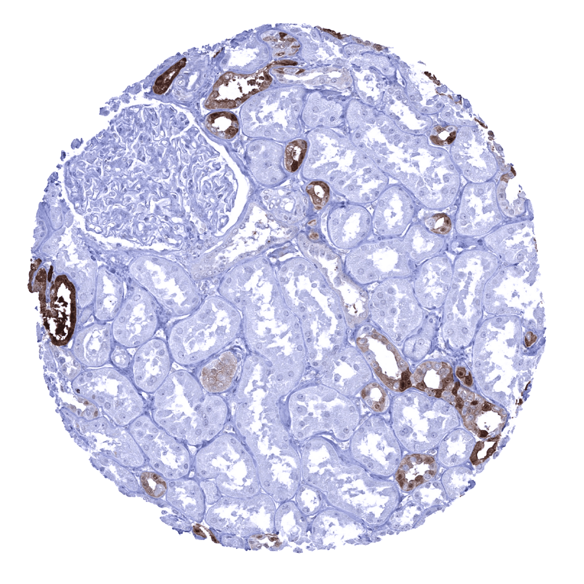
Positive control = Kidney: A subset of the cells of distal tubuli should show an intense cytoplasmic calbindin 1 immunostaining.
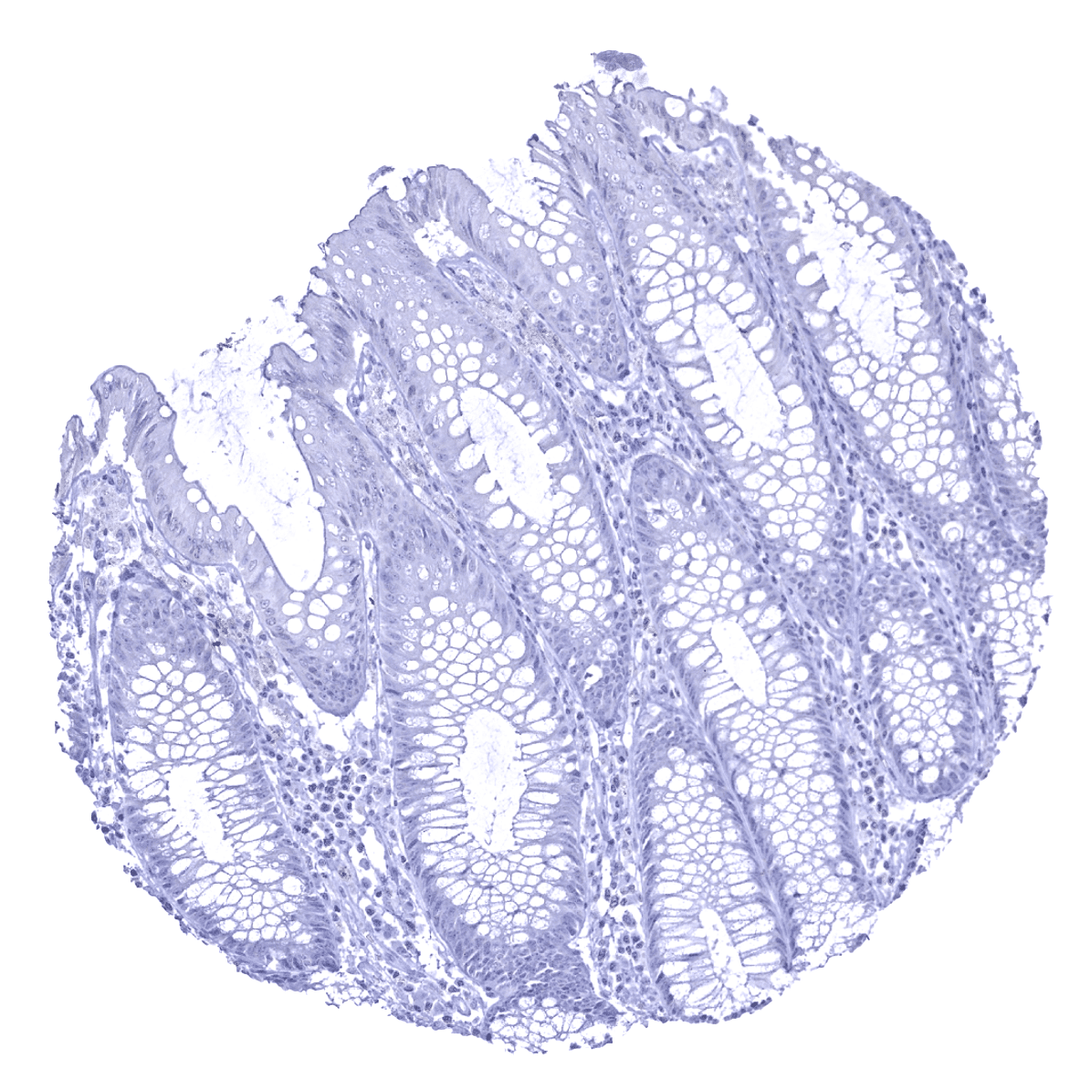
Negative control = Colon: Calbindin 1 immunostaining should be completely absent in all cells.
Cellular localization = Cytoplasmic.
Reactivity = Human
Application = Immunohistochemistry
Dilution = 1:50
Intended Use = Research Use Only
Relevance of Antibody
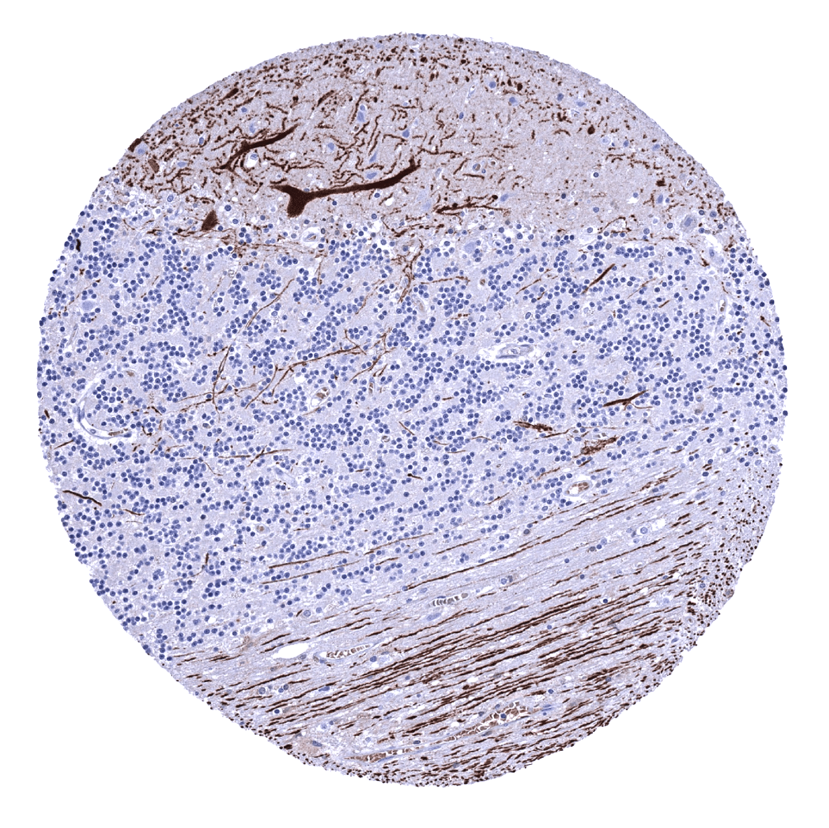
Calbindin 1 is highly expressed in Purkinje cells of the cerebellum.
Biology Behind
Calbindin 1 (CALB1) is coded by the CALB1 gene at 8q21.3. It belongs to the calbindin family of calcium-binding proteins, along with calretinin (CALB2). Calbindin 1 is a major calcium-buffering cytoplasmic protein that is thought to buffer entry of calcium upon stimulation of glutamate receptors. It is expressed at particularly high levels in the central nervous system (CNS) where it makes up for 1.5% of the soluble protein mass and appears to have neuroprotective properties. Calbindin 1 expression in the brain is significantly diminished in various neurological disorders associated with epileptiform activity and seizures. In Alzheimer’s disease (AD) and epilepsy, the severity of cognitive deficits reflects the degree of calbindin 1 reduction. Calbindin 1 interacts with a variety of proteins including caspase-3 and inositol monophosphatase (IMPase), the putative target of lithium therapy in bipolar disorder.
Staining Pattern in Normal Tissues
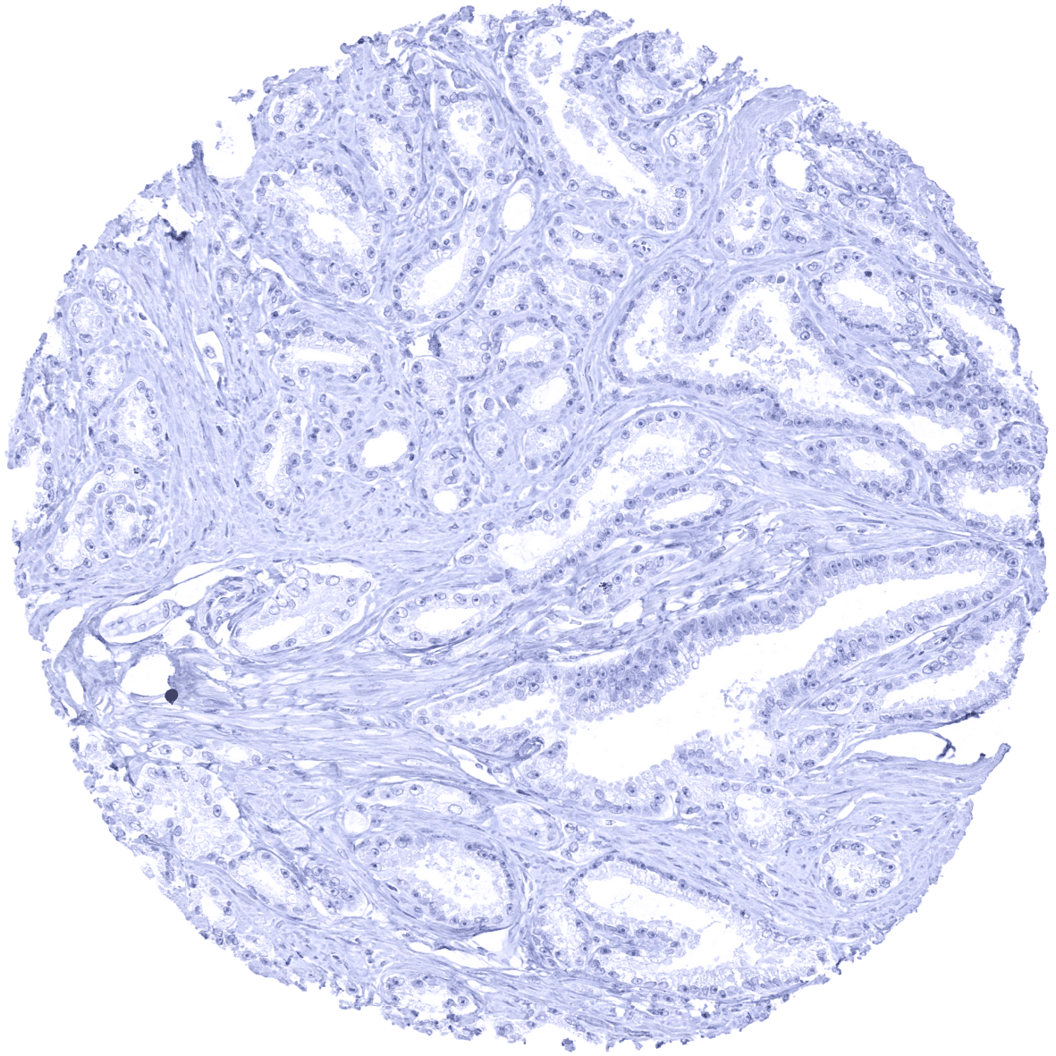
Calbindin 1 is strongly expressed in Purkinje cells and associated axonal fibres of the cerebellum. A weaker expression occurs in neuronal cells of the cerebrum. Calbindin 1 is also seen in the cytoplasm of a fraction of distal tubuli of the kidney. Calbindin 1 immunostaining is not seen in the gastrointestinal tract, pancreas, liver, gallbladder, salivary glands, Brunner glands, prostate, seminal vesicles, testis, thyroid gland, parathyroid gland, adrenal gland, respiratory epithelium, bronchial glands, lung, breast gland, endometrium, endocervix, fallopian tube, ovary (including follicular cysts and corpora lutea), placenta, keratinizing and nonkeratinizing squamous epithelia, sebaceous glands, urothelium, mesenchymal tissues, thymus, spleen, lymph nodes, tonsil, bone marrow, and the pituitary gland.
These findings are largely consistent with the RNA and protein data described in the Human Protein Atlas (Tissue expression Calbindin 1)
Positive control: Kidney: A subset of the cells of distal tubuli should show an intense cytoplasmic calbindin 1 immunostaining.
Negative control: Colon: Calbindin 1 immunostaining should be completely absent in all cells.
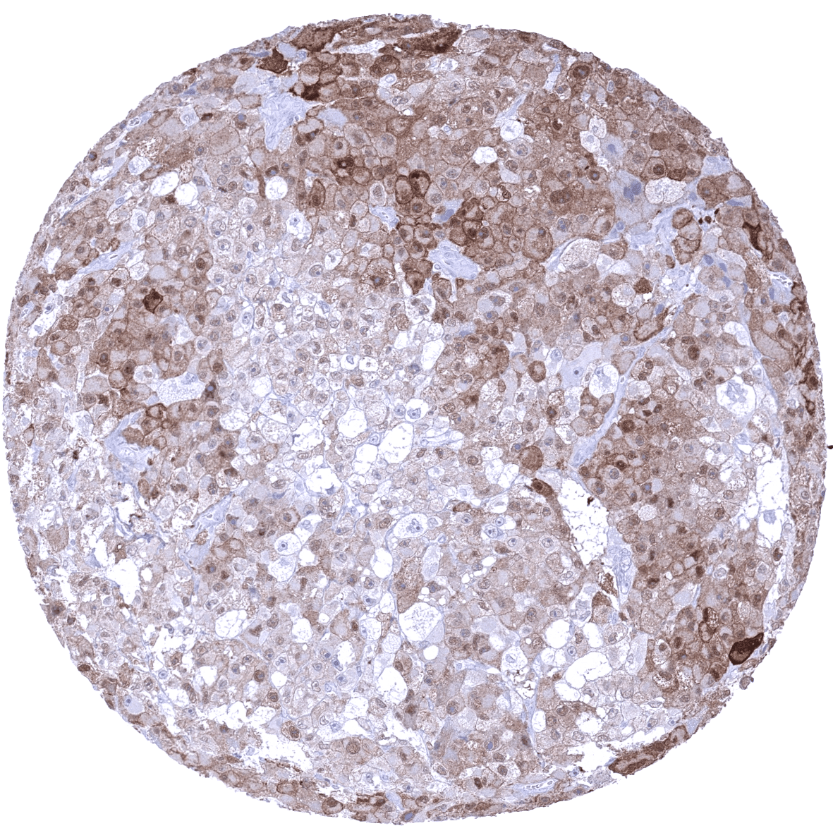
Staining Pattern in Relevant Tumor Types
Calbindin 1 immunostaining is rare in tumors. The TCGA database on RNA expression in cancer has described upregulation of Calbindin 1 in a fraction of lung, colorectal, head and neck, urothelial, cervical and endometrial cancers.
The TCGA findings on Calbindin 1 RNA expression in different tumor categories have been summarized in the Human Protein Atlas.
Compatibility of Antibodies
No data available at the moment
Protocol Recommendations
IHC users have different preferences on how the stains should look like. Some prefer high staining intensity of the target stain and even accept some background. Others favor absolute specificity and lighter target stains. Factors that invariably lead to more intense staining include higher concentration of the antibody and visualization tools, longer incubation time, higher temperature during incubation, higher temperature and longer duration of the heat induced epitope retrieval (slide pretreatment). The impact of the pH during slide pretreatment has variable effects and depends on the antibody and the target protein.
All images and data shown here and in our image galleries are obtained by the manual protocol described below. Other protocols resulting in equivalent staining are described as well.
-Manual protocol
Freshly cut sections should be used (less than 10 days between cutting and staining). Heat-induced antigen retrieval for 5 minutes in an autoclave at 121°C in pH 7,8 Target Retrieval Solution buffer. Apply MSVA-471M at a dilution of 1:50 at 37°C for 60 minutes. Visualization of bound antibody by the EnVision Kit (Dako, Agilent) according to the manufacturer’s directions.
Potential Research Applications
- The suitability of calbindin 1 as a marker for visualization of Purkinje cells and associated axons should be evaluated.
- The clinical significance of calbindin 1 protein expression levels in the brain and in specific areas of the brain should be further investigated.
- The role of calbindin 1 in renal cell cancer and in other cancer entities is unclear.
Evidence for Antibody Specificity in IHC
There are two ways how the specificity of antibodies can be documented for immunohistochemistry on formalin fixed tissues. These are: 1. comparison with a second independent method for target expression measurement across a large number of different tissue types (orthogonal strategy), and 2. Comparison with one or several independent antibodies for the same target and showing that all positive staining results are also seen with other antibodies for the same target (independent antibody strategy).
Orthogonal validation: For the antibody MSVA-471M specificity is suggested by the complete concordance of the immunostaining data with data from three independent RNA screening studies, including the Human Protein Atlas (HPA) RNA-seq tissue dataset, the FANTOM5 project, and the Genotype-Tissue Expression (GTEx) project, which are all summarized in the Human Protein Atlas (Tissue expression Calbindin 1).
Calbindin 1 immunostaining by using MSVA-471M was exclusively detected in organs with documented CALB1 RNA expression (brain and kidney).
Comparison of antibodies: True expression of calbindin 1 in specific cell types of the brain and the kidney is confirmed by comparable findings obtained by the antibody CAB002138. The respective images are depicted in the protein atlas for the kidney and the cerebellum
