
195,00 € – 695,00 €
Product details
Synonyms = STF1; STFA; AREI; cstA; Stefin A
Antibody type = Mouse monoclonal / IgG
Clone = MSVA-461M
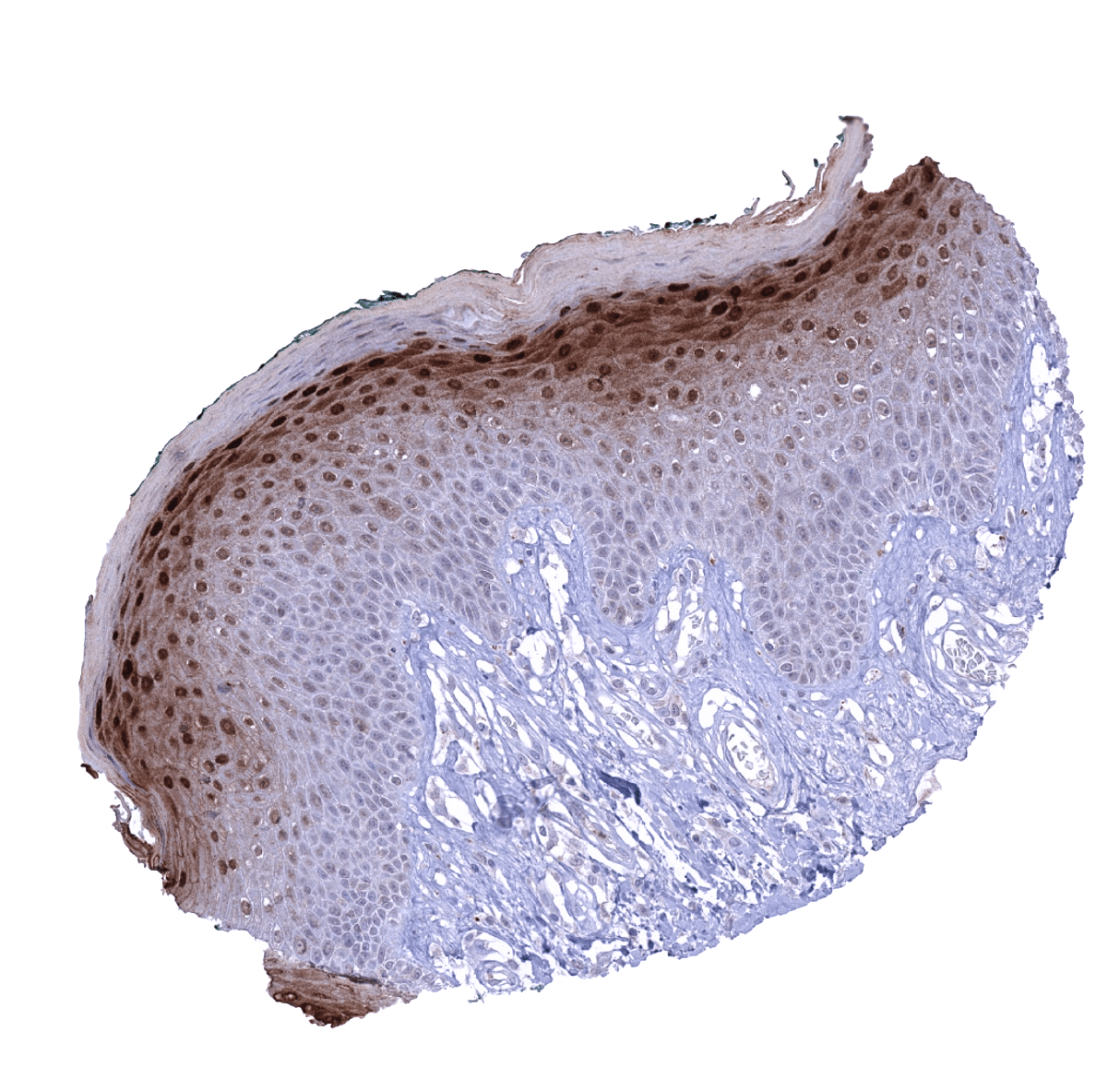
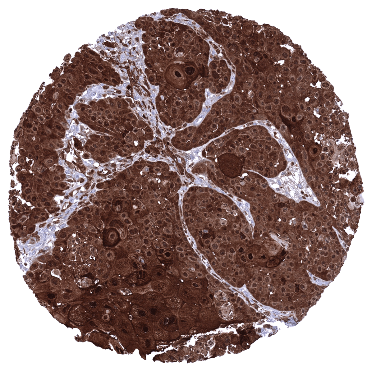
Positive control = Skin: A strong nuclear and cytoplasmic Cystatin A immunostaining should be seen in the granular layer.
Negative control = Colon: No epithelial cell staining should be seen. Few inflammatory cells may show Cystatin A positivity.
Cellular localization = Cytoplasmic and nuclear.
Reactivity = Human
Application = Immunohistochemistry
Dilution = 1:100 – 200
Intended Use = Research Use Only
Relevance of Antibody
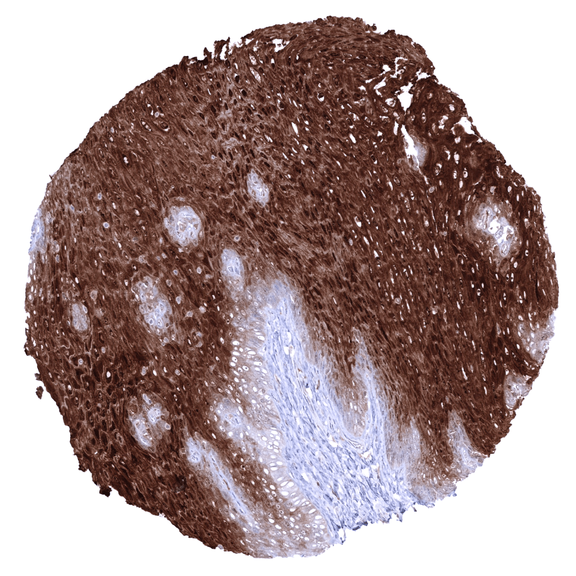
Cystatin A is expressed in squamous epithelial cells.
Biology Behind
Cystatin-A is an 11 kDa protein that is coded by the CSTA gene located on chromosome 3q21.1. Cystatin A is a type 1 cystatin (stefin) that functions as a cysteine protease inhibitor. The protein is a precursor protein of the cornified cell envelope in keratinocytes and plays a role in the development and maintenance of squamous epithelia. Cystatin A forms tight complexes with papain and the cathepsins B, H, and L. Cystatin A has been suggested to play a role in barrier function and in targeting dust mite proteases.
Staining Pattern in Normal Tissues
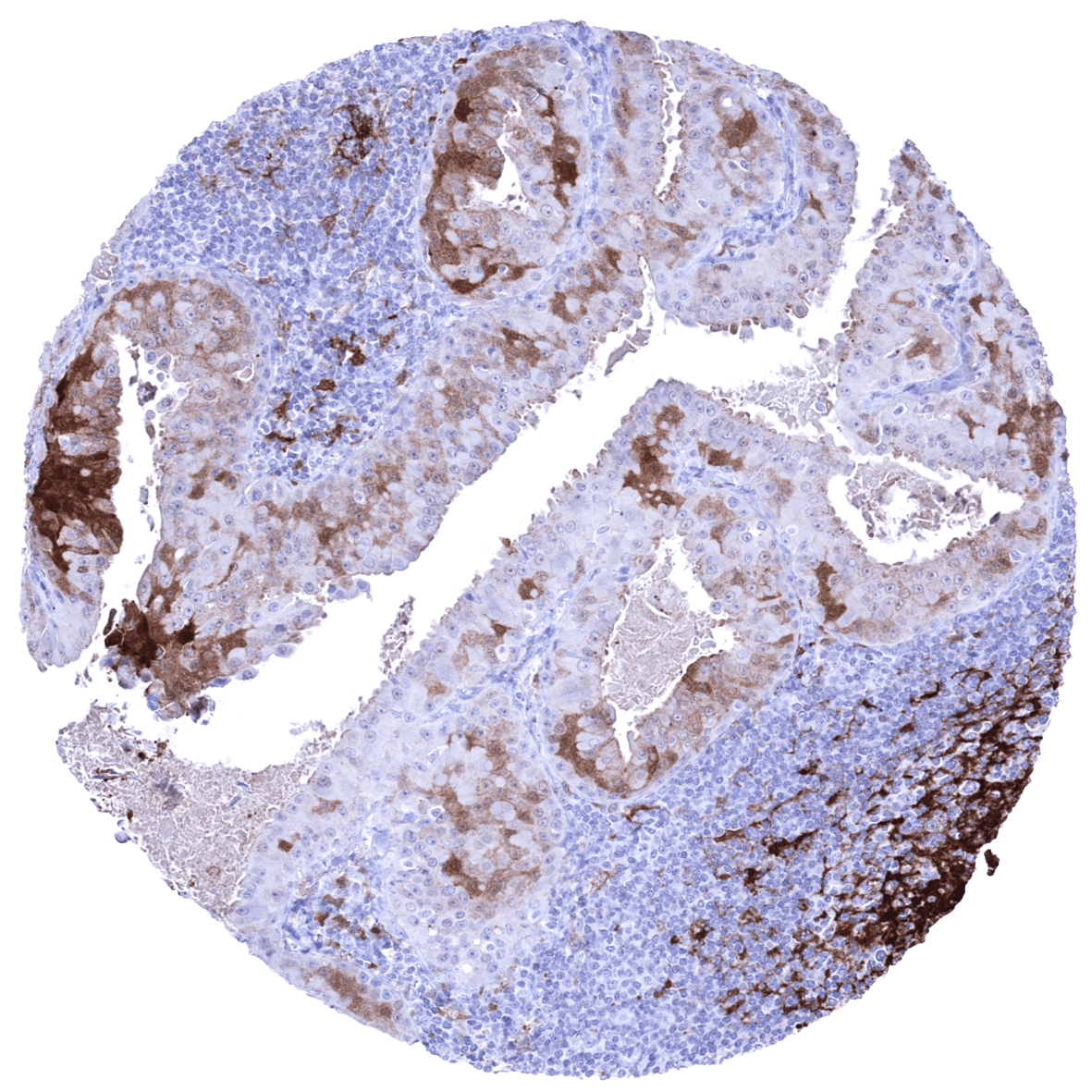
Cystatin A immunostaining predominantly occurs in squamous epithelia where a cytoplasmic and also nuclear staining pattern can be observed. The extent of staining is variable in these tissues and ranges from a staining of the entire epithelium to a staining of only the most superficial layers which especially occurs in the skin (granular layer). If the basal cell layer is stained, its staining intensity is always the least. Squamous cell staining also includes corpuscles of Hassall’s and a fraction of cells in tonsil crypt epithelium. A less intense cystatin A immunostaining is also seen in surface epithelial cells of the stomach (weak), basal cells of the prostate (moderate intensity), seminal vesicle, and the epididymis, myoepithelial cells and mucinous glands in sublingual glands, mucinous cells in bronchial glands (weak), and few cells of the respiratory epithelium. In the liver a faint cystatin A staining that exhibits a zonal distribution is seen in some samples. Cystatin A immunostaining also occurs in dendritic cells of germinal centres, macrophages, granulocytes, and a fraction of bone marrow cells.
These findings are largely consistent with the RNA and protein data described in the Human Protein Atlas (Tissue expression Cystatin A)
Positive control: Skin: A strong nuclear and cytoplasmic Cystatin A immunostaining should be seen in the granular layer.
Negative control: Colon: No epithelial cell staining should be seen. Few inflammatory cells may show Cystatin A positivity.


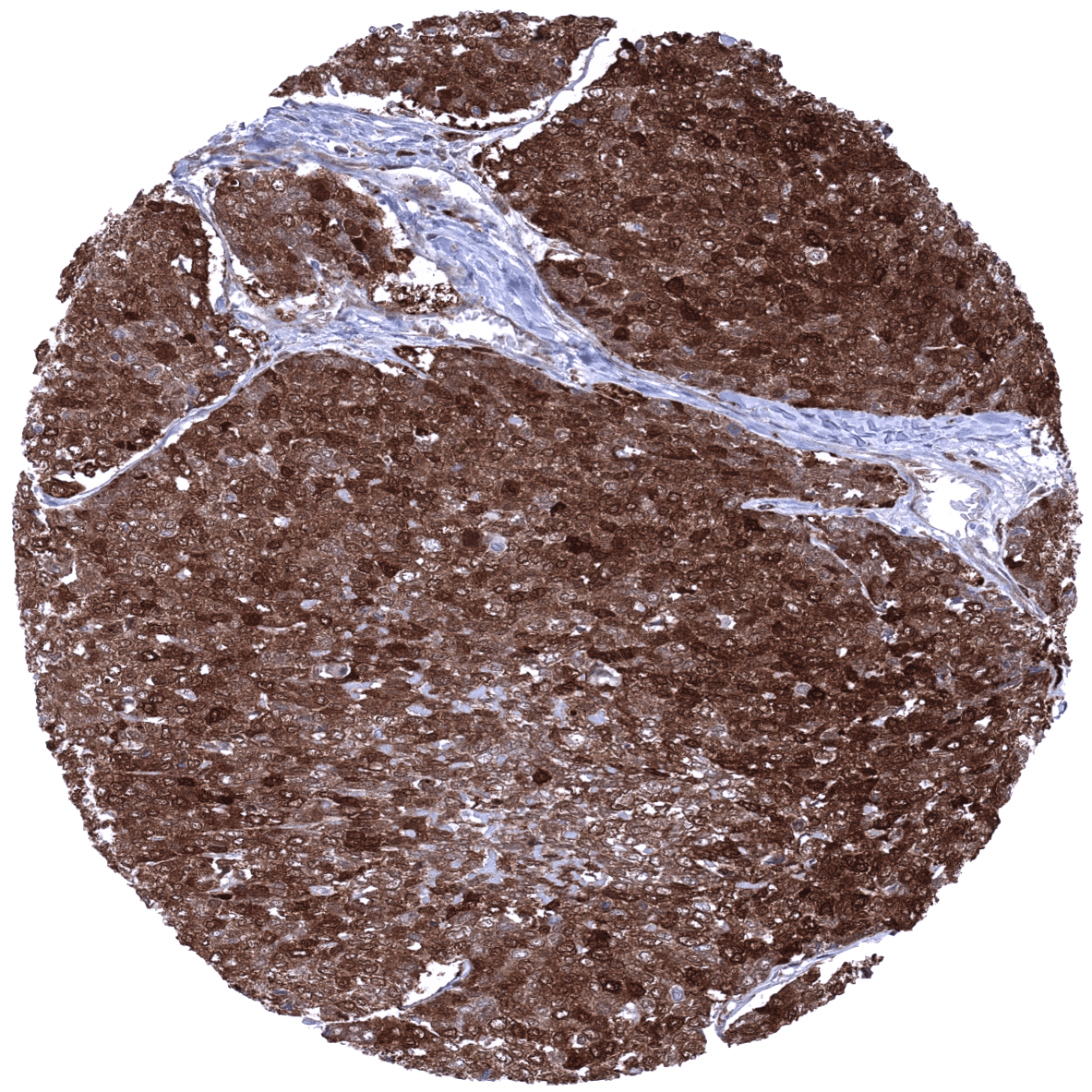
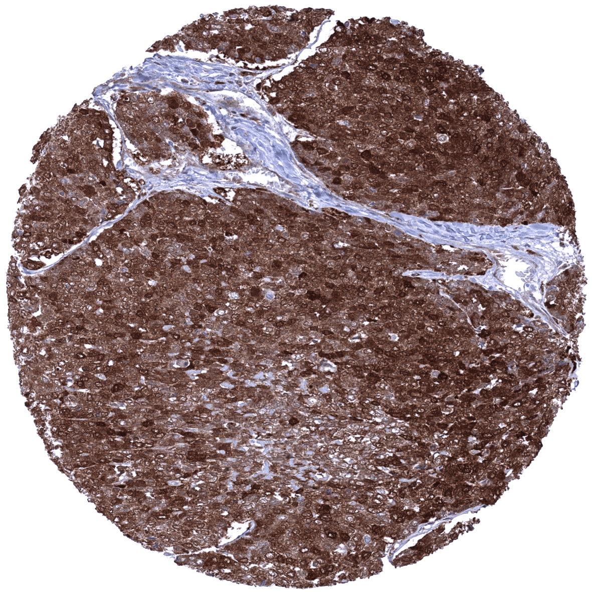
Staining Pattern in Relevant Tumor Types
A positive Cystatin A immunostaining is usually seen in squamous cell carcinomas. Cystatin A expression can – at lower frequency – also occur in various other tumor entities.
The TCGA findings on Cystatin A RNA expression in different tumor categories have been summarized in the Human Protein Atlas.
Compatibility of Antibodies
No data available at the moment
Protocol Recommendations
IHC users have different preferences on how the stains should look like. Some prefer high staining intensity of the target stain and even accept some background. Others favor absolute specificity and lighter target stains. Factors that invariably lead to more intense staining include higher concentration of the antibody and visualization tools, longer incubation time, higher temperature during incubation, higher temperature and longer duration of the heat induced epitope retrieval (slide pretreatment). The impact of the pH during slide pretreatment has variable effects and depends on the antibody and the target protein.
All images and data shown here and in our image galleries are obtained by the manual protocol described below. Other protocols resulting in equivalent staining are described as well.
-Manual protocol
Freshly cut sections should be used (less than 10 days between cutting and staining). Heat-induced antigen retrieval for 5 minutes in an autoclave at 121°C in pH 7,8 Target Retrieval Solution buffer. Apply MSVA-461M at a dilution of 1:150 at 37°C for 60 minutes. Visualization of bound antibody by the EnVision Kit (Dako, Agilent) according to the manufacturer’s directions.
Potential Research Applications
- The biological and clinical significance of Cystatin A expression in tumors is unknown.
- The role of Cystatin A in the tumor stroma and its cells is unclear.
- The role of Cystatin A in dendritic cells and in other inflammatory cells in unclear.
Evidence for Antibody Specificity in IHC
There are two ways how the specificity of antibodies can be documented for immunohistochemistry on formalin fixed tissues. These are: 1. comparison with a second independent method for target expression measurement across a large number of different tissue types (orthogonal strategy), and 2. Comparison with one or several independent antibodies for the same target and showing that all positive staining results are also seen with other antibodies for the same target (independent antibody strategy).
Orthogonal validation: For the antibody MSVA-461M, specificity is suggested by the strong concordance of the immunostaining data with data from three independent RNA screening studies, including the Human Protein Atlas (HPA) RNA-seq tissue dataset, the FANTOM5 project, and the Genotype-Tissue Expression (GTEx) project, which are all summarized in the Human Protein Atlas (Tissue expression Cystatin A). Immunostaining by using MSVA-461M was detected in all organs with documented RNA expression such as organs covered by squamous epithelium (tongue, esophagus, uterine cervix, vagina, skin and tonsil), salivary glands, macrophages, dendritic cells, granulocytes, and bone marrow. In addition, CSTA immunostaining was seen in surface epithelial cells of the stomach, basal cells of the prostate, seminal vesicle, and the epididymis, mucinous cells in bronchial glands (weak), and few cells of the respiratory epithelium, and the liver. These cell types may represent too small subsets of their respective organs to be detected in RNA analyses.
Comparison of antibodies: True expression of CSTA in surface epithelial cells of the stomach, basal cells of the prostate, seminal vesicle, and the epididymis, mucinous cells in bronchial glands (weak), cells of the respiratory epithelium, and the liver is suggested by similar findings obtained by the antibody HPA001031. The respective images are depicted in the protein atlas for:
- prostate
- epididymis
- seminal vesicle
- inflammatory cells in the colon
- bronchial glands and the respiratory epithelium
- liver
The only CSTA finding obtained by MSVA-461M and not confirmed by HPA001031 involves corpuscles of Hassall’s which were not analyzed by HPA001031 in the human protein atlas.
