
295,00 € – 1.095,00 €
Product details
Synonyms = ATP hydrolyzing DNA topoisomerase II alfa; DNA gyrase; DNA topoisomerase (ATP hydrolyzing); DNA topoisomerase 2 alpha; DNA topoisomerase II 170kD; DNA topoisomerase II alpha; Topoisomerase DNA II alpha 170kDa; TP2A
Antibody type = Recombinant Rabbit monoclonal / IgG
Clone = MSVA-802R
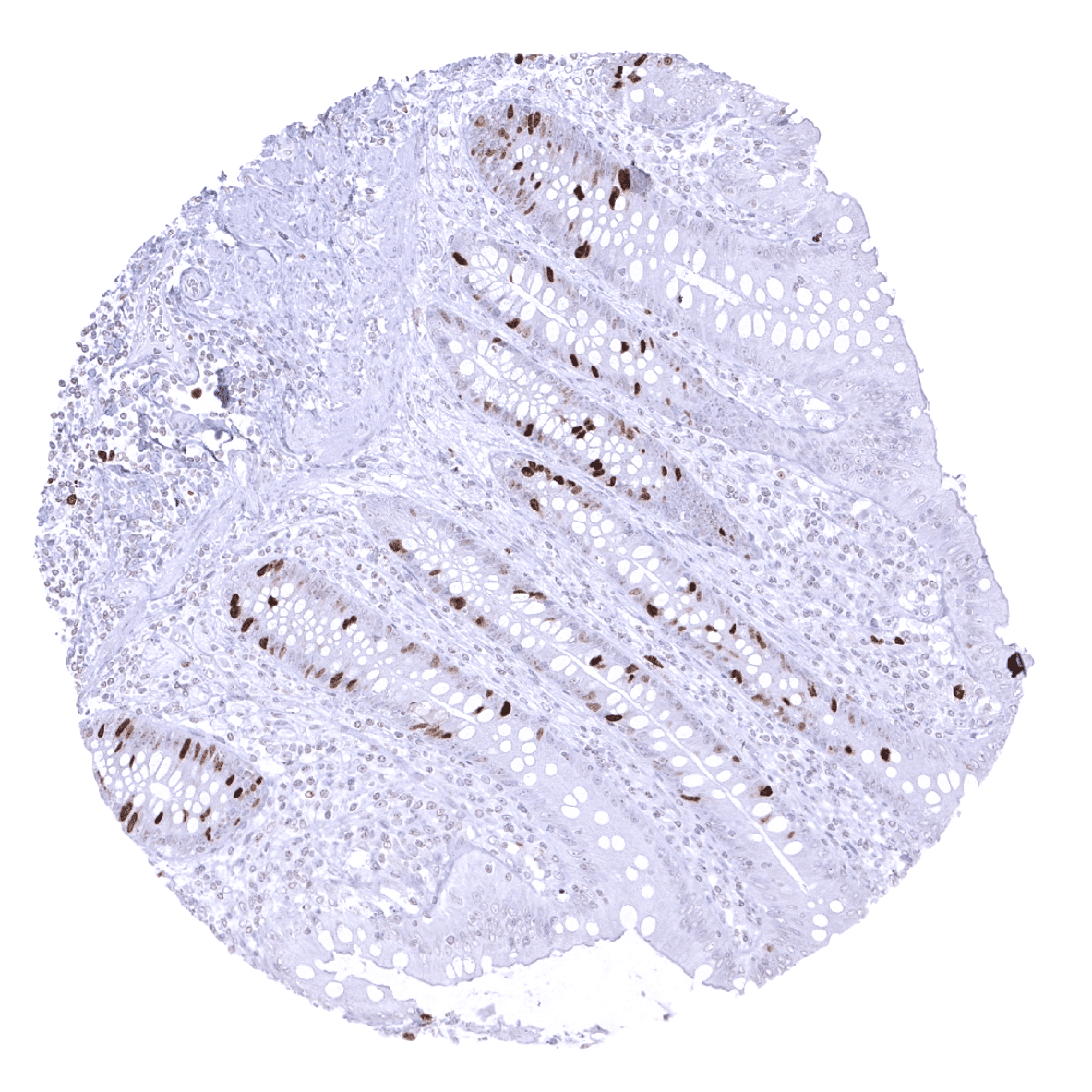
Positive control = Colon: A fraction of cells, especially in the crypt basis should show a moderate to strong nuclear TOPO2A immunostaining.
Negative control = Colon: Epithelial cells must not show any TOPO2A staining in the cytoplasm and in most nuclei of the surface epithelium.
Cellular localization = Nuclear
Reactivity = Human
Application = Immunohistochemistry
Dilution = 1:100 – 1:200
Intended Use = Research Use Only
Relevance of Antibody
Topoisomerase 2-alpha is a proliferation marker and anthracycline target.
Biology Behind
DNA topoisomerase 2-alpha (TOP2A) coded by the gene TOP2A at 17q21 is an enzyme localized to cell nuclei that influences the topologic states of DNA during RNA transcription. TOP2A is thus regularly expressed in proliferating cells in late S phase and G2-M phase where it specifically affects chromosome condensation and chromatid separation. It catalyzes the necessary breaking and rejoining of two strands of duplex DNA if altering the topology of DNA requires the DNA strands to pass through one another. TOP2A is the target for several anticancer agents including anthracycline and etoposide drugs. The TOP2A gene is located near the HER2 locus at 17q12-q21 and is often coamplified with HER2. TOP2A amplification has been suspected to raise susceptibility of cancers to anthracyclines. Several TOP2A mutations have been associated with the development of drug resistance. Reduced TOP2A activity may play a role in ataxia-telangiectasia.
Staining Pattern in Normal Tissues
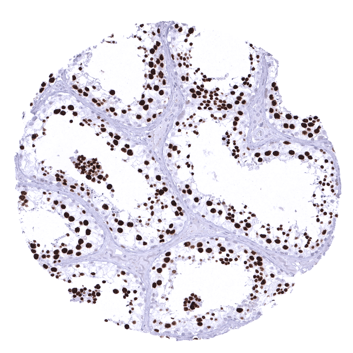
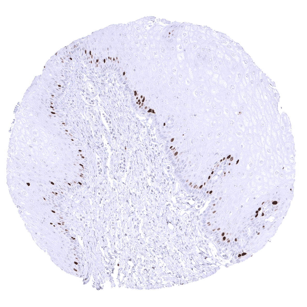
In normal tissues, TOPO2A immunostaining can regularly be seen in the vast majority of organs with a distribution pattern consistent with proliferating cells. Accordingly, the pattern largely parallels the display of Ki67 immunostaining. Spermatocytes of the testis – in which a special type of cell division occurs (meiosis) – is the only normal cell type with a consistent strong TOPO2A expression in virtually all cells albeit Ki67 is hardly seen in parallel sections.
These findings are largely comparable to the RNA and protein data described in the Human Protein Atlas (Tissue expression TOP2 alpha)
Suggested positive tissue control: Colon: A fraction of cells, especially in the crypt basis should show a moderate to strong nuclear TOPO2A immunostaining.
Suggested negative tissue control: Colon: Epithelial cells must not show any TOPO2A staining in the cytoplasm and in most nuclei of the surface epithelium.

Staining Pattern in Relevant Tumor Types
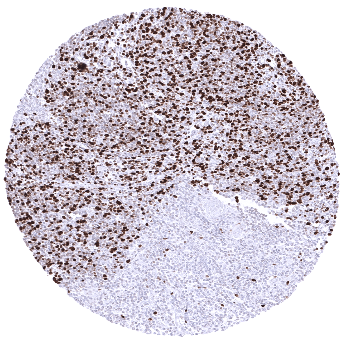
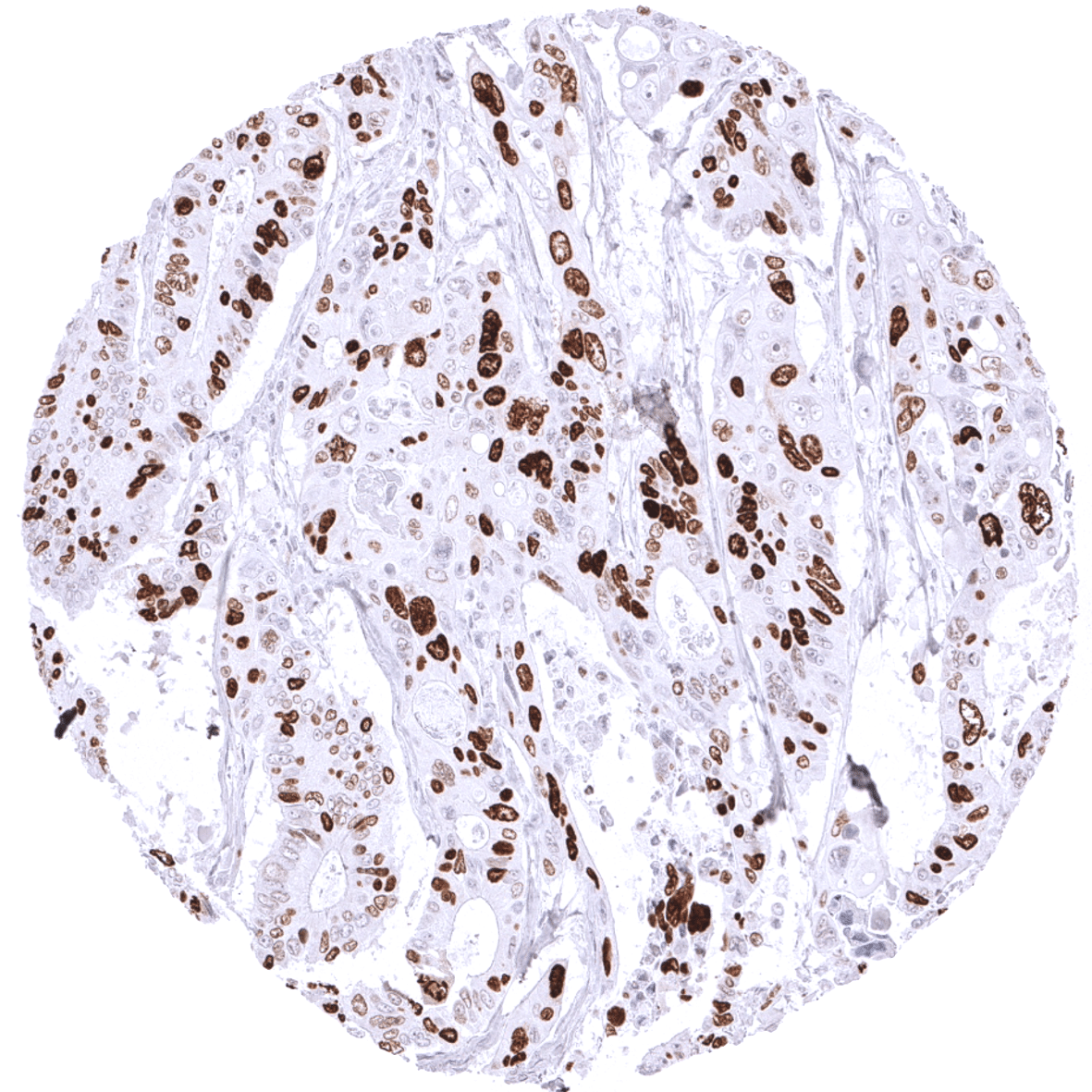
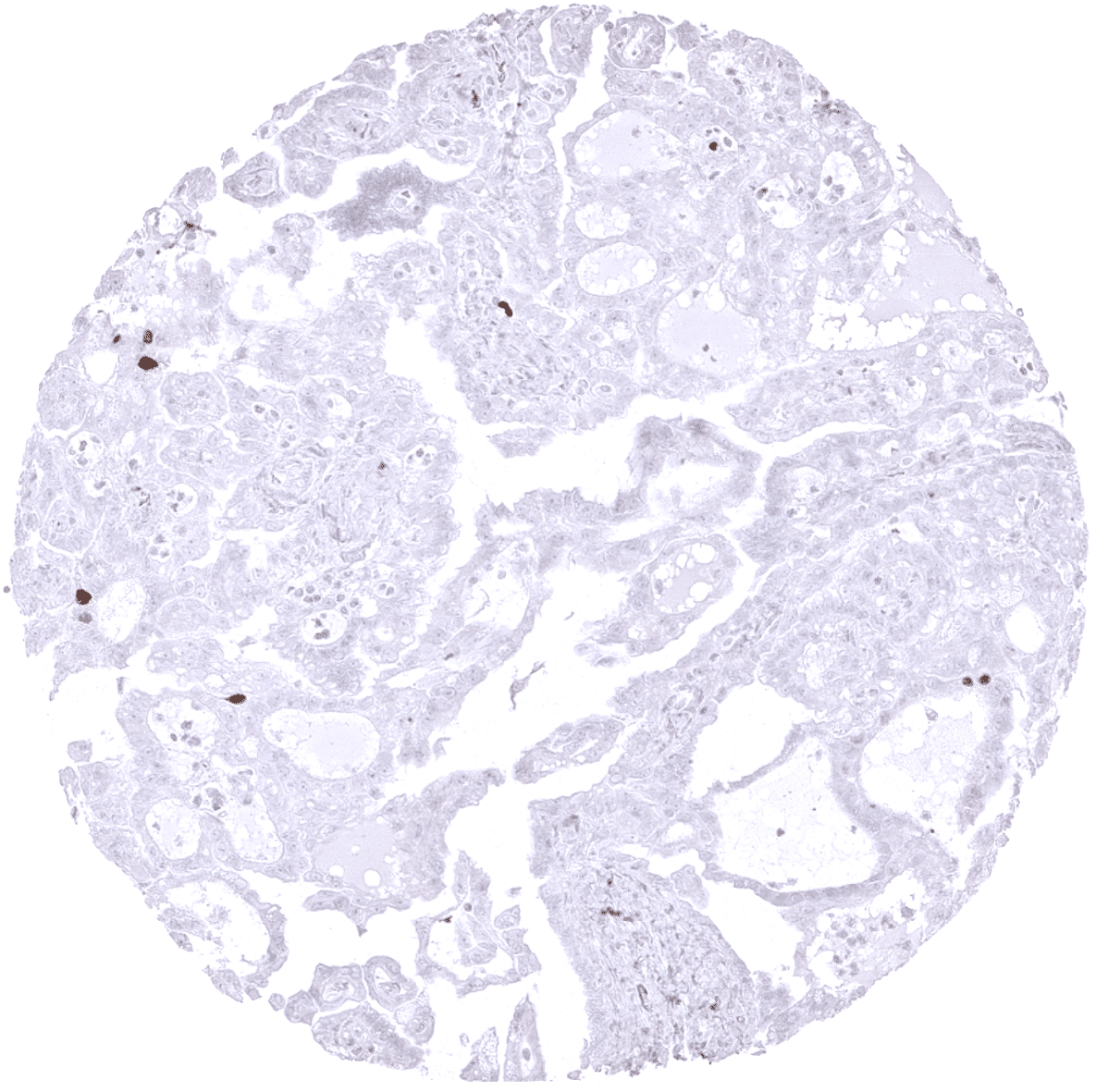
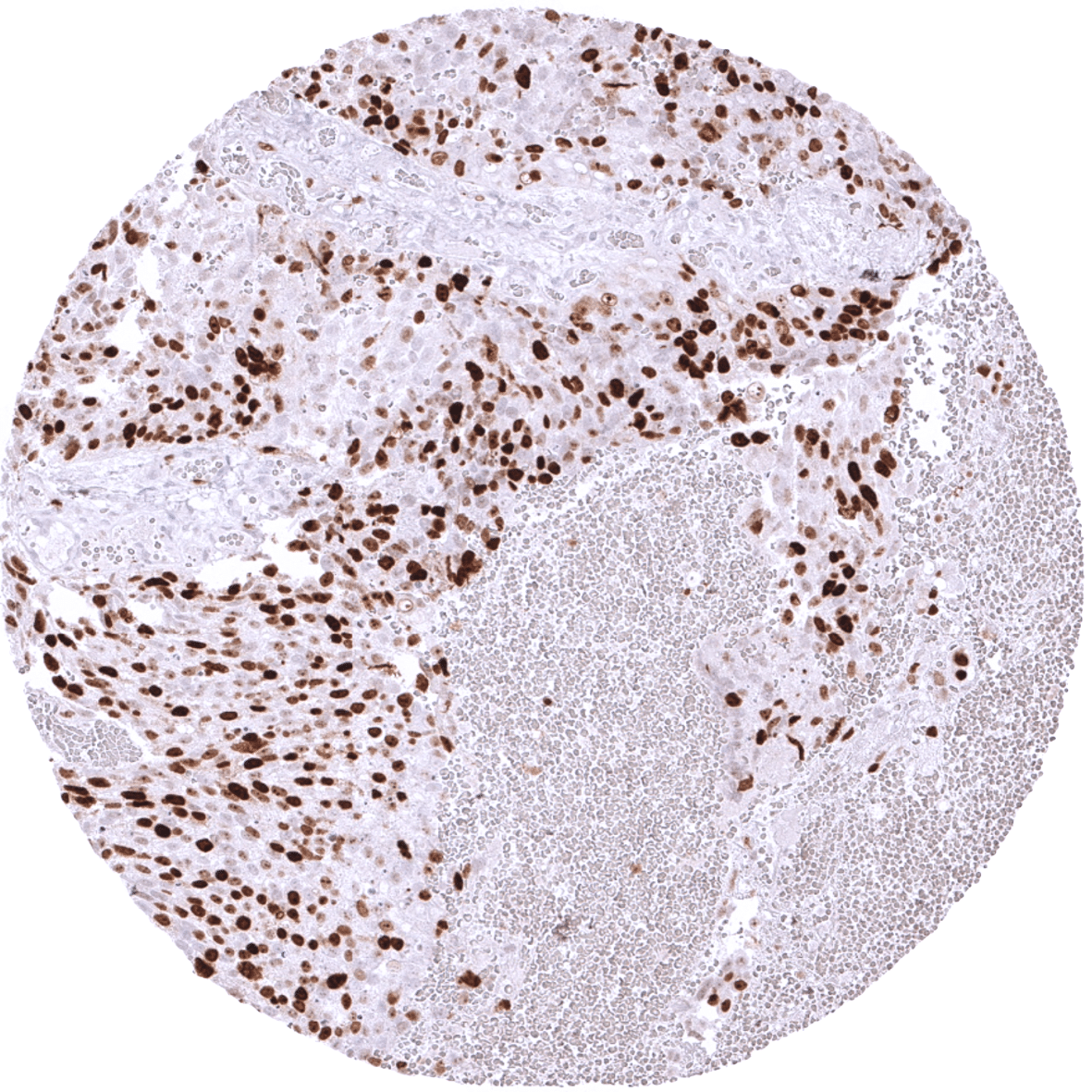
At least a fraction of tumor cells with TOPO2A immunostaining can be seen in the vast majority of cancers, with an intratumoral distribution consistent with proliferating cells. A TOPO2A positivity of near 100% of tumor cells occurs in tumors with very high proliferative activity or may be indicative of TOPO2A amplification, the latter most commonly occurring in combination with HER2 amplification, preferably in breast, stomach, and urothelial cancer.
The TCGA findings on TOP2 alpha RNA expression in different tumor categories have been summarized in the Human Protein Atlas.
Compatibility of Antibodies
No data available at the moment
Protocol Recommendations
IHC users have different preferences on how the stains should look like. Some prefer high staining intensity of the target stain and even accept some background. Others favor absolute specificity and lighter target stains. Factors that invariably lead to more intense staining include higher concentration of the antibody and visualization tools, longer incubation time, higher temperature during incubation, higher temperature and longer duration of the heat induced epitope retrieval (slide pretreatment). The impact of the pH during slide pretreatment has variable effects and depends on the antibody and the target protein.
All images and data shown here and in our image galleries are obtained by the manual protocol described below. Other protocols resulting in equivalent staining are described as well.
-Manual protocol
Freshly cut sections should be used (less than 10 days between cutting and staining). Heat-induced antigen retrieval for 5 minutes in an autoclave at 121°C in pH 7,8 Target Retrieval Solution buffer. Apply MSVA-802R at a dilution of 1:150 at 37°C for 60 minutes. Visualization of bound antibody by the EnVision Kit (Dako, Agilent) according to the manufacturer’s directions.
Potential Research Applications
- TOP2A expression may be predictive of prognosis and response to TOP2A inhibitors in various malignancies.
- The TOP2A or Topo II labeling index representing the percentage of TOP2A positive staining nuclei may be similarly prognostic as the Ki67 LI.
- The best tool and cutoff values for TOP2A assessment are unknown.
Evidence for Antibody Specificity in IHC
There are two ways how the specificity of antibodies can be documented for immunohistochemistry on formalin fixed tissues. These are: 1. comparison with a second independent method for target expression measurement across a large number of different tissue types (orthogonal strategy), and 2. Comparison with one or several independent antibodies for the same target and showing that all positive staining results are also seen with other antibodies for the same target (independent antibody strategy).
Orthogonal validation: For the antibody MSVA-802R specificity is suggested by the strong concordance of the immunostaining with RNA expression data derived from the Human Protein Atlas (HPA) RNA-seq tissue dataset, the FANTOM5 project, and the Genotype-Tissue Expression (GTEx) project which are all summarized in the Human Protein Atlas (Tissue expression TOP2alpha). TOPO2A immunostaining by using MSVA-802R was strongest in the thymus and the testis which are the organs with the highest documented RNA expression.
Comparison of antibodies: Specificity of the TOPO2A findings obtained by MSVA-802R is suggested by similar findings obtained by the independent antibody HPA006458 which was used within the human protein atlas project.
