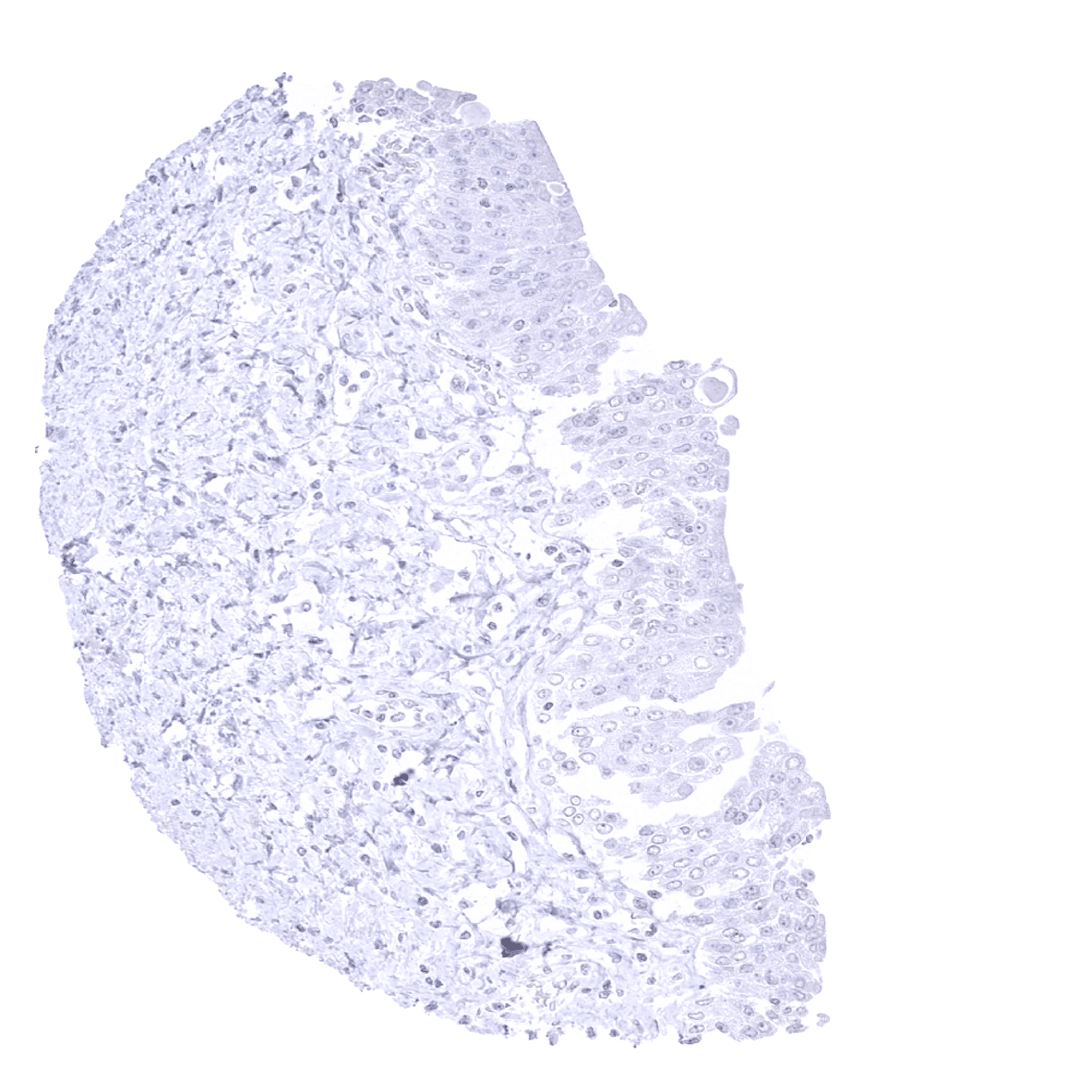
Adrenal gland

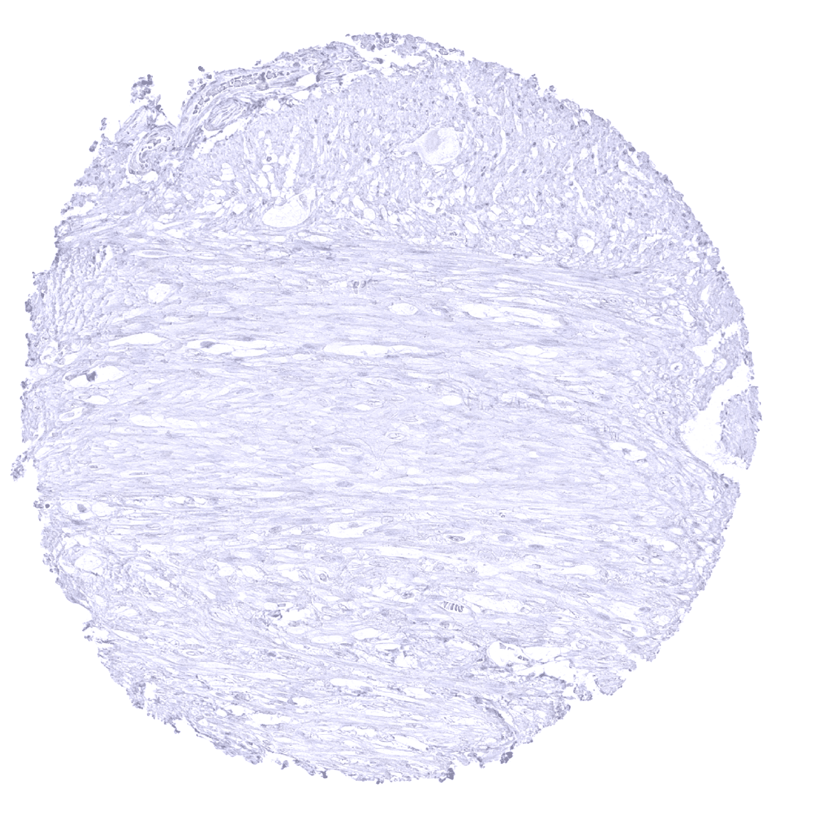
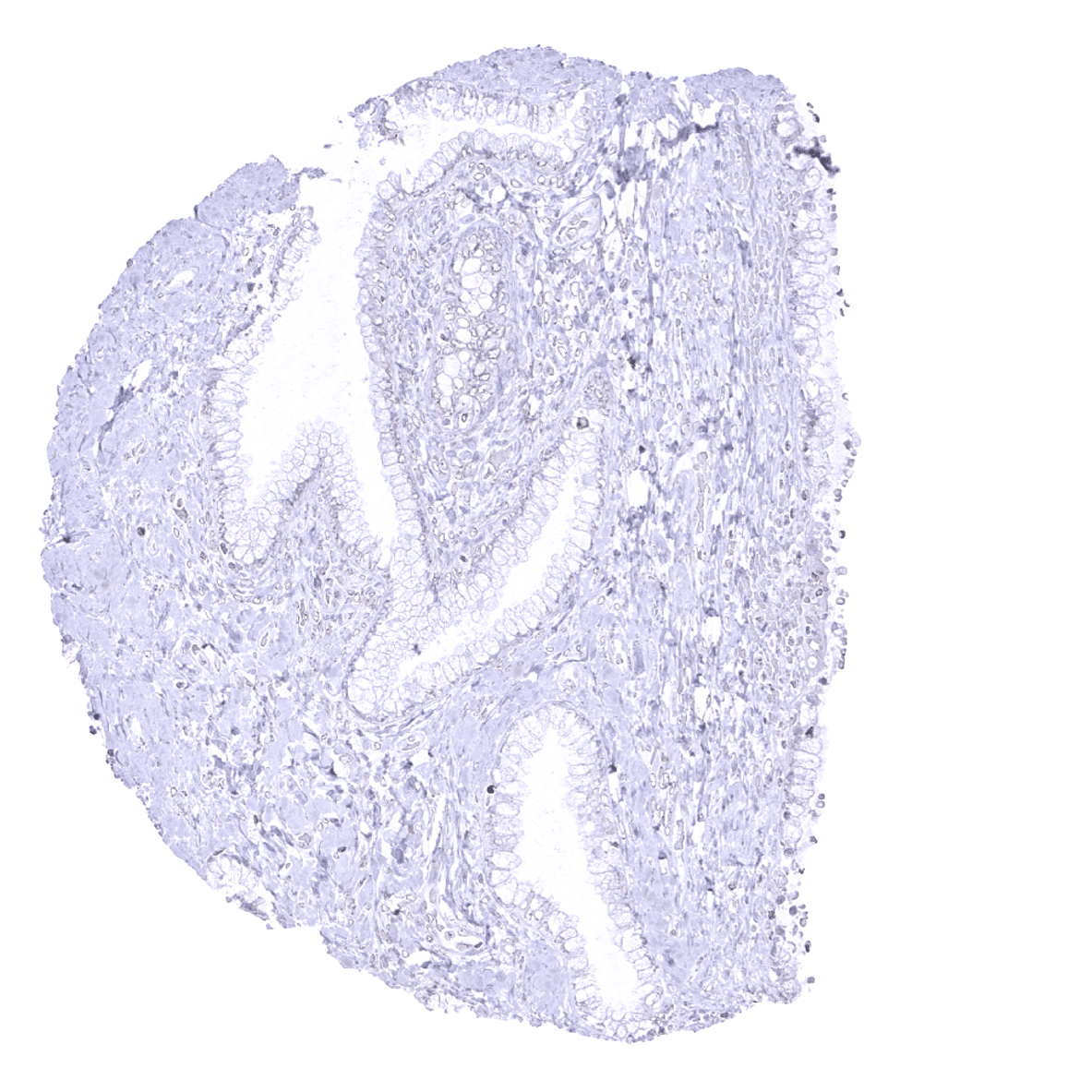
Aorta, media
Appendix, mucosa - TOPO2A positive cells in the appendix mucosa reflect proliferating cells.
Appendix, muscular wall

Bone marrow
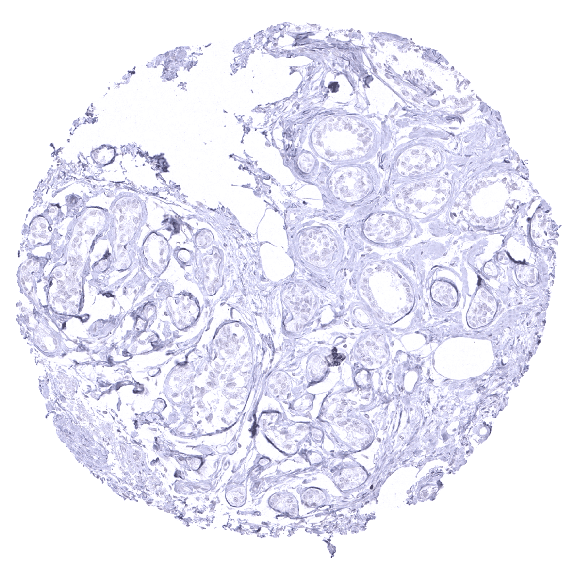
Breast
Bronchus, mucosa
Cerebellum (molecular layer, Purkinje cell layer, granule cell layer)
Cerebellum (molecular layer, Purkinje cell layer, granule cell layer, white matter)
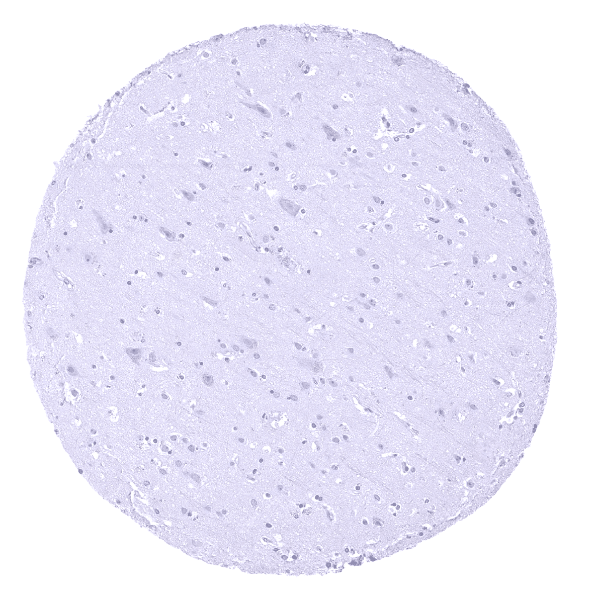
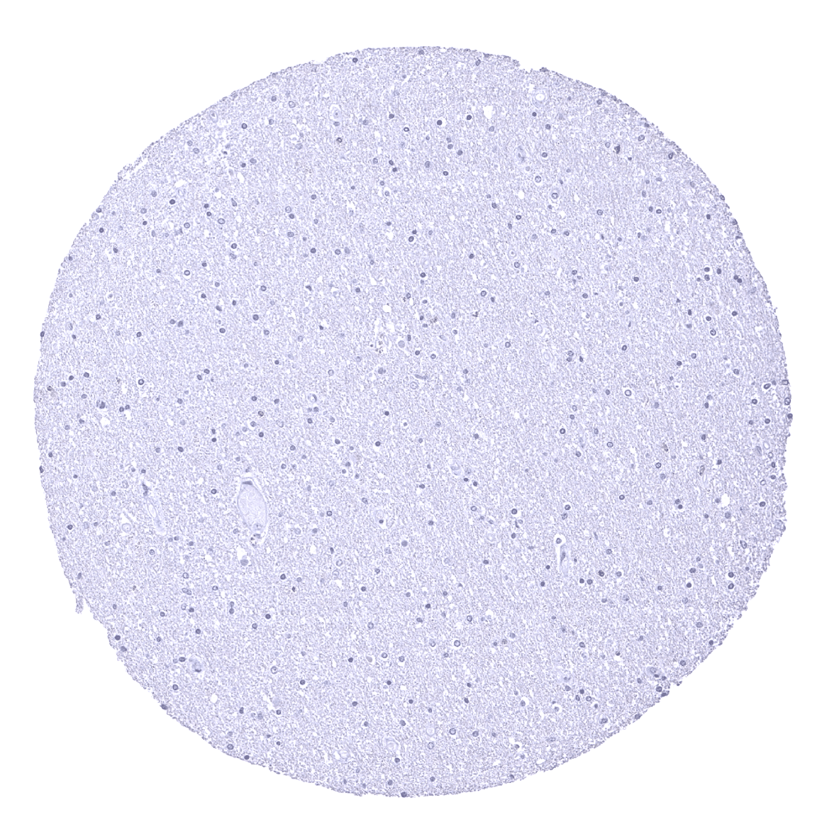
Cerebrum, grey matter
Cerebrum, white matter
Colon descendens, muscular wall

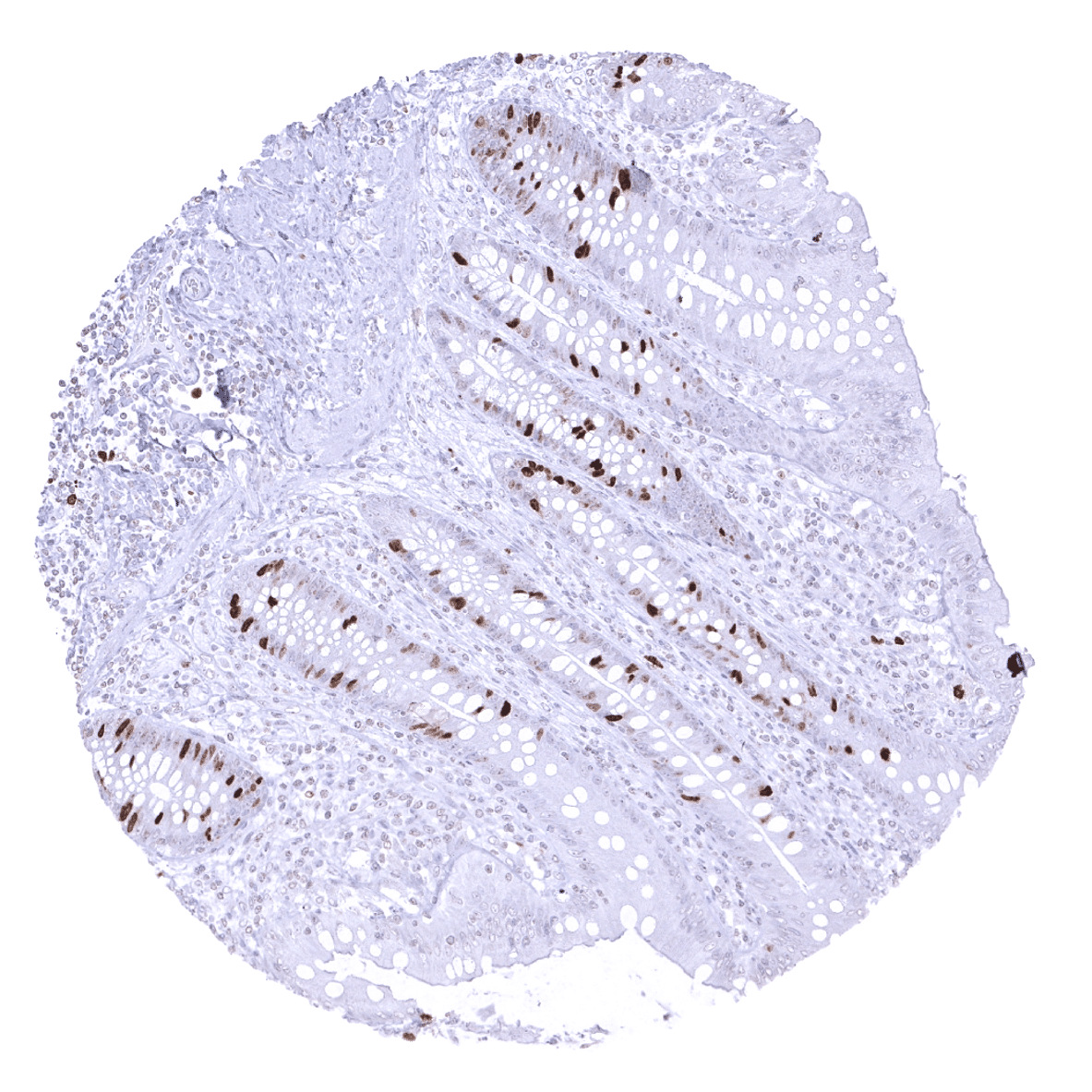
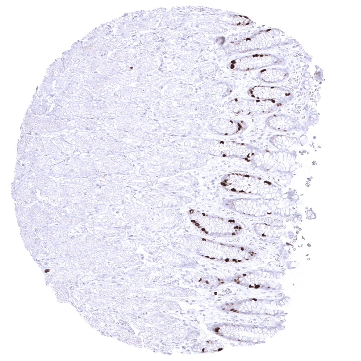
Colon, mucosa
Duodenum, Brunner gland

Duodenum, mucosa
Epididymis

Esophagus, squamous epithelium
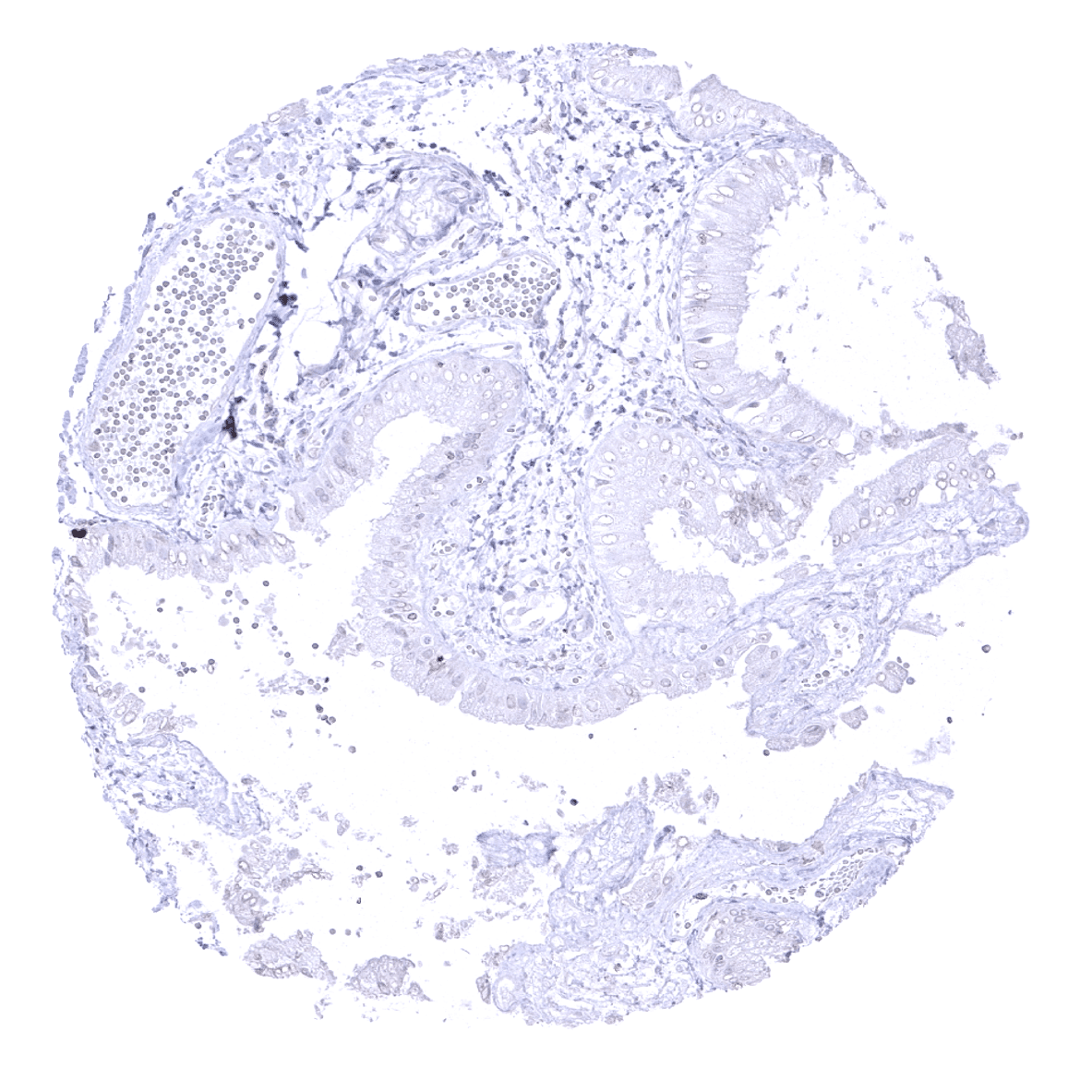
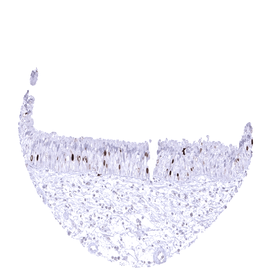
Fallopian tube, mucosa
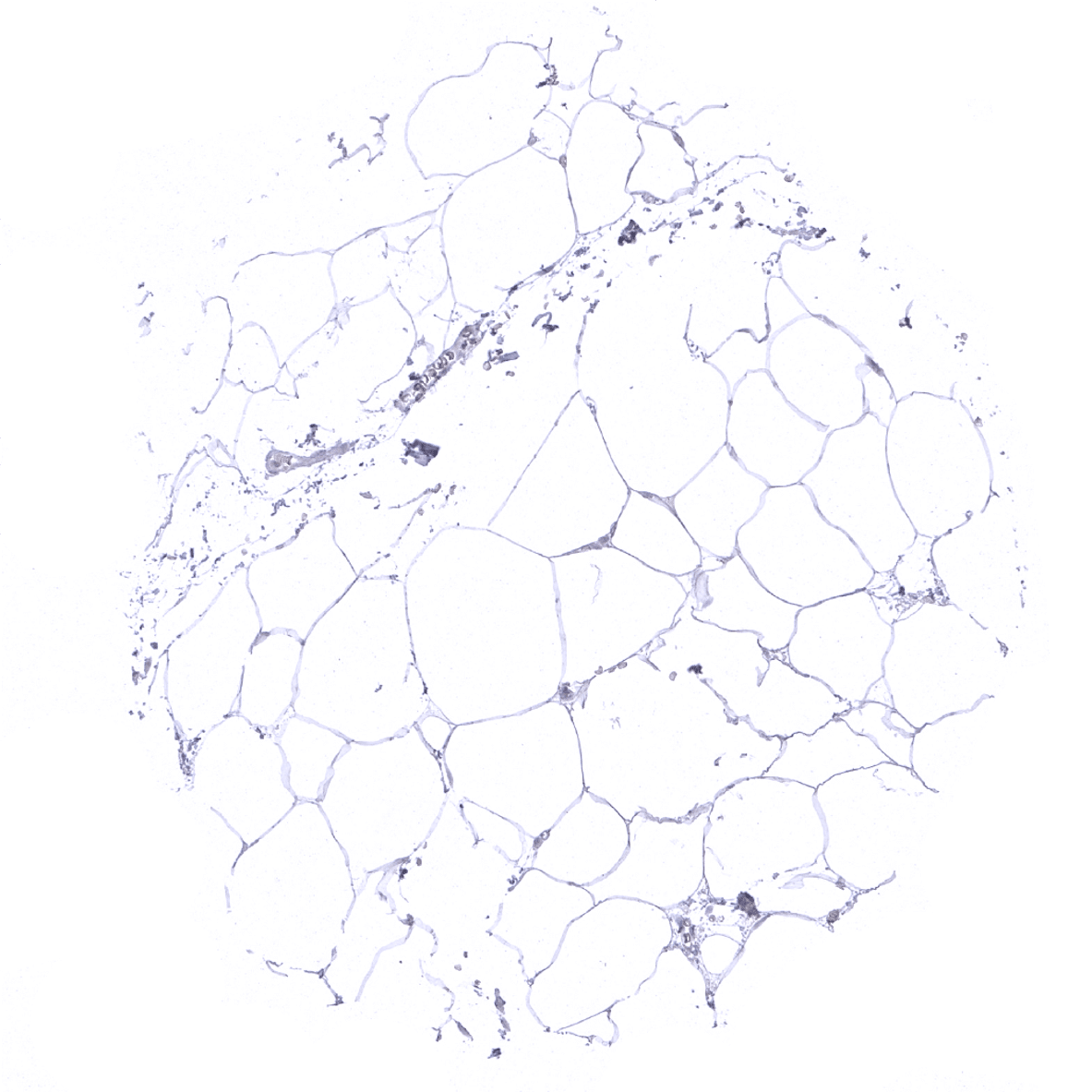
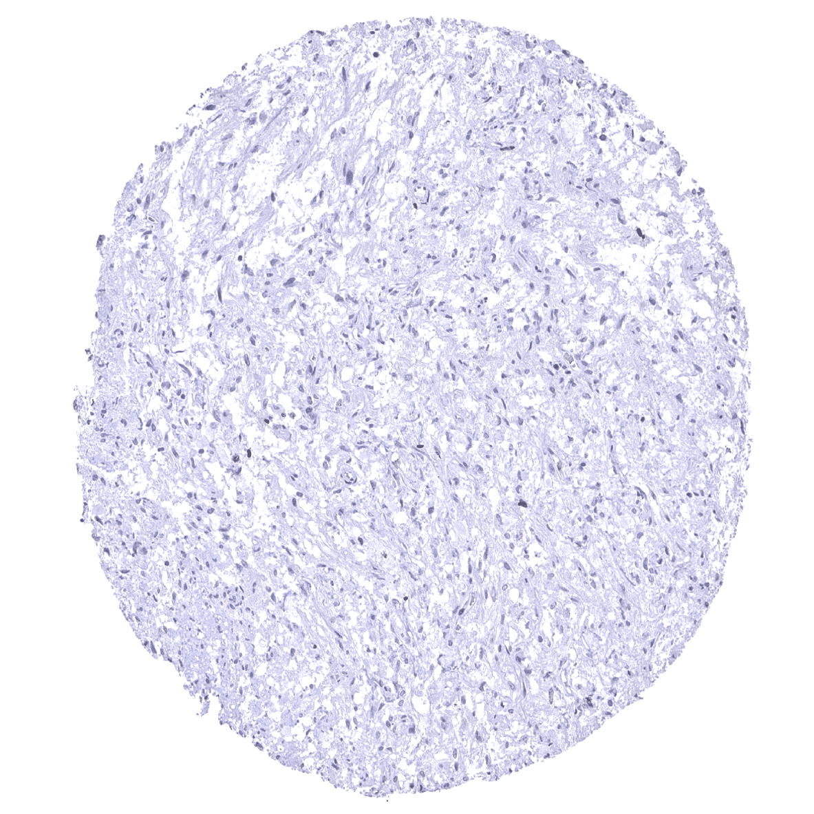
Fat
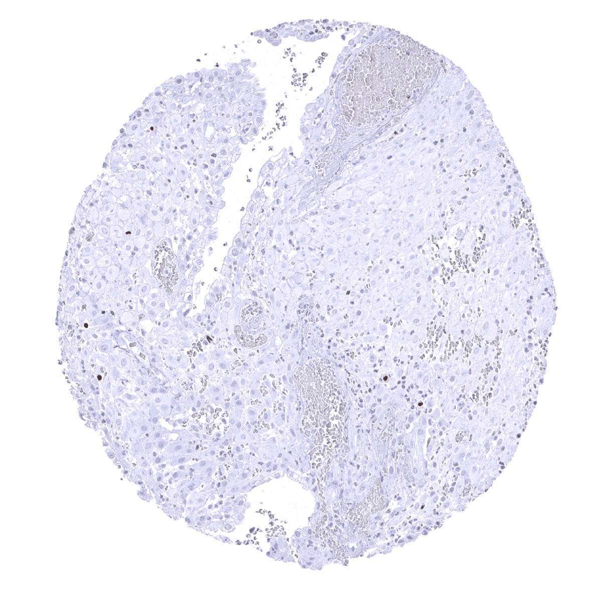
Gallbladder, epithelium
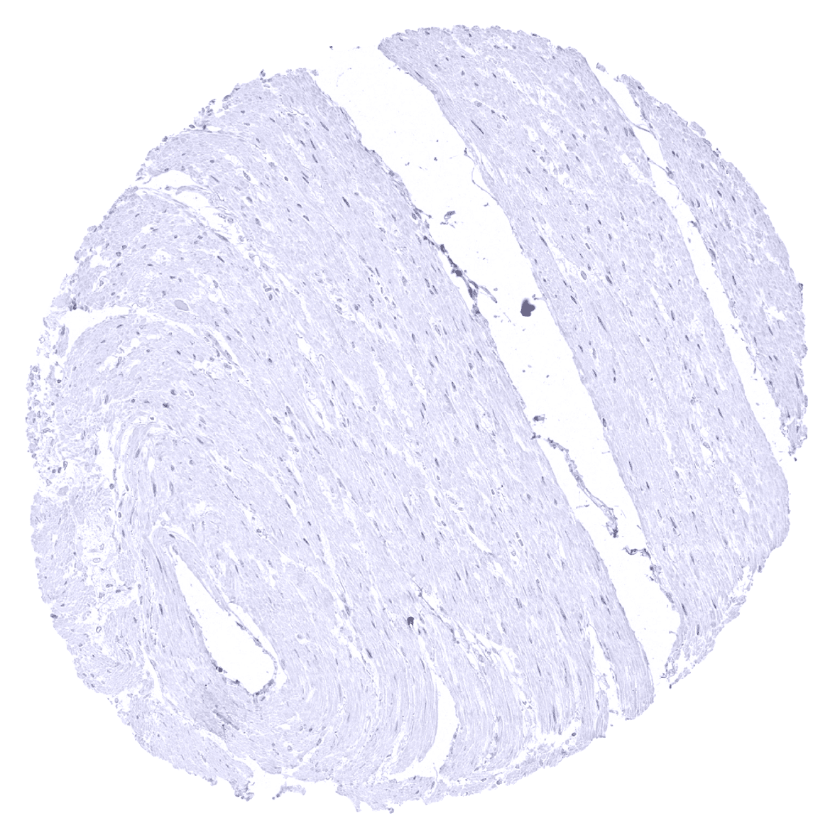
Heart muscle

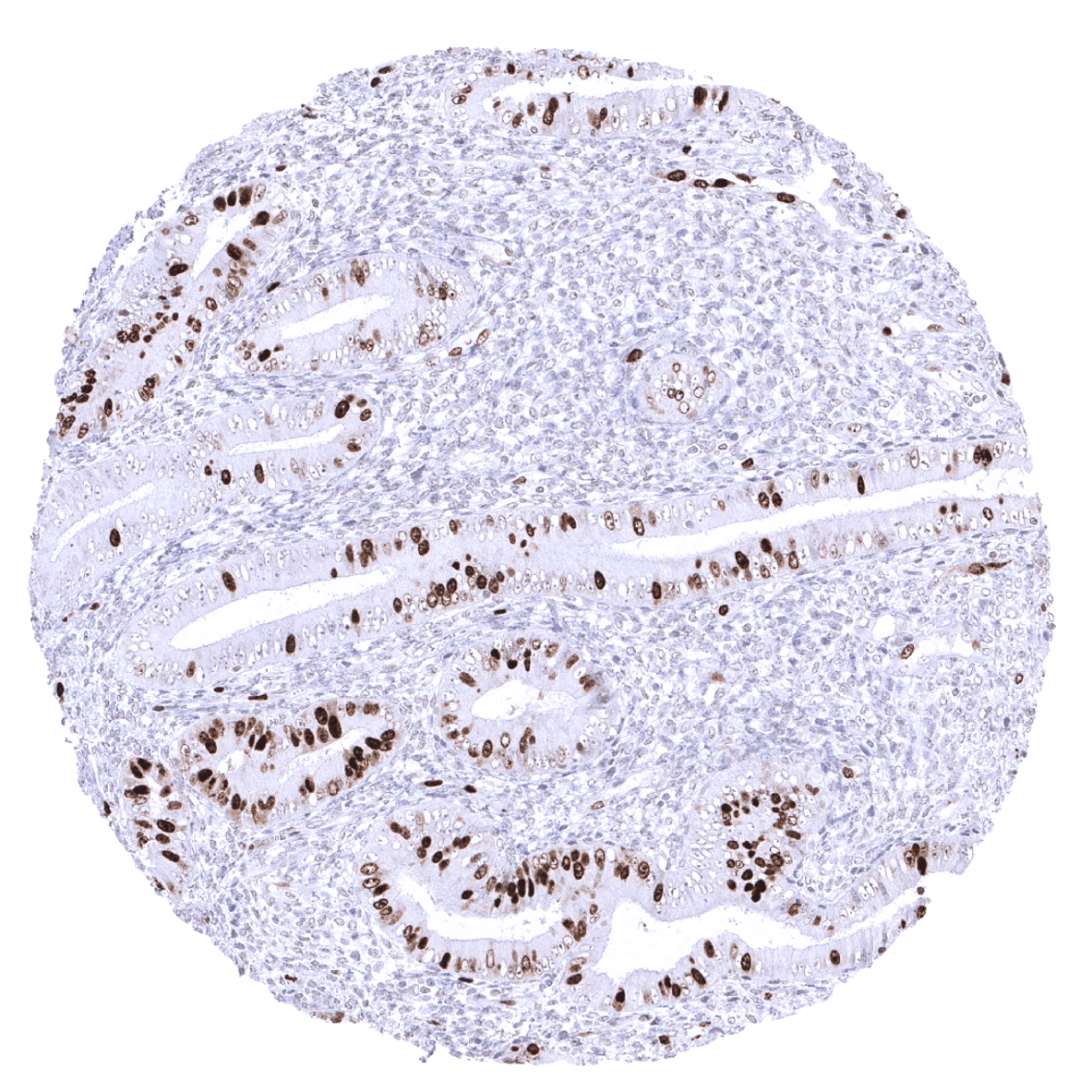
Ileum, mucosa - TOPO2A positive cells in the ileum mucosa reflect proliferating cells.
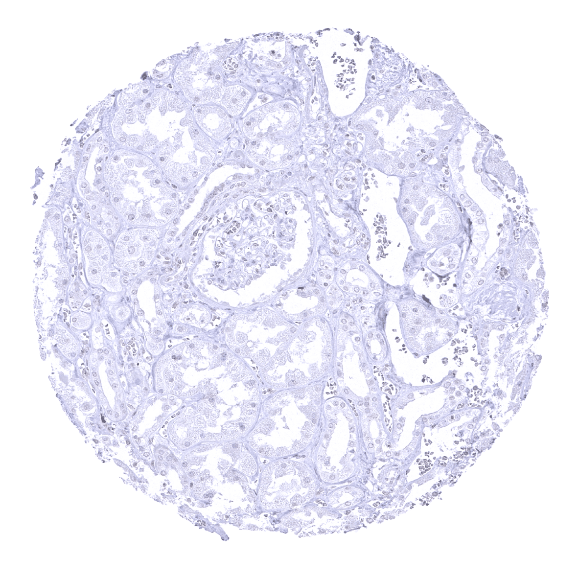
Kidney, cortex
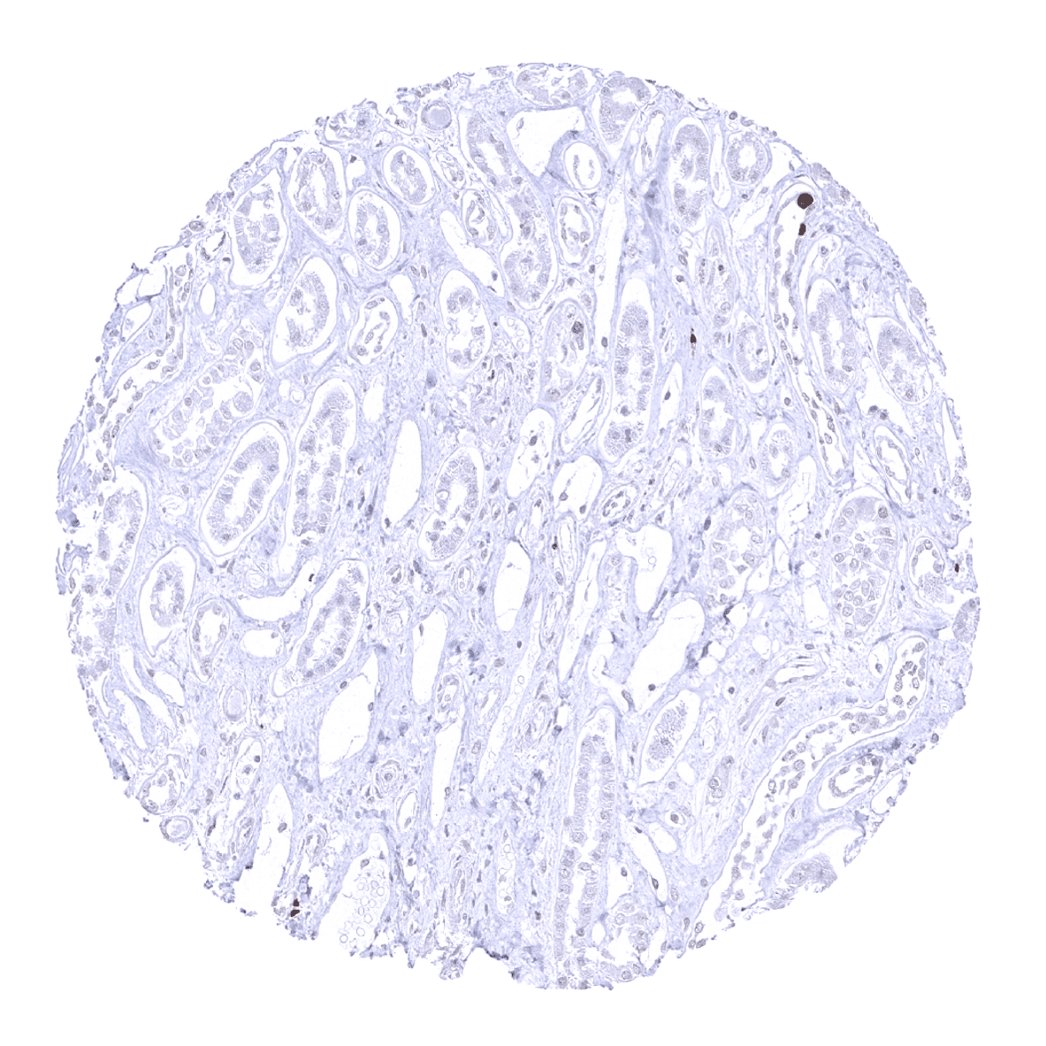
Kidney, medulla
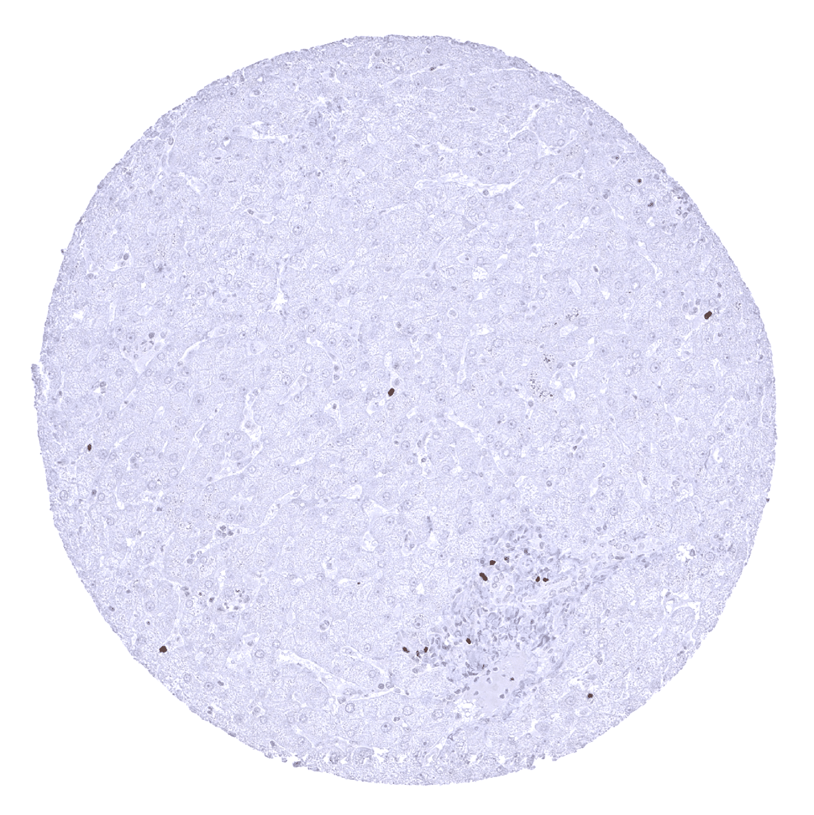
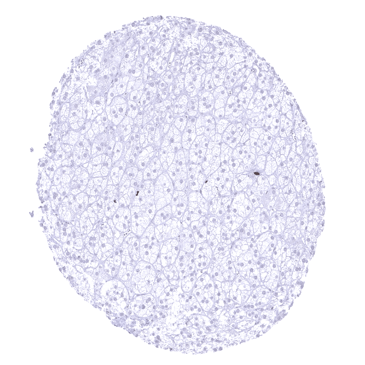
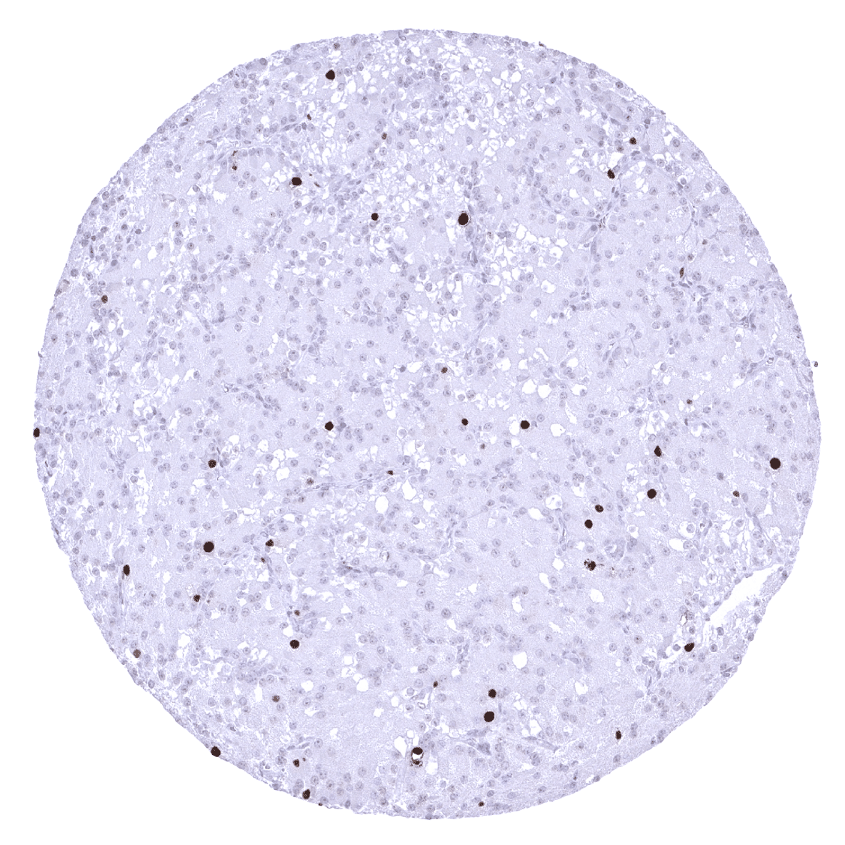
Liver

Lung

Lymph node
Ovary, stroma
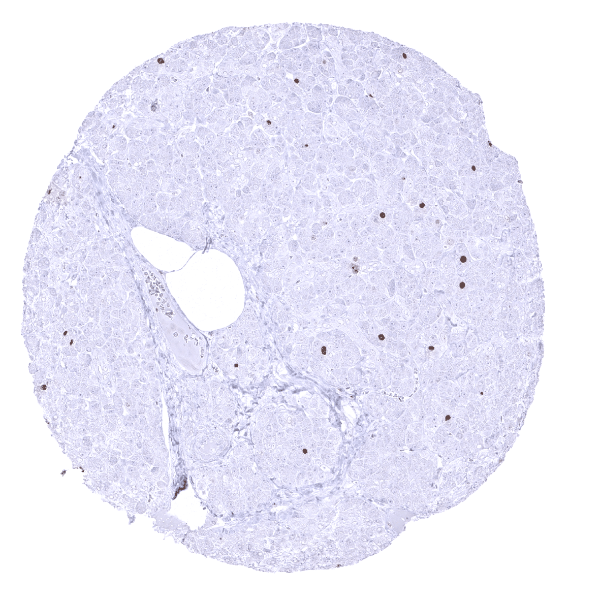
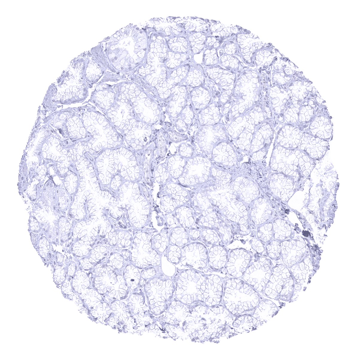
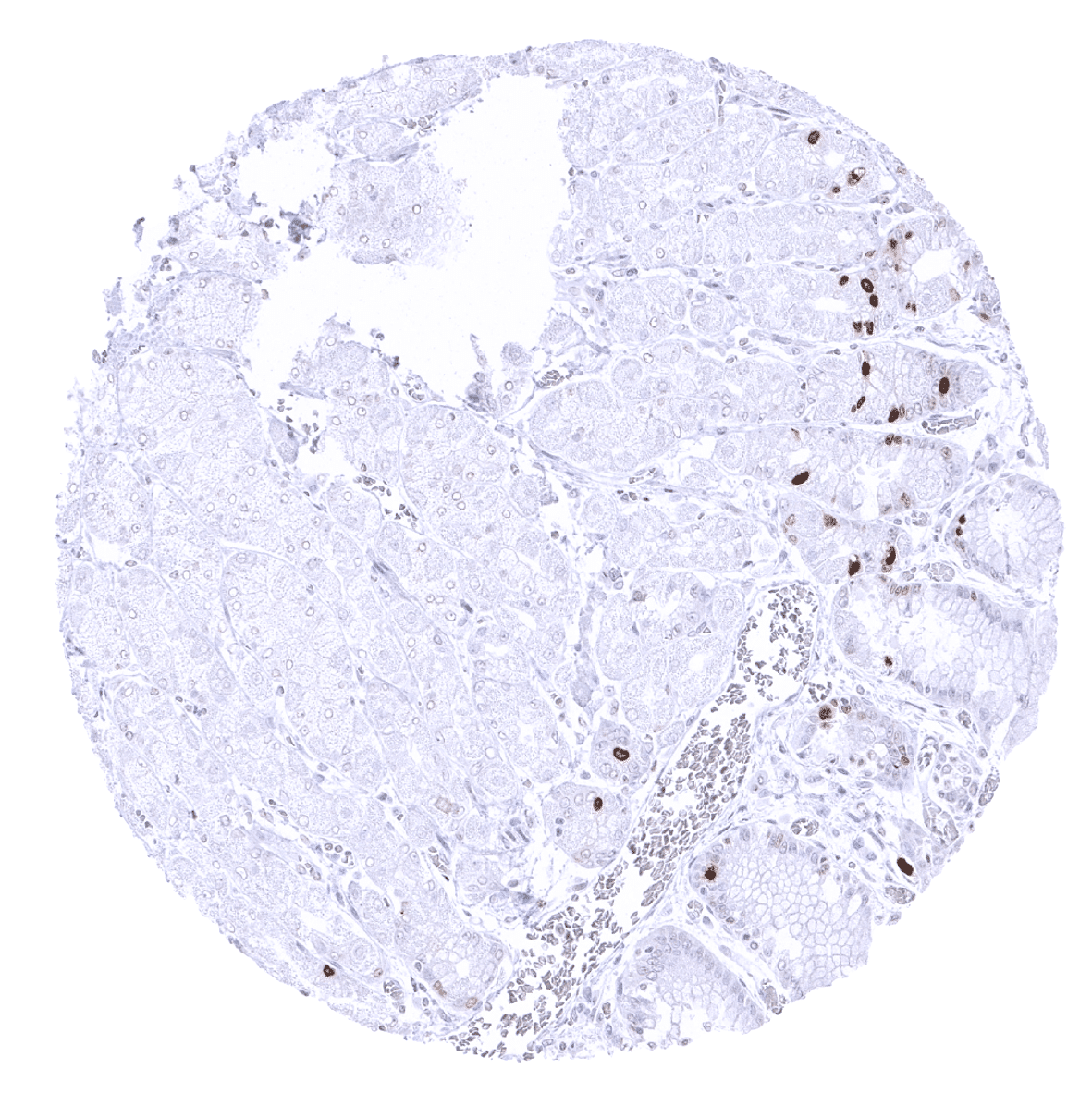
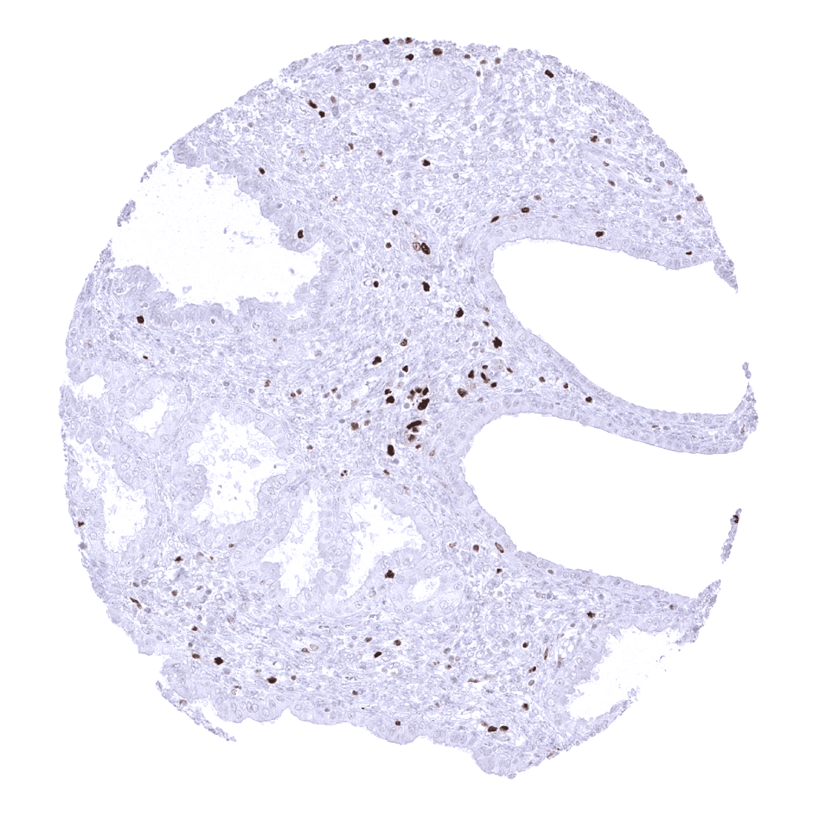
Pancreas
Parathyroid gland
Parotid gland
Pituitary gland, anterior lobe
Pituitary gland, posterior lobe
Placenta (amnion and chorion)
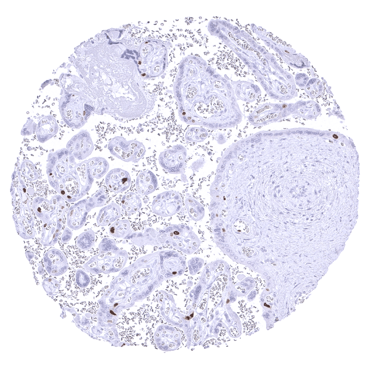
Placenta, early
Pregnant uterus (decidua)
Placenta, mature
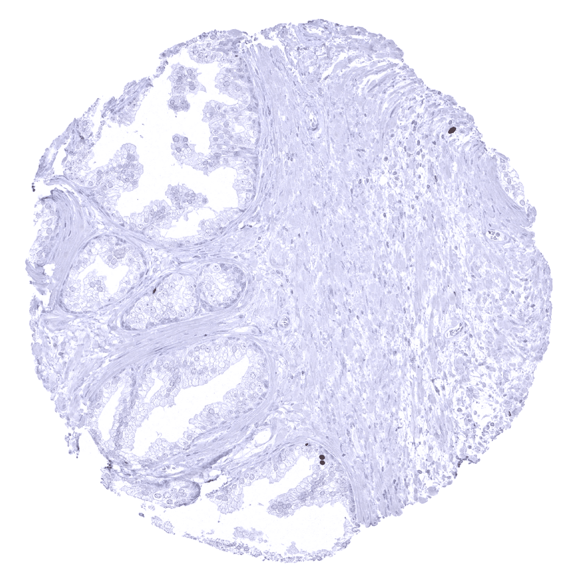
Prostate

Rectum, mucosa
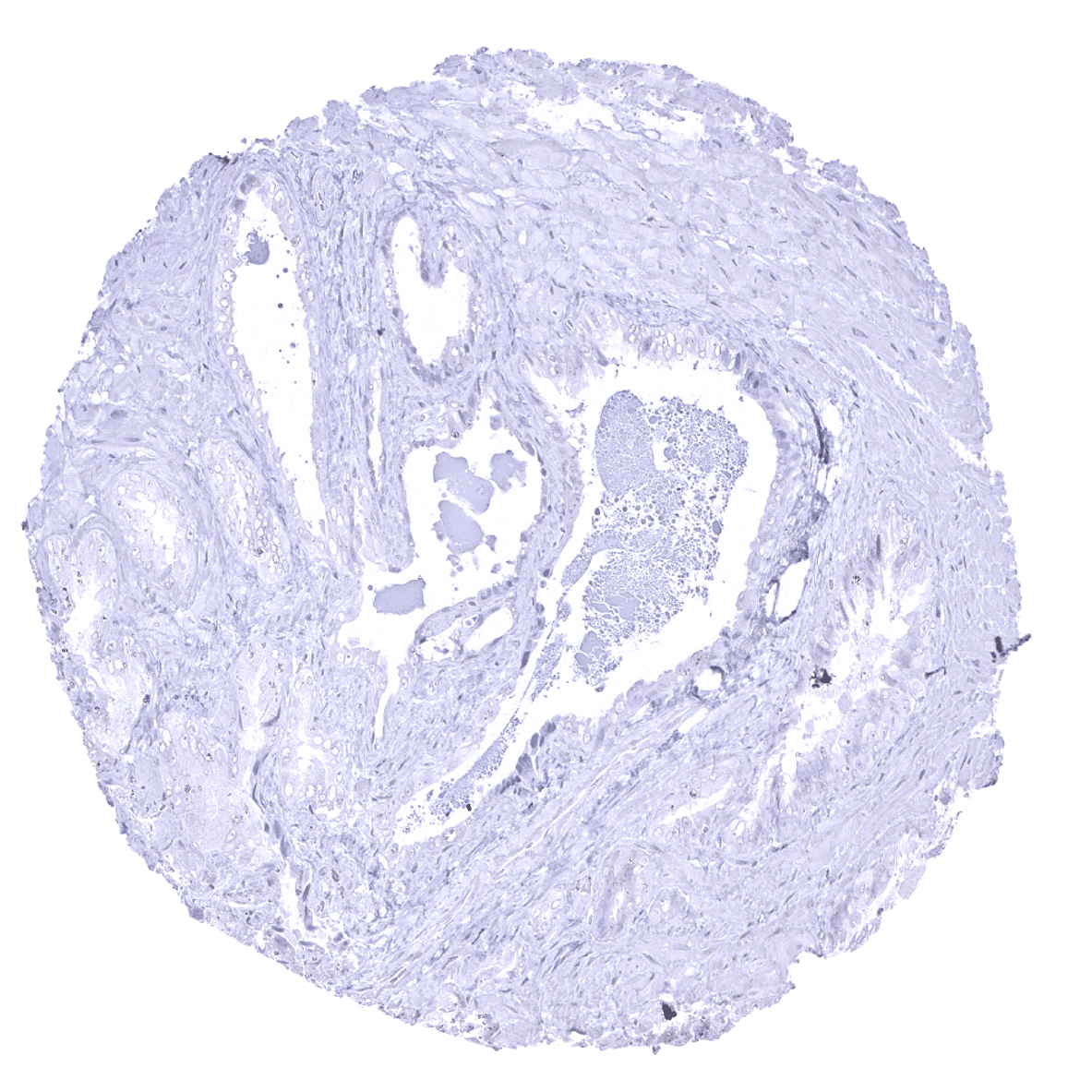
Seminal vesicle
Sinus paranasales, epithelium

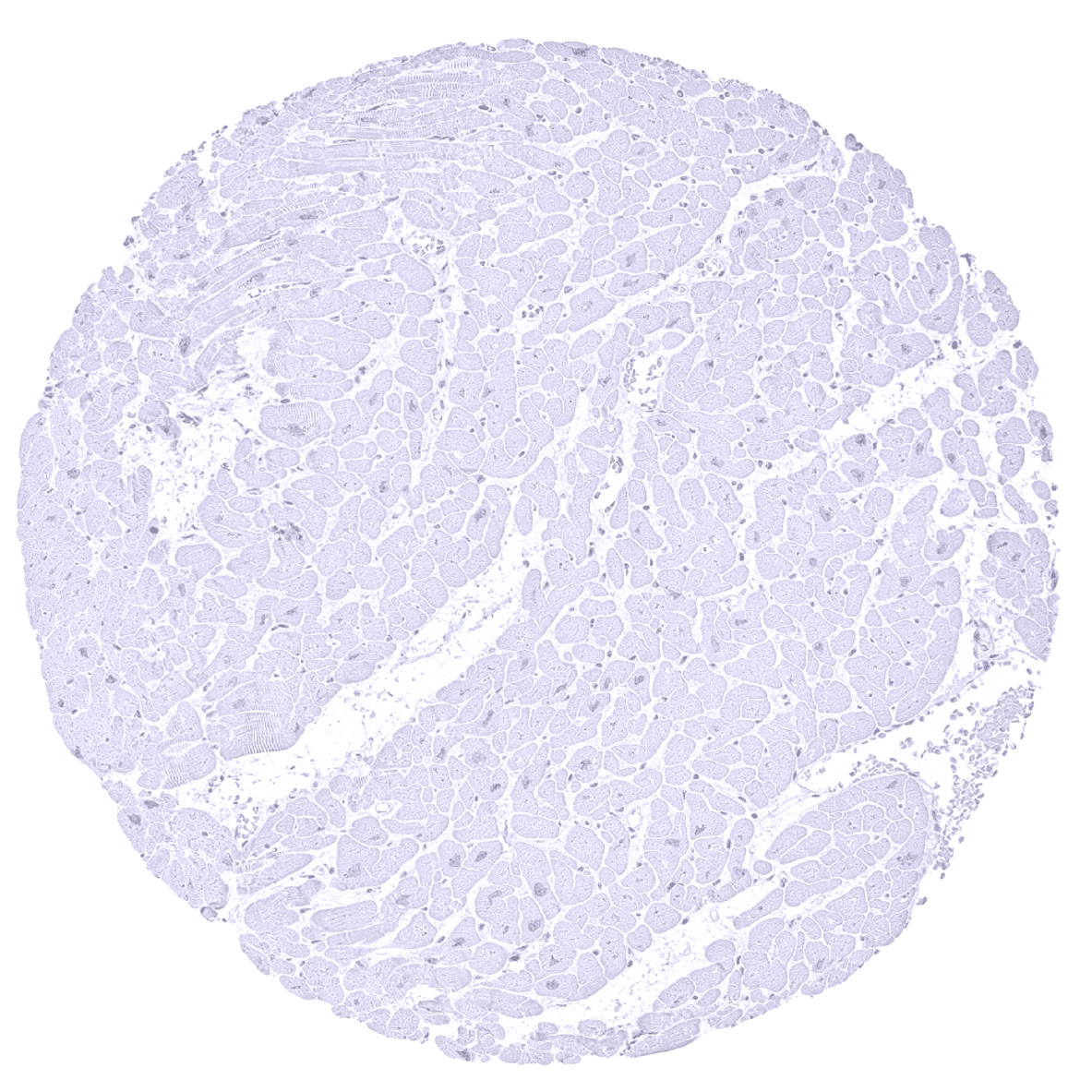
Skeletal muscle
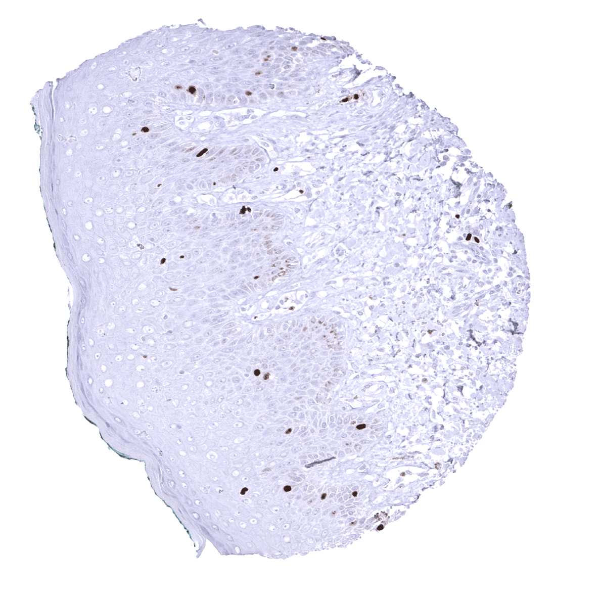
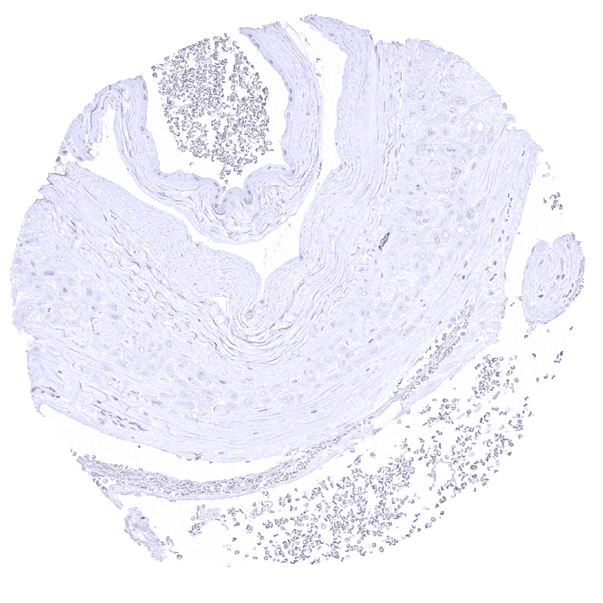
Skin - TOPO2A immunostaining is seen seen in proliferating cells.
Spleen
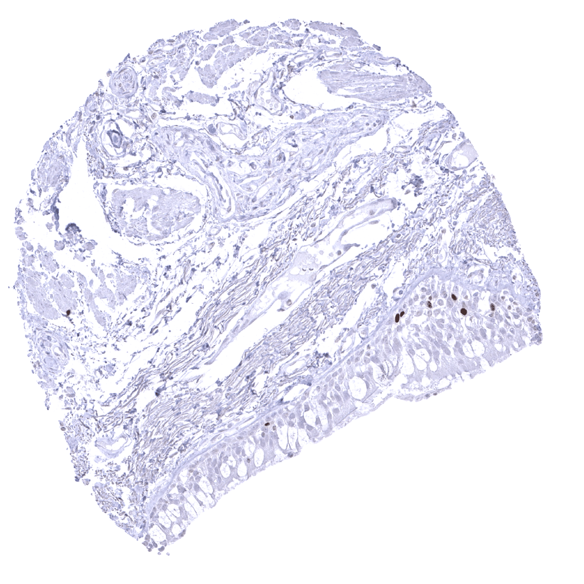
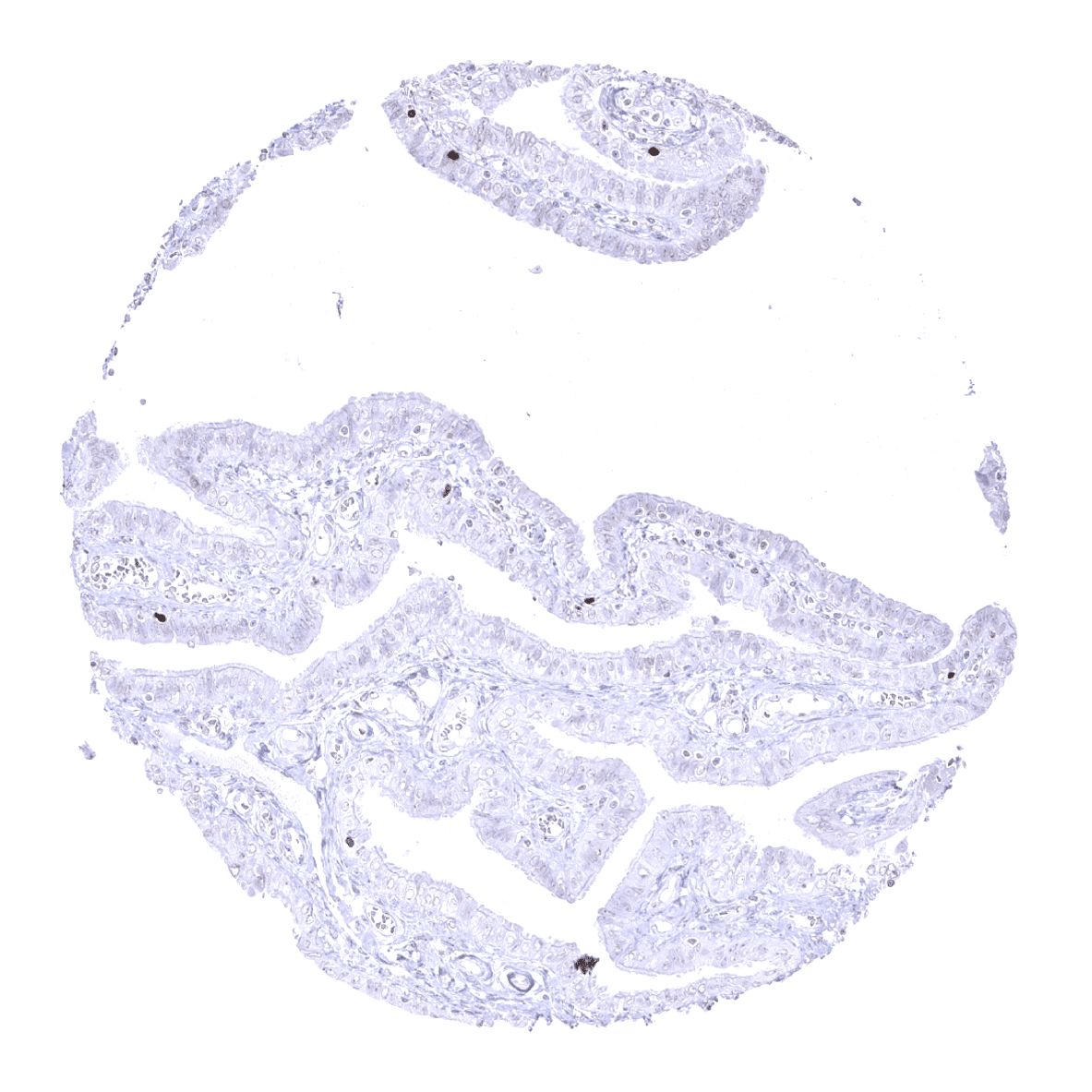
Stomach, antrum - TOPO2A positive cells in the stomach antrum reflect proliferating cells.
Stomach, corpus - TOPO2A positive cells in the stomach corpus reflect proliferating cells.

Testis - Spermatocyte of the testis – subject to meiosis - is the only normal cell type with a consistent strong TOPO2A expression in virtually all cells.
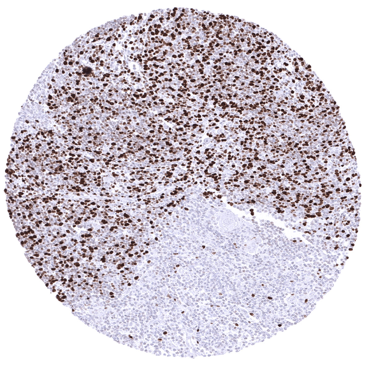
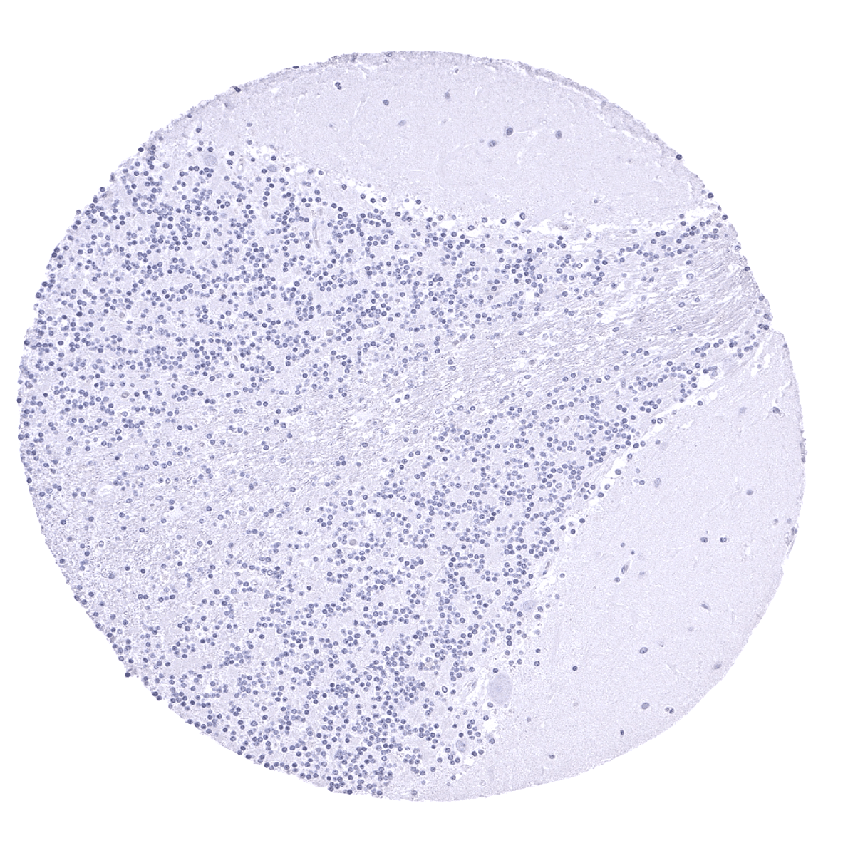
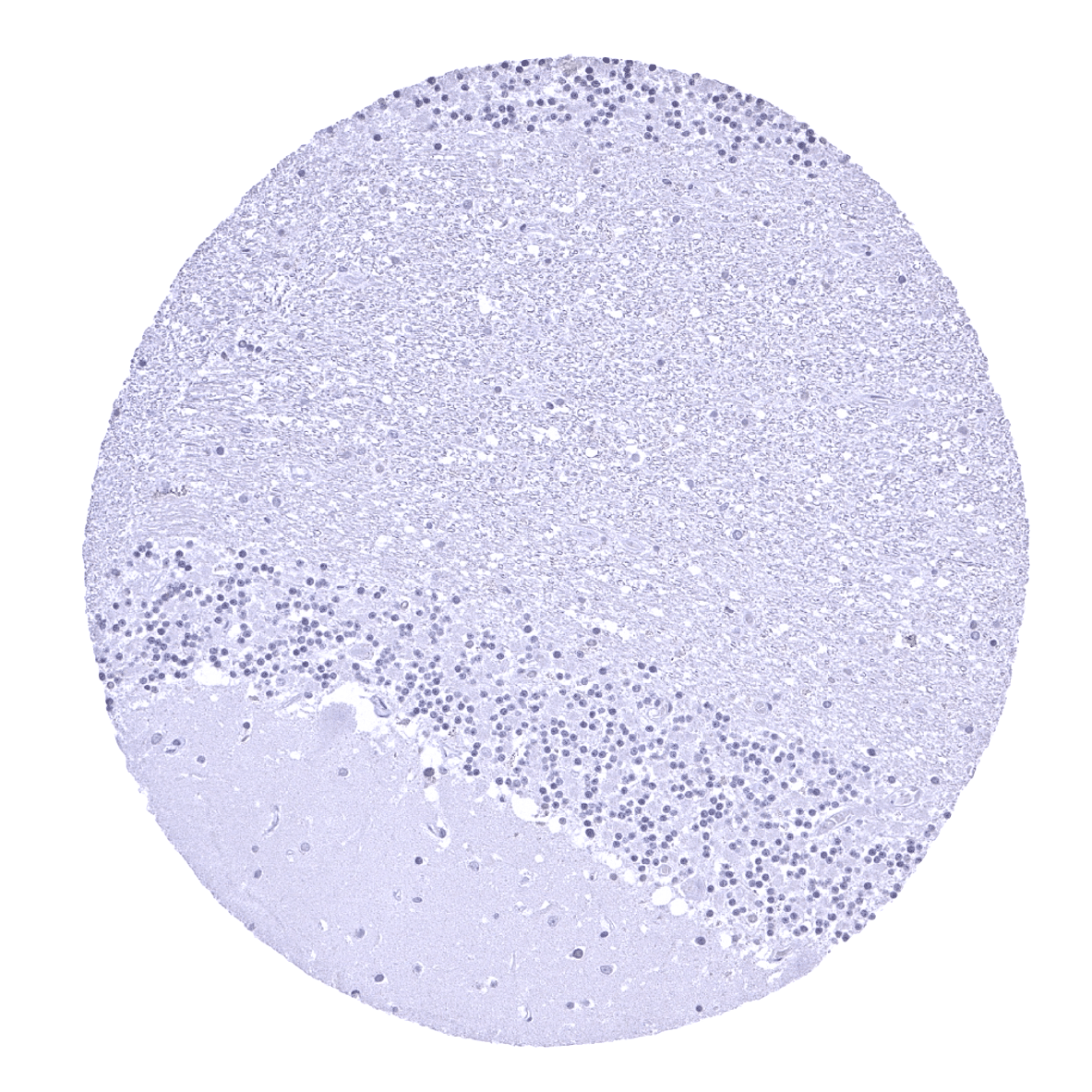
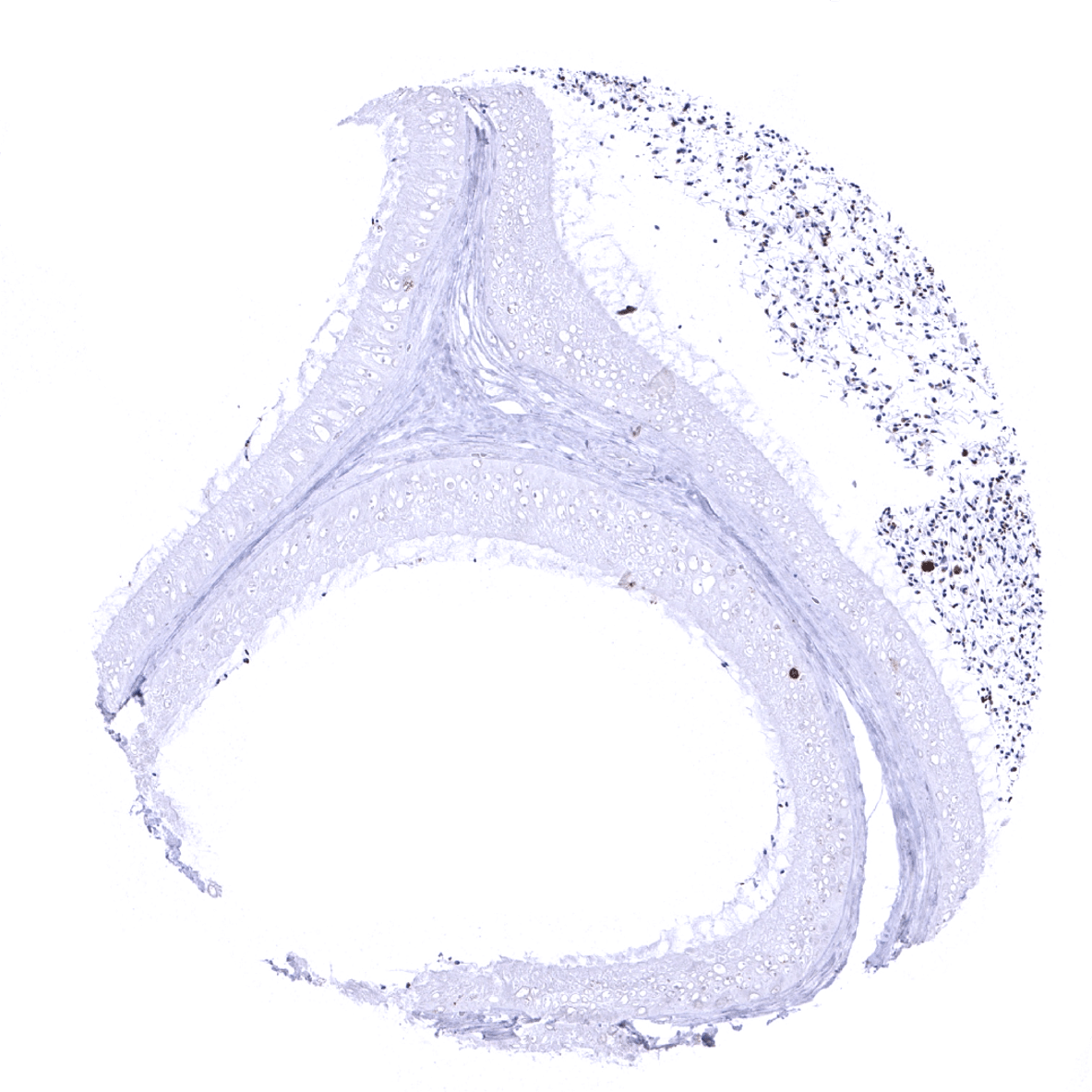
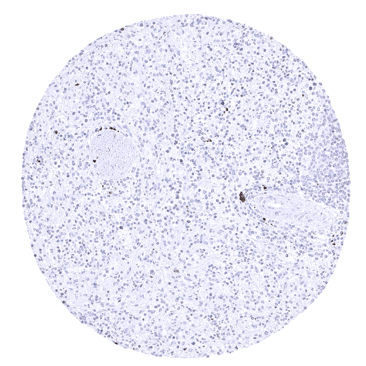
Thymus - The highest rate of TOPO2A positive lymphocytes is seen in the thymus (cortex).
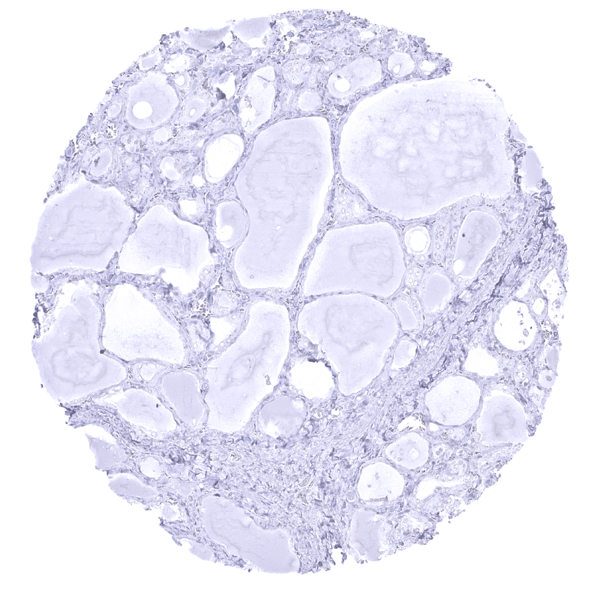
Thyroid gland

Tonsil - In the tonsil, TOPO2A positive lymphocytes are predominantly seen in germinal centres.
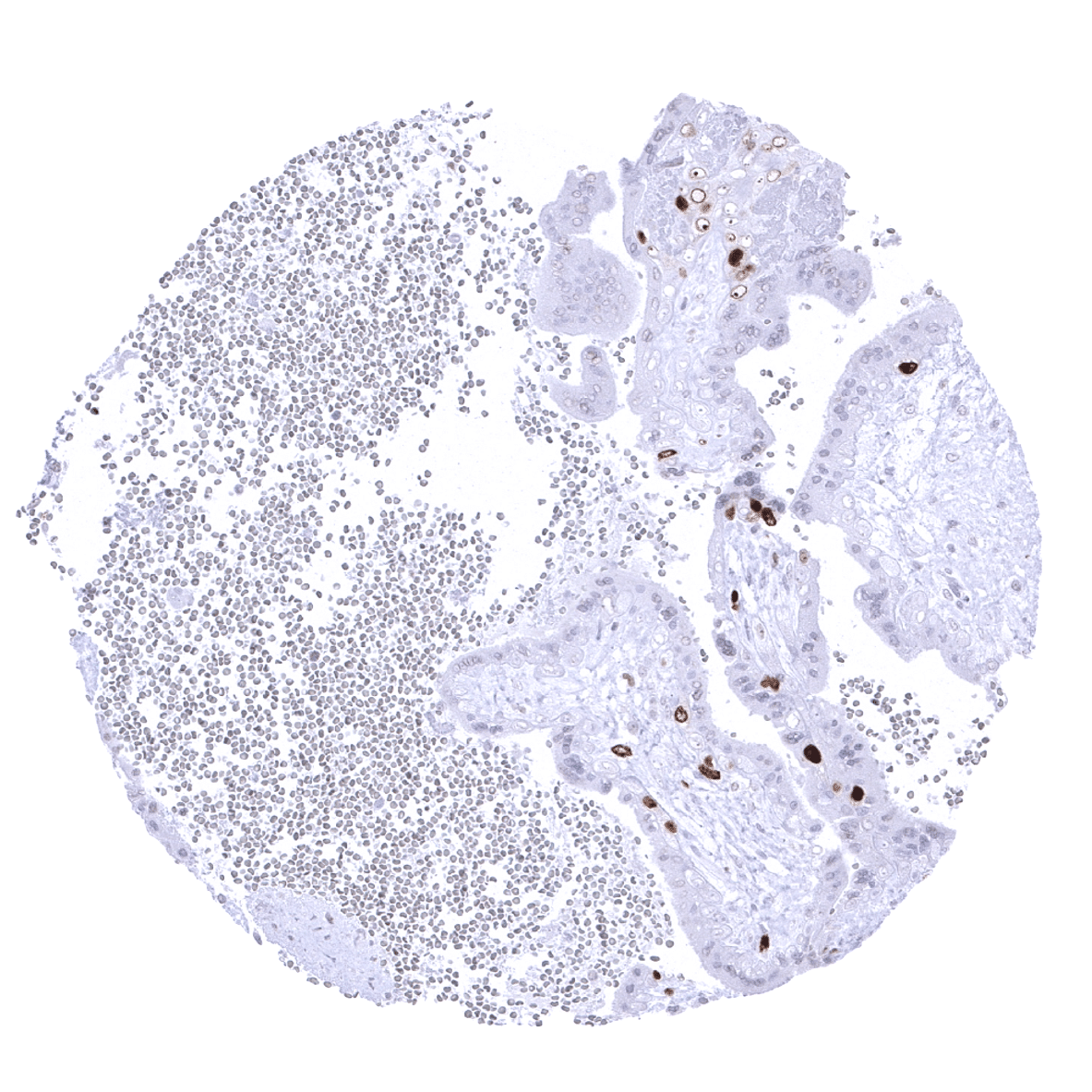
Tonsil, surface epithelium - In the tonsil, TOPO2A immunostaining can regularly be seen in epithelial cells and lymphocytes with a distribution pattern consistent with proliferating cells.
Urinary bladder, muscular wall
Urinary bladder, urothelium

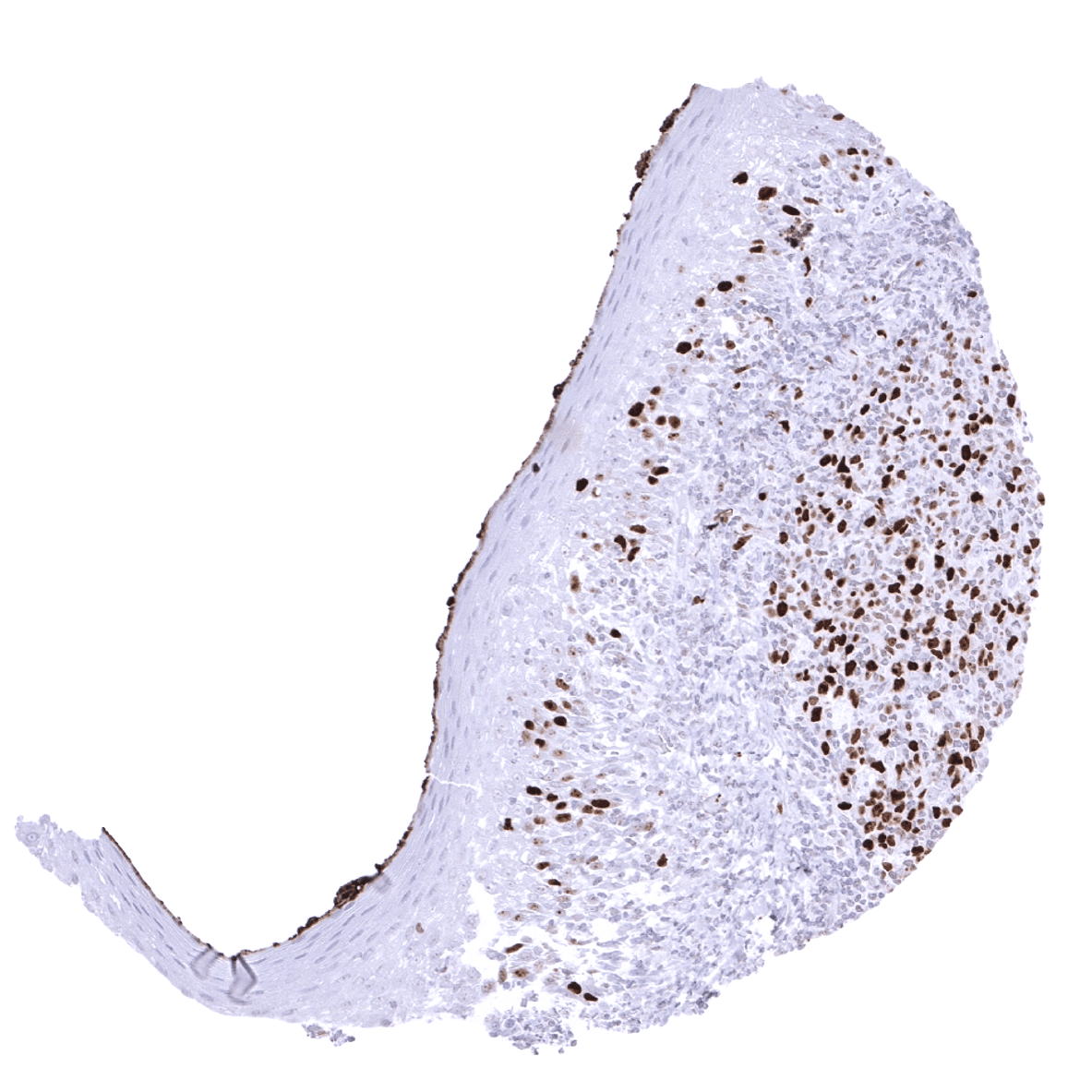
Uterus, ectocervix - Few proliferating cells show TOPO2A immunostaining.
Uterus, endocervix
Uterus, endometrium (proliferation)
Uterus, endometrium (secretion)

Uterus, myometrium