

195,00 € – 695,00 €
Product details
Synonyms = NEF; Protein S100-B; S-100 protein beta chain; S100 calcium binding protein beta (neural); S100 calcium-binding protein B; S100 protein beta chain; S100B; S100beta
Antibody type = Recombinant Rabbit monoclonal / IgG
Clone = MSVA-490R
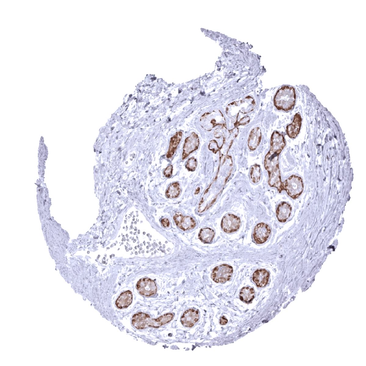
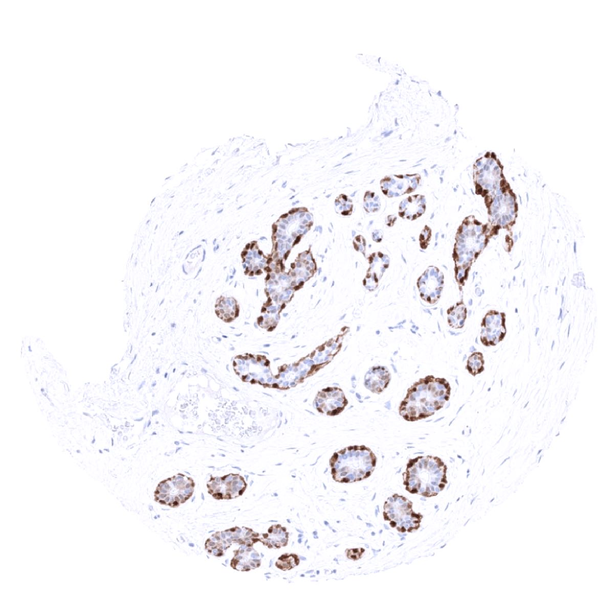
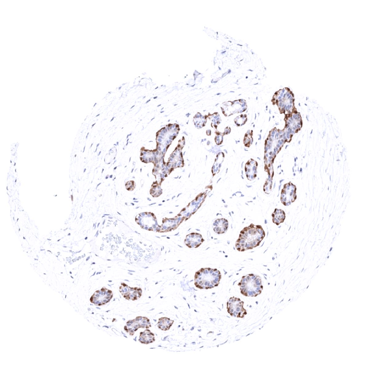
Positive control = Appendix: Schwann cells of peripheral nerves and adipocytes should show a strong, predominantly cytoplasmic S100B staining.
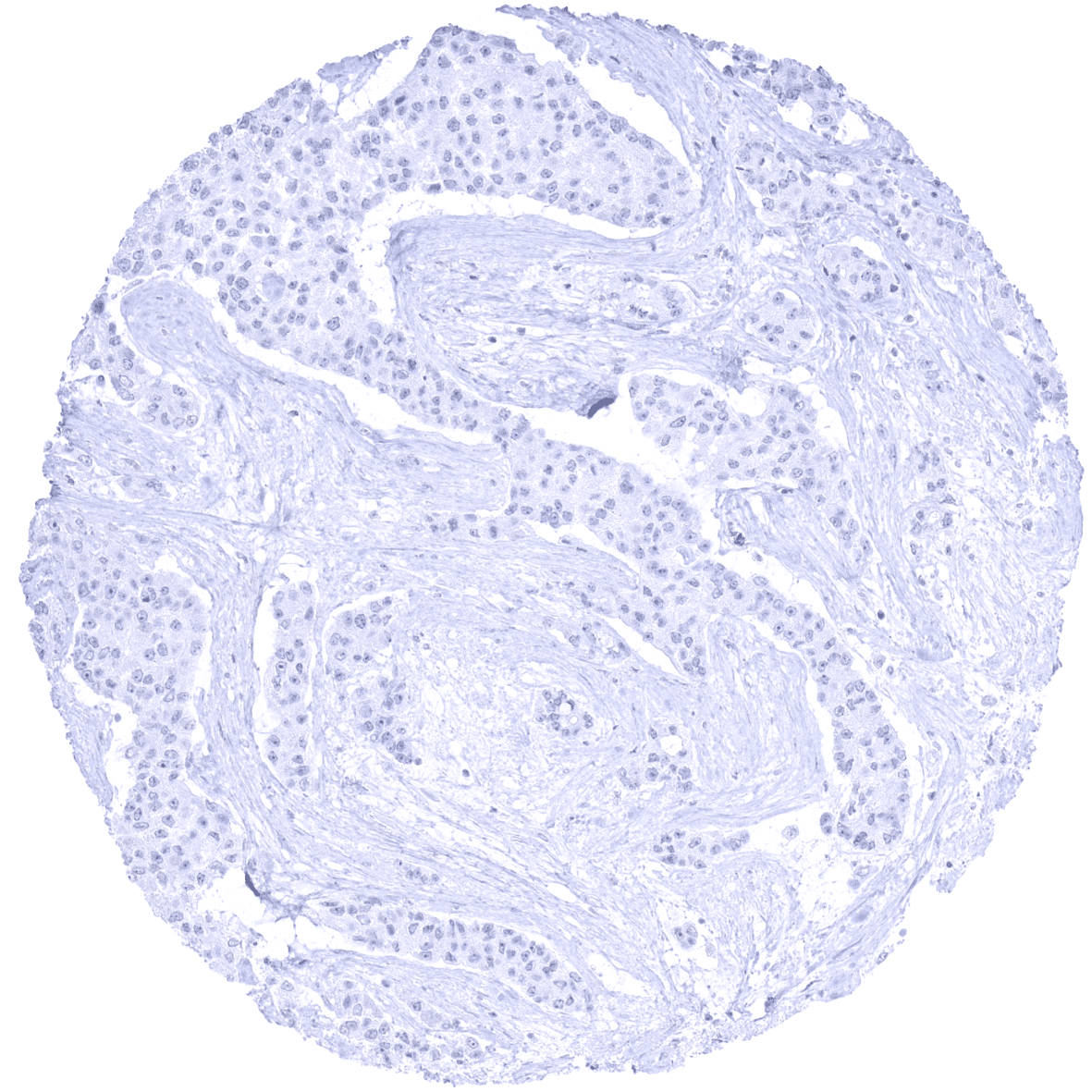
Negative control = Appendix: Smooth muscle and epithelial cells must not show any S100B staining.
Cellular localization = Cytoplasmic (and membranes/nuclei)
Reactivity = Human
Application = Immunohistochemistry
Dilution = 1:100 – 1:200
Intended Use = Research Use Only
Relevance of Antibody
Biology Behind
The S100 genes are a group of water soluble low-molecular-weight proteins characterized by two calcium-binding sites that have a specific helix-loop-helix (“EF-hand type”) conformation. The “S100” gene name is derived from the fact that these proteins are soluble in 100%. There are at least 21 family members but because most cells containing S100 protein also express the beta chain, S100B has become almost synonymous with S100 protein. S100 beta is coded by a gene at chromosome 21q22. Its functions involve microtubule assembly, cation diffusion across lipid membranes, and RNA polymerase activity. In diagnostic pathology, S100B – also termed “S100” – is used as a marker for Schwann cells and melanocytes. In these cell types, S100B is highly expressed, however, S100B is also regularly seen in various other cell types.
Staining Pattern in Normal Tissues
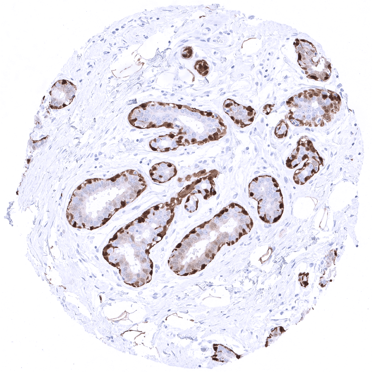
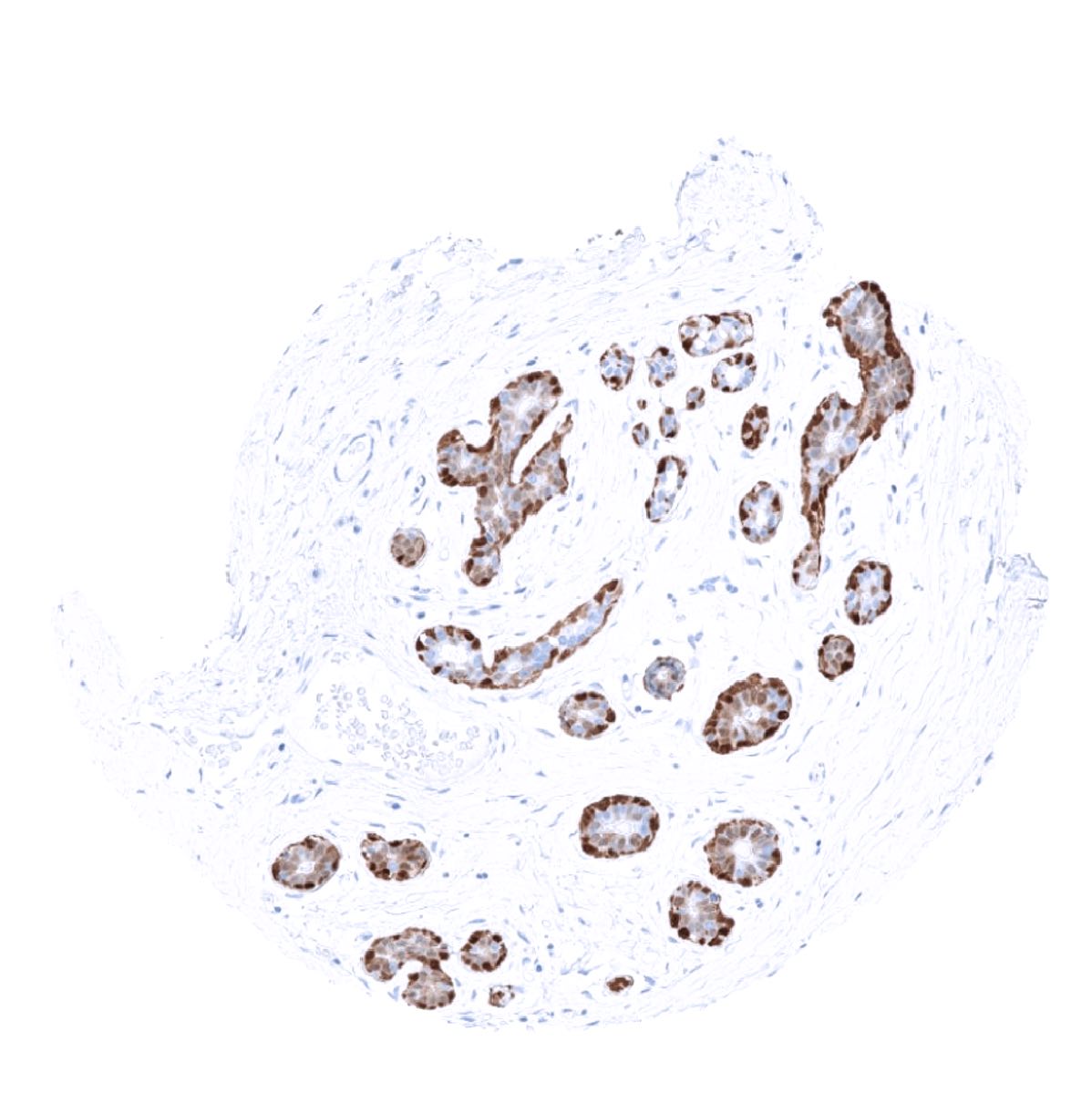
S100 protein can be found in cell membranes, cytoplasm and nuclei. S100 immunostaining shows strong positivity in cerebrum, cerebellum, neurohypophysis, peripheral nerves (Schwann cells) which are visible in most organs, myoepithelial cells of salivary glands and breast glands, fat cells, Langerhans cells, sustentacular cells, melanocytes, chondrocytes, subsets of dendritic cells and lymphocytes as well as serous cells in bronchial glands. A weaker S100 staining is seen in adrenal medullary cells, a fraction of cells in the adenohypophysis, and occasionally also in Sertoli cells of the testis, a fraction of islet cells of the pancreas, and breast luminal cells.
These findings are largely consistent with RNA and protein data summarized in the Human Protein Atlas (Tissue expression S100).
Suggested positive tissue control: Appendix: Schwann cells of peripheral nerves and adipocytes should show a strong, predominantly cytoplasmic S100 staining.
Suggested negative tissue control: Appendix: Smooth muscle and epithelial cells must not show any S100 staining.

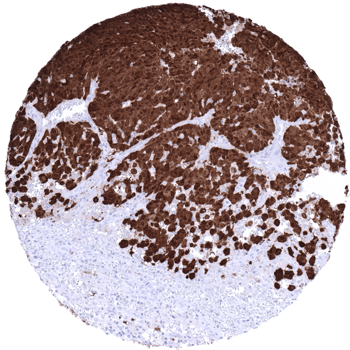
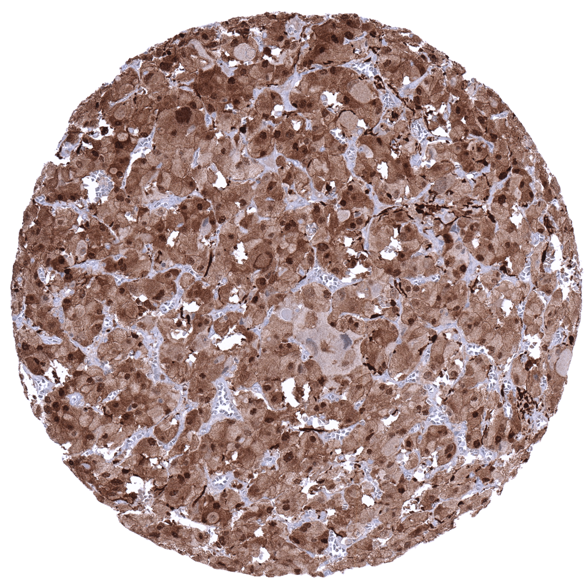
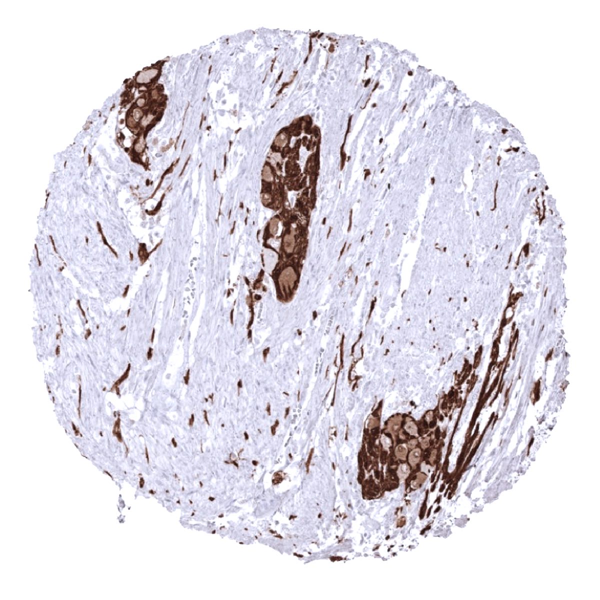
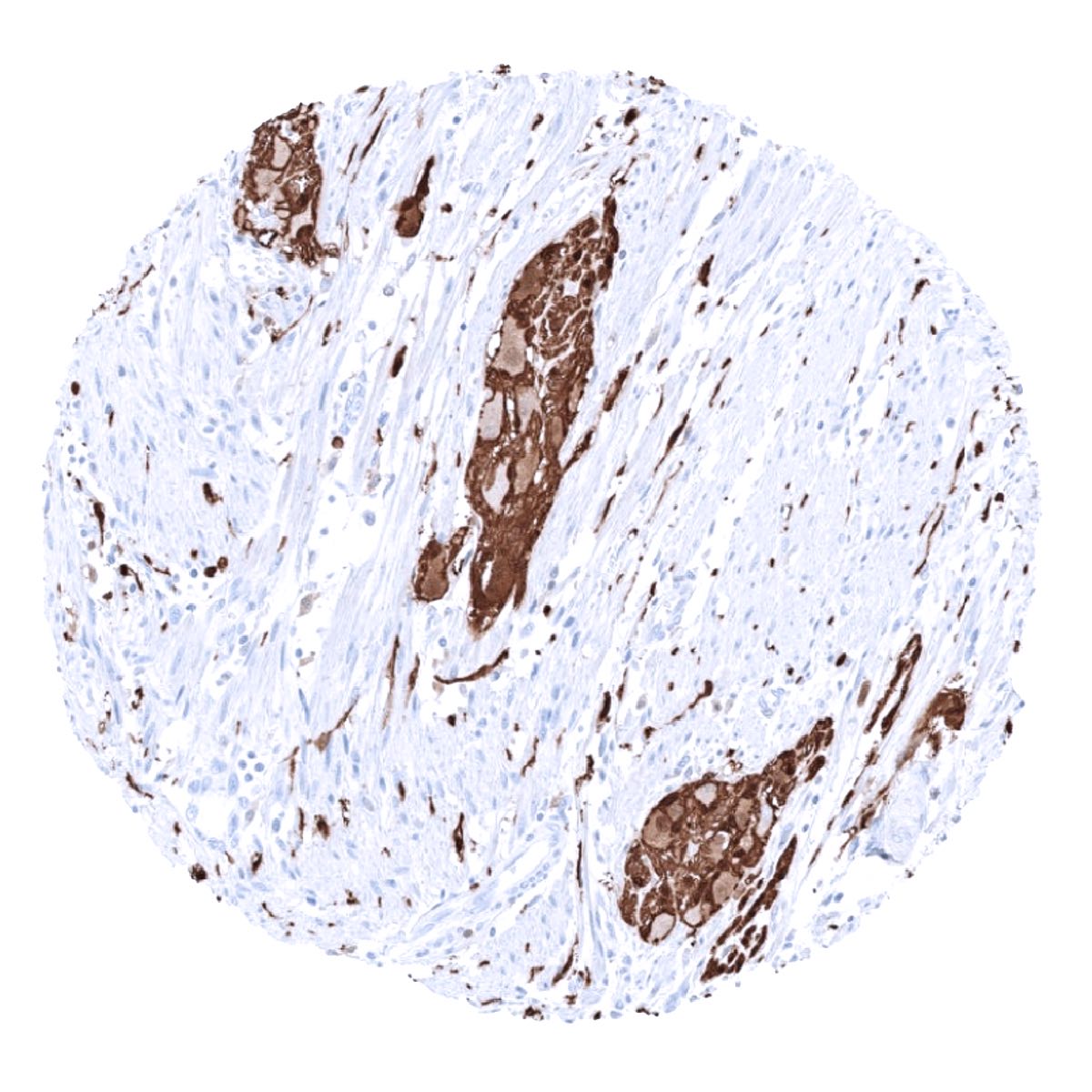
Staining Pattern in Relevant Tumor Types
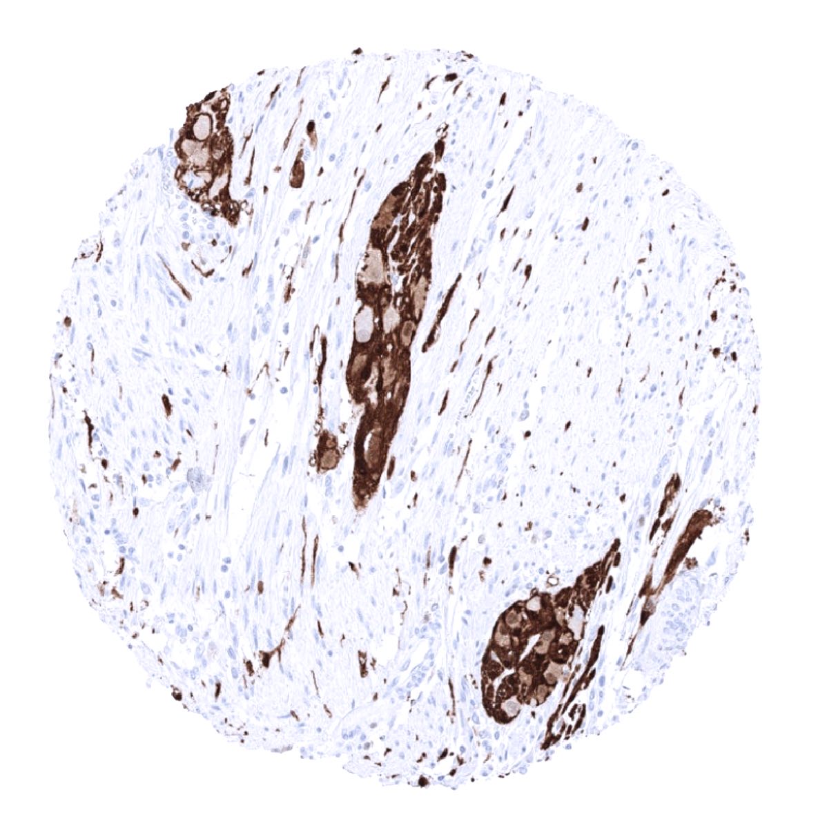
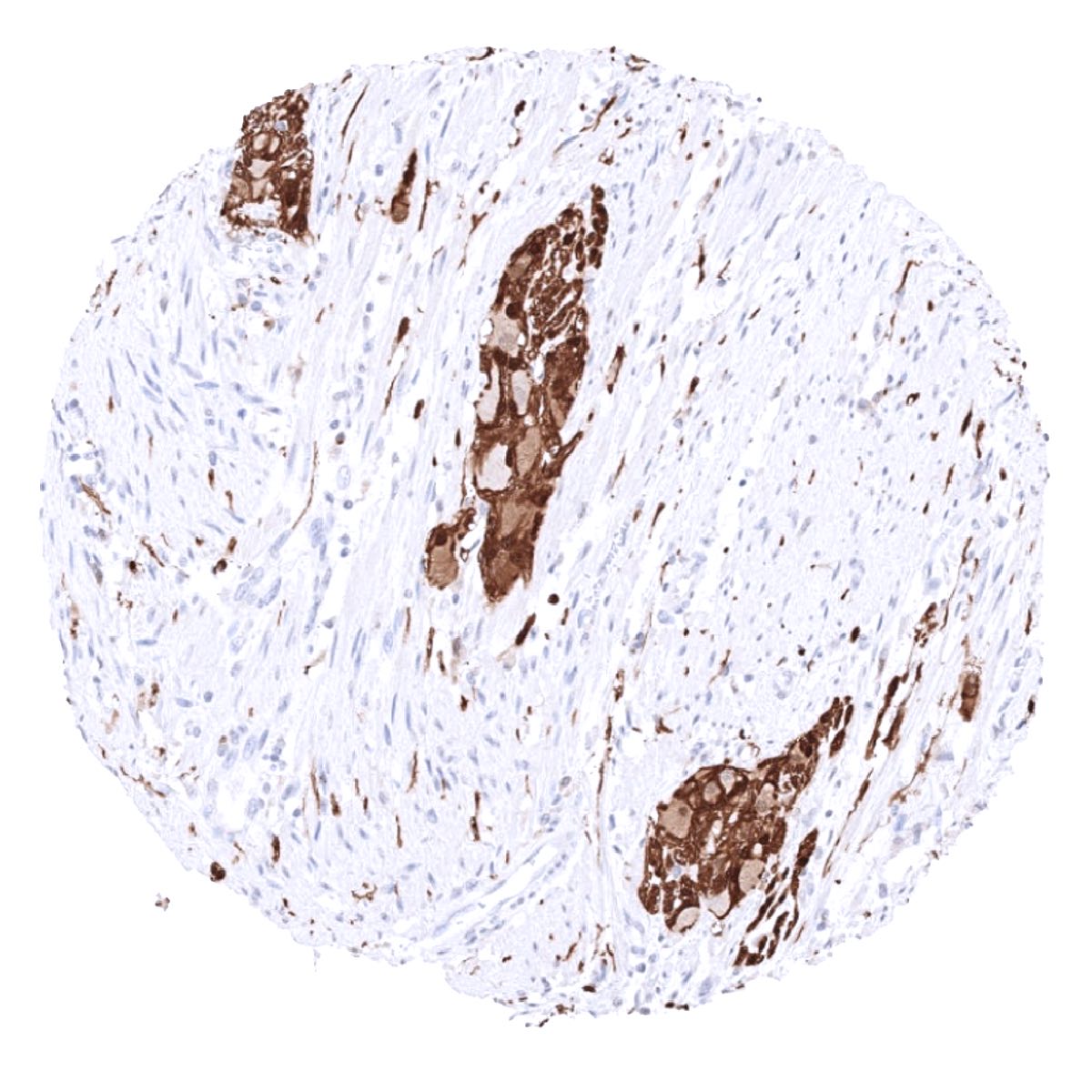
S100 expression is seen in many different tumor types. A positive S100 immunostaining is particularly frequent in brain tumors of various types, benign and malignant melanocytic tumors, Schwannoma, neurofibroma, granular cell tumor, myoepithelial tumors, Langerhans cell histiocytosis, benign and malignant lipomatous tumors, primitive neuroectodermal tumors, neuroblastoma, clear cell sarcoma, rhabdomyosarcoma, chordoma, chondroid tumors, sweat gland carcinoma, Sertoli-Leydig cell tumors, synovial sarcoma, Ewing sarcoma, meningioma, and neuroendocrine tumors. S100 positivity also occurs in a variable fraction of various different epithelial tumors.
The TCGA findings on S100 RNA expression in different tumor categories have been summarized in the Human Protein Atlas.
Compatibility of Antibodies
No data available at the moment
Protocol Recommendations
IHC users have different preferences on how the stains should look like. Some prefer high staining intensity of the target stain and even accept some background. Others favor absolute specificity and lighter target stains. Factors that invariably lead to more intense staining include higher concentration of the antibody and visualization tools, longer incubation time, higher temperature during incubation, higher temperature and longer duration of the heat induced epitope retrieval (slide pretreatment). The impact of the pH during slide pretreatment has variable effects and depends on the antibody and the target protein. Accordingly, multiple different protocols can generate identical staining results.
All images and data shown here and in our image galleries are obtained by the manual protocol described below. Other protocols resulting in equivalent staining are described as well.
Manual protocol
Freshly cut sections should be used (less than 10 days between cutting and staining). Heat-induced antigen retrieval for 5 minutes in an autoclave at 121°C in pH 9 Target Retrieval Solution buffer. Apply MSVA-490R at a dilution of 1:200 at 37°C for 60 minutes. Visualization of bound antibody by the EnVision Kit (Dako, Agilent) according to the manufacturer’s directions.
Agilent / DAKO – Autostainer Link 48
Pretreatment in PT-Link for 30 minutes at 95°C (pH high); FLEX peroxidase blocking for 5 minutes (room temperature), MSVA-490R 1:150 for 20 minutes (room temperature), FLEX+ mouse/rabbit (LINKER) for 15 minutes (room temperature), horseradish peroxidase (HRP) for 20 minutes (room temperature), FLEX DAB+Sub-Chromo for 10 minutes (room temperature), FLEX hematoxylin for 5 minutes (room temperature).
These images reflect stainings by the protocol described above. It is of note that a comparable staining result can also be obtained by different protocols. In general, a longer pretreatment, a longer incubation time of the primary antibody, a higher antibody concentration, and a longer incubation time of FLEX+LINKER result in stronger staining, potentially at the cost of more background staining. Modifications of the protocol with a strengthening effect on staining intensity in combination with changes of other parameters that result in lower staining intensity can result in a comparable result as shown above.
Leica – BOND RX
Dewax at 72°C for 30 seconds; Pretreatment in Bond Epitope Retrieval Solution (ER2 – EDTA pH9) for 20 minutes at 100°C; Peroxidase blocking for 5 minutes (room temperature), MSVA-490R 1:150 for 15 minutes (room temperature), Post primary (rabbit anti mouse) for 8 minutes (room temperature), Polymer (goat anti rabbit) for 8 minutes (room temperature), mixed DAB refine for 10 minutes (room temperature), hematoxylin for 5 minutes (room temperature).

These images reflect stainings by the protocol described above. It is of note that a comparable staining result can also be obtained by different protocols. In general, a longer pretreatment, a longer incubation time of the primary antibody, a higher antibody concentration, a higher temperature during incubation, and a longer incubation time of Post primary and or the Polymer result in stronger staining, potentially at the cost of more background staining. Modifications of the protocol with a strengthening effect on staining intensity in combination with changes of other parameters that result in lower staining intensity can result in a comparable result as shown above.
Roche – Ventana Discovery ULTRA
Pretreatment for 64 minutes at 100°C (pH 8,4); CM peroxidase blocking for 12 minutes (room temperature), MSVA-490R 1:150 for 20 minutes at 36°C, secondary antibody (anti-rabbit HQ) for 12 minutes at 36°C, anti-HQ HRP for 12 minutes at room temperature, DAB at room temperature, hematoxylin II at room temperature for 8 minutes, bluing reagent at room temperature for 4 minutes.
These images depict staining results obtained by the protocol described above. It is of note, that the Ventana machines generally require higher antibody concentrations than other commonly used autostainers because the antibodies are automatically diluted during the procedure. Various other protocols can result in an identical result as shown above. A longer pretreatment, a longer incubation time of the primary antibody, a higher antibody concentration, a higher temperature during incubation, and a longer incubation time of secondary antibody and or the anti-HQ HRP result in stronger staining, potentially at the cost of more background staining.
Potential Research Applications
- The diagnostic utility of S100 expression analysis should be further investigated in a large cohort of tumors from different entities.
- The clinical/biological significance of S100 expression in epithelial tumors is unknown.
- The role of S100 positive lymphocytes and dendritic cells is not clear.
Evidence for Antibody Specificity in IHC
Specificity of MSVA-490R is documented by strong positive staining in cell types that are well documented to express S100 such as peripheral nerves, myoepithelial cells, fat cells, Langerhans cells, or sustentacular cells in combination with absence of staining in all tissues known to not express S100 including tissues notorious for non-specific IHC background such as kidney and colonic mucosa.
