
195,00 € – 695,00 €
Product details
Synonyms = CK6A, CK6B, CK6C, CK6D, CK6E, Keratin Type II Cytoskeletal 6A, Keratin Type II Cytoskeletal 6B, Keratin Type II Cytoskeletal 6C, Keratin Type II Cytoskeletal 6D, Keratin Type II Cytoskeletal 6E, KRT6, KRT6A, KRT6B, KRT6C, KRT6D, KRT6E
Antibody type = Recombinant Rabbit monoclonal / IgG1
Clone = MSVA-606R
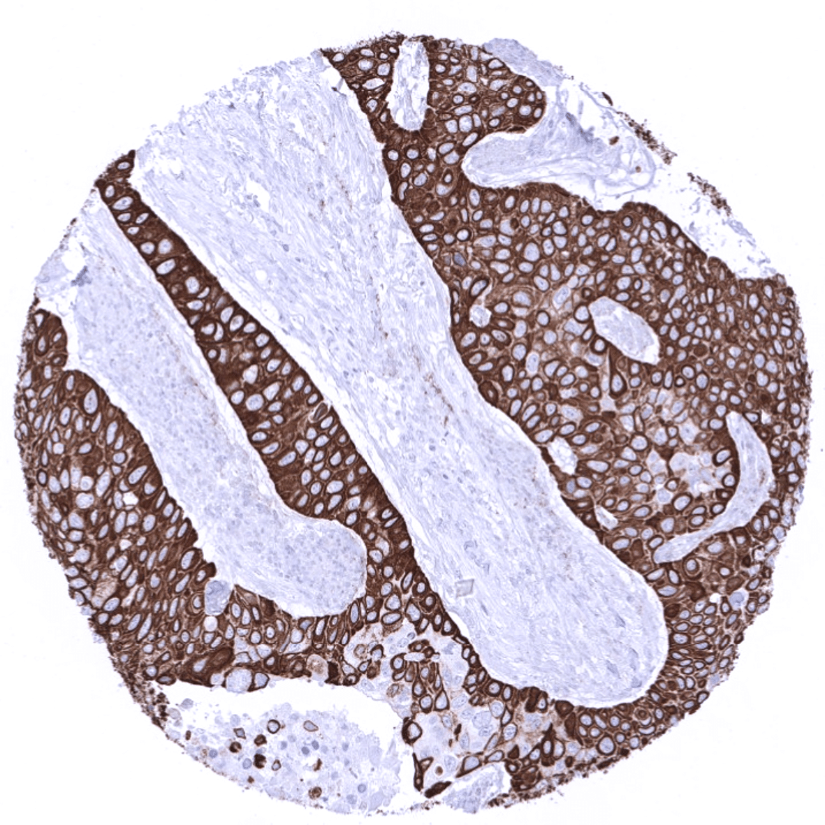
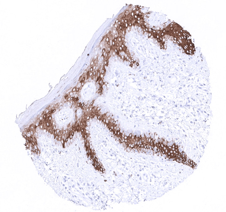
Positive control = Tonsil: A strong CK6 staining should be seen in all epithelial cells of the surface epithelium except the basal cell layer where staining should be absent or weak.
Negative control = Tonsil: CK6 staining should be absent in lymphocytes and all other mesenchymal cells.
Cellular localization = Cytoplasmic
Reactivity = Human
Application = Immunohistochemistry
Dilution = 1:100 – 1:200
Intended Use = Research Use Only
Relevance of Antibody
Biology Behind
Cytokeratin 6 (CK6) or keratin 6 (KRT6) is a basic high molecular weight Type II cytokeratin. It is an integral part of the cytoskeleton of various mostly squamous epithelial cell types in various organs. There are 3 keratin 6 isoforms which are all coded by neighboring genes located within the type II keratin gene cluster on chromosome 12q. The Keratins 6A, 6B and 6C genes consist of 9 exons and share more than 99% similarity in their DNA coding sequences. Most keratins form heteropolymers consisting of a type I and a type II keratin. All keratin 6 isoforms pair with keratin 16 and/or keratin 17. Keratin 6A has antimicrobial properties. Mutations of keratin 6 lead to the PC-K6A subtype of pachyonychia congenita with structural abnormalities of the nails, the epidermis of the palms and soles, and oral epithelia.[1]
[1] Völkel et al. “Cytokeratin 5 and cytokeratin 6 expressions are unconnected in normal and cancerous tissues and have separate diagnostic implications” Virchows Archiv published online; September 24th 2021
Staining Pattern in Normal Tissues
Cytokeratin 6 staining pattern in Normal Tissues with antibody MSVA-606R (images are shown in our “Normal Tissue Gallery”)
| Brain | Cerebrum | Negative. |
| Cerebellum | Negative. | |
| Endocrine Tissues | Thyroid | Negative. |
| Parathyroid | Negative. | |
| Adrenal gland | Negative. | |
| Pituitary gland | Negative. | |
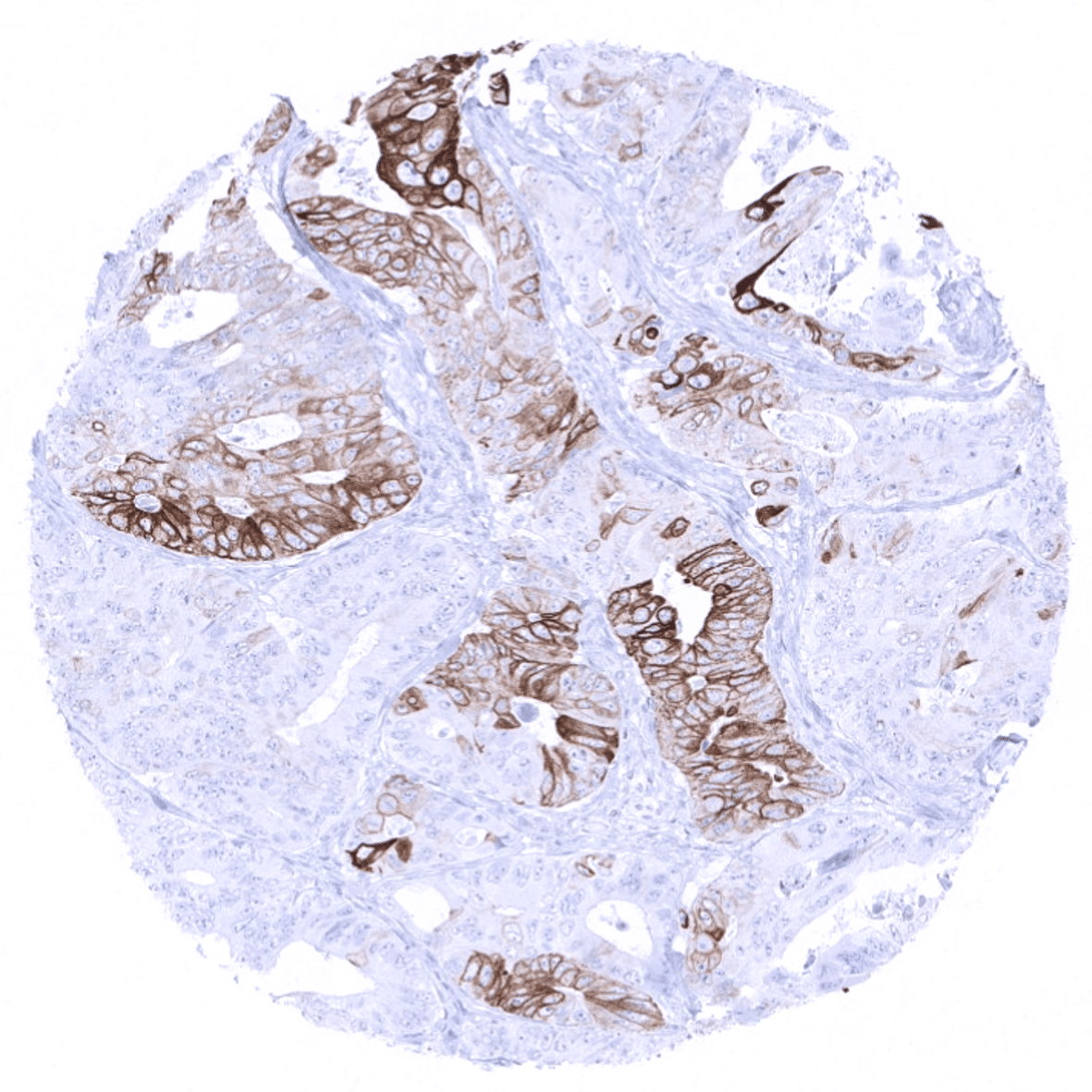
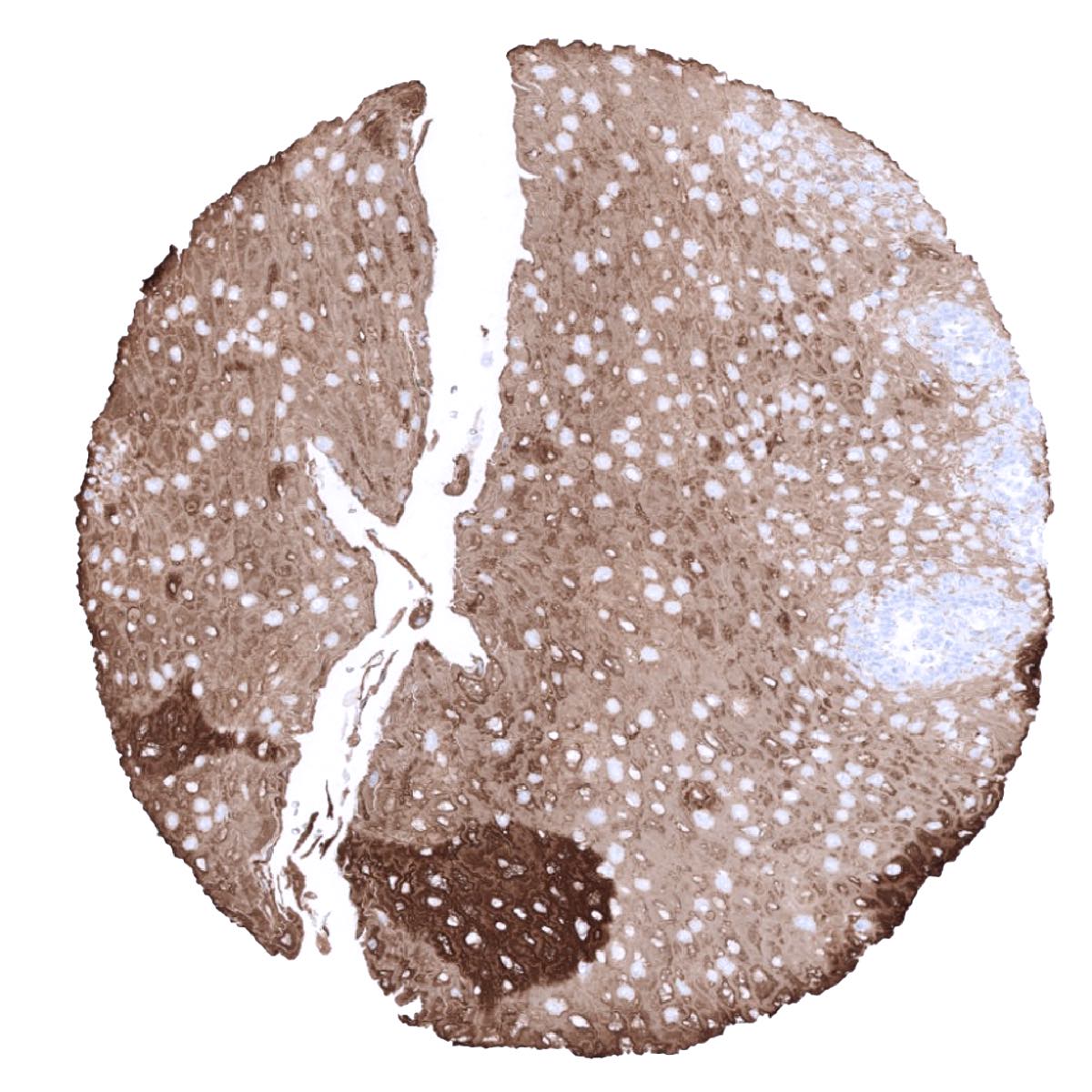
| Respiratory system | Respiratory epithelium | Moderate to strong CK6 staining of basal cells and sometimes also ciliated cells of respiratory epithelium. Moderate to strong positivity also in intercalated ducts of bronchial glands. |
| Lung | Negative. | |
| Gastrointestinal Tract | Salivary glands | Moderate to strong CK6 positivity in intercalated ducts. |
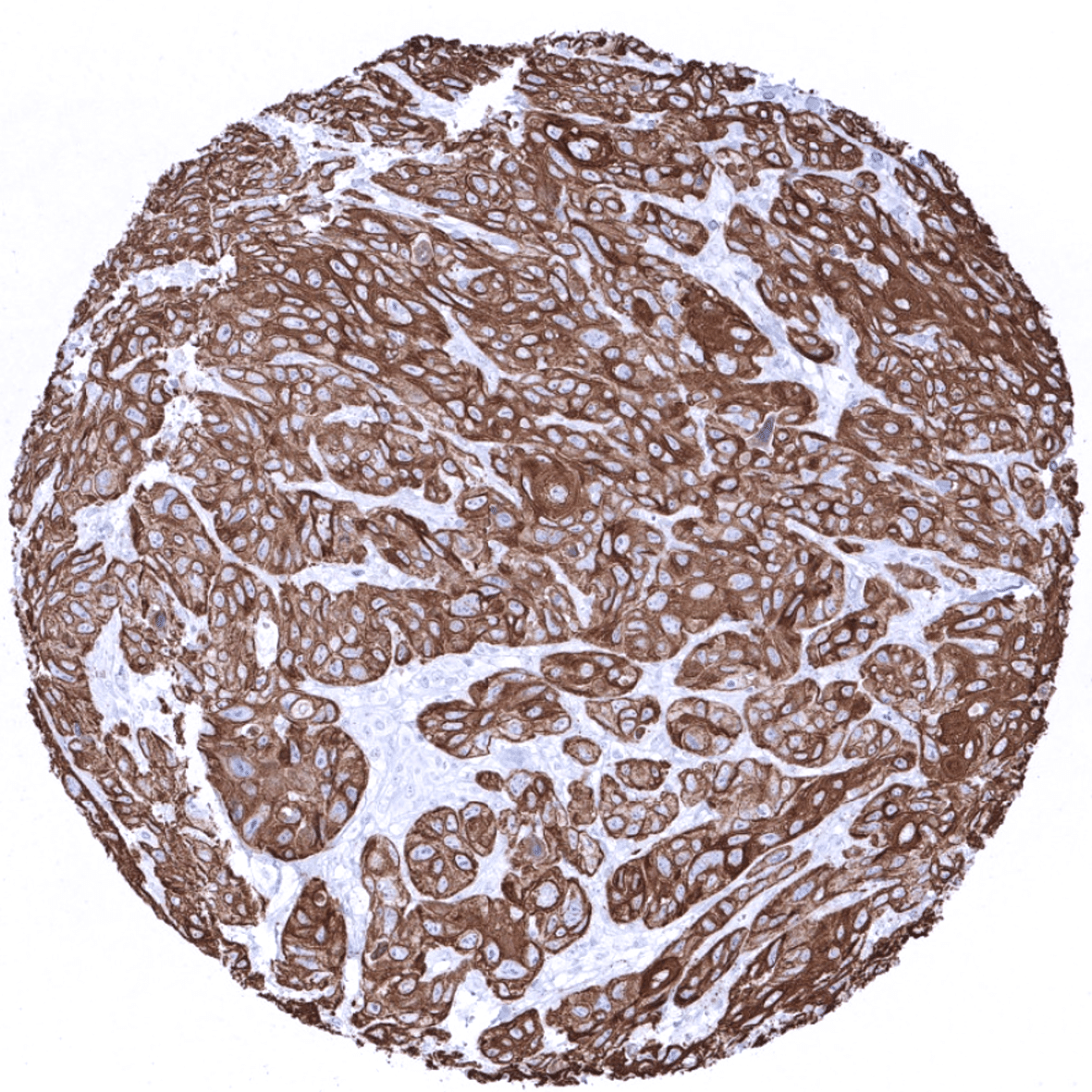
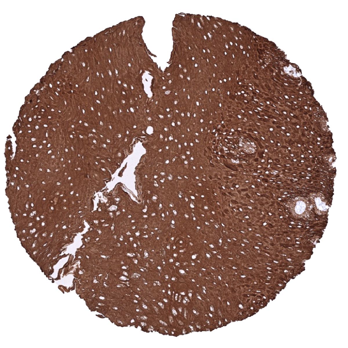
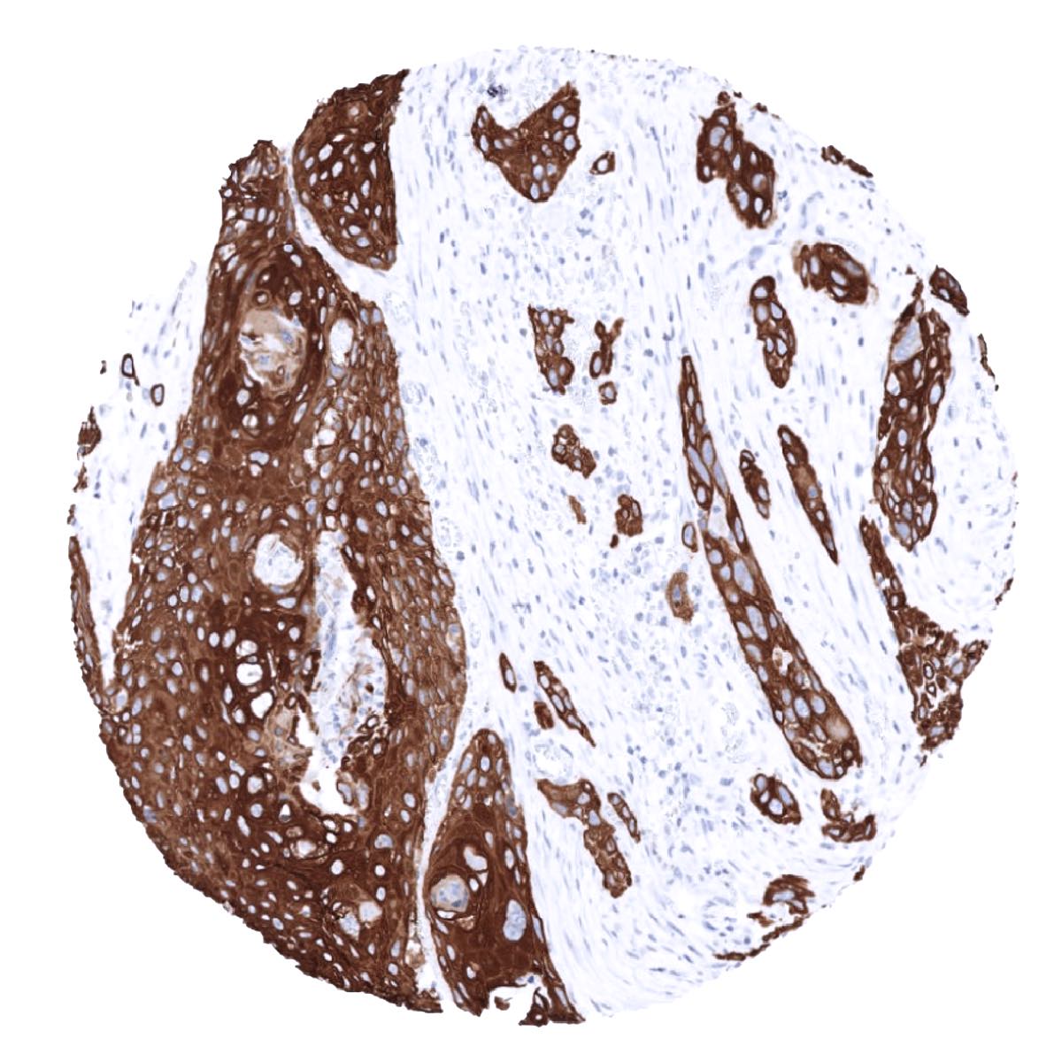
| Esophagus | Strong CK6 positivity in the squamous epithelium but the basal cell layer is negative. | |
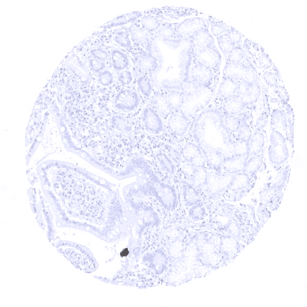
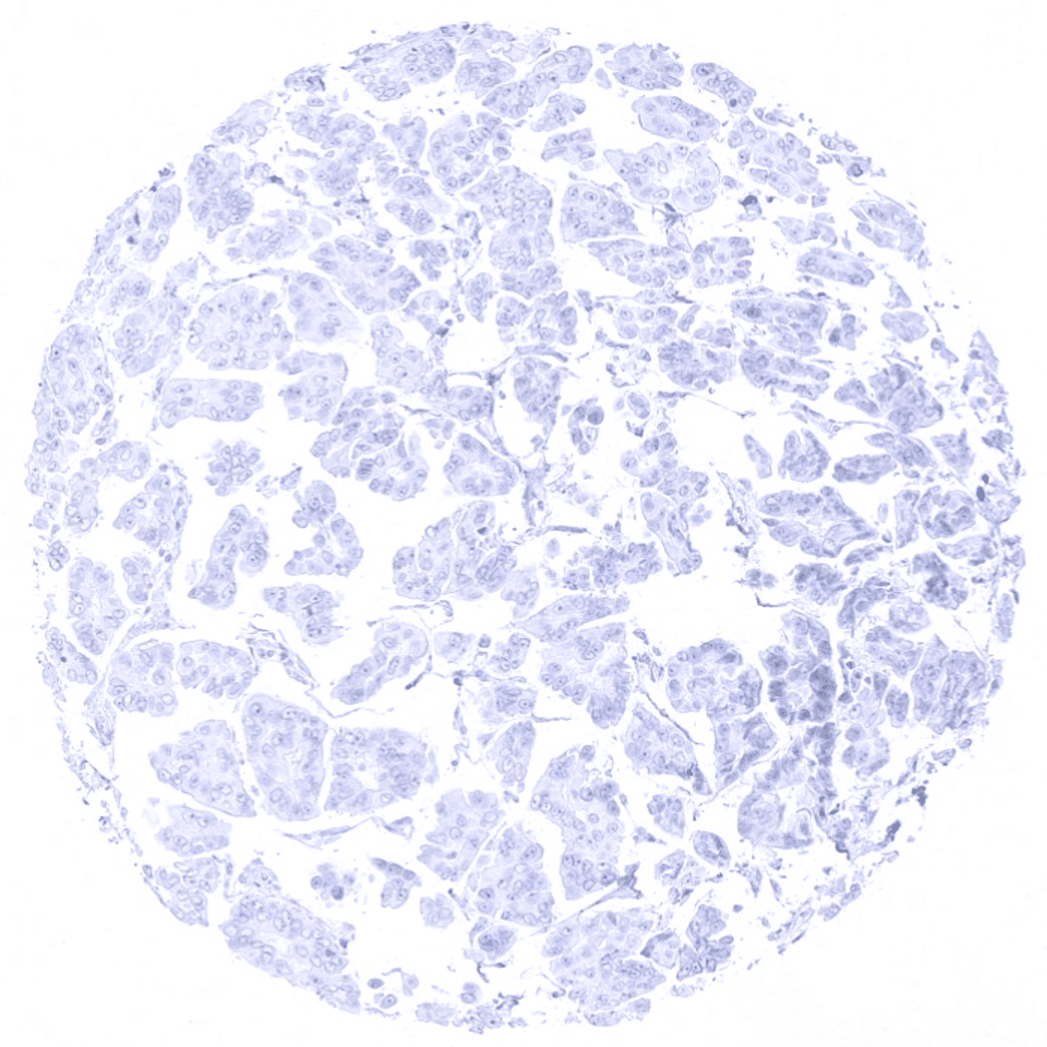
| Stomach | Negative. | |
| Duodenum | Negative. | |
| Small intestine | Negative. | |
| Appendix | Negative. | |
| Colon | Negative. | |
| Rectum | Negative. | |
| Liver | Negative. | |
| Gallbladder | Negative. | |
| Pancreas | Negative. | |
| Genitourinary | Kidney | Negative. |
| Urothelium | Negative. | |
| Male genital | Prostate | Negative. |
| Seminal vesicles | Negative. | |
| Testis | Negative. | |
| Epididymis | Negative. | |
| Female genital | Breast | Negative. |
| Uterus, myometrium | Negative. | |
| Uterus, ectocervix | Strong CK6 positivity in the squamous epithelium but the basal cell layer is negative. | |
| Uterus endocervix | Negative. | |
| Uterus, endometrium | Focal CK6 positivity can be seen in scattered endometrial cells of some samples. | |
| Fallopian Tube | Negative. | |
| Ovary | Negative. | |
| Placenta early | Negative. | |
| Placenta mature | Negative. | |
| Amnion | Strong CK6 positivity of amnion cells. | |
| Chorion | Strong CK6 positivity of chorion cells. | |
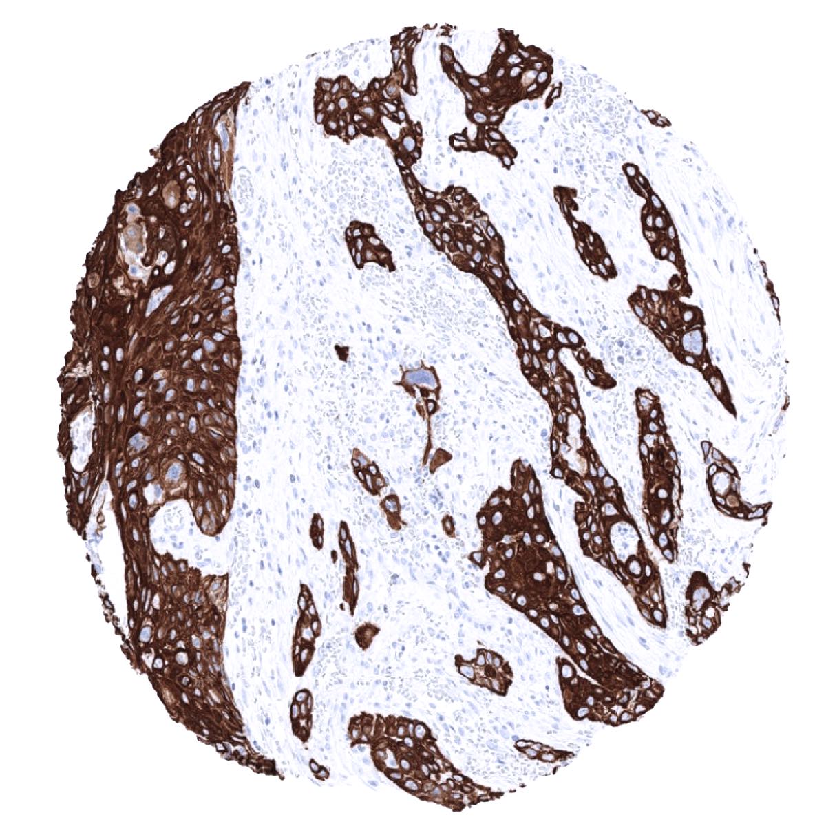
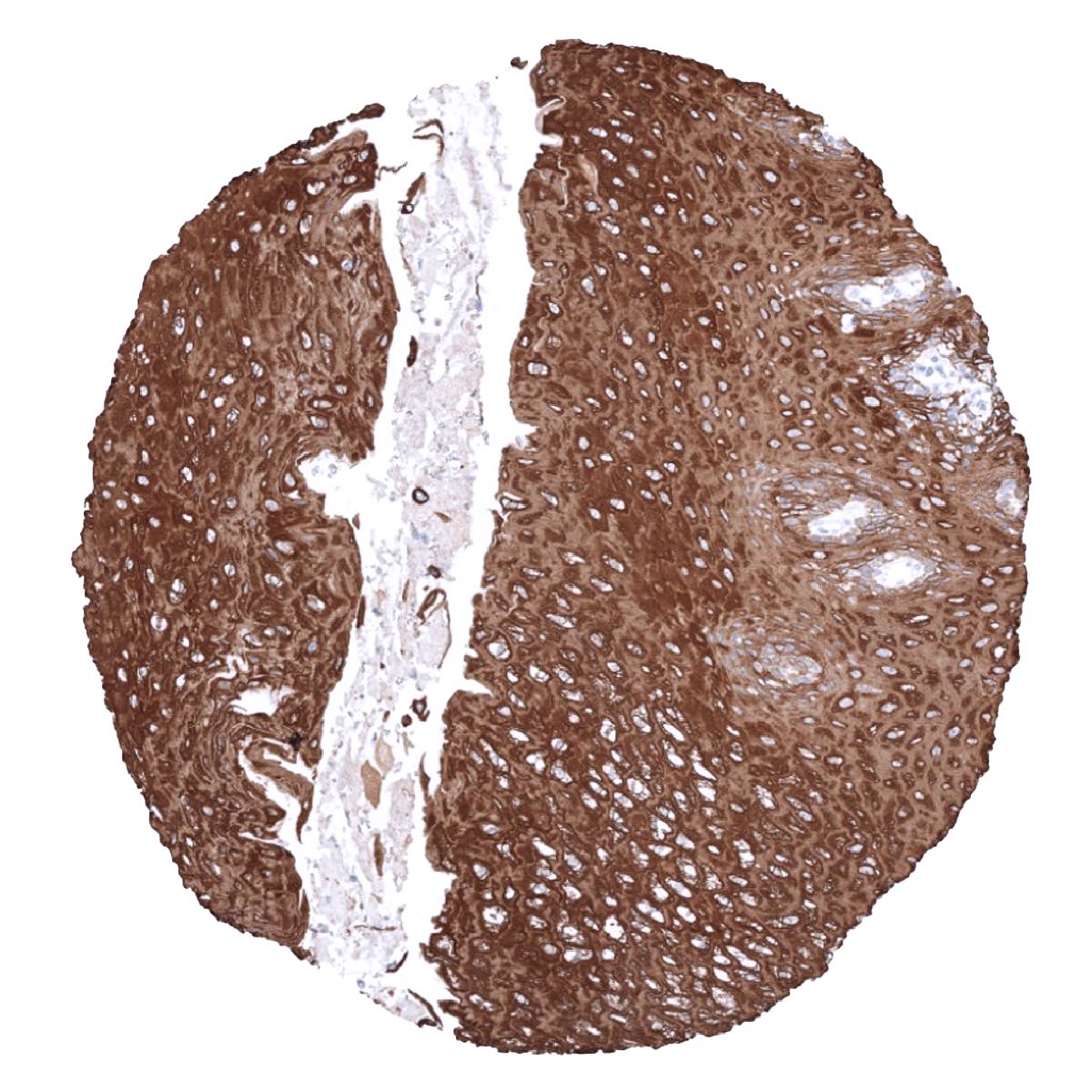
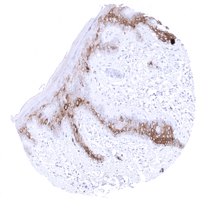
| Skin | Epidermis | Strong CK6 positivity in the squamous epithelium but the basal cell layer is negative. |
| Sebaceous glands | Strong CK6 positivity. | |
| Muscle/connective tissue | Heart muscle | Negative. |
| Skeletal muscle | Negative. | |
| Smooth muscle | Negative. | |
| Vessel walls | Negative. | |
| Fat | Negative. | |
| Stroma | Negative. | |
| Endothelium | Negative. | |
| Bone marrow/ lymphoid tissue | Bone marrow | Negative. |
| Lymph node | Negative. | |
| Spleen | Negative. | |
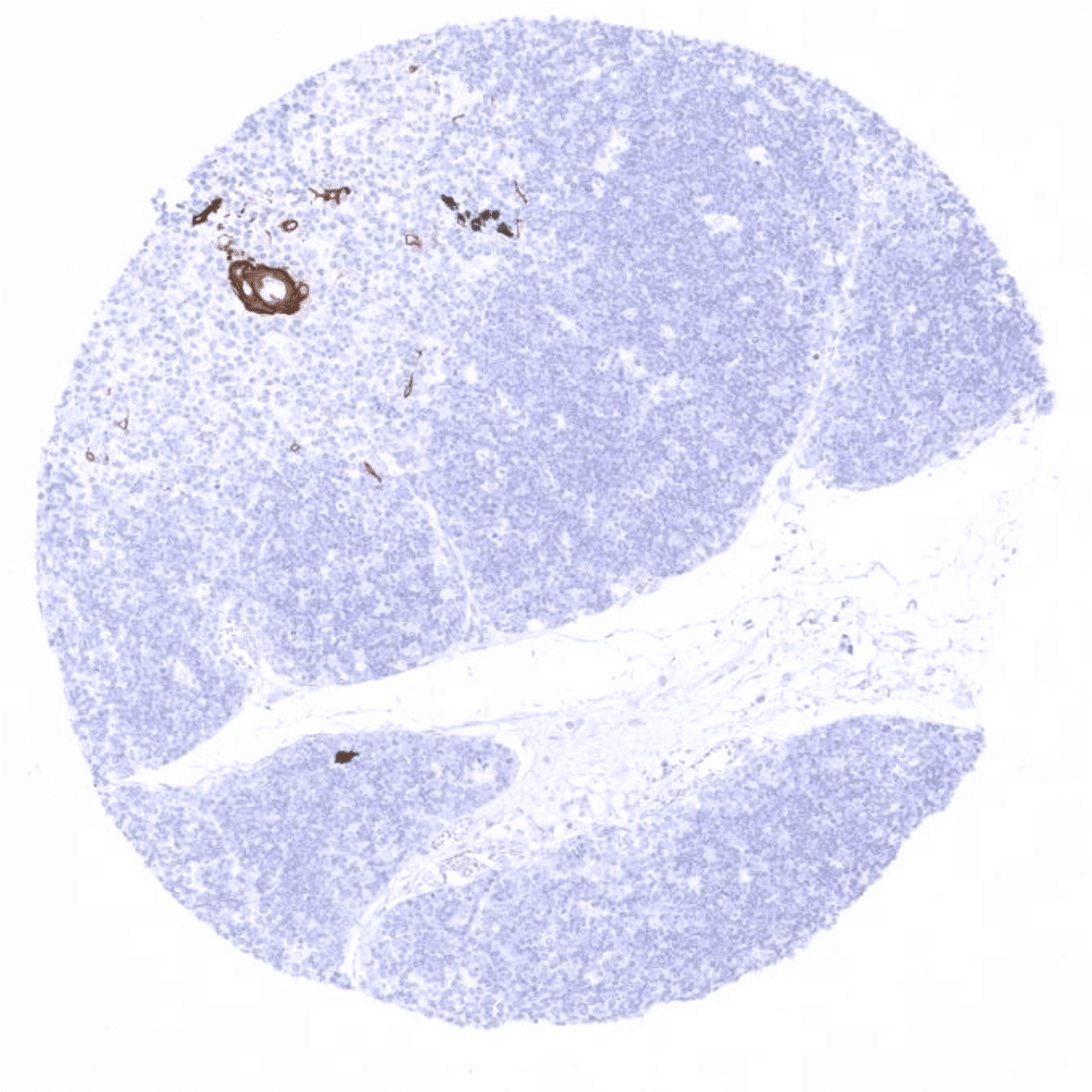
| Thymus | Strong CK6 positivity in a fraction of the squamous epithelial cells of corpuscles of Hassall’s. | |
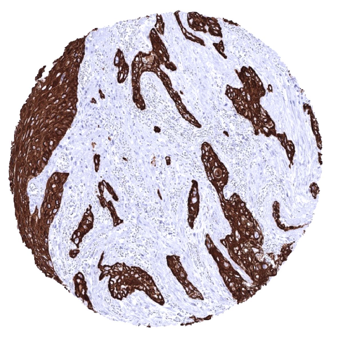
| Tonsil | Strong CK6 positivity in the squamous epithelium of the surface, where only the basal cell layer is negative. In the crypts, a fraction of squamous epithelial cells is positive. | |
| Remarks |
Suggested positive tissue control: Tonsil: A strong CK6 staining should be seen in all epithelial cells of the surface epithelium except the basal cell layer where staining should be absent or weak.
Suggested negative tissue control: Tonsil: CK6 staining should be absent in lymphocytes and all other mesenchymal cells.

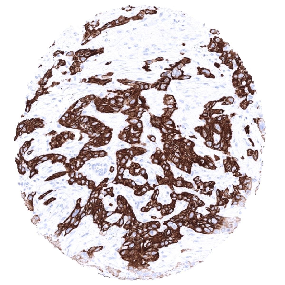
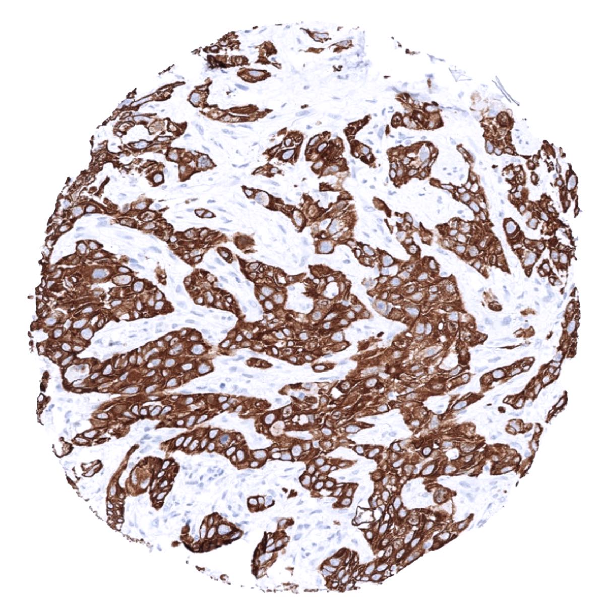
Staining Pattern in Relevant Tumor Types
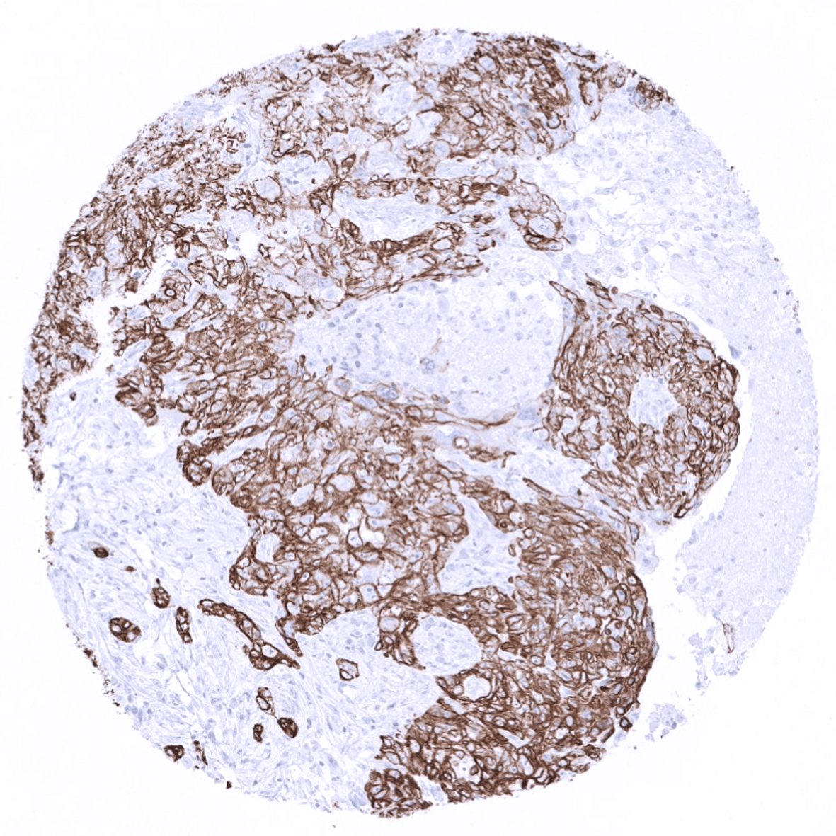
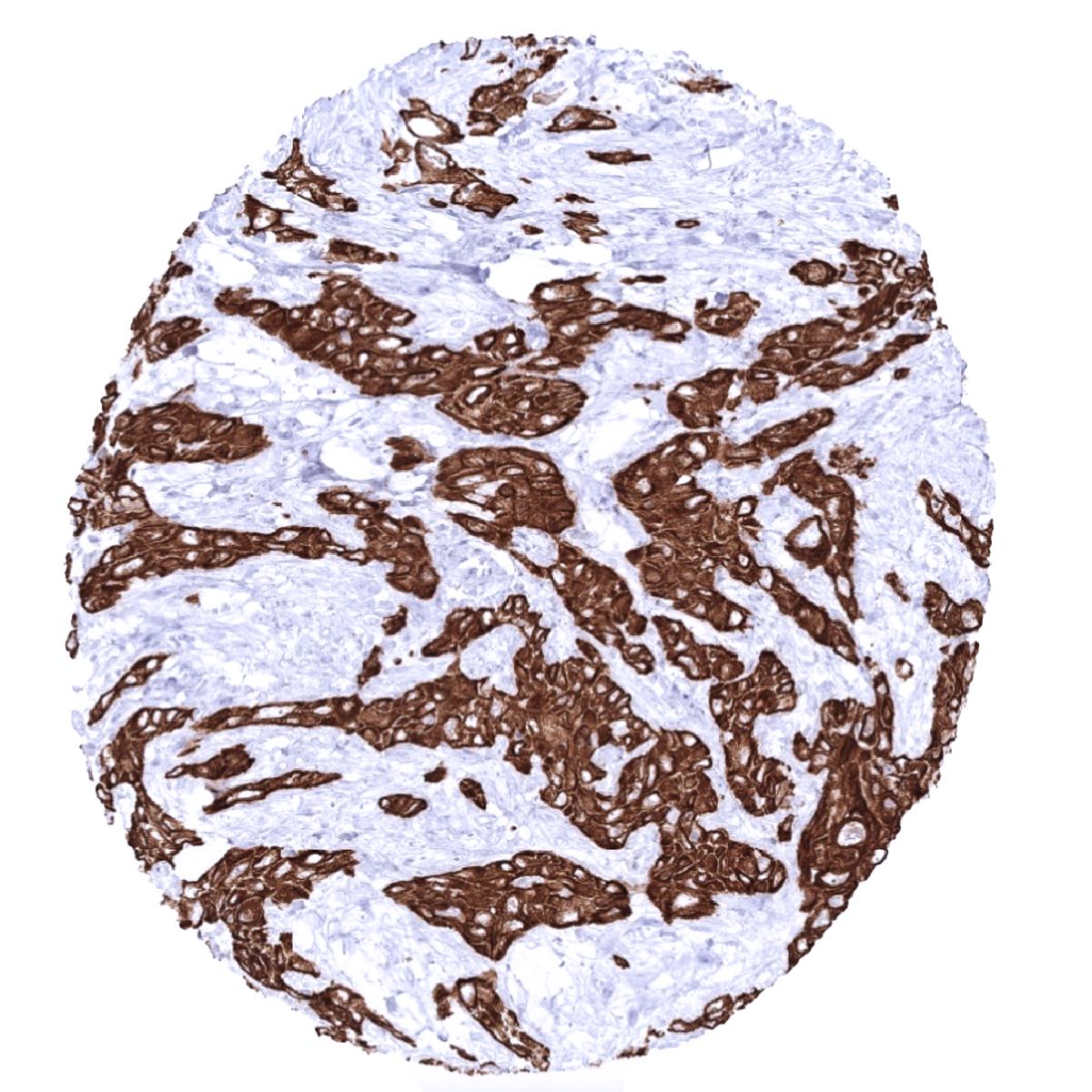
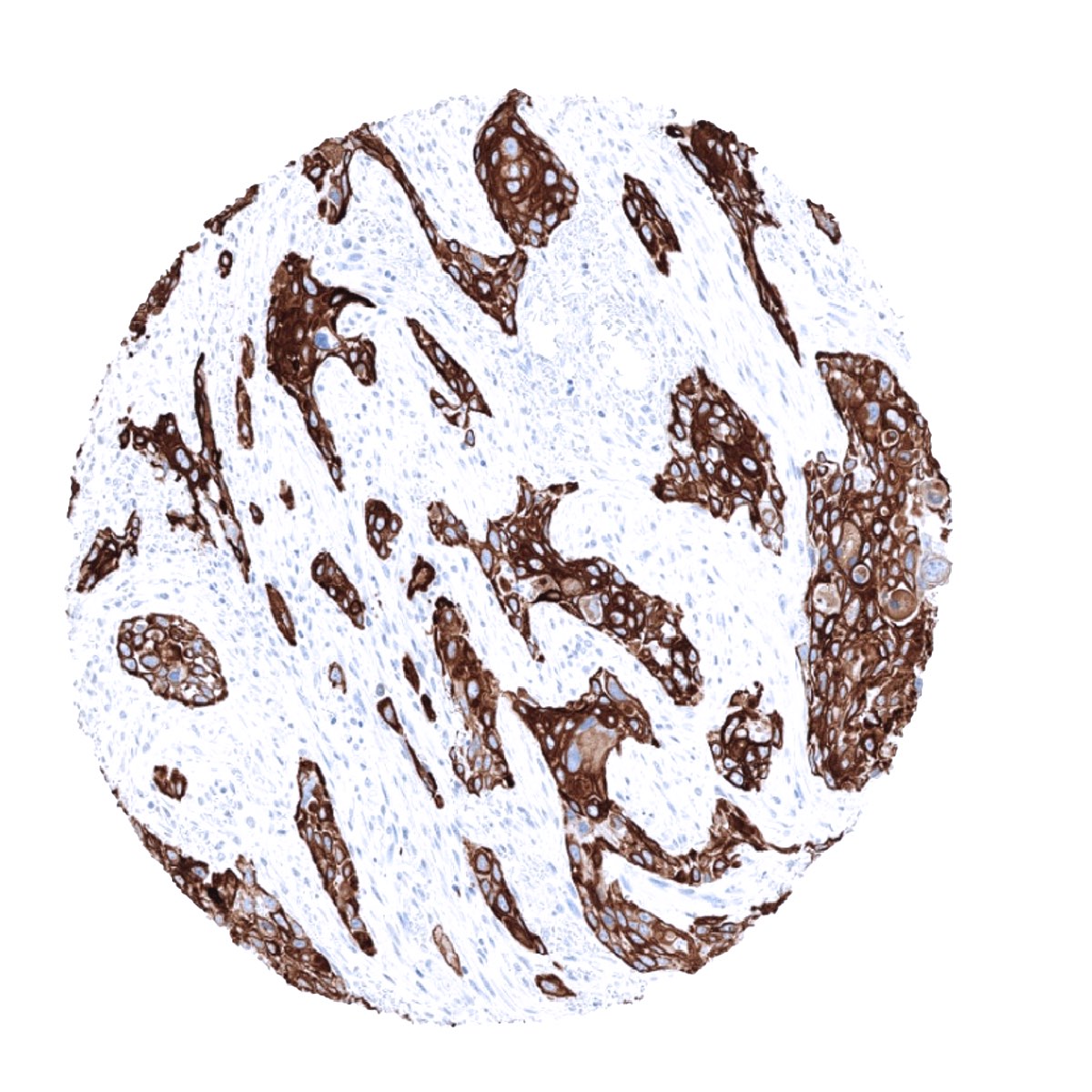
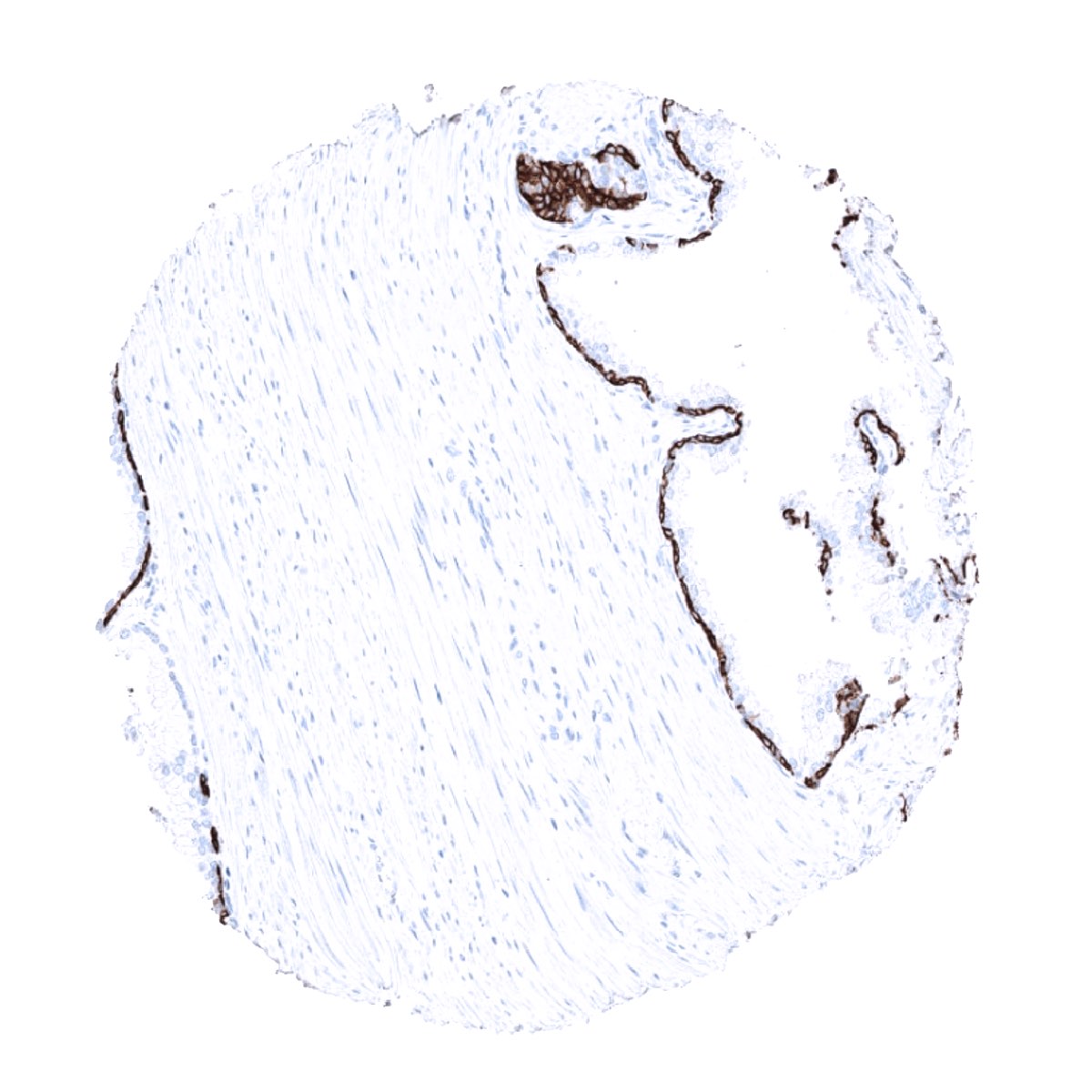
CK6 expression is seen in almost all squamous cell carcinomas irrespective of their origin. CK6 is also seen in 30-50% of urothelial carcinomas of the urinary bladder, 30-50% of pancreatic adenocarcinomas, 20-30% of endometroid carcinomas of endometrium and ovary, 10-20% of cholangiocarcinomas of the liver, 10-20% of adenocarcinomas of the lung, and in other cancers. KRT6 immunostaining can usually be seen in only a few cells of basaliomas.
Detailed data on CK6 staining by MSVA-606R obtained from an analysis of 12,898 tumors from 120 different tumor types and subtypes have recently been published by Völkel et al. “Cytokeratin 5 and cytokeratin 6 expressions are unconnected in normal and cancerous tissues and have separate diagnostic implications”
The TCGA findings on Cytokeratin 6 RNA expression in different tumor categories have been summarized in the Human Protein Atlas.
Compatibility of Antibodies
CK6 staining in tumors with antibody MSVA-606R. Data from the publication “Cytokeratin 5 and cytokeratin 6 expressions are unconnected in normal and cancerous tissues and have separate diagnostic implications” published by Völkel et al. in Virchows Archive September 2021. Summarized in own graphics:
- CK6 staining in tumors with antibody MSVA-606R

2. CK6 staining in tumors with antibody MSVA-606R ranked by positivity.

Figure 3. Clinico-pathological associations described by Völkel et al. (p-value)

Protocol Recommendations
IHC users have different preferences on how the stains should look like. Some prefer high staining intensity of the target stain and even accept some background. Others favor absolute specificity and lighter target stains. Factors that invariably lead to more intense staining include higher concentration of the antibody and visualization tools, longer incubation time, higher temperature during incubation, higher temperature and longer duration of the heat induced epitope retrieval (slide pretreatment). The impact of the pH during slide pretreatment has variable effects and depends on the antibody and the target protein. Accordingly, multiple different protocols can generate identical staining results.
All images and data shown here and in our image gallery are obtained by the manual protocol described below. Other protocols resulting in equivalent staining are described as well.
Manual protocol
Freshly cut sections should be used (less than 10 days between cutting and staining). Heat-induced antigen retrieval for 5 minutes in an autoclave at 121°C in pH 9 Target Retrieveal Solution buffer. Apply MSVA-606Rat a dilution of 1:150 at 37°C for 60 minutes. Visualization of bound antibody by the EnVision Kit (Dako, Agilent) according to the manufacturer’s directions.
Agilent / Dako – Autostainer Link 48
Pretreatment in PT-Link for 30 minutes at 95°C (pH high); FLEX peroxidase blocking for 5 minutes (room temperature), MSVA-606R 1:150 for 20 minutes (room temperature), FLEX+ mouse/rabbit (LINKER) for 15 minutes (room temperature), horseradish peroxidase (HRP) for 20 minutes (room temperature), FLEX DAB+Sub-Chromo for 10 minutes (room temperature), FLEX hematoxylin for 5 minutes (room temperature).
These images reflect stainings by the protocol described above. It is of note that a comparable staining result can also be obtained by different protocols. In general, a longer pretreatment, a longer incubation time of the primary antibody, a higher antibody concentration, and a longer incubation time of FLEX+LINKER result in stronger staining, potentially at the cost of more background staining. Modifications of the protocol with a strengthening effect on staining intensity in combination with changes of other parameters that result in lower staining intensity can result in a comparable result as shown above.
Leica – BOND RX
Dewax at 72°C for 30 seconds; Pretreatment in Bond Epitope Retrieval Solution (ER2 – EDTA pH9) for 20 minutes at 100°C; Peroxidase blocking for 5 minutes (room temperature), MSVA-606R 1:150 for 15 minutes (room temperature), Post primary (rabbit anti mouse) for 8 minutes (room temperature), Polymer (goat anti rabbit) for 8 minutes (room temperature), mixed DAB refine for 10 minutes (room temperature), hematoxylin for 5 minutes (room temperature).
Roche – Ventana Discovery ULTRA
Pretreatment for 64 minutes at 100°C (pH 8,4); CM peroxidase blocking for 12 minutes (room temperature), MSVA-606R 1:150 for 20 minutes at 36°C, secondary antibody (anti-rabbit HQ) for 12 minutes at 36°C, anti-HQ HRP for 12 minutes at room temperature, DAB at room temperature, hematoxylin II at room temperature for 8 minutes, bluing reagent at room temperature for 4 minutes.

These images depict staining results obtained by the protocol described above. It is of note, that the Ventana machines generally require higher antibody concentrations than other commonly used autostainers because the antibodies are automatically diluted during the procedure. Various other protocols can result in an identical result as shown above. A longer pretreatment, a longer incubation time of the primary antibody, a higher antibody concentration, a higher temperature during incubation, and a longer incubation time of secondary antibody and or the anti-HQ HRP result in stronger staining, potentially at the cost of more background staining.
Impact of pH

MSVA-606R results in strongest staining if pH9,0 is used for slide pretreatment. pH7,8 is acceptable but lower pH results in a significant reduction of sensitivity.
Potential Research Applications
- The potential diagnostic utility of KRT6 expression analysis (as compared to KRT5/6 analysis) needs to be investigated.
- The clinical significance of KRT6 expression needs to be evaluated in tumor types containing significant subgroups of KRT6 positive and negative tumors.
Evidence for Antibody Specificity in IHC
Specificity of MSVA-606R is documented by strong positive staining in cell types that are well documented to express CK6 such as squamous epithelia of all origins and absence of staining in all tissues known to not express CK6 including tissues notorious for non-specific IHC background such as kidney, colonic mucosa, and epidermis.
In a recent publication, the specificity of MSVA-606R was demonstrated in a comparison vs another independent antibody. Völkel et al. “Cytokeratin 5 and cytokeratin 6 expressions are unconnected in normal and cancerous tissues and have separate diagnostic implications”
