
295,00 € – 995,00 €
Product details
Synonyms = Fasciclin-I like; Osteoblast specific factor 2 (OSF2); PDLPOSTN; Periodontal ligament specific periostin; Periostin isoform thy2 / th4 / thy6 / thy8; Periostin osteoblast specific factor; PN; POSTN
Antibody type = Mouse monoclonal / IgG
Clone = MSVA-649M
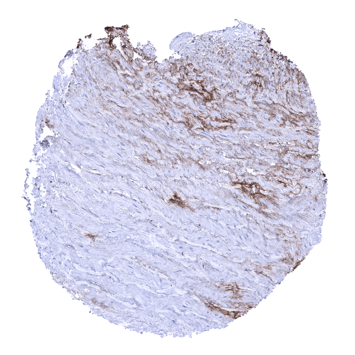
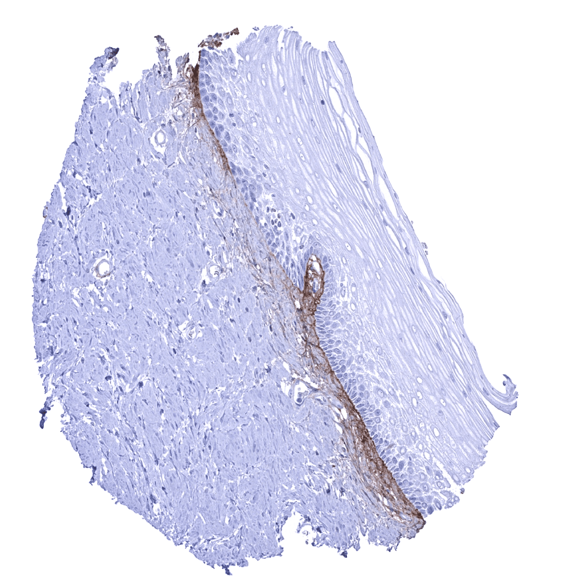
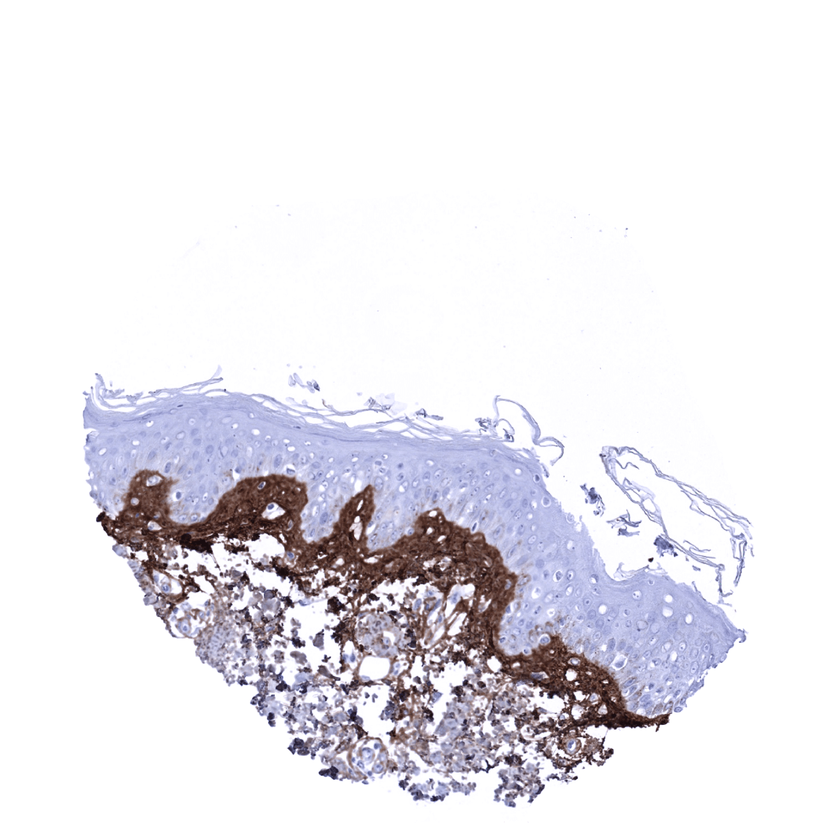
Positive control = Skin: A strong periostin immunostaining should be seen in the skin, forming a subepithelial band of variable thickness.
Negative control = Skin: Periostin immunostaining should be lacking in epithelial cells.
Cellular localization = Extracellular space.
Reactivity = Human
Application = Immunohistochemistry
Dilution = 1:100 – 1:200
Intended Use = Research Use Only
Relevance of Antibody
Periostin is a Marker for tissue damage and epithelial-mesenchymal transition.
Biology Behind
Periostin (POSTN) also termed osteoblast-specific factor (OSF-2) is a 90kDa protein coded by the POSTN gene at 13q13.3. Periostin is a secreted extracellular matrix protein that was originally identified in cells from the mesenchymal lineage (osteoblasts, osteoblast-derived cells, the periodontal ligament, and periosteum). Periostin plays a wide variety of roles in tissue development and disease. It is pivotal in multiple different mechanisms of tissue remodeling in response to injury. Periostin is transiently upregulated during various cell fate changes, irrespective of whether these are physiological or caused by disease or injury. Periostin impacts extracellular matrix restructuring, tissue remodeling, and the epithelial-mesenchymal transition, all of which can be related to tissue healing, development, cancer, and other diseases. Thus, periostin functions as a mediator, balancing appropriate and inappropriate responses to tissue damage. In many cancers, periostin binds to integrins on cancer cells, activating the Akt/PKB- and FAK-mediated signaling pathways. This leads to increased cell survival, migration, invasion, angiogenesis, metastasis, and the epithelial-mesenchymal transition.
Staining Pattern in Normal Tissues
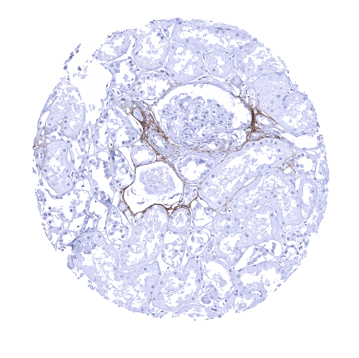
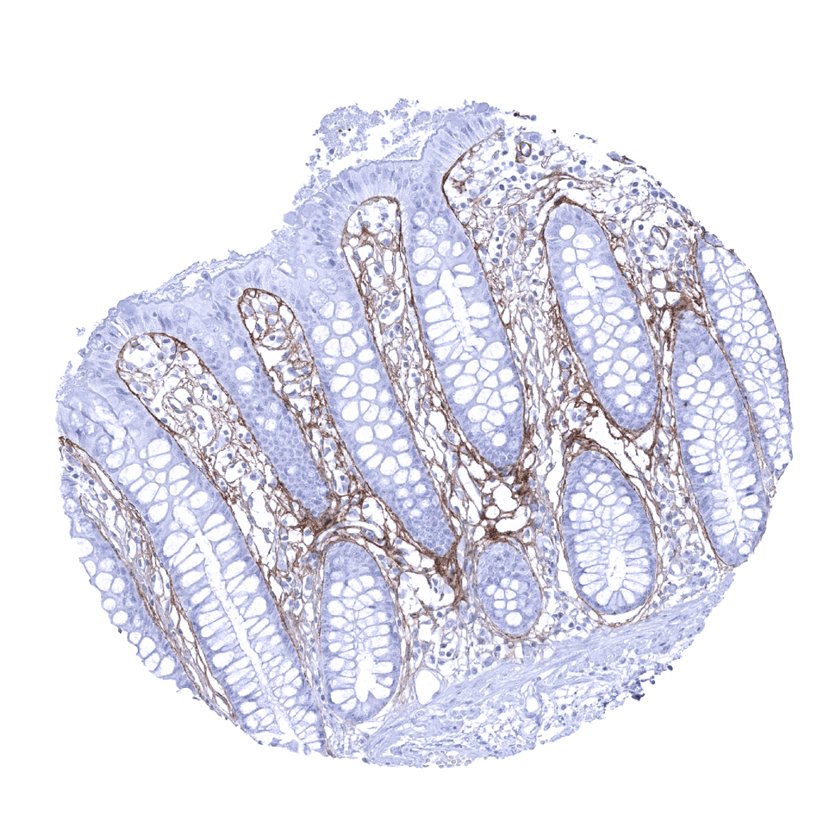
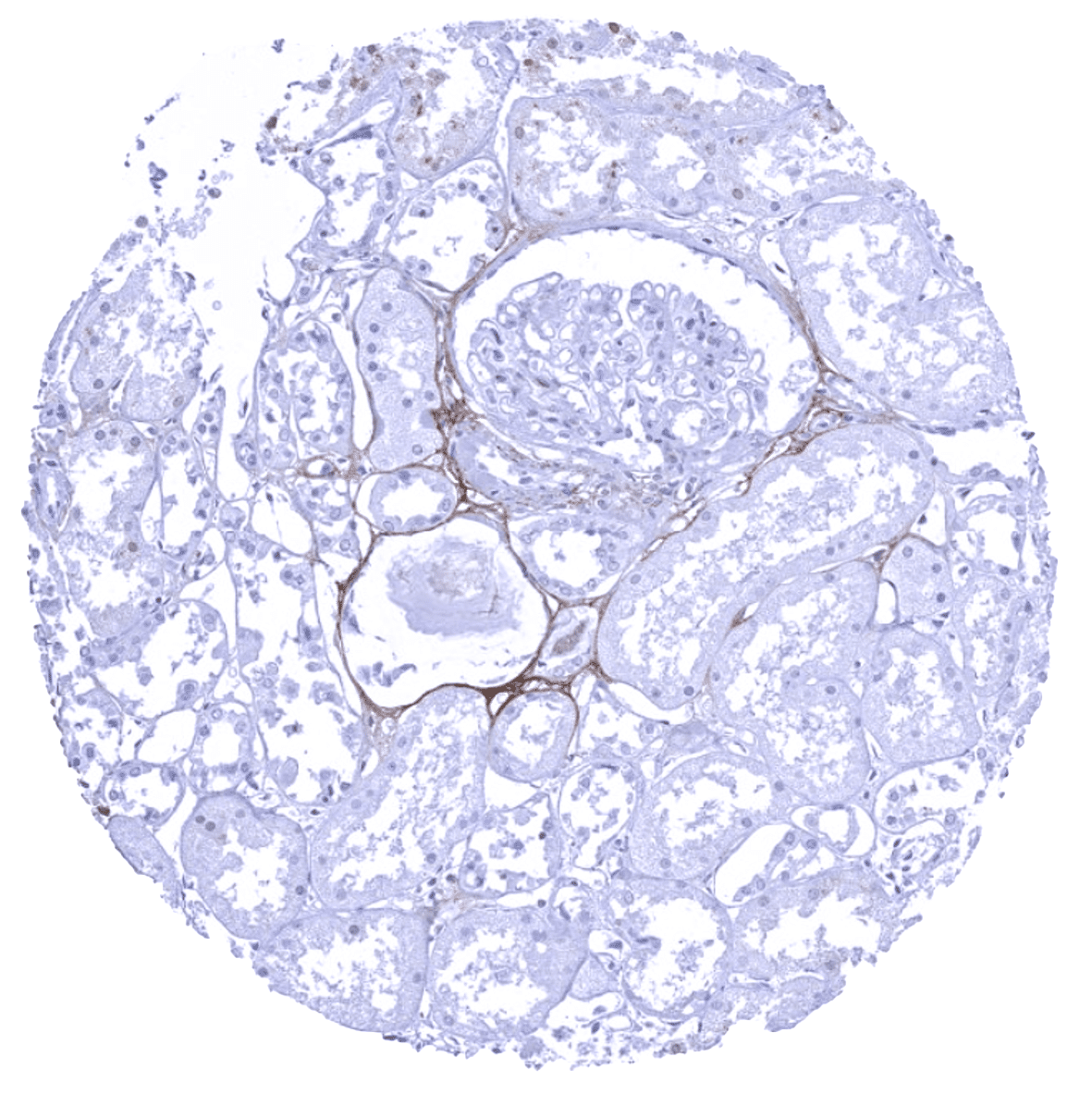
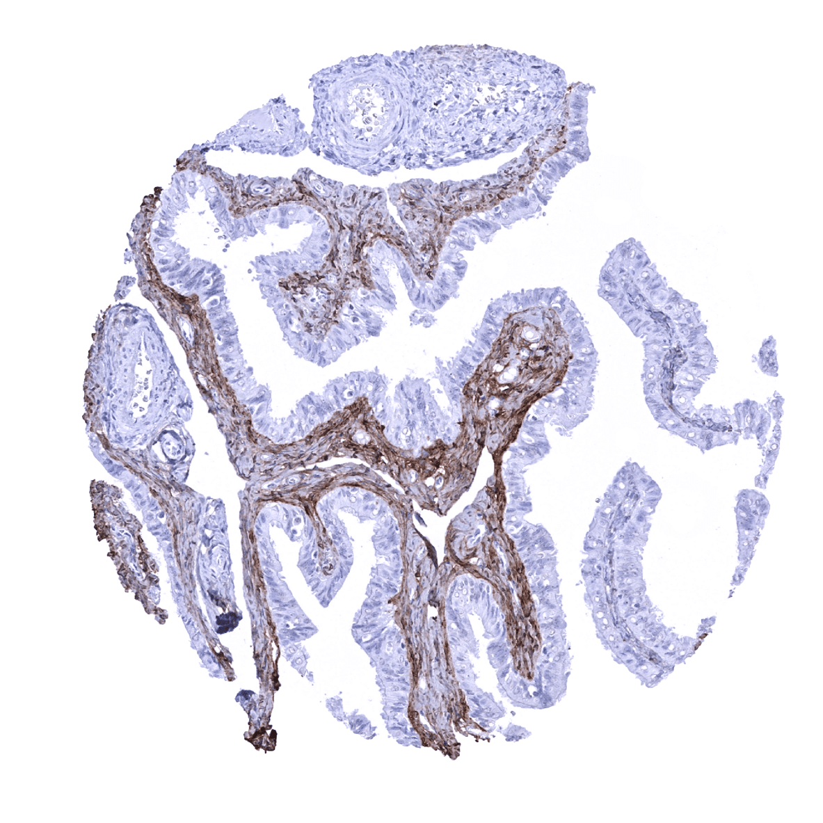
Periostin is found in variable quantities in tissues. Periostin immunostaining is not seen in normal epithelial cells but represents a variable fraction of the extracellular matrix and may also occur in fibroblasts and fibrocytes. Detectable levels of periostin can especially occur along surface epithelia. In these tissues, periostin immunostaining ranges from “absent” to significant subepithelial masses. Most commonly, periostin occurs as a thin band along the basement membrane. Periostin is most regularly seen in the stomach, colorectum, fallopian tube, the skin and the aorta but – at least to some extent – it can occur in virtually all organs. Increased levels of periostin can be seen in areas of tissue damage such as in atrophic areas in the kidney. Periostin immunostaining is particularly rare in the first trimenon placenta, small intestine mucosa, and in lymphatic tissues.
These findings are largely consistent with the RNA and protein data described in the Human Protein Atlas (Tissue expression Periostin)
Positive control: Skin: A strong periostin immunostaining should be seen in the skin, forming a subepithelial band of variable thickness.
Negative control: Skin: Periostin immunostaining should be lacking in epithelial cells.


Staining Pattern in Relevant Tumor Types
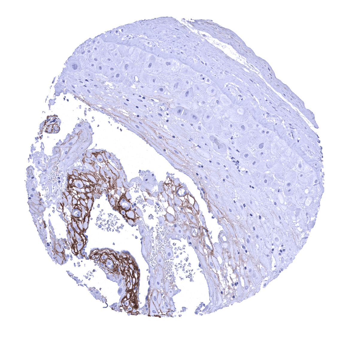
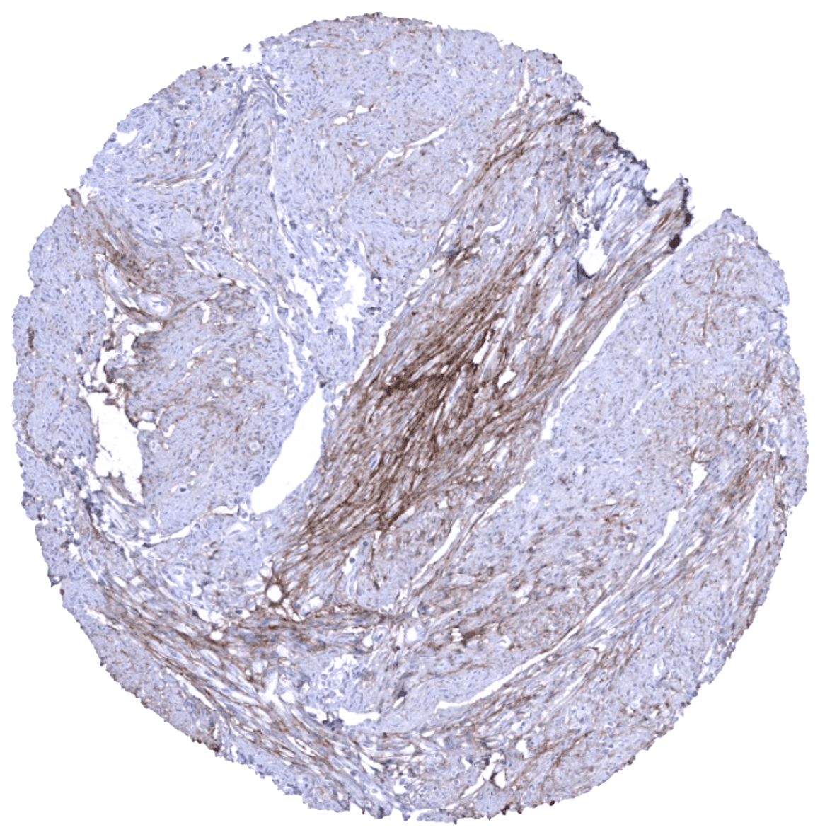
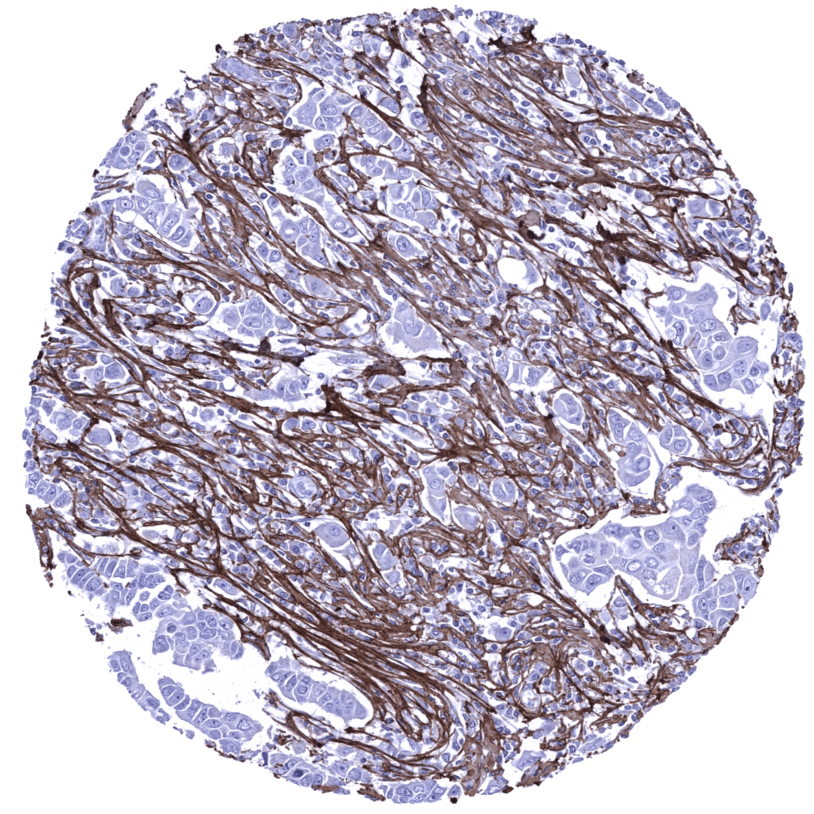
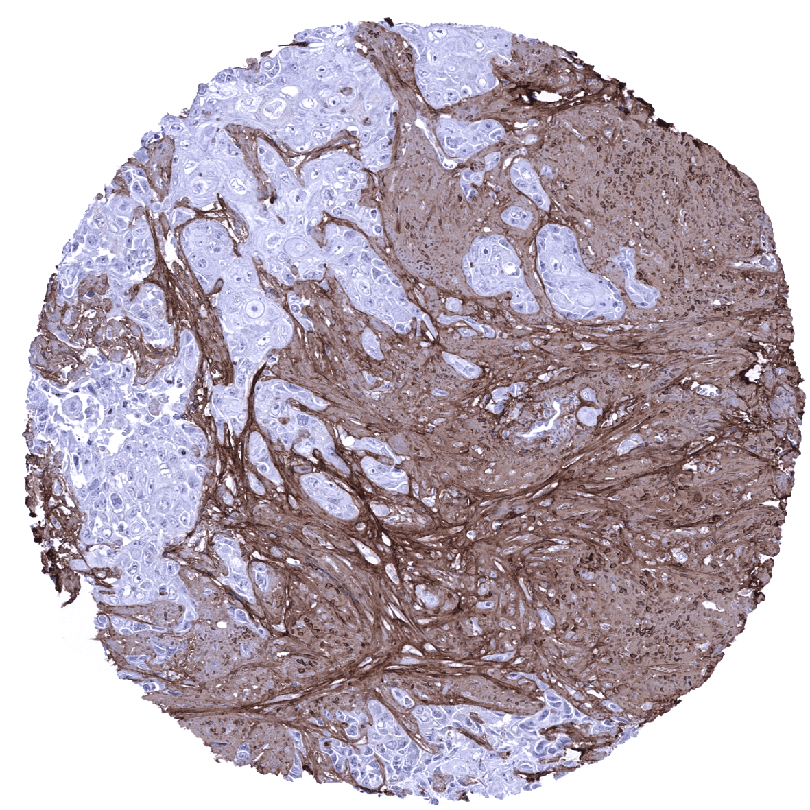
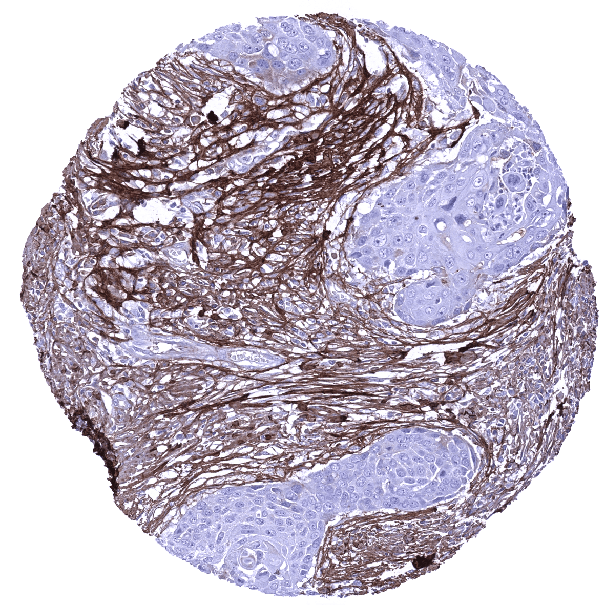
In tumors, periostin immunostaining occurs in the tumor associated stroma. Its extent is highly variable and ranges from completely absent to excessive.
The TCGA findings on Periostin RNA expression in different tumor categories have been summarized in the Human Protein Atlas.

Compatibility of Antibodies
No data available at the moment
Protocol Recommendations
IHC users have different preferences on how the stains should look like. Some prefer high staining intensity of the target stain and even accept some background. Others favor absolute specificity and lighter target stains. Factors that invariably lead to more intense staining include higher concentration of the antibody and visualization tools, longer incubation time, higher temperature during incubation, higher temperature and longer duration of the heat induced epitope retrieval (slide pretreatment). The impact of the pH during slide pretreatment has variable effects and depends on the antibody and the target protein.
All images and data shown here and in our image galleries are obtained by the manual protocol described below. Other protocols resulting in equivalent staining are described as well.
-Manual protocol
Freshly cut sections should be used (less than 10 days between cutting and staining). Heat-induced antigen retrieval for 5 minutes in an autoclave at 121°C in pH 7,8 Target Retrieval Solution buffer. Apply MSVA-649M at a dilution of 1:150 at 37°C for 60 minutes. Visualization of bound antibody by the EnVision Kit (Dako, Agilent) according to the manufacturer’s directions.
Potential Research Applications
- The level of periostin immunostaining in the stroma of tumors may have clinical significance and should be further investigated.
- The clinical and diagnostic impact of variable quantities of periostin in a tissue should be evaluated in various disease types including inflammation and heart disease
Evidence for Antibody Specificity in IHC
There are two ways how the specificity of antibodies can be documented for immunohistochemistry on formalin fixed tissues. These are: 1. comparison with a second independent method for target expression measurement across a large number of different tissue types (orthogonal strategy), and 2. Comparison with one or several independent antibodies for the same target and showing that all positive staining results are also seen with other antibodies for the same target (independent antibody strategy).
Orthogonal validation is not suited for validation of targets such as extracellular matrix markers that are expressed in virtually every organ.
Comparison of antibodies: Specific periostin staining by MSVA-649M was suggested by a complete absence of elastin immunostaining in epithelial cells and the fact, that all structures identified by a MSVA-649M were also stained by the antibody “MSVA_periostin_cand5” which was also evaluated. Selected images of this comparison are shown below.
Antibody Comparison: MSVA-649M vs another MSVA Periostin antibody candidate called “MSVA-cand5f”