Adrenal gland

Aorta, media - Focal periostin immunostaining is usually seen in normal appearing media of the aorta of the elderly.

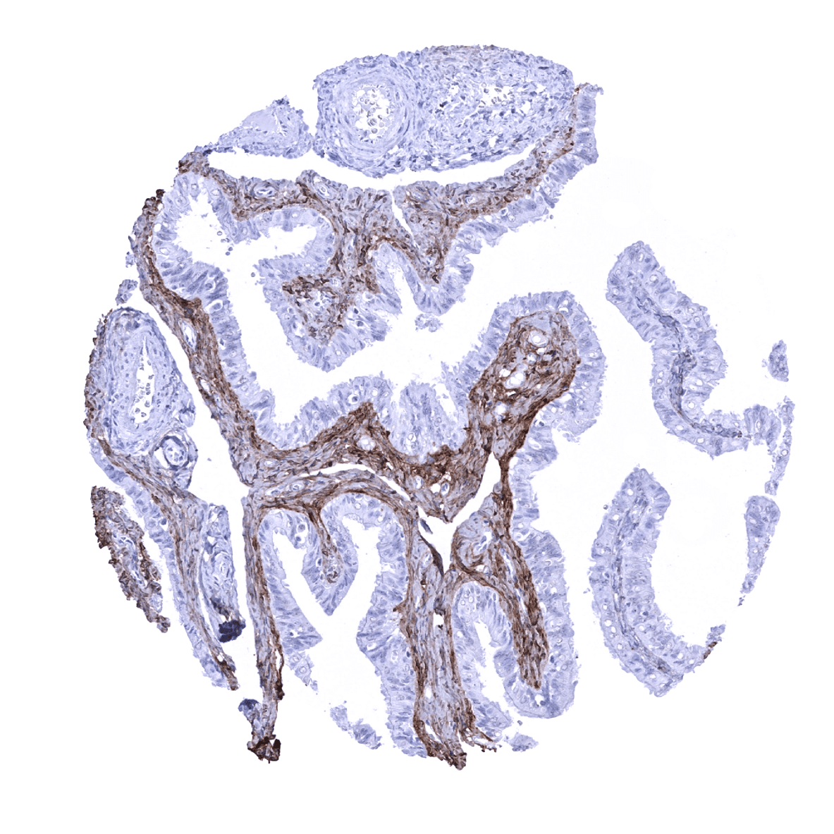
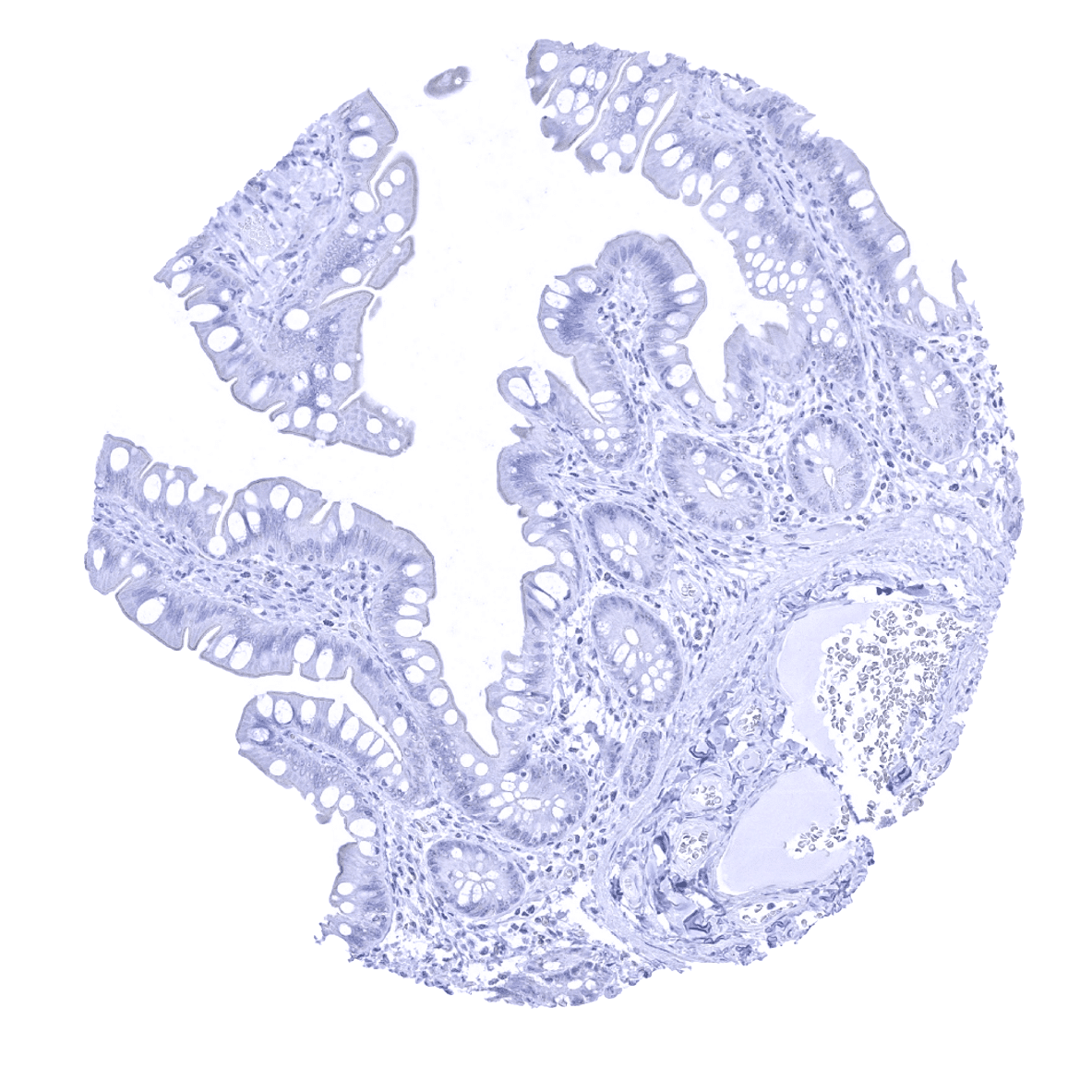
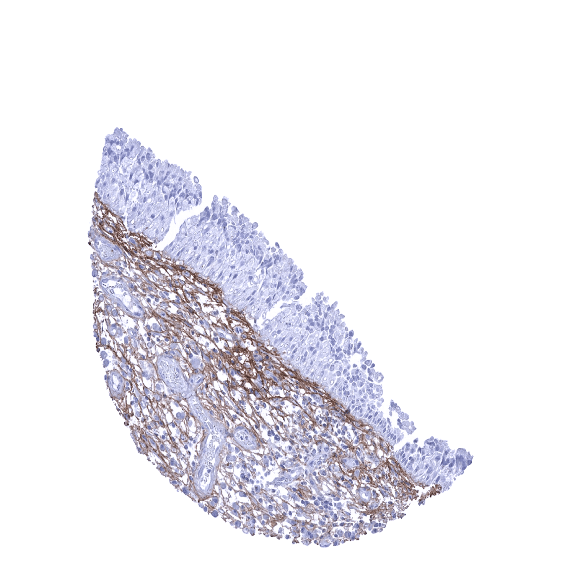
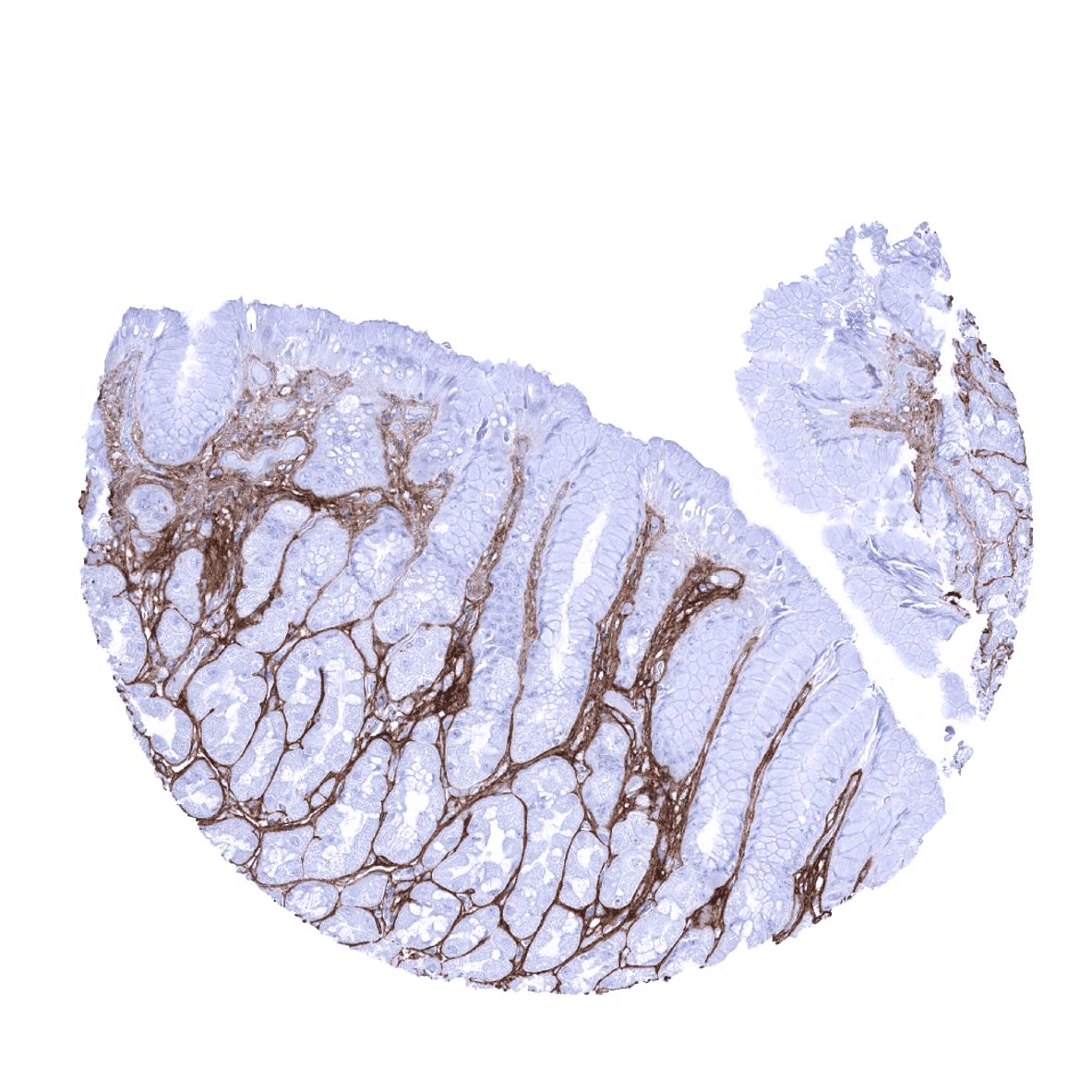
Appendix, mucosa - Periostin is regularly seen in the colorectal mucosa where it forms a characteristic subepithelial band. Additional periostin deposits occur in the lamina propria.

Appendix, muscular wall
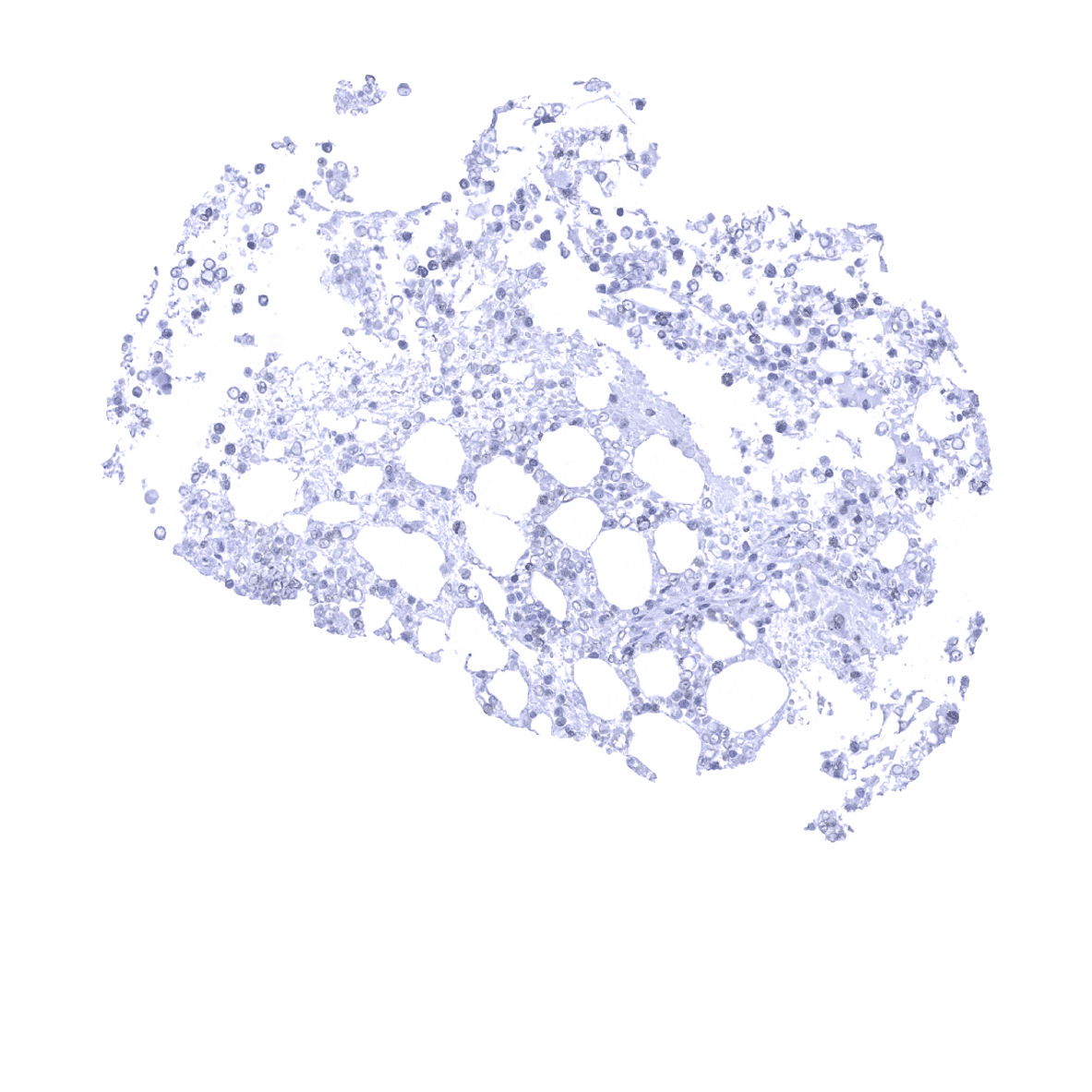
Bone marrow

Breast

Bronchus, mucosa - Periostin immunostaining is regularly seen in respiratory epithelium where it forms a subepithelial band of variable thickness.

Cerebellum (molecular, Purkinje cell, granule cell layers; white matter)
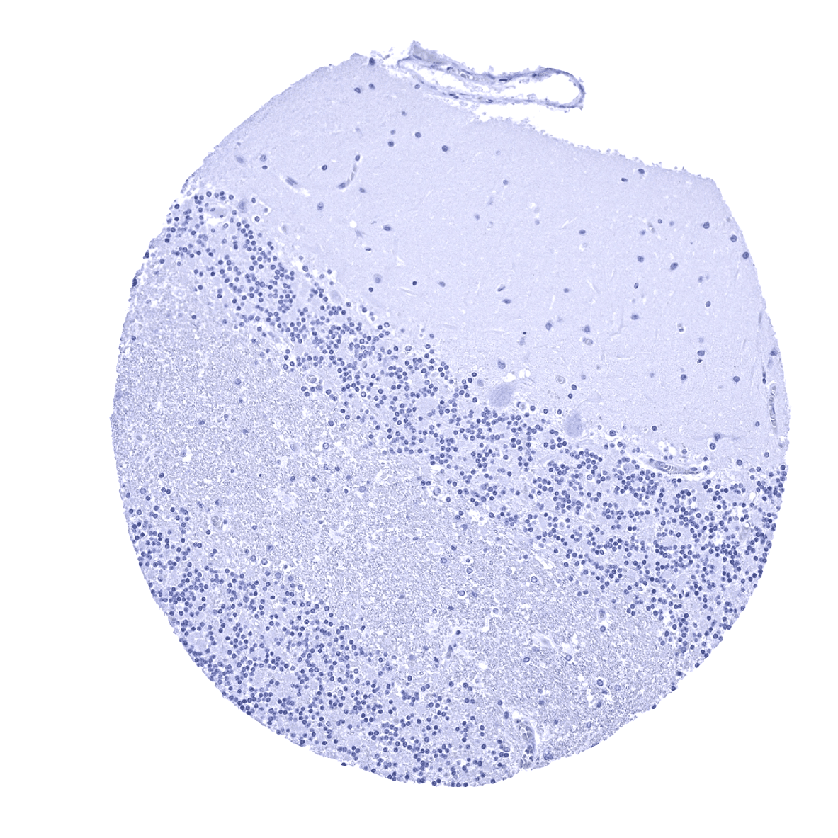
Cerebellum (molecular layer, Purkinje cell layer, granule cell layer, white matter)

Cerebrum, grey matter

Cerebrum, white matter
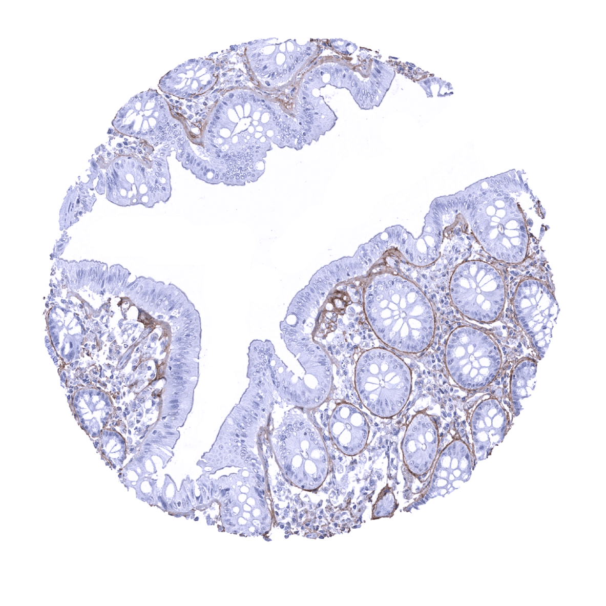
Colon descendens, mucosa - Periostin is regularly seen in the colorectal mucosa where it forms a characteristic subepithelial band. Additional periostin deposits occur in the lamina propria.

Colon descendens, muscular wall

Duodenum, Brunner gland
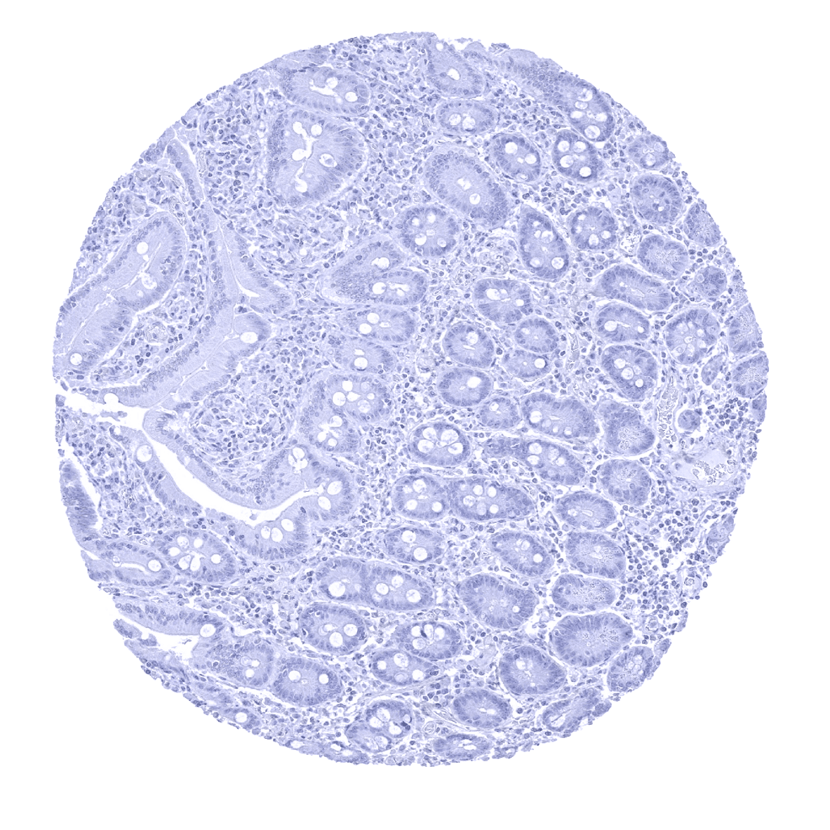
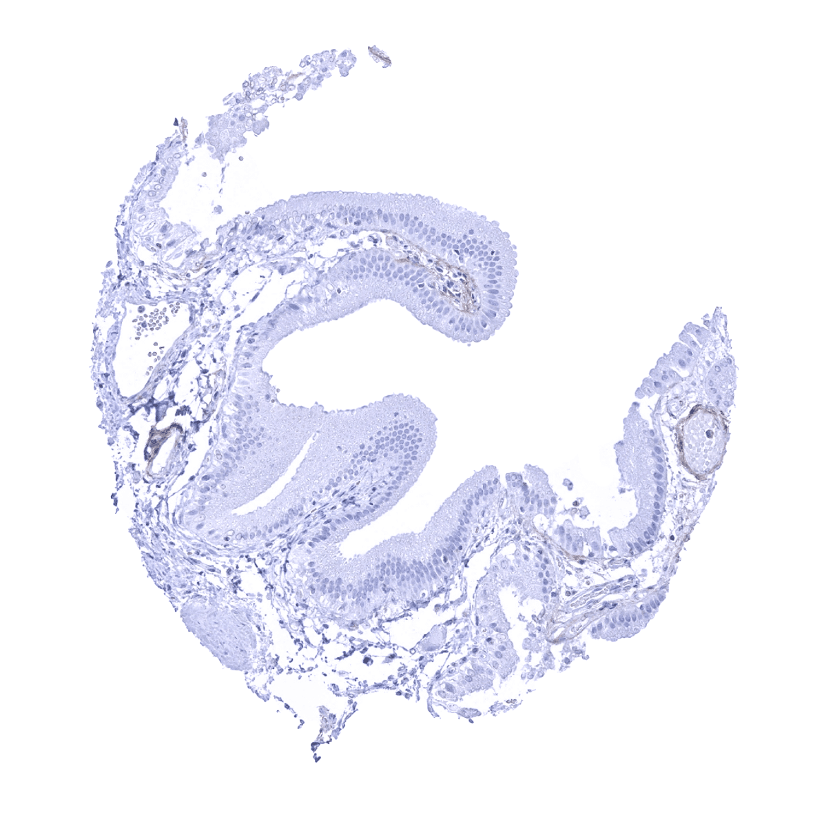
Duodenum, mucosa - Prominent periostin immunostaining is usually lacking in the mucosa of the small intestine.
Epididymis
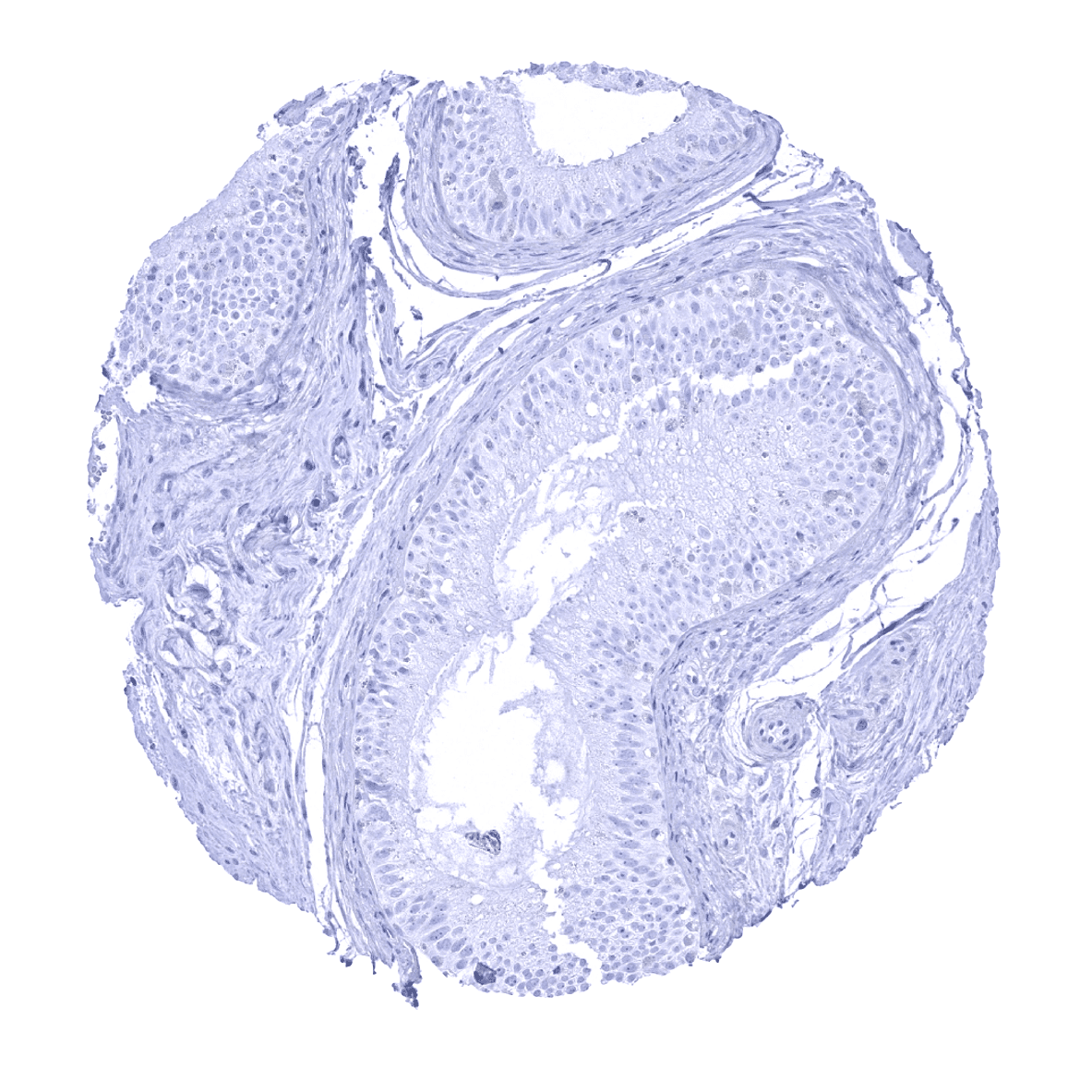
Esophagus, squamous epithelium

Fallopian tube, mucosa - Periostin immunostaining is typically seen in the fallopian tube where it forms a subepithelial band of variable thickness.
Fallopian tube, mucosa - Periostin immunostaining is regularly seen in the fallopian tube where it forms a subepithelial band of variable thickness.
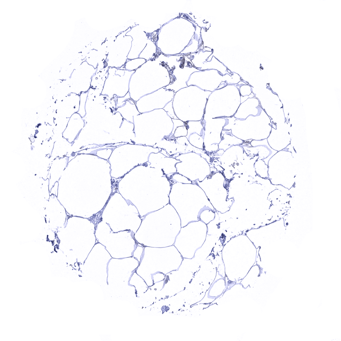
Fat
Gallbladder, epithelium

Heart muscle
Ileum, mucosa - Prominent periostin immunostaining is usually lacking in the mucosa of the small intestine.
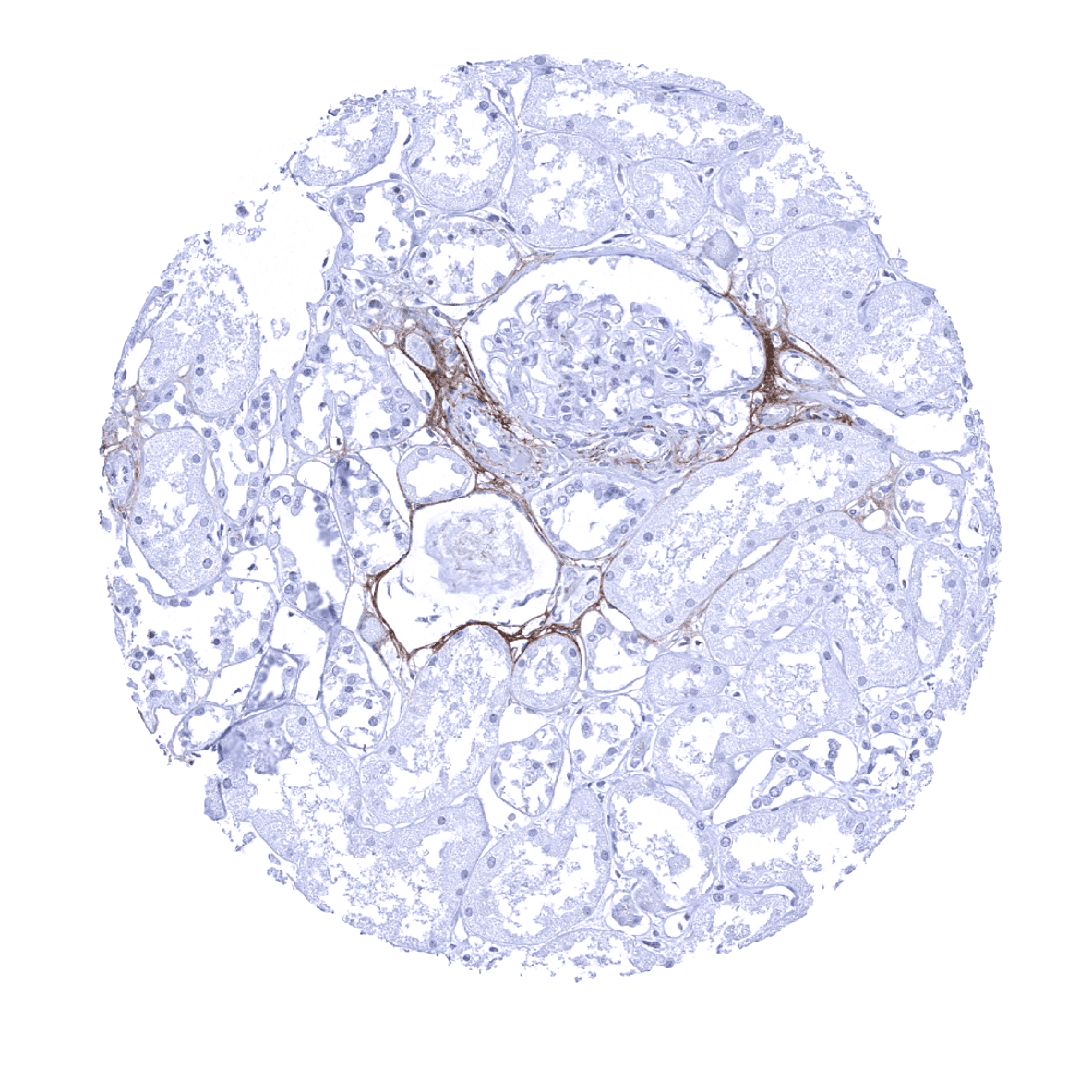
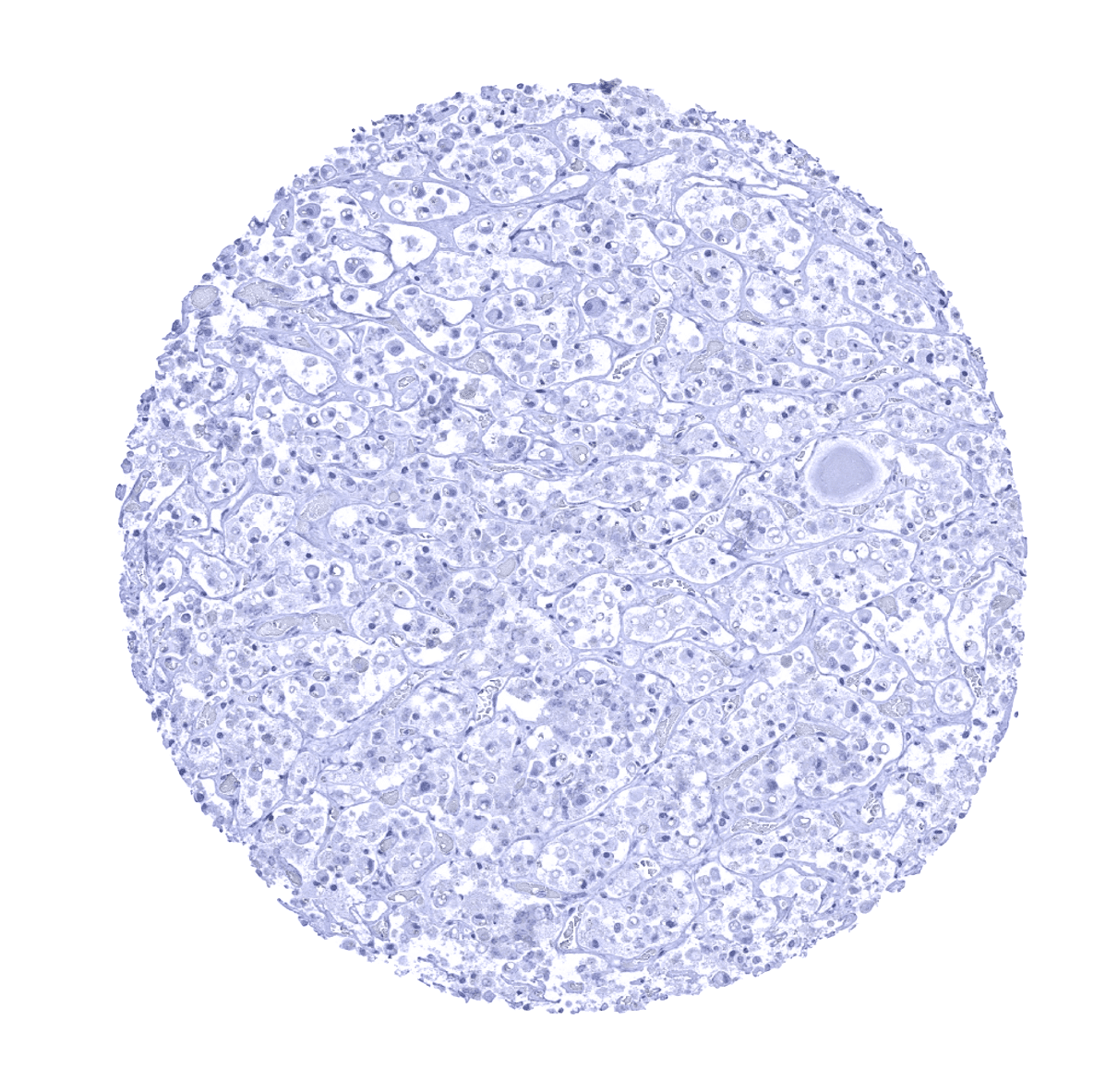
Kidney, cortex
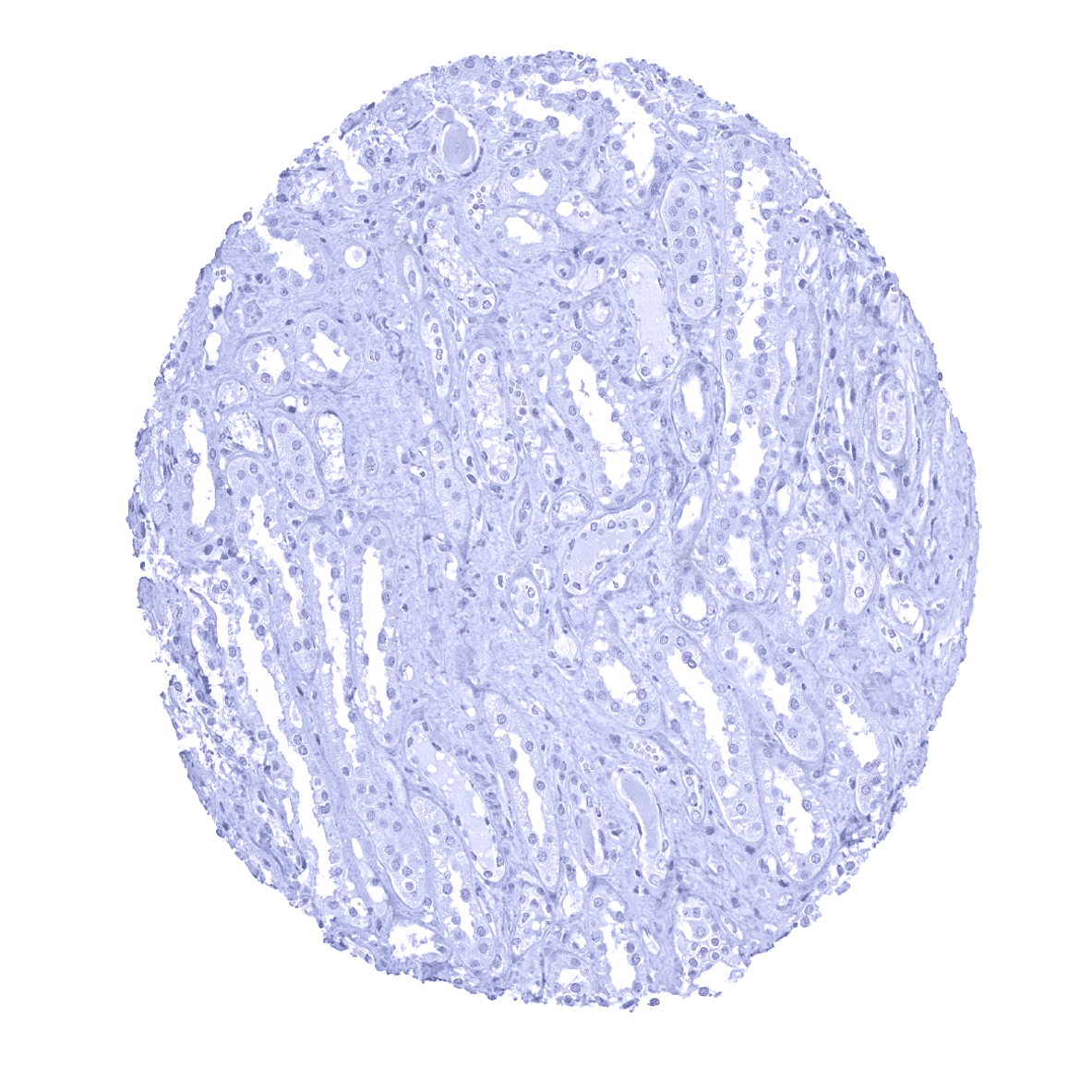
Kidney, medulla
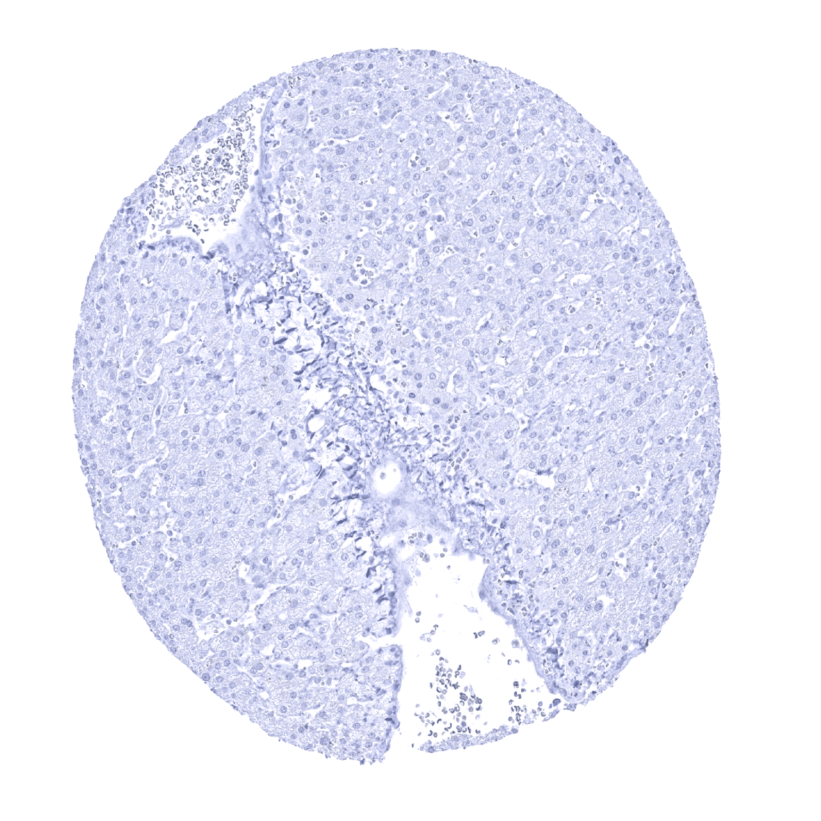
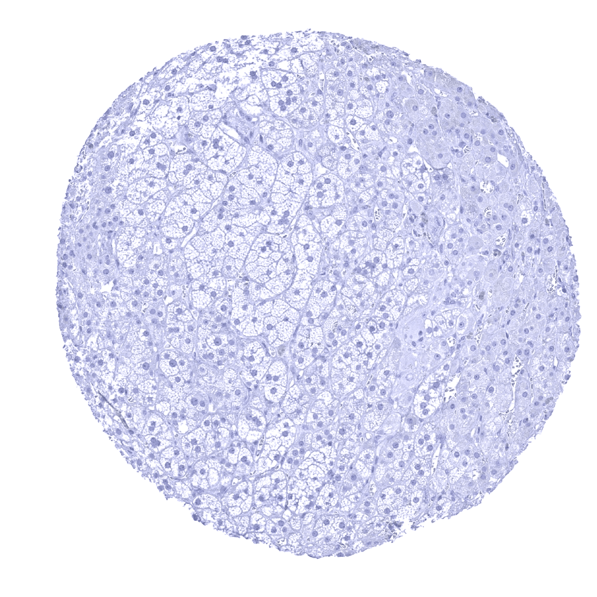
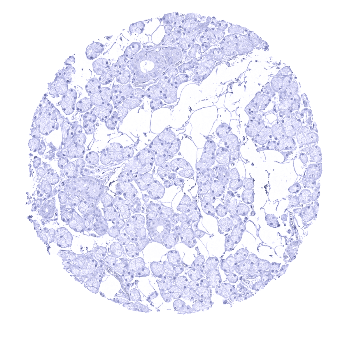
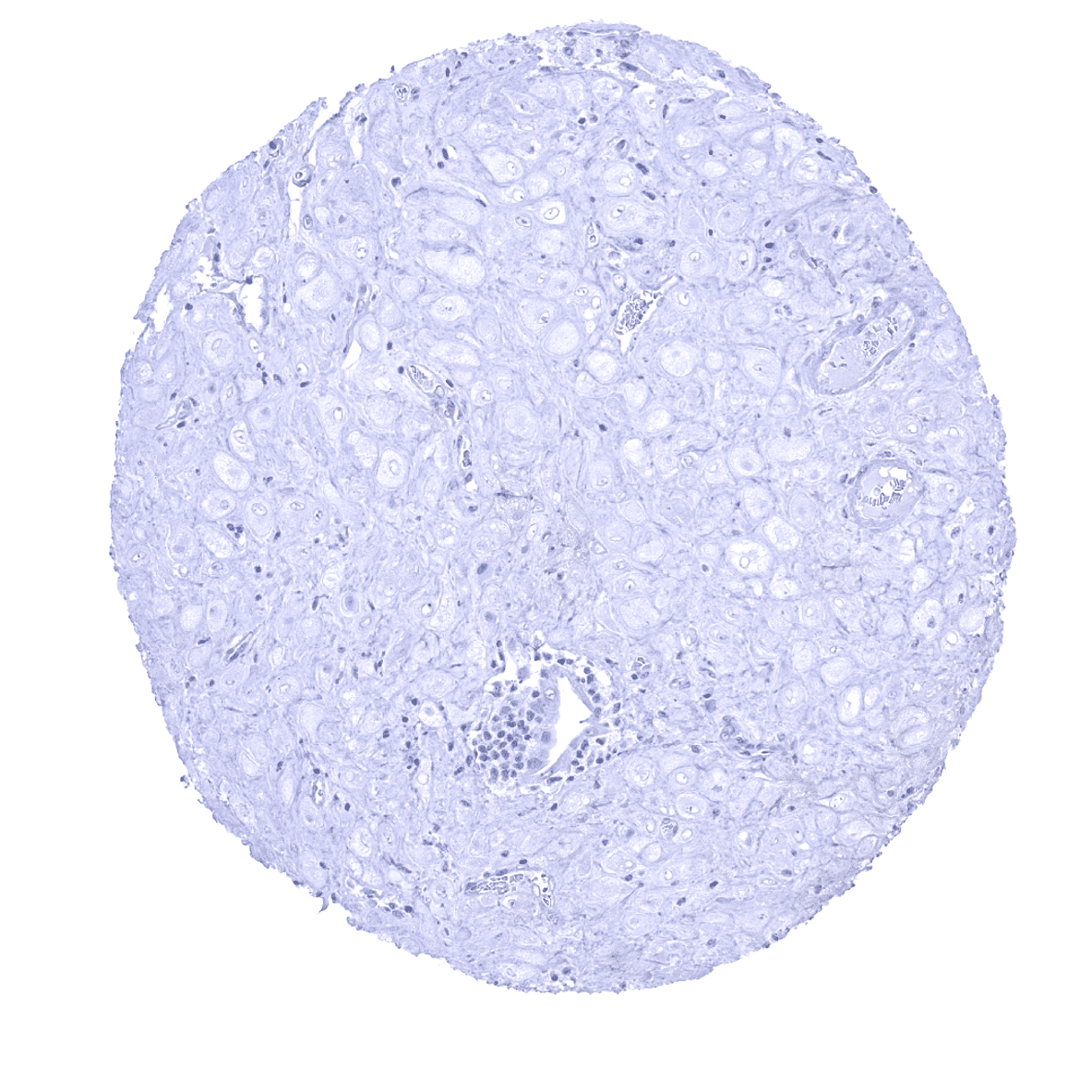
Liver
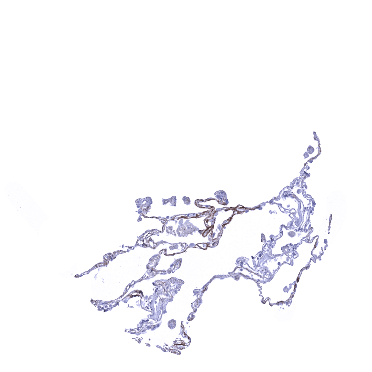
Lung - Periostin immunostaining can be observed in alveoli of the lung.
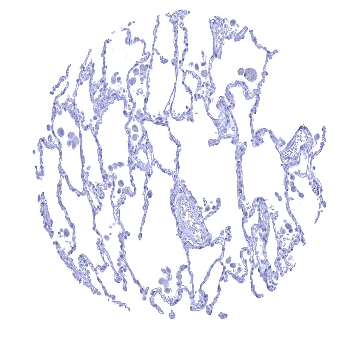
Lung - Periostin immunostaining is usually lacking in alveoli of the lung.
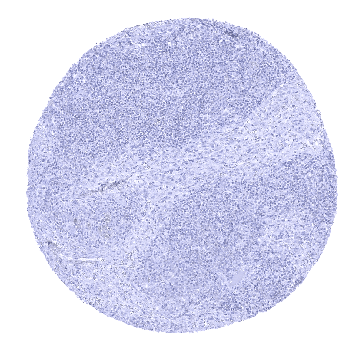
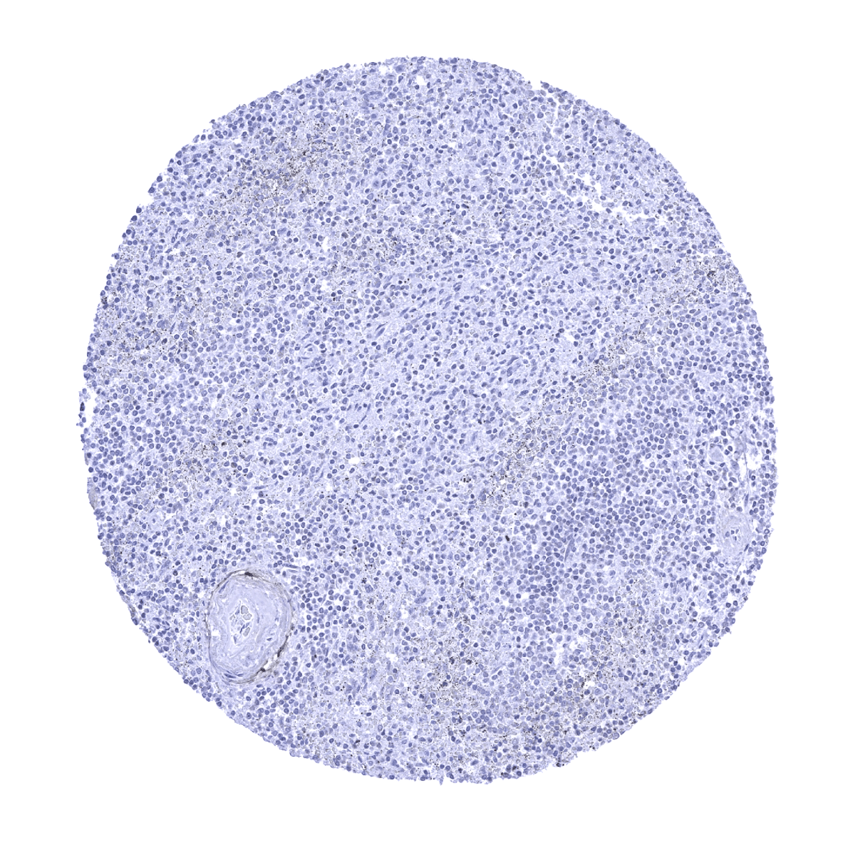
Lymph node
Ovary, stroma

Pancreas - Periostin immunostaining can be abundant in the pancreas.

Pancreas - Periostin expression is typically lacking in the pancreas.

Parathyroid
Parotid gland

Pituitary gland, anterior lobe - Focal periostin immunostaining can be found in samples from the adenohypophysis
Pituitary gland, anterior lobe - Periostin immunostaining is often lacking in samples from the pituitary gland.
Pituitary gland, posterior lobe

Placenta (amnion and chorion) - Periostin immunostaining can be absent in the placenta.
Placenta (chorion) - Periostin immunostaining can be observed in the chorion plate of the placenta in some samples.

Placenta, early
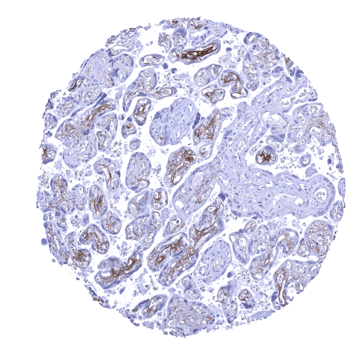
Placenta, mature - Periostin immunostaining can be observed in the mature placenta.
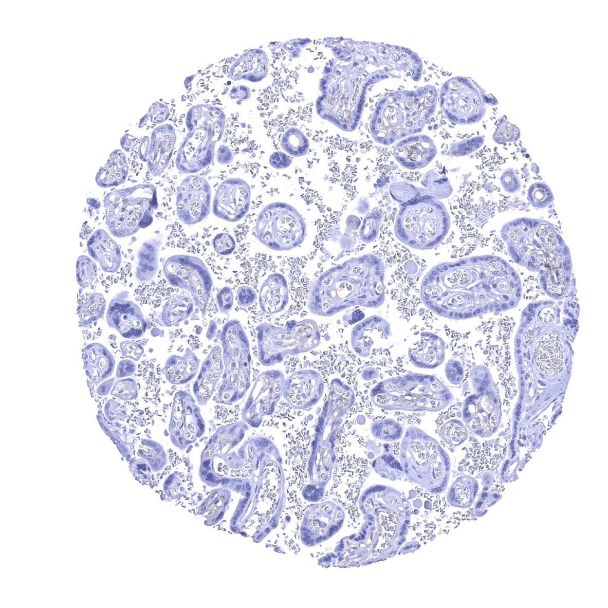
Placenta, mature - Periostin immunostaining is often lacking in samples from the mature placenta.
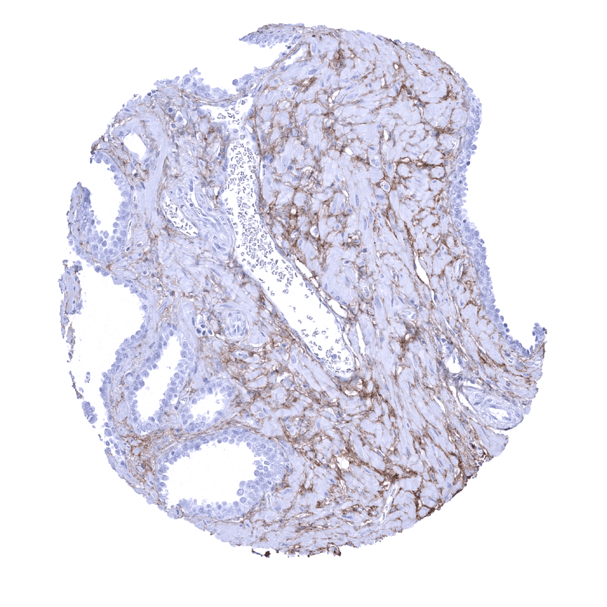
Prostate - Periostin immunostaining can be focally abundant in the prostate.
Prostate - Periostin immunostaining can be lacking in the prostate.
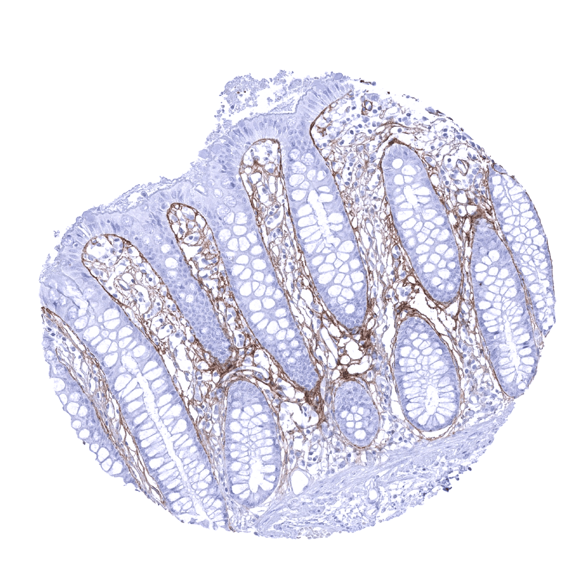
Rectum, mucosa - Periostin is regularly seen in the colorectal mucosa where it forms a characteristic subepithelial band. Additional periostin deposits occur in the lamina propria.
Seminal vesicle
Sinus paranasales - Periostin immunostaining is regularly seen in respiratory epithelium where it forms a subepithelial band of variable thickness.

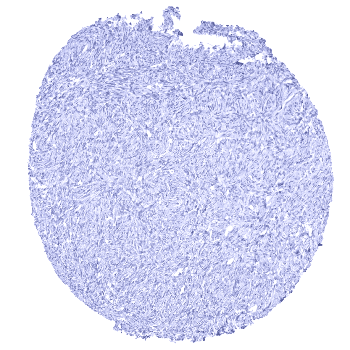
Skeletal muscle
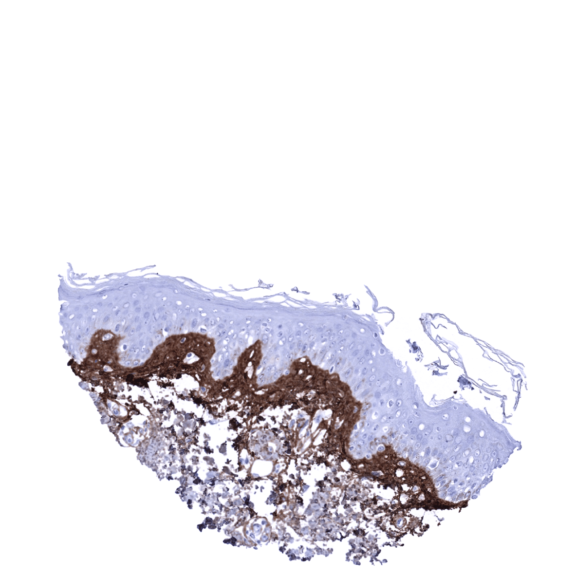
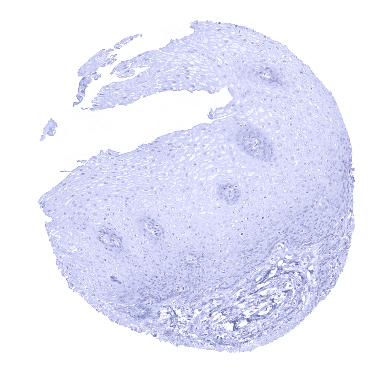
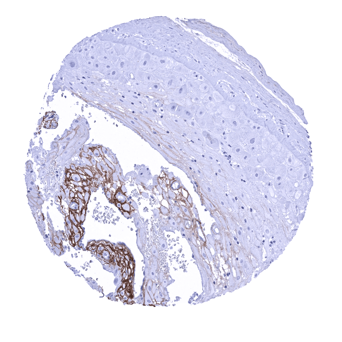
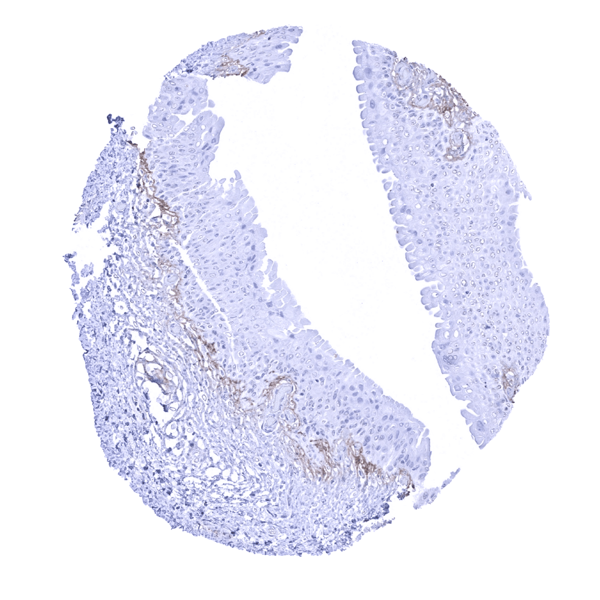
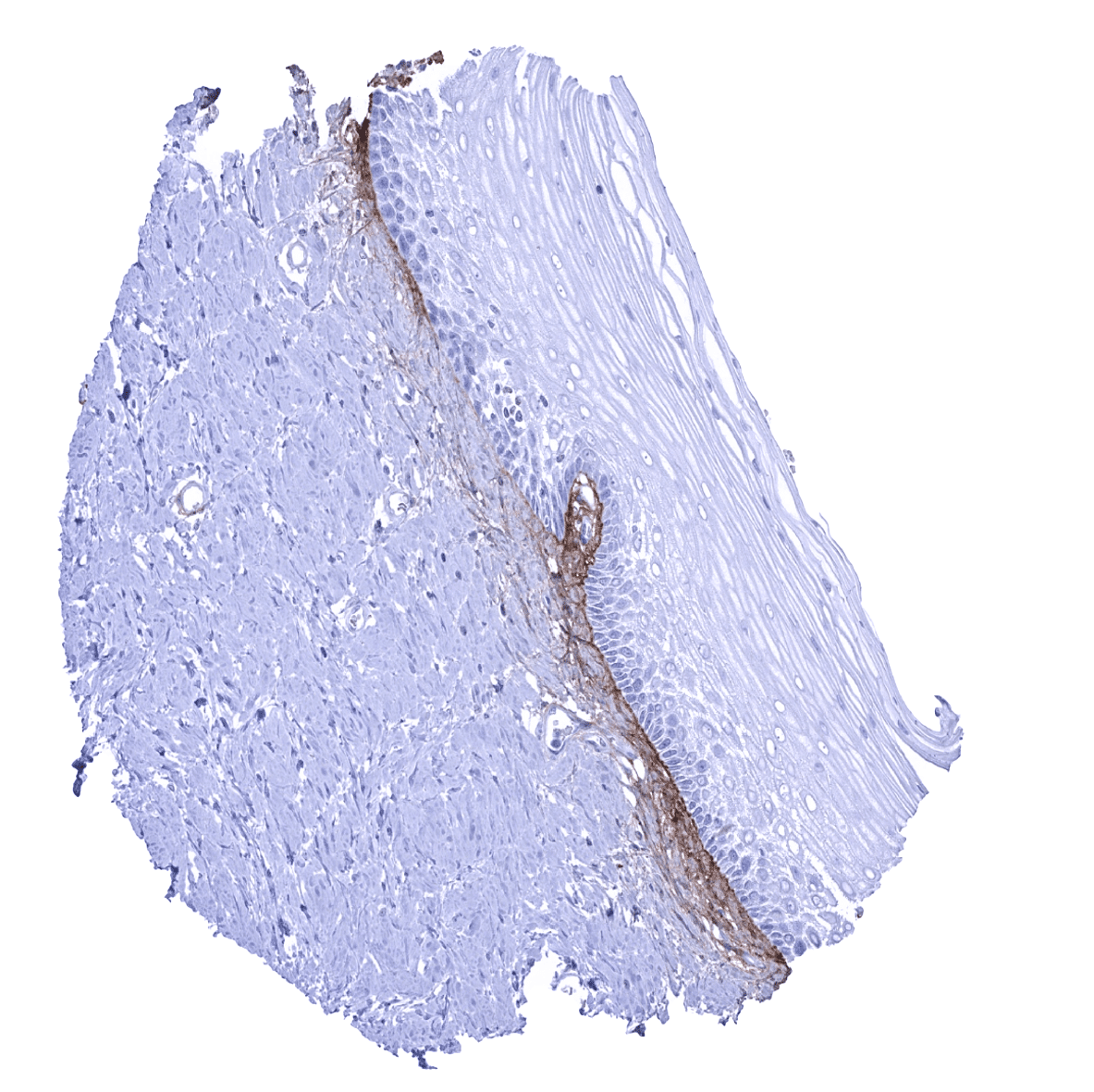
Skin - Periostin immunostaining is most regularly seen in the skin where it forms a subepithelial band of variable thickness.
Spleen
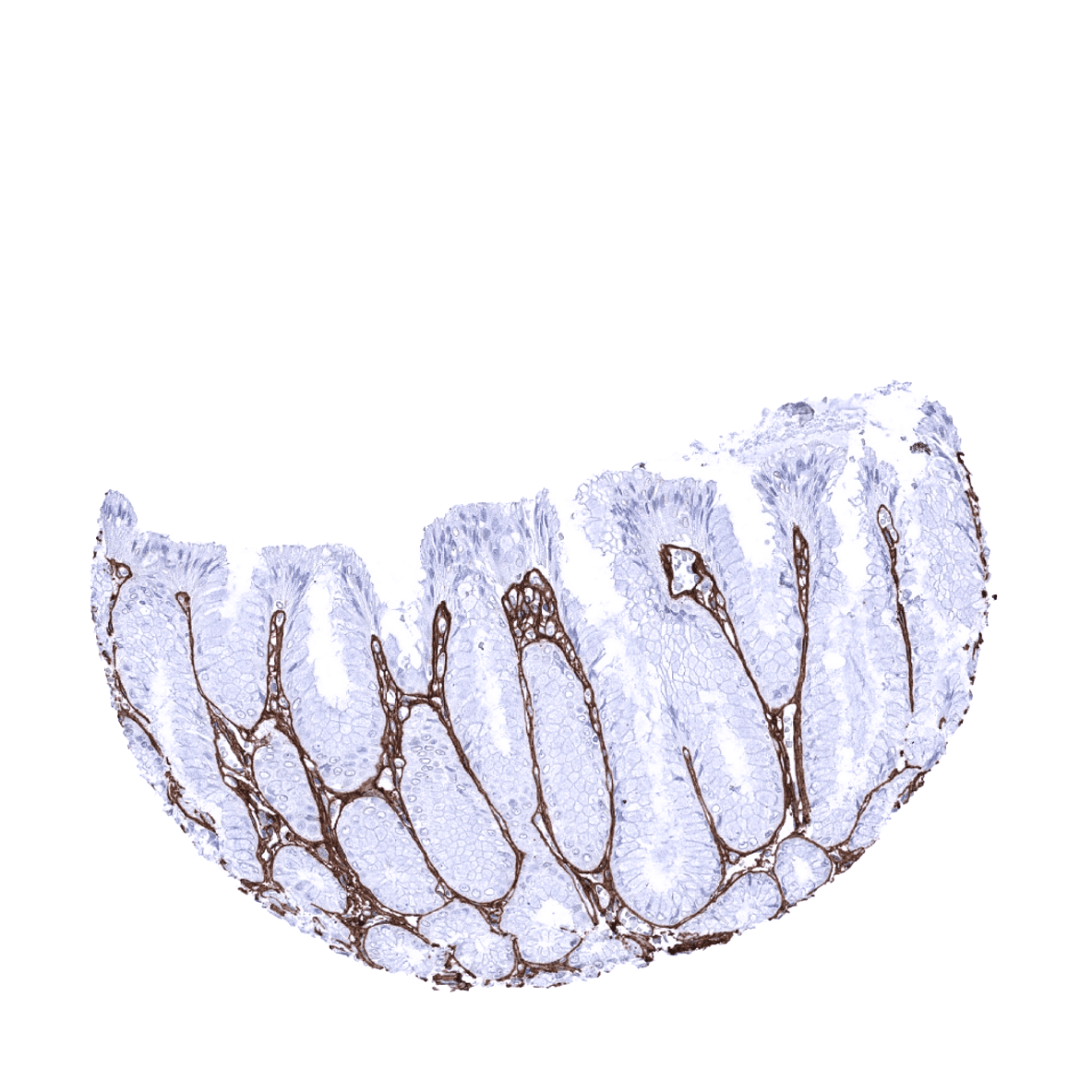
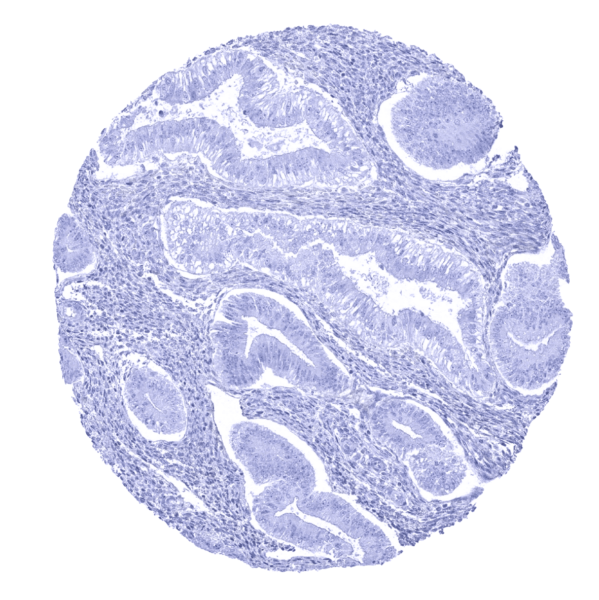
Stomach, antrum - Periostin is regularly seen in the gastrointestinal mucosa where it forms a rather thin subepithelial band.
Stomach, corpus - Periostin is regularly seen in the stomach mucosa where it forms a rather thin subepithelial band.

Testis
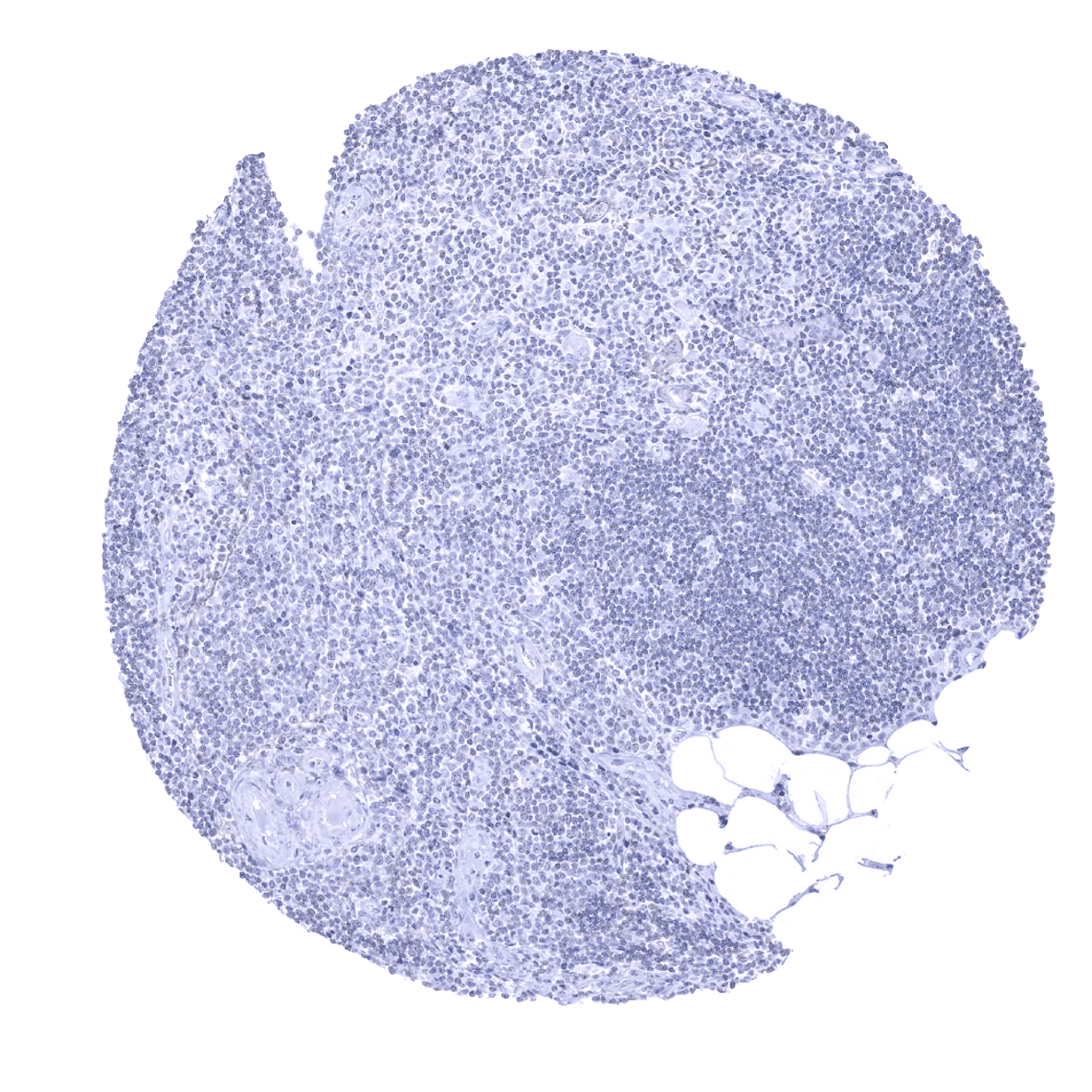
Thymus

Thyroid gland
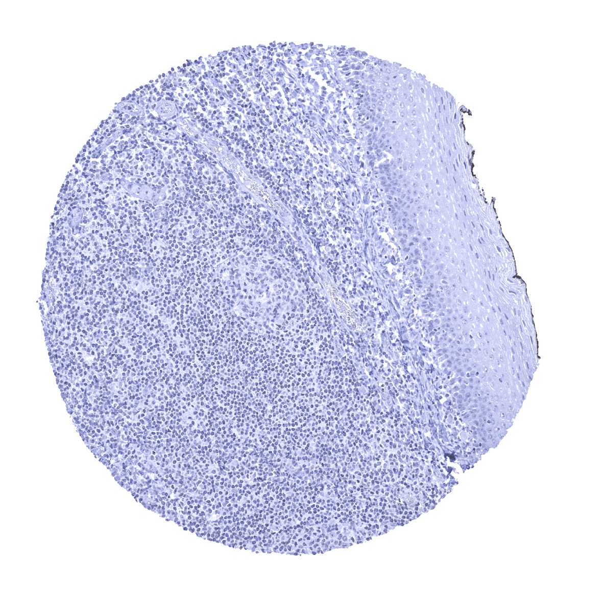
Tonsil, surface epithelium
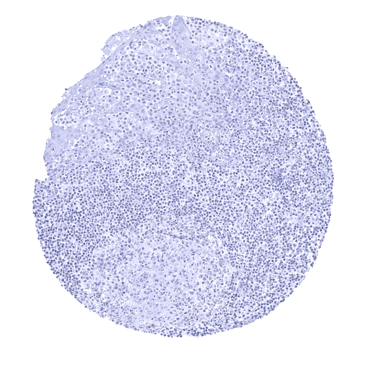
Tonsil

Urinary bladder, muscular wall
Urinary bladder, urothelium - A thin band of periostin immunostaining immediately below the suburothelial basement membrane can be seen in the urinary bladder.
Urinary bladder, urothelium - Bladder mucosa can lack periostin immunostaining.
Uterus, ectocervix - A thin band of periostin immunostaining immediately below the subepithelial basement membrane is often seen in the ectocervix.
Uterus, endocervix
Uterus, endometrium (pregnancy)

Uterus, endometrium (proliferation)
Uterus, endometrium (secretion)
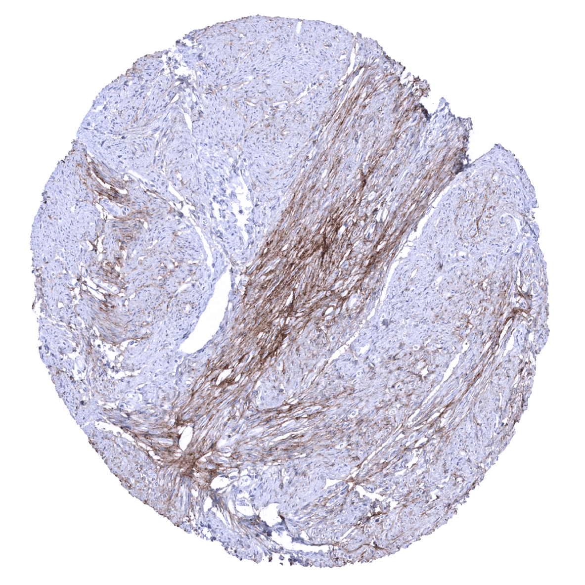
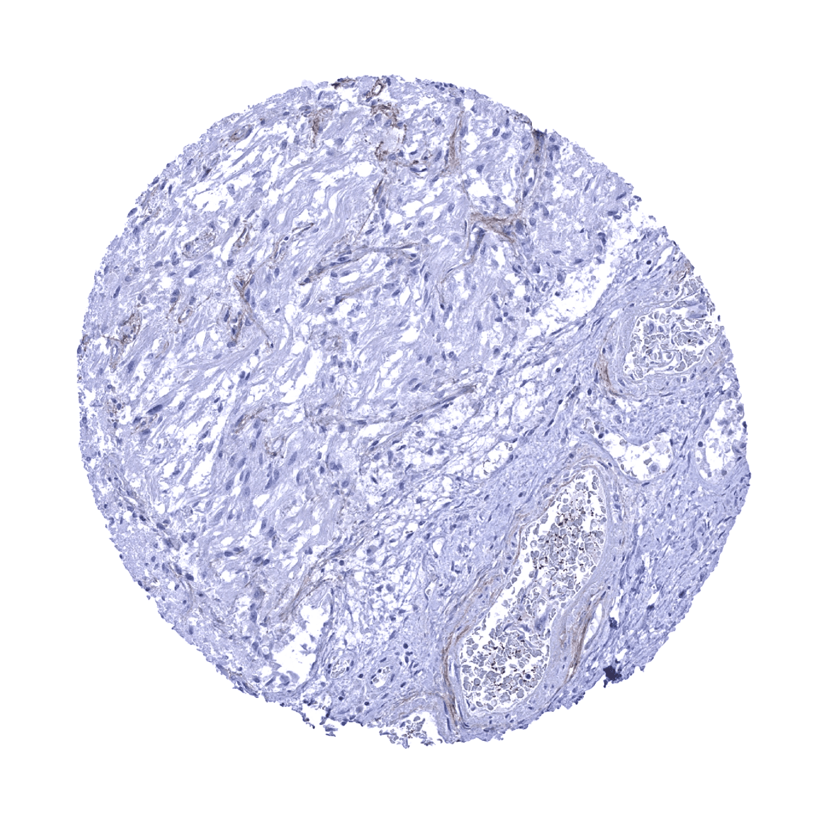
Uterus, myometrium - Focal periostin immunostaining can be observed in the myometrium.
Uterus, myometrium - Myometrium usually lacks periostin immunostaining.