
295,00 € – 995,00 €
Product details
Synonyms = BLCPMG; Occludin; OCLN; Phosphatase 1 regulatory subunit 115; PPP1R115; PTORCH1; Tight junction protein occludin
Antibody type = Mouse monoclonal / IgG
Clone = MSVA-415M
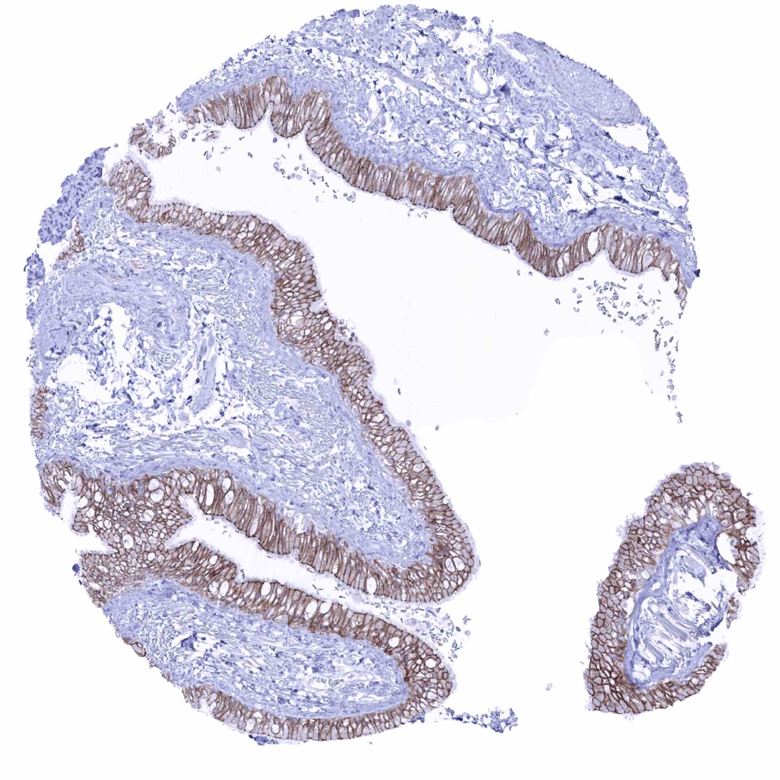
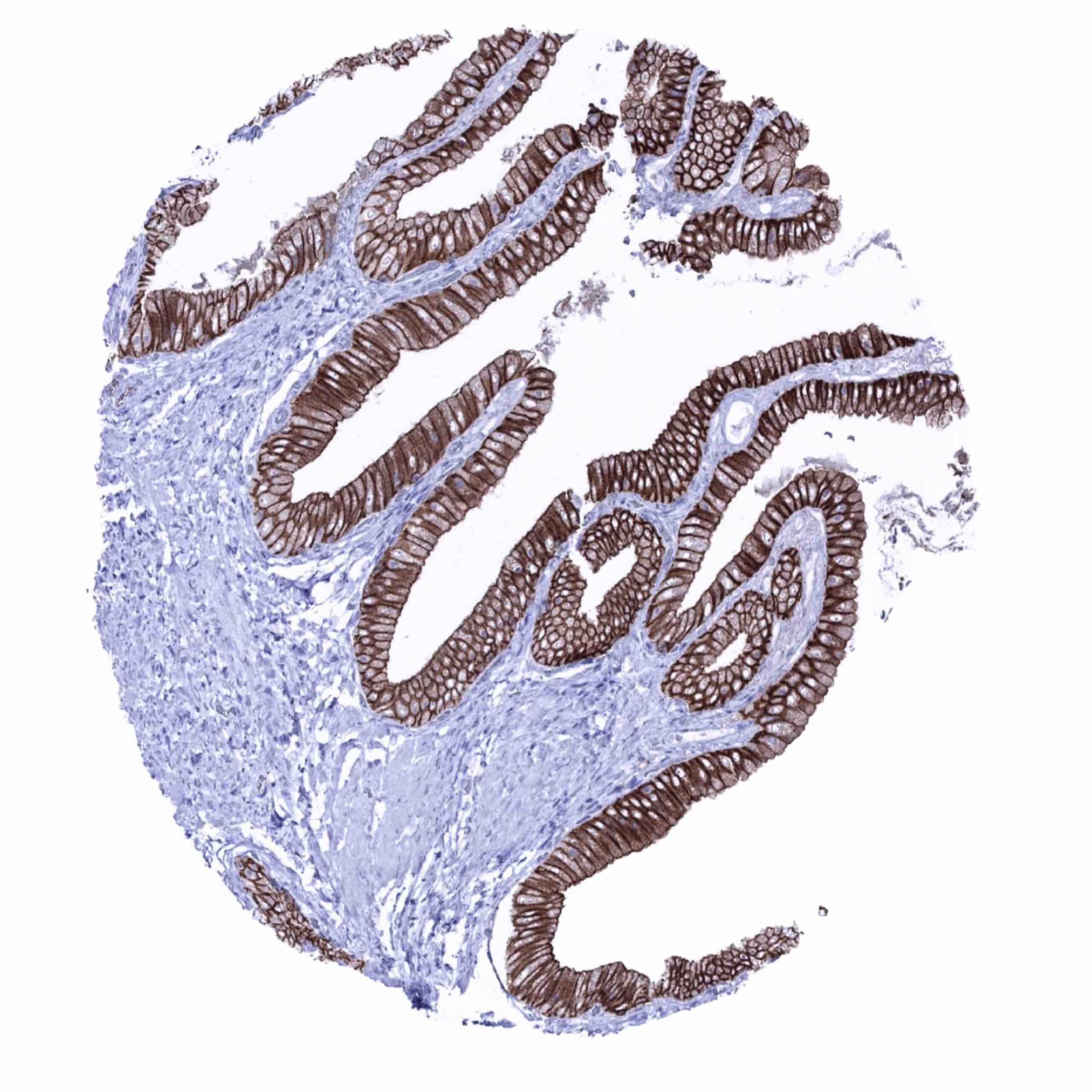
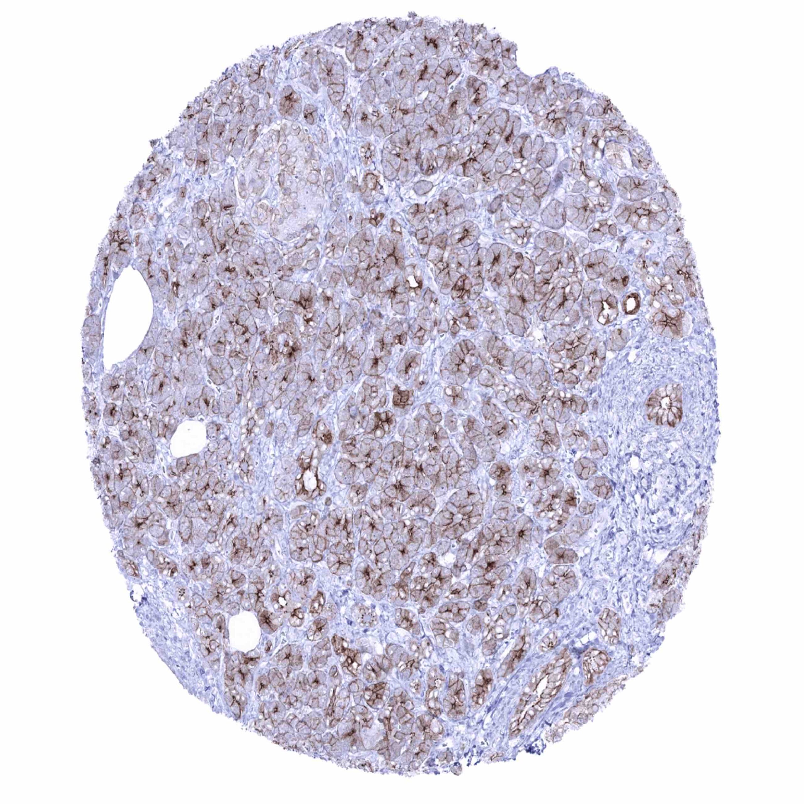
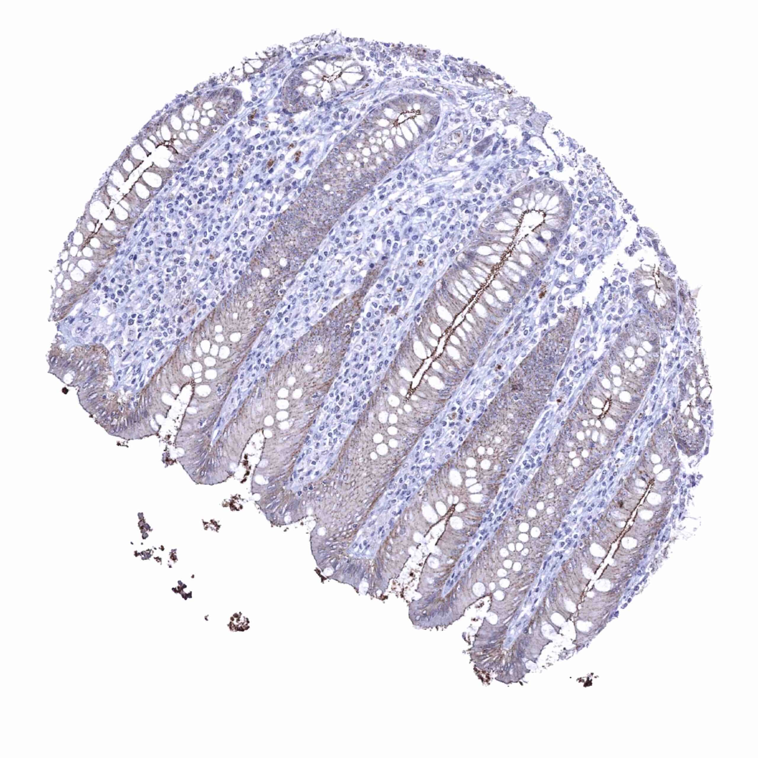
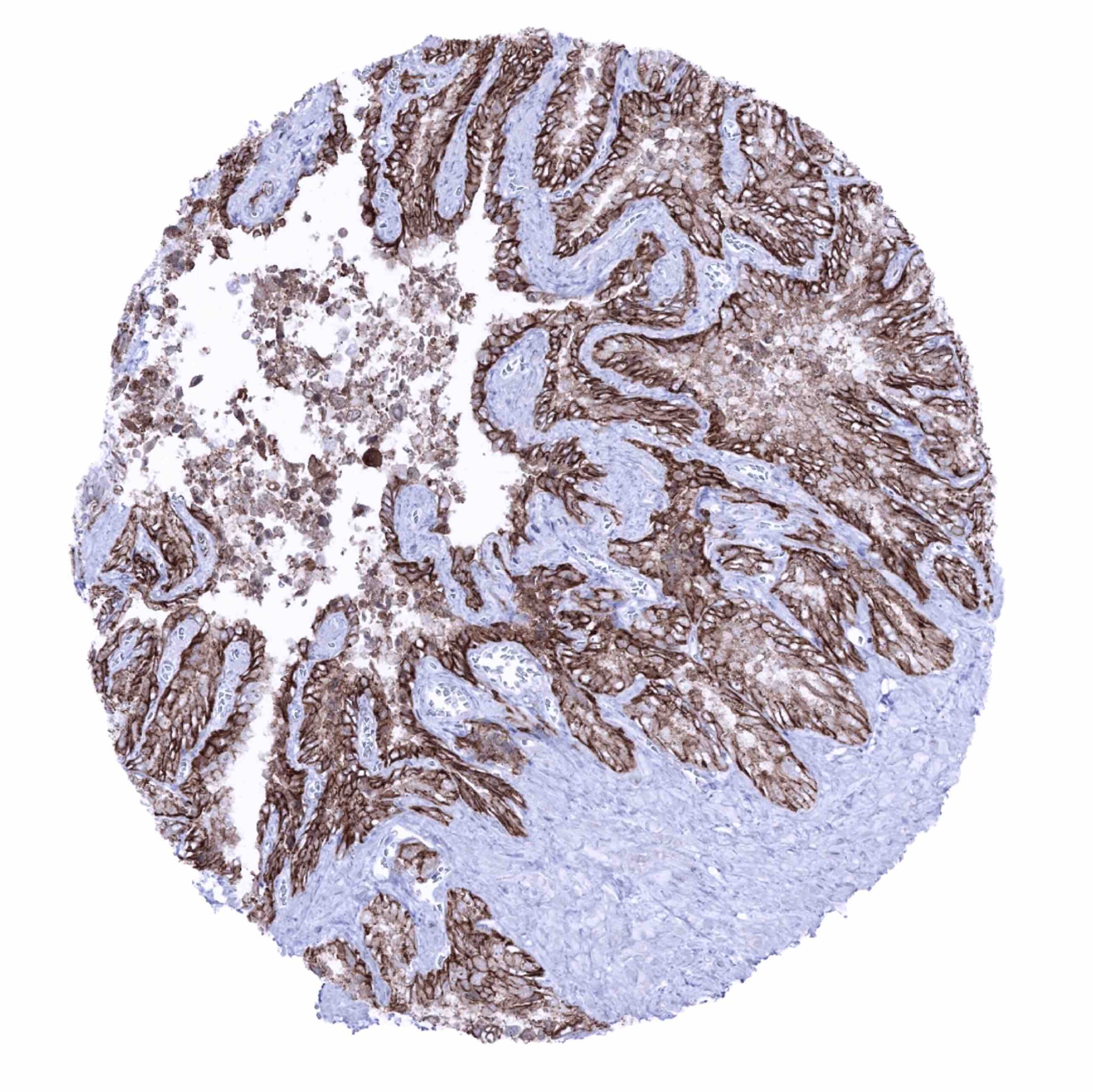
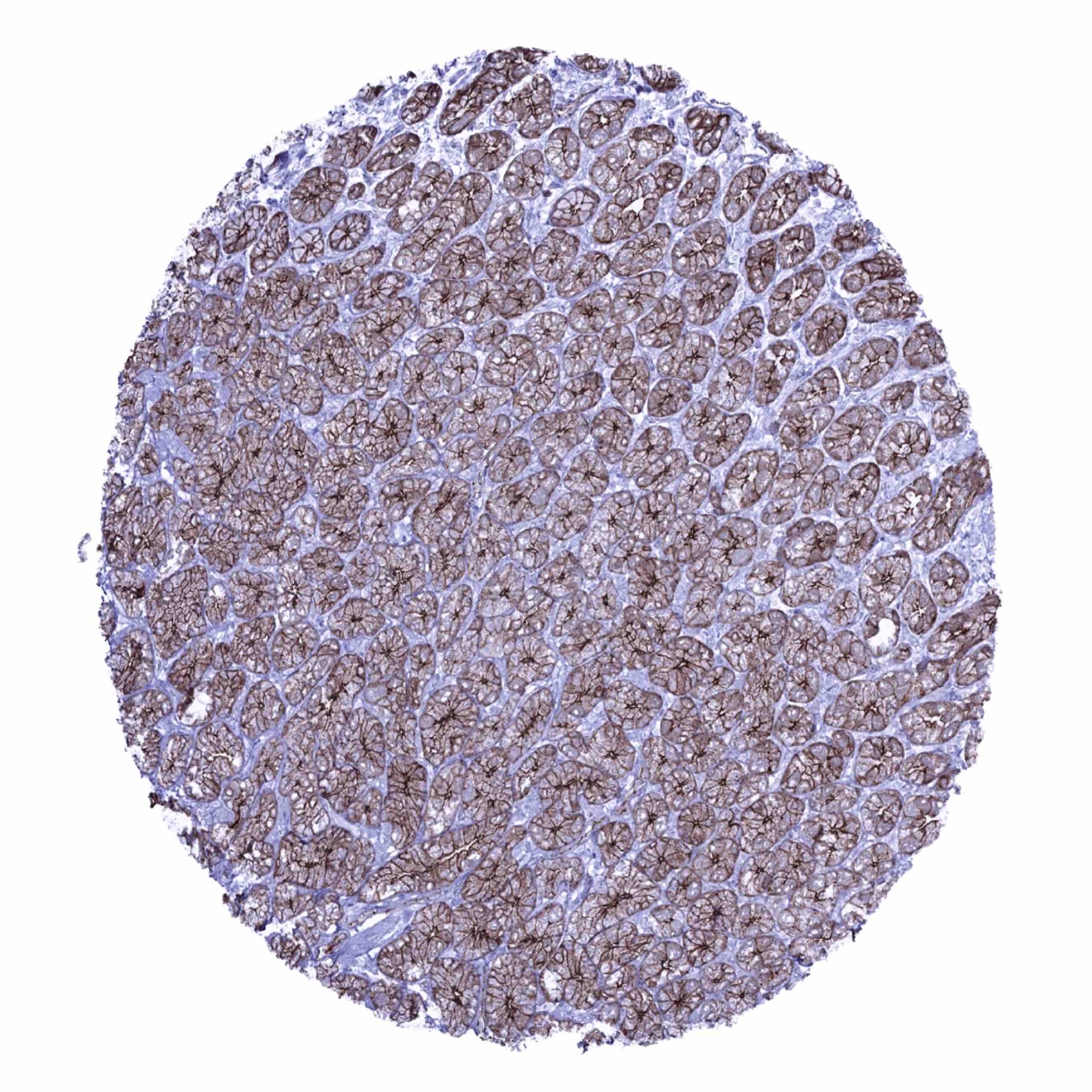
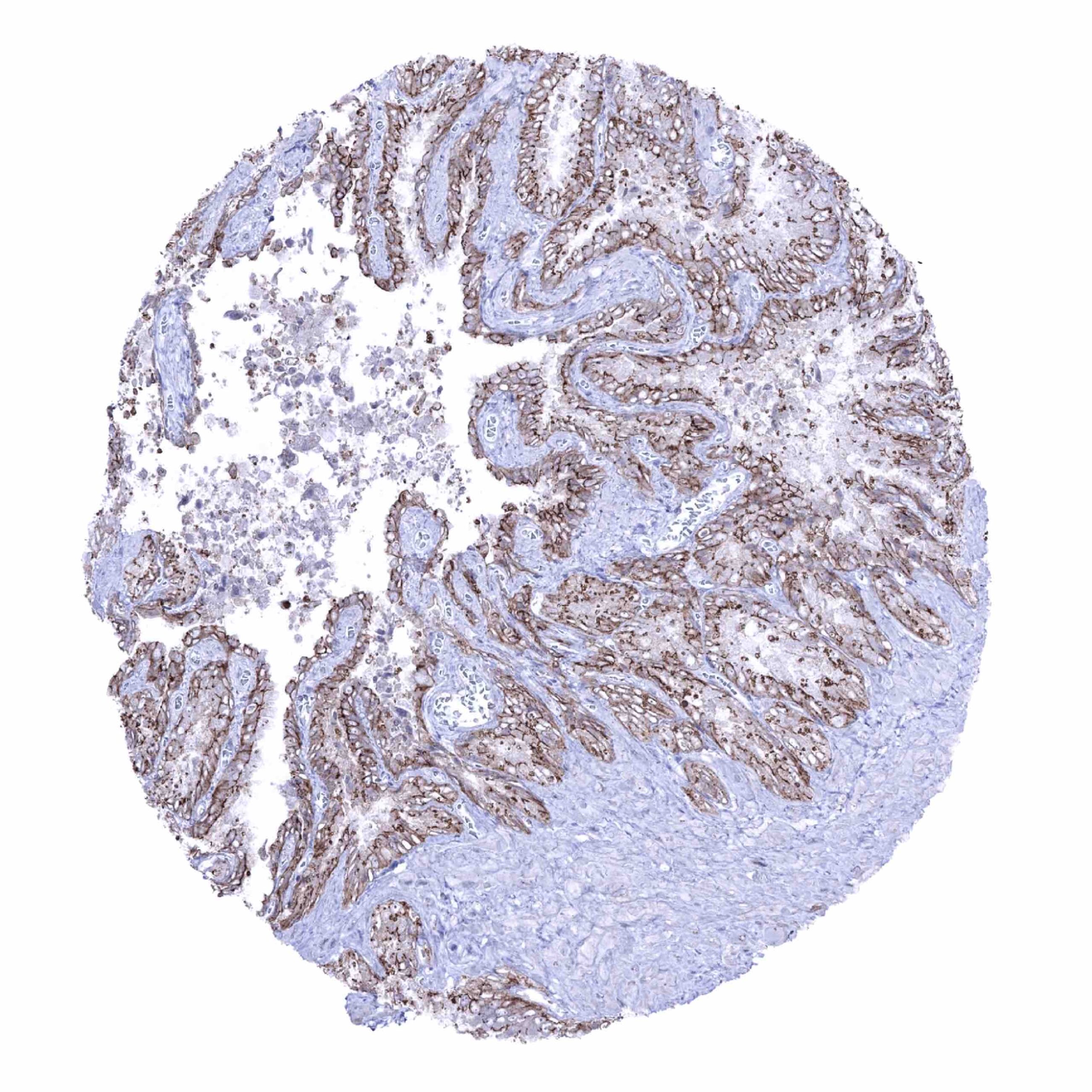
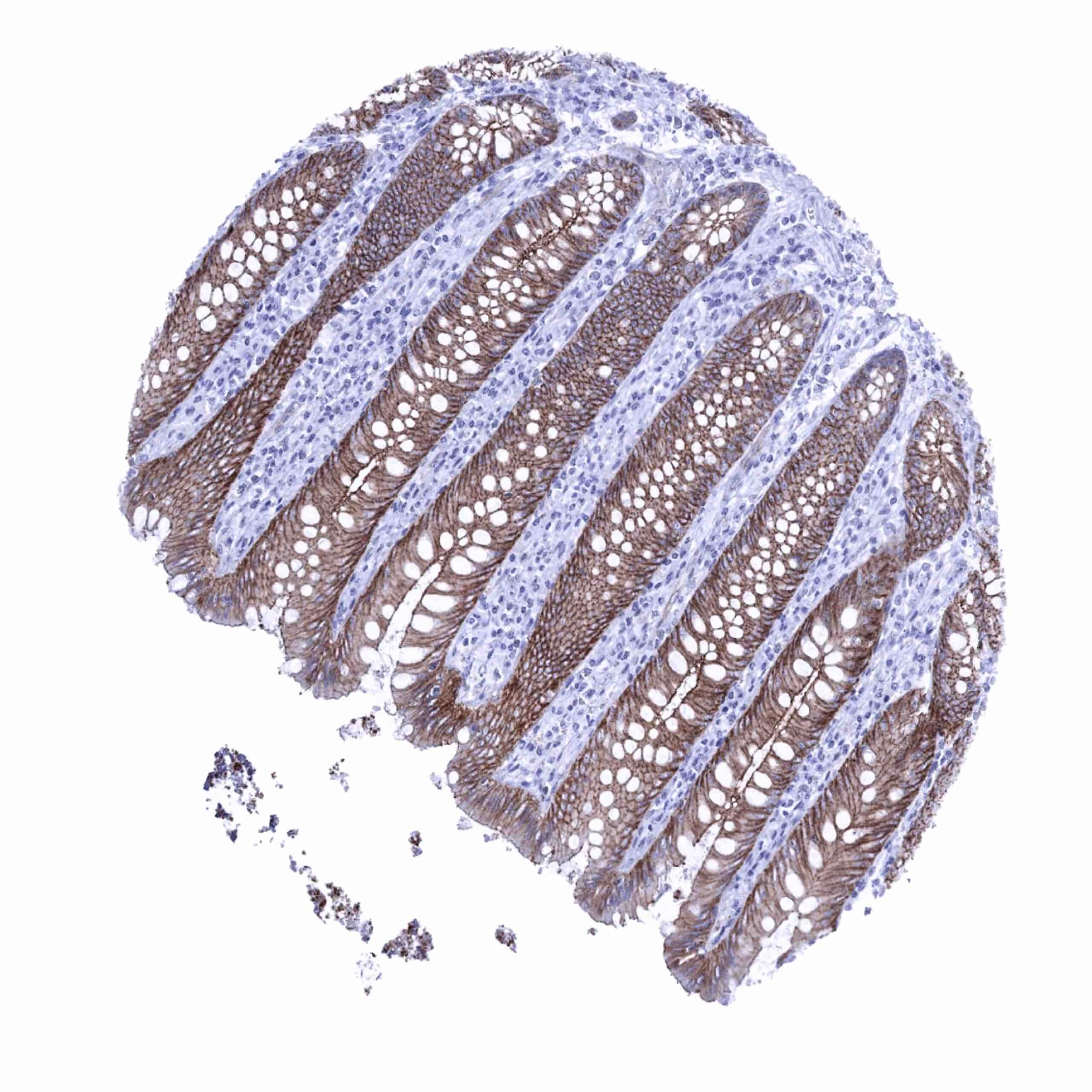
Positive control = Colon: A strong membranous occludin staining of epithelial cells should be seen with a particularly strong staining at the apical/luminal membranes.
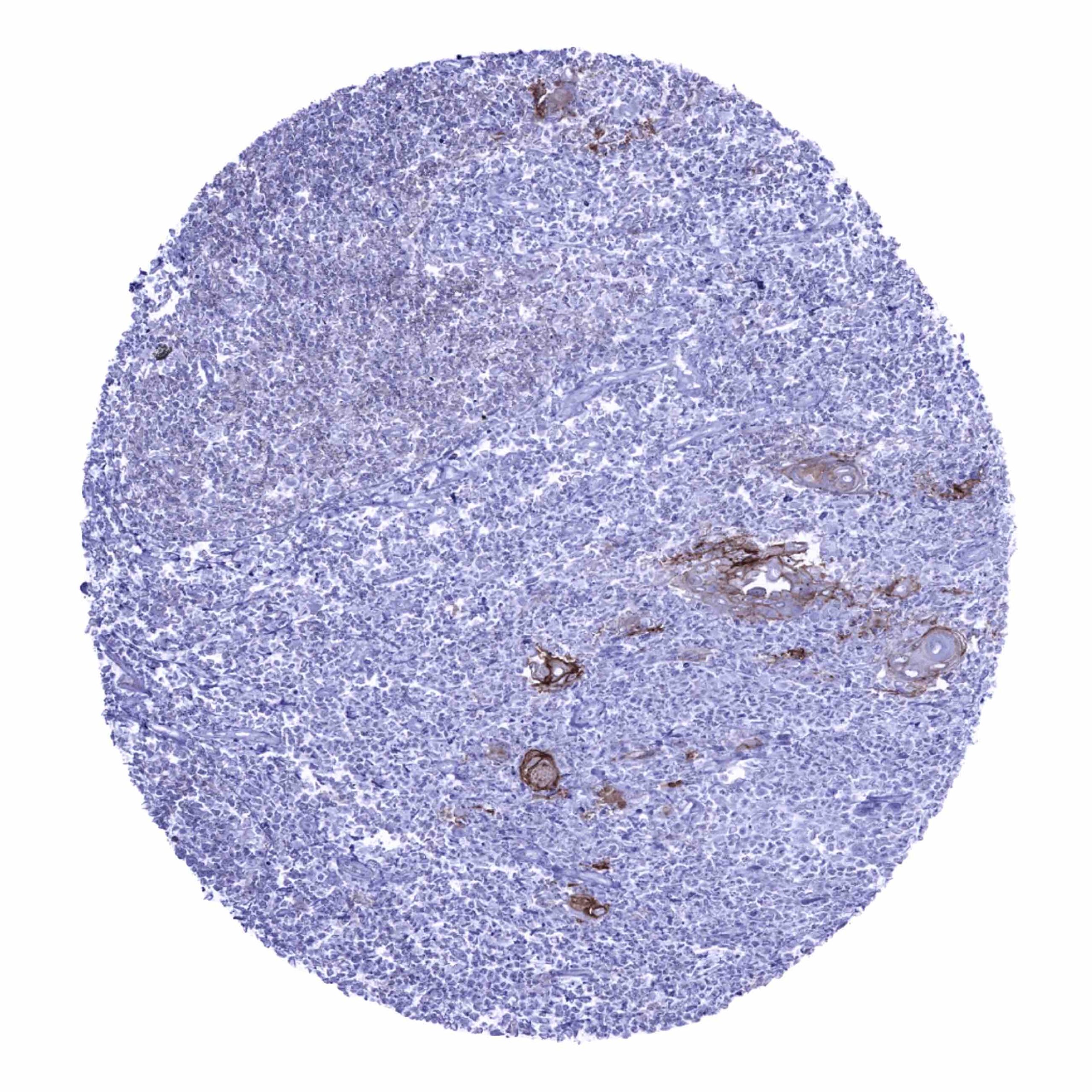
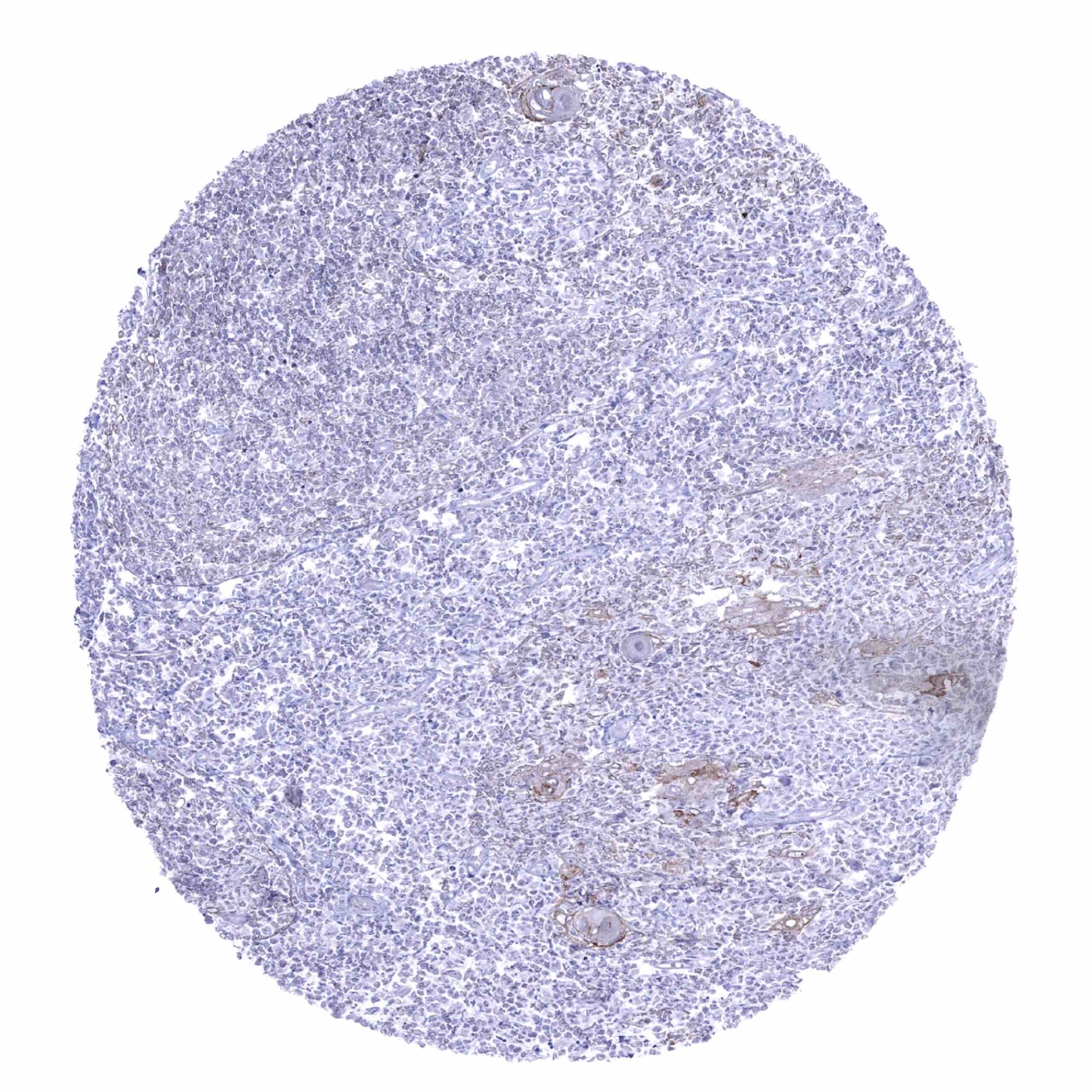
Negative control = Colon: All non-epithelial and non-endothelial cells must not show any occludin staining.
Cellular localization = Cell Surface and Cytoplasmic
Reactivity = Human
Application = Immunohistochemistry
Dilution = 1:100 – 1:200
Intended Use = Research Use Only
Relevance of Antibody
Occludin is a Pivotal component of tight junctions.
Biology Behind
Occludin (OCLN) is a 65 kDa plasma-membrane protein coded by the OCLN gene at chromosome 5q13.1. The Occludin protein is preferentially located at the tight junctions of epithelial and endothelial cells. Together with other proteins such as claudins and zonula occludens-1 (ZO-1), occludin regulates the formation, maintenance, and function of tight junctions. In particular, occludin may be important for tight junction stability and barrier function. As a NADH oxidase, occludin influences critical aspects of cell metabolism like glucose uptake, ATP production and gene expression. Manipulation of the occludin content in human cells affects the expression of glucose transporters, and the activation of transcription factors such as NFkB. In cancer, loss of occludin has been linked to increased invasion, reduced adhesion and metastasis.
Staining Pattern in Normal Tissues
Occludin staining pattern in Normal Tissues with antibody MSVA-415M (images are shown in our “Normal Tissue Gallery”)
| Brain | Cerebrum | Negative. |
| Cerebellum | Negative. | |
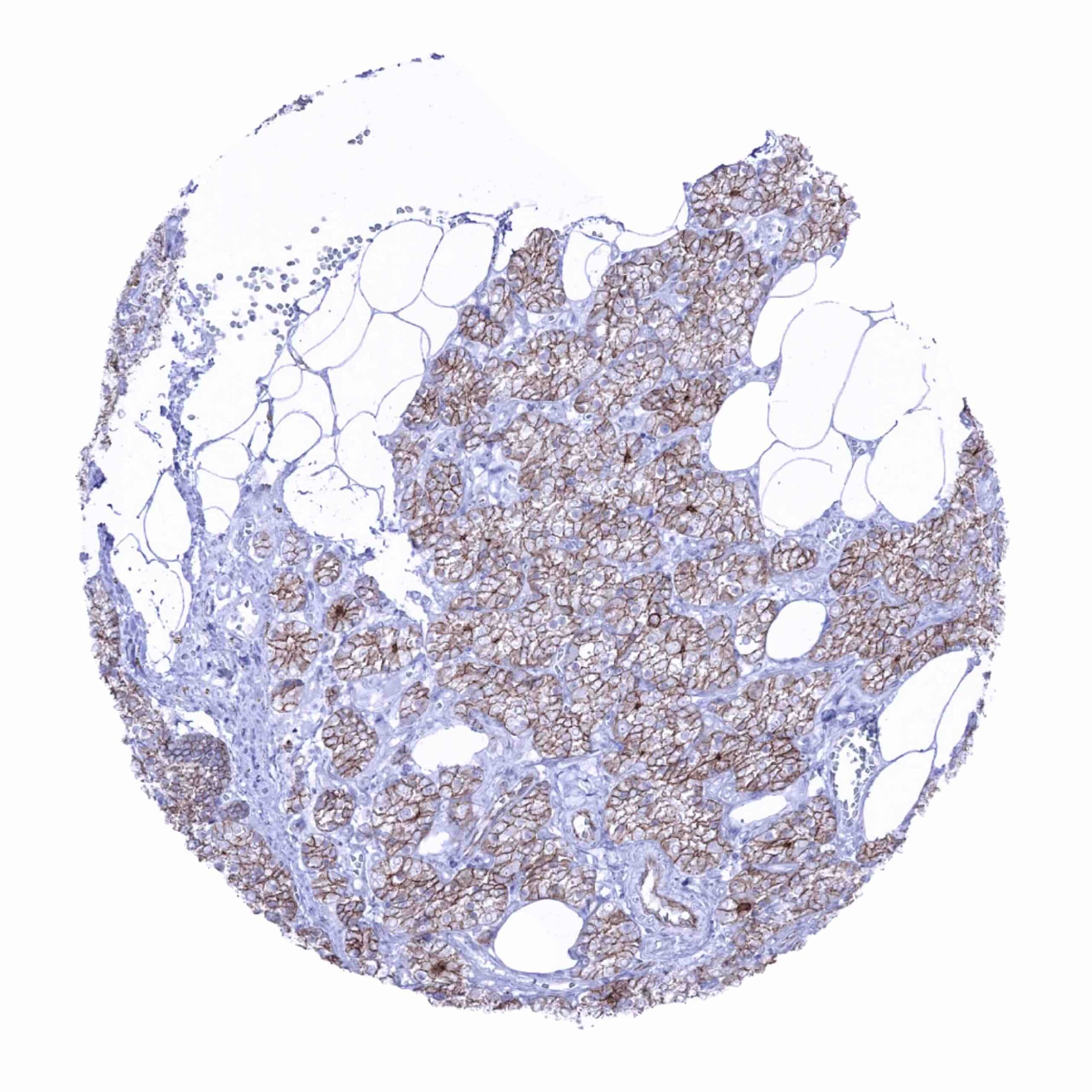
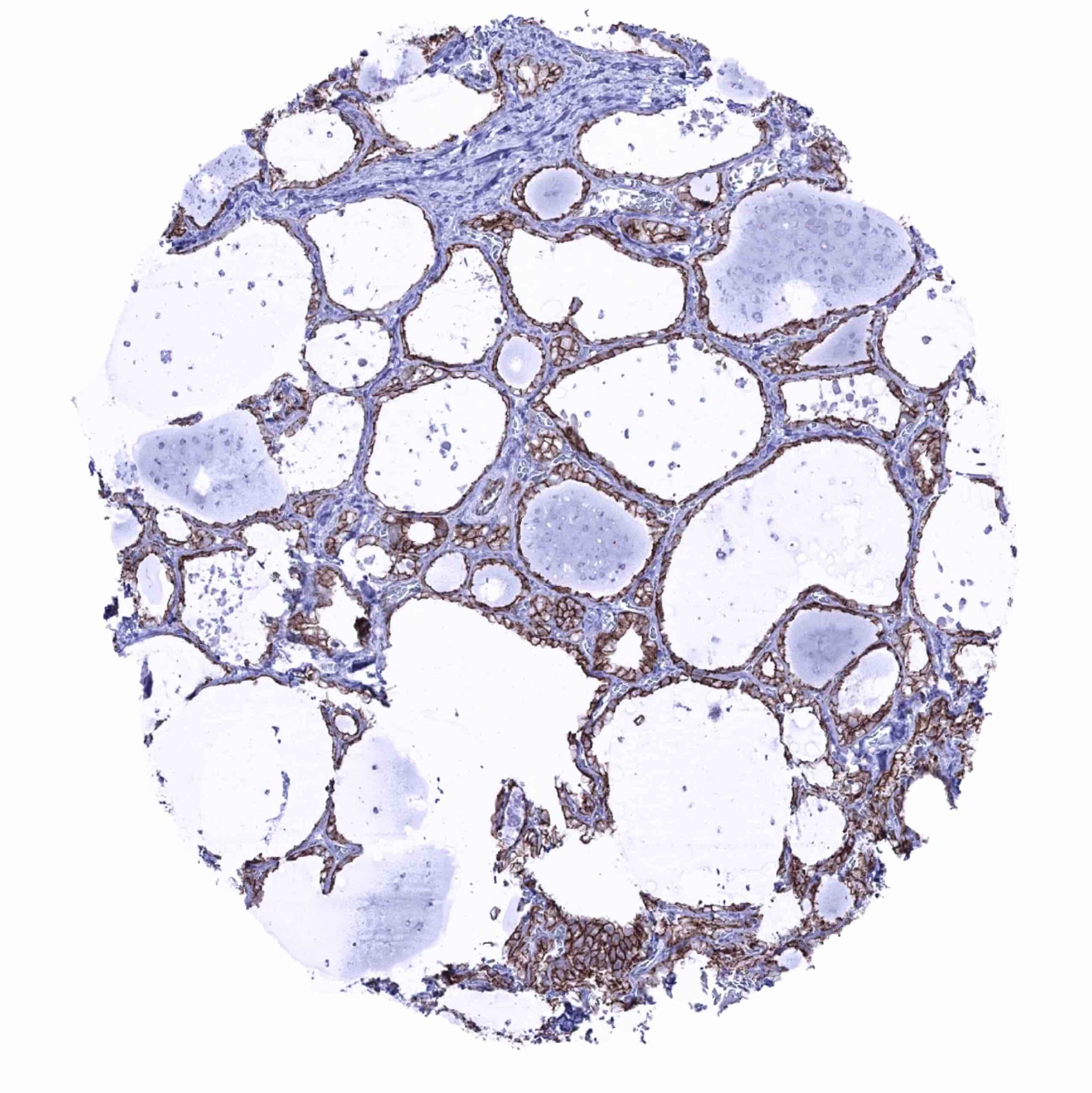
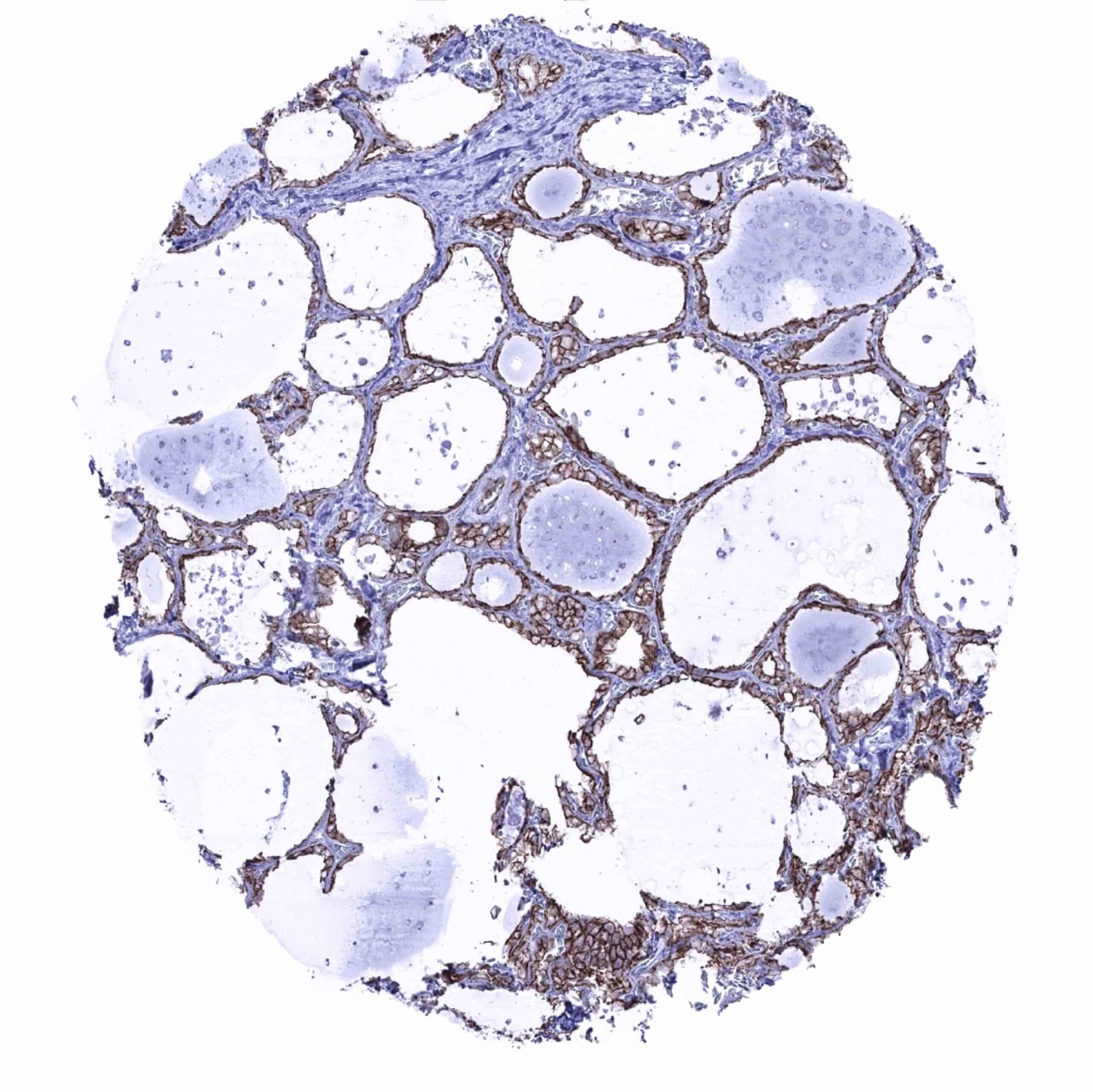
| Endocrine Tissues | Thyroid | Intense membranous occludin immunostaining of follicular cells. |
| Parathyroid | Moderate membranous occludin staining of epithelial cells. | |
| Adrenal gland | Negative. | |
| Pituitary gland | Only few epithelial cells show a detectable membranous occluding positivity in the adenohypophysis. The neurohypophysis is occludin negative. | |
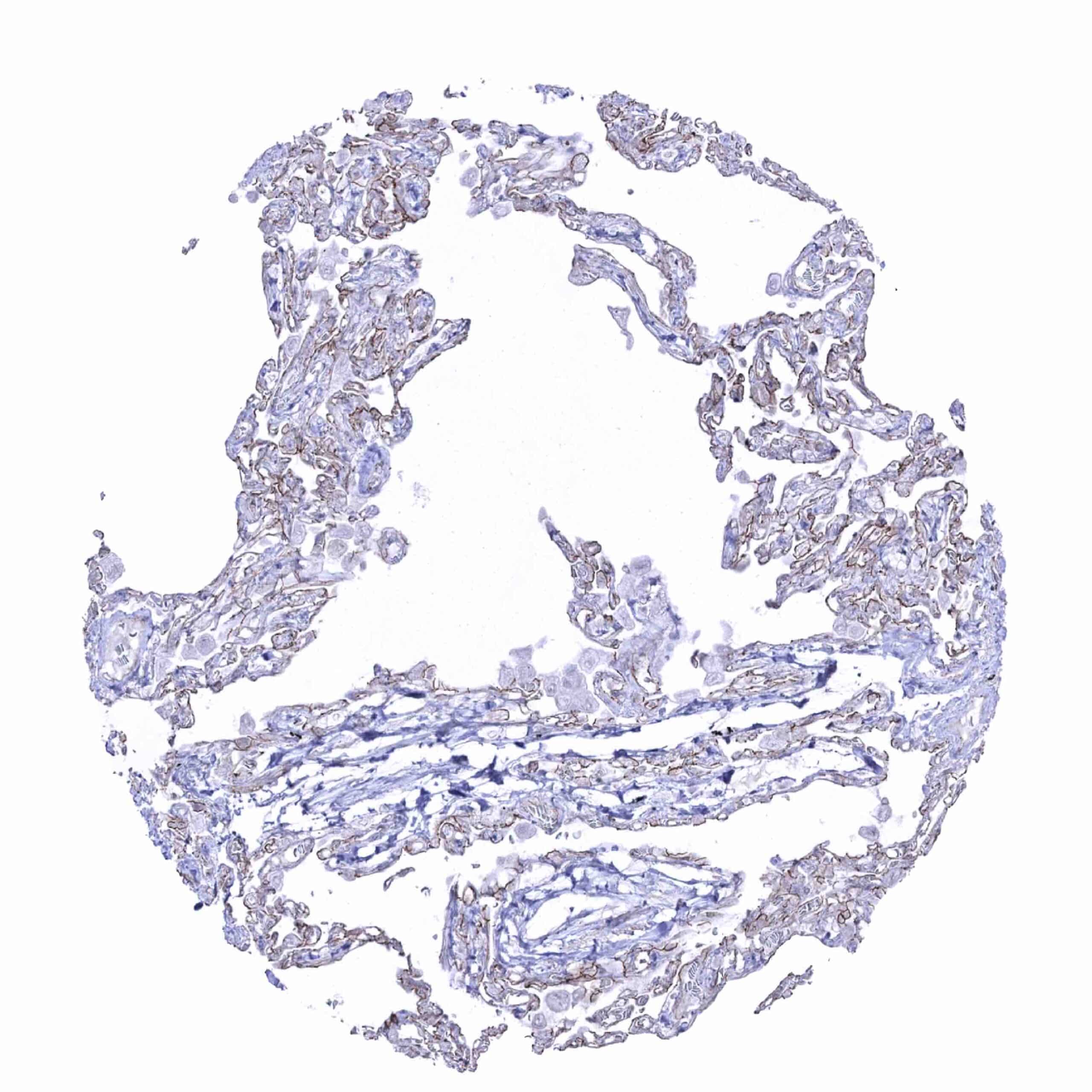
| Respiratory system | Respiratory epithelium | Significant membranous occludin staining. |
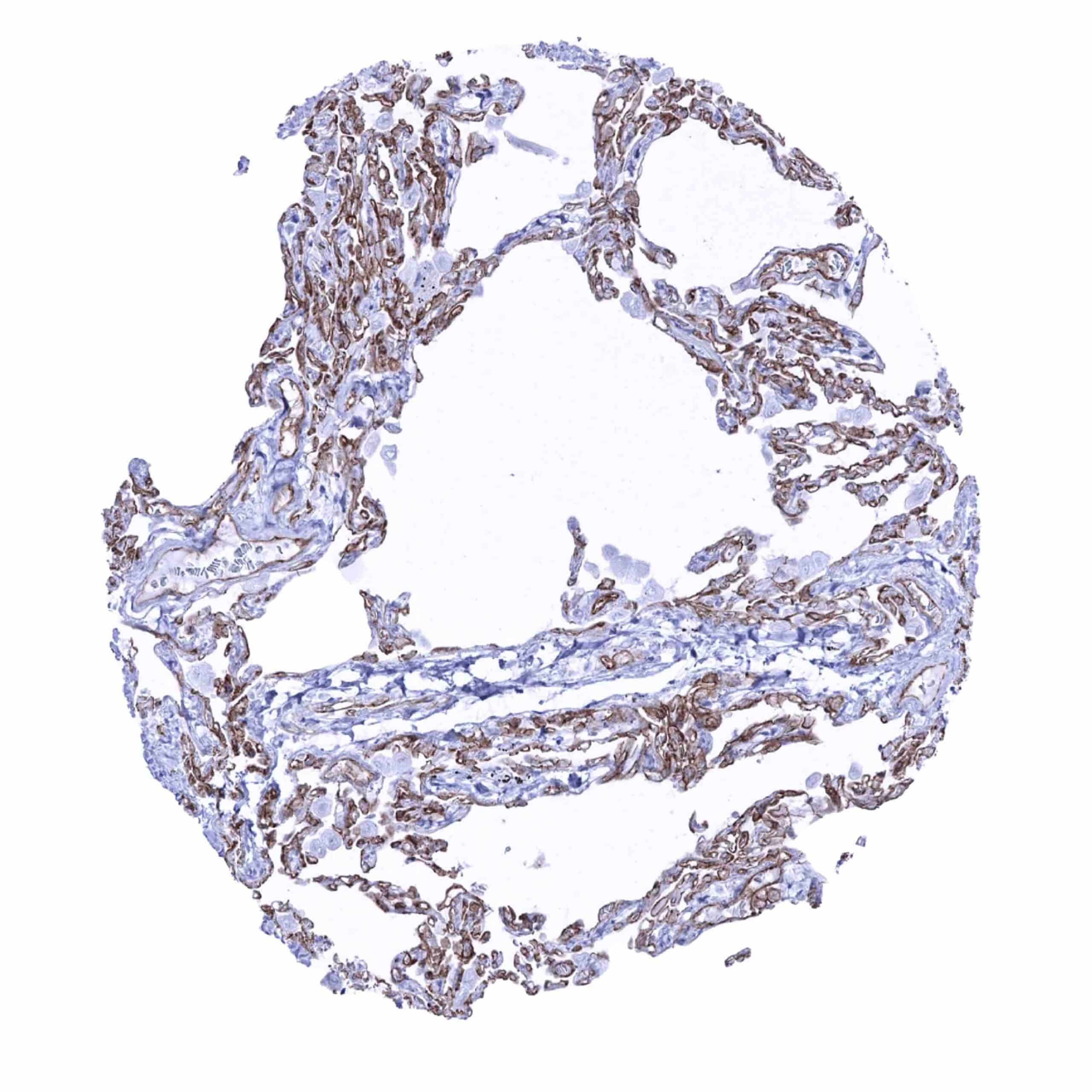
| Lung | Both pneumocytes and endothelial cells show a significant membranous occludin staining while macrophages remain negative. | |
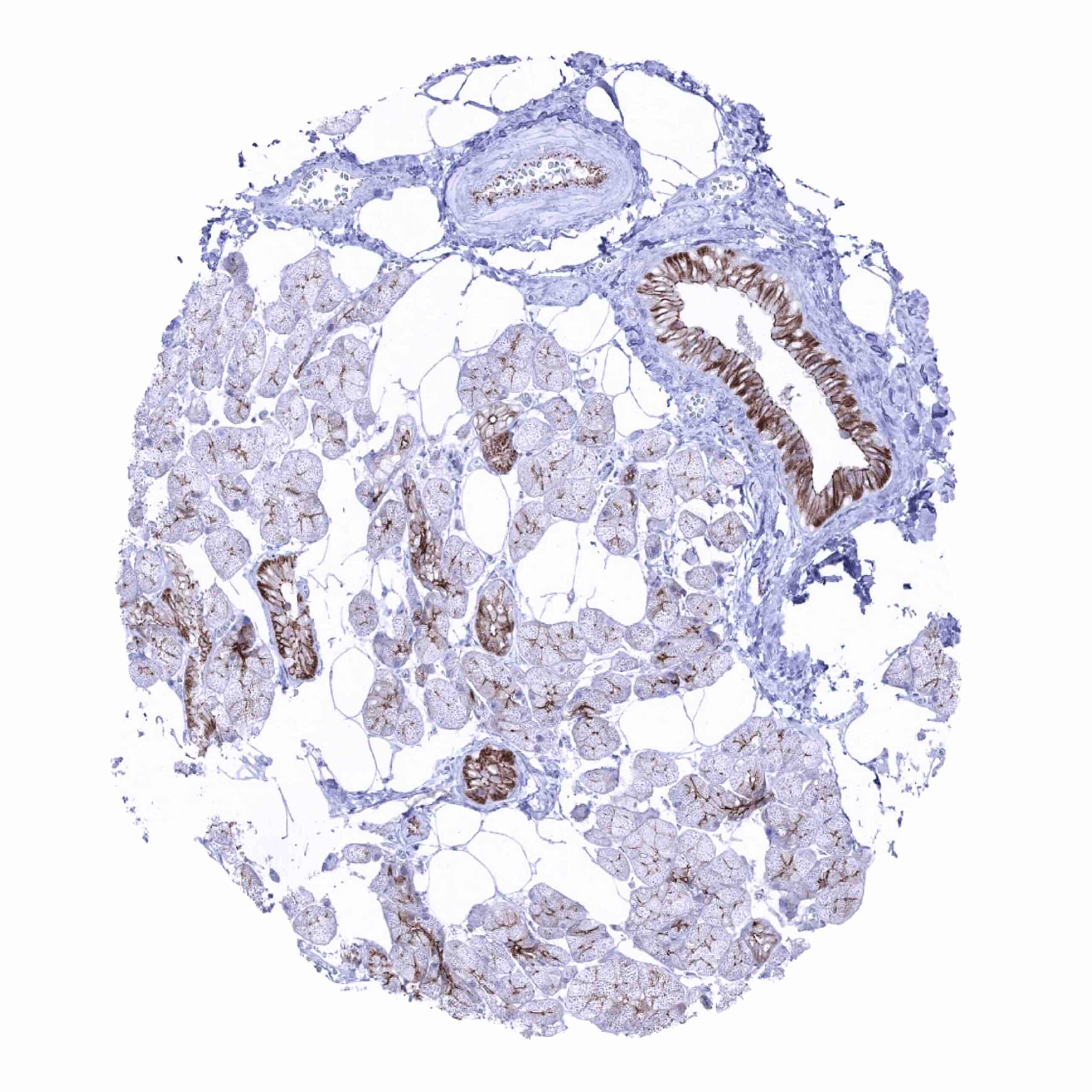
| Gastrointestinal Tract | Salivary glands | Membranous occludin staining is most intense in excretory ducts and only faint in glandular cells. |
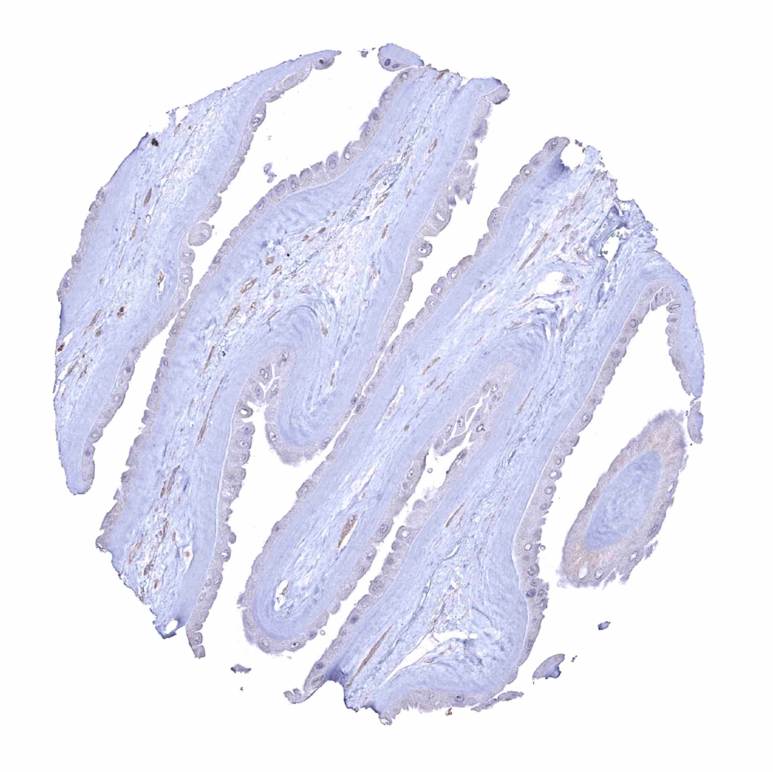
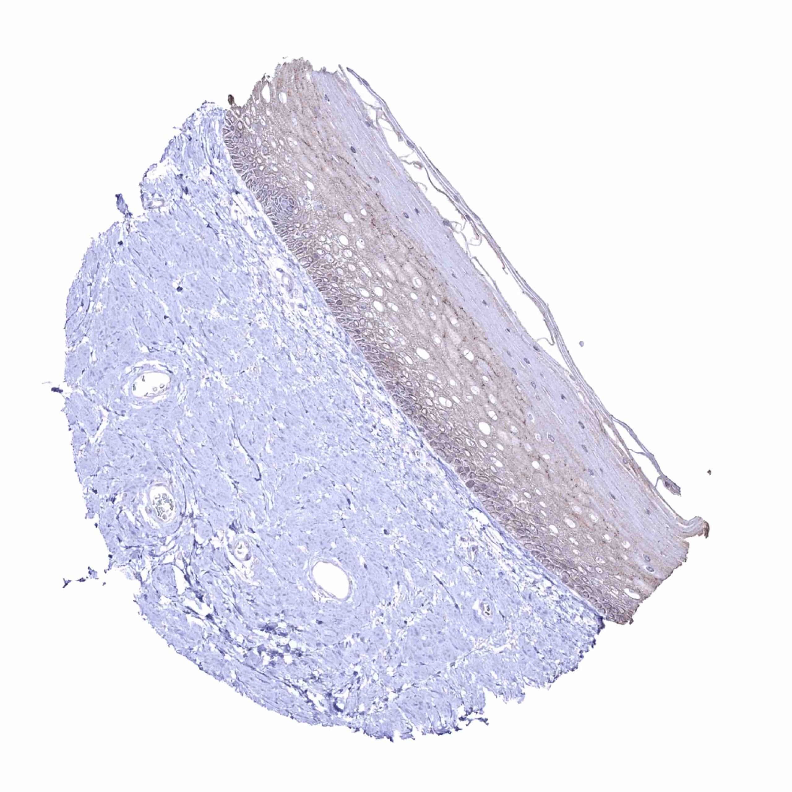
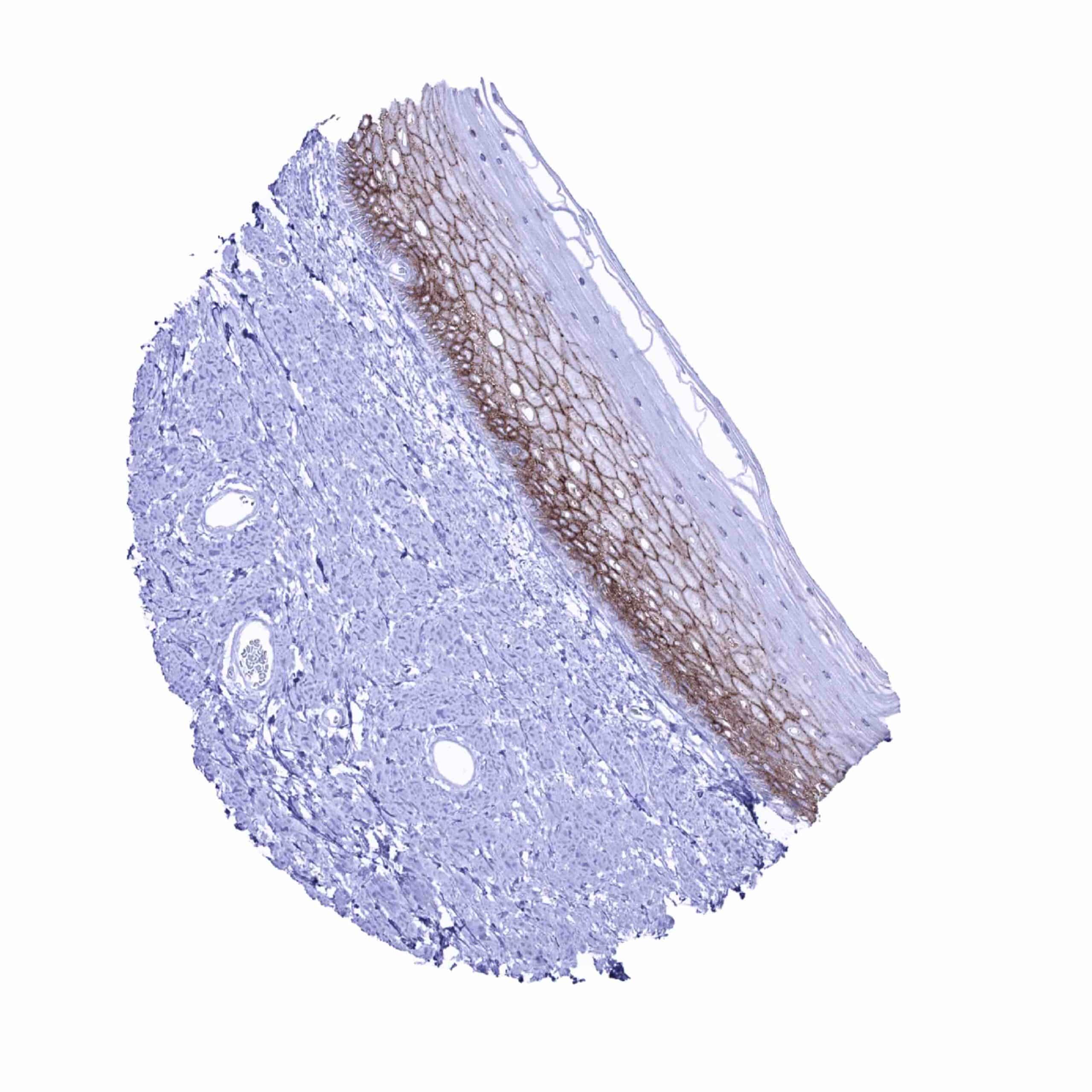
| Esophagus | Occludin staining predominates in intermediate cell layers of the squamous epithelium, while occludin positivity is either absent or reduced in basal and superficial cell layers. | |
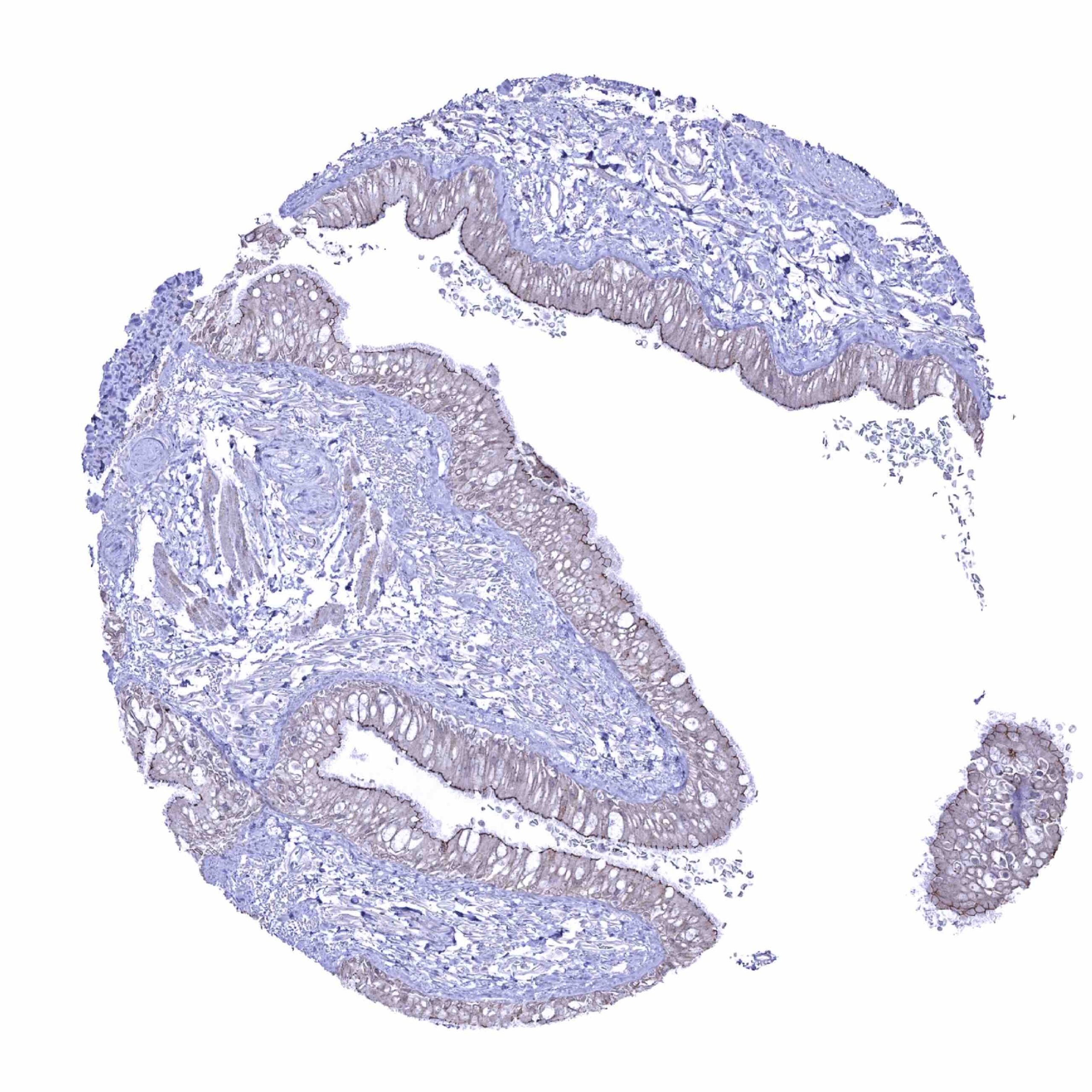
| Stomach | Strong occludin staining of all epithelial cells. Staining is often strongest at the apical membranes. | |
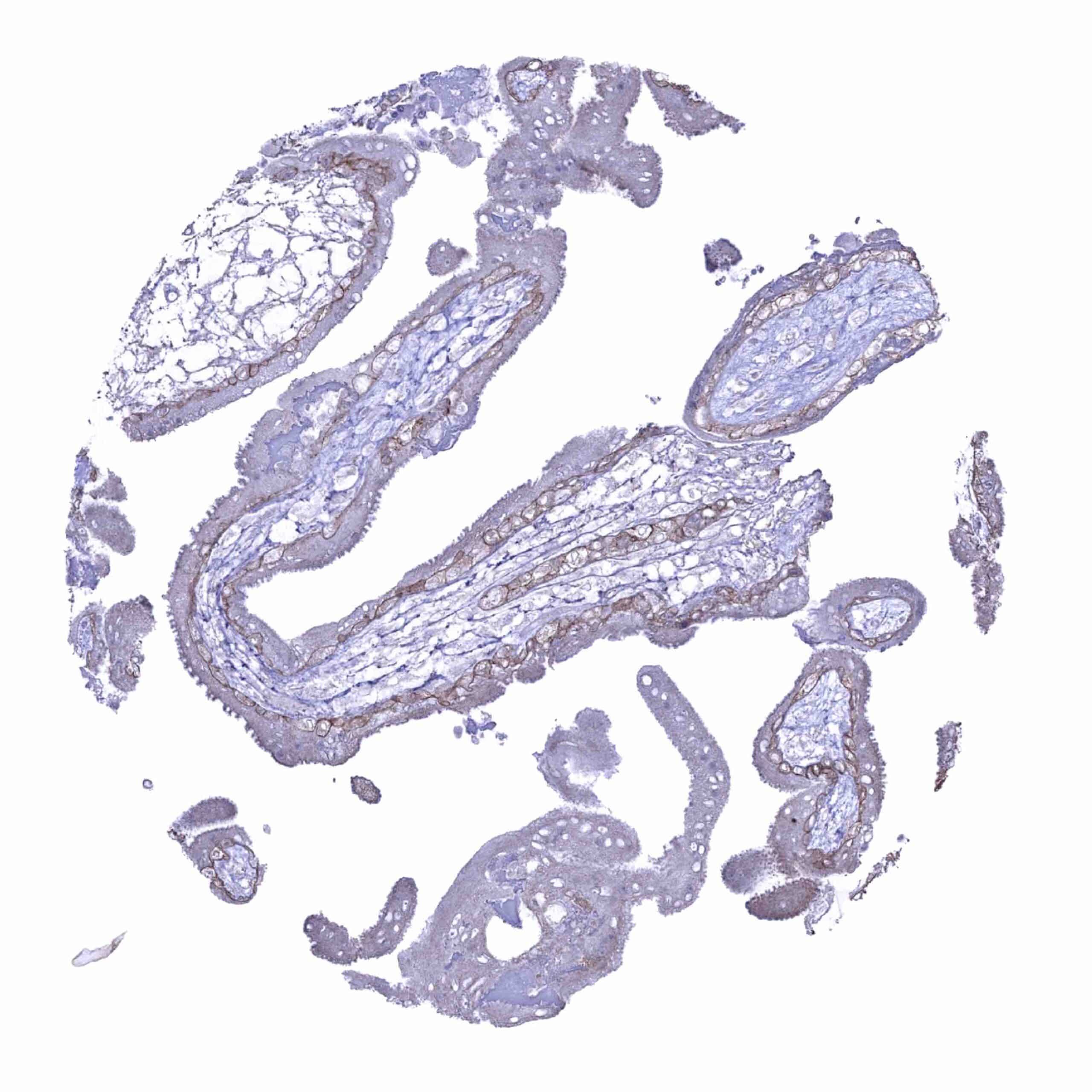
| Duodenum | Strong occludin staining of all intestinal epithelial cells. Staining is often strongest at the apical membranes. Membranous occludin staining of moderate intensity in glandular cells of Brunner glands. | |
| Small intestine | Strong occludin staining of all epithelial cells. Staining is often strongest at the apical membranes. | |
| Appendix | Strong occludin staining of all epithelial cells. Staining is often strongest at the apical membranes. | |
| Colon | Strong occludin staining of all epithelial cells. Staining is often strongest at the apical membranes. | |
| Rectum | Strong occludin staining of all epithelial cells. Staining is often strongest at the apical membranes. | |
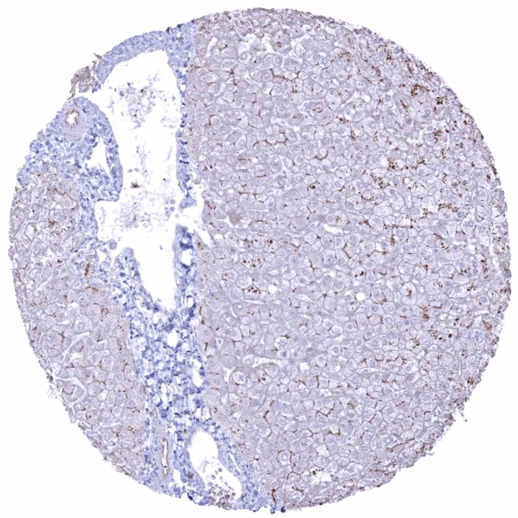
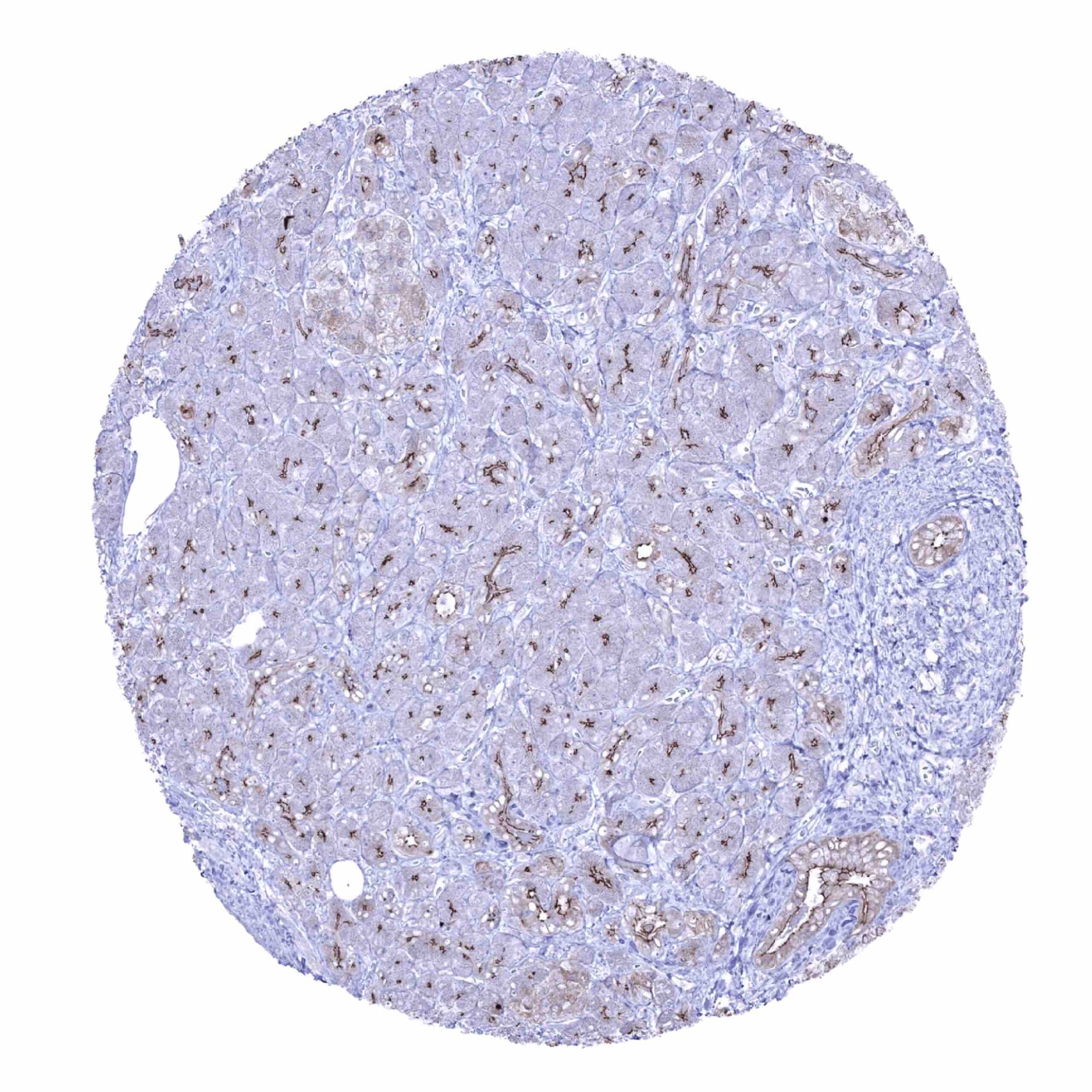
| Liver | Significant membranous occludin staining. Positivity is stronger in bile duct epithelia than in hepatocytes. | |
| Gallbladder | Strong occludin staining of all epithelial cells. | |
| Pancreas | Membranous occludin staining of all cells. Occludin positivity is strongest at apical membranes of acinar cells and only weak in islet cells. | |
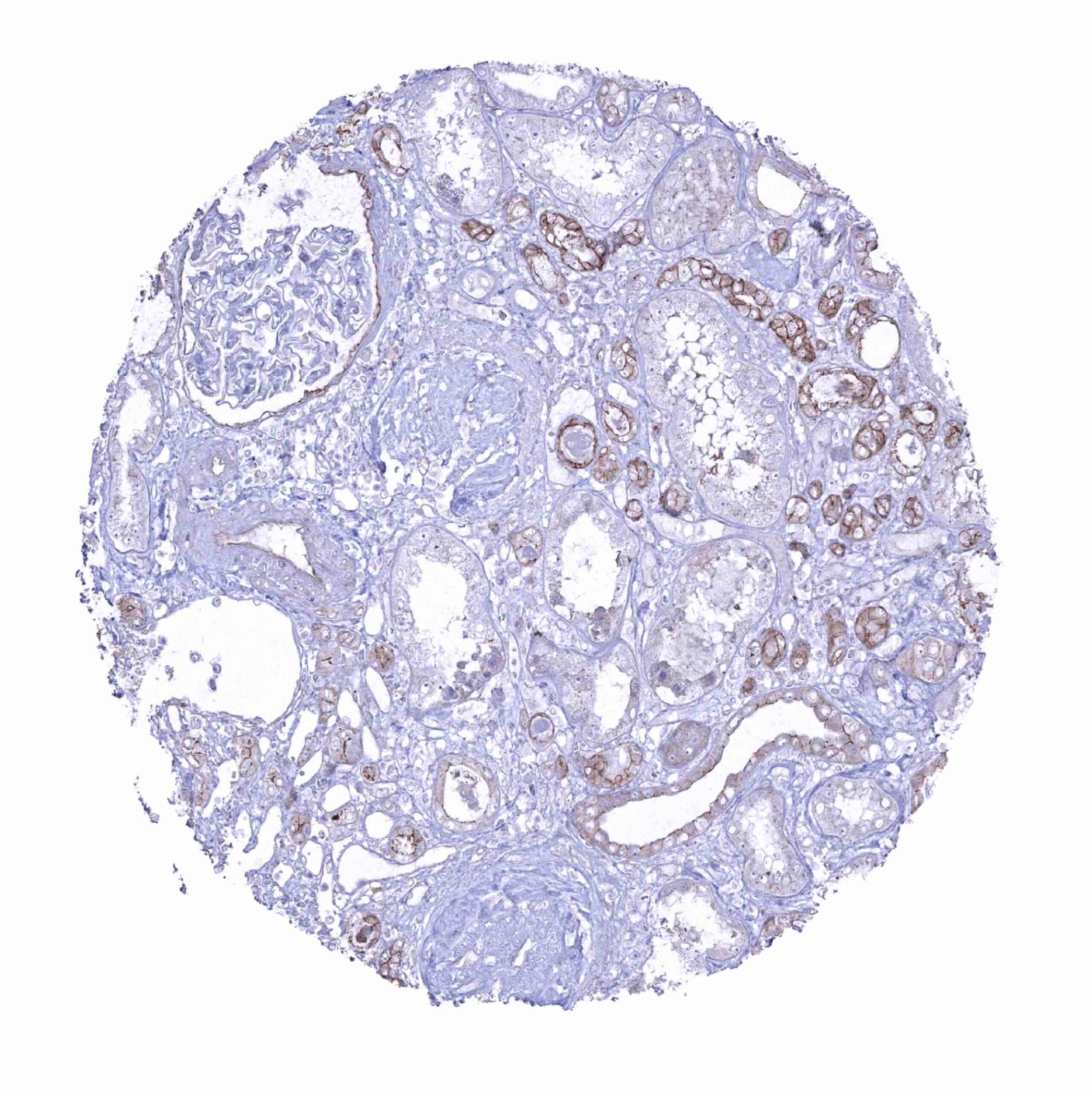
| Genitourinary | Kidney | Occludin staining is strongest in collecting ducts, weaker in distal tubuli and weakest in proximal tubuli of the kidney. |
| Urothelium | Strong occludin staining of all urothelial cell layers. | |
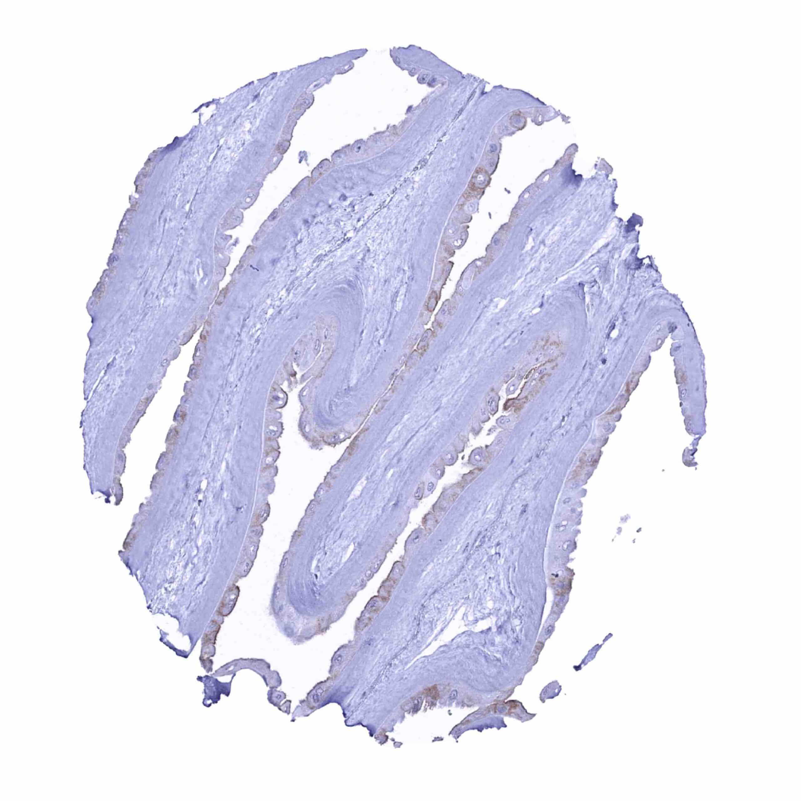
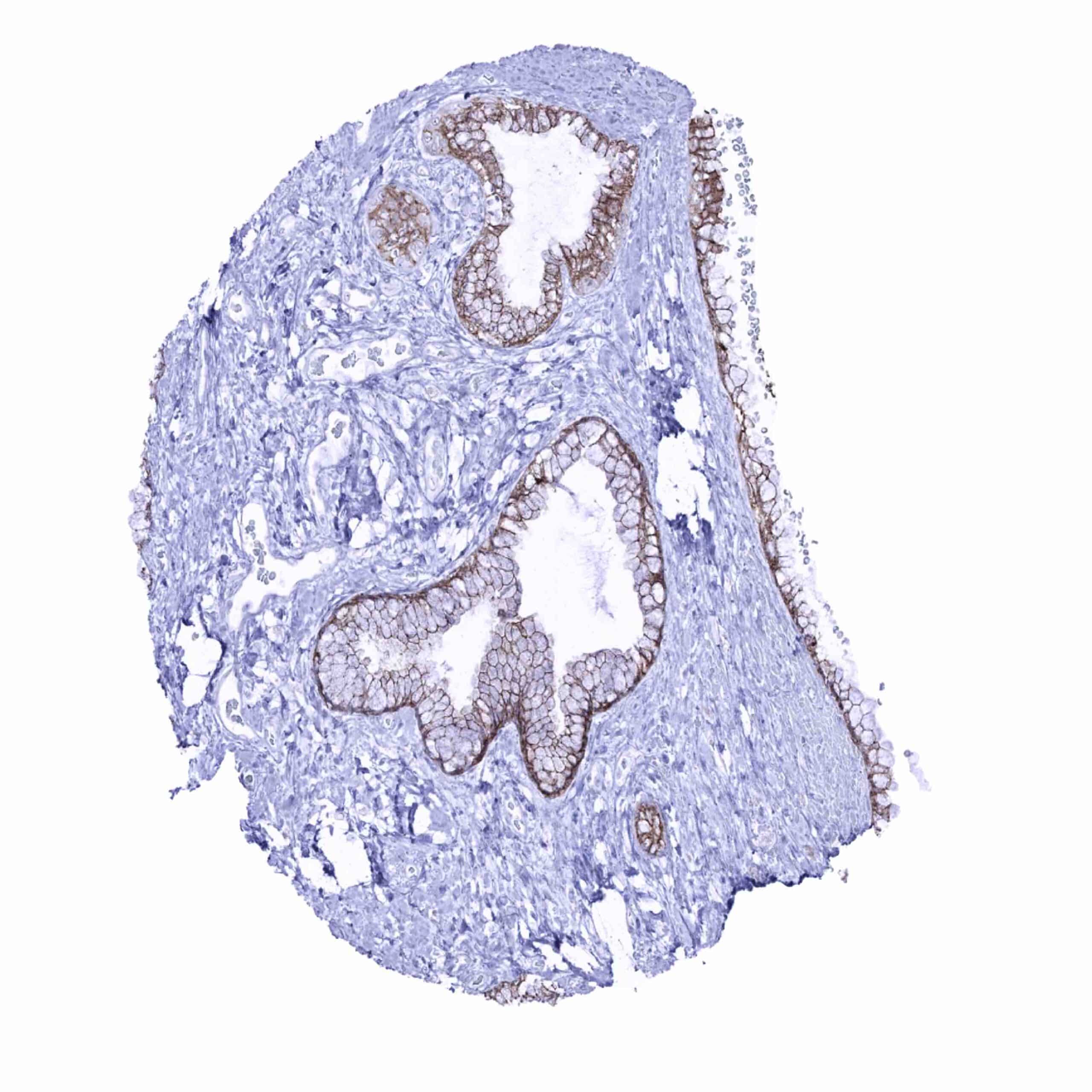
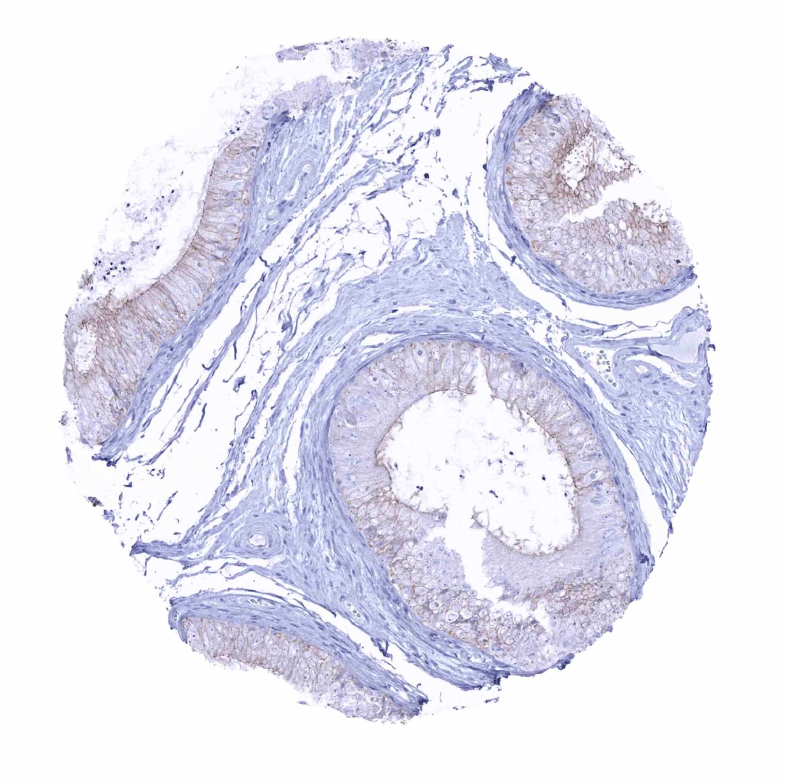
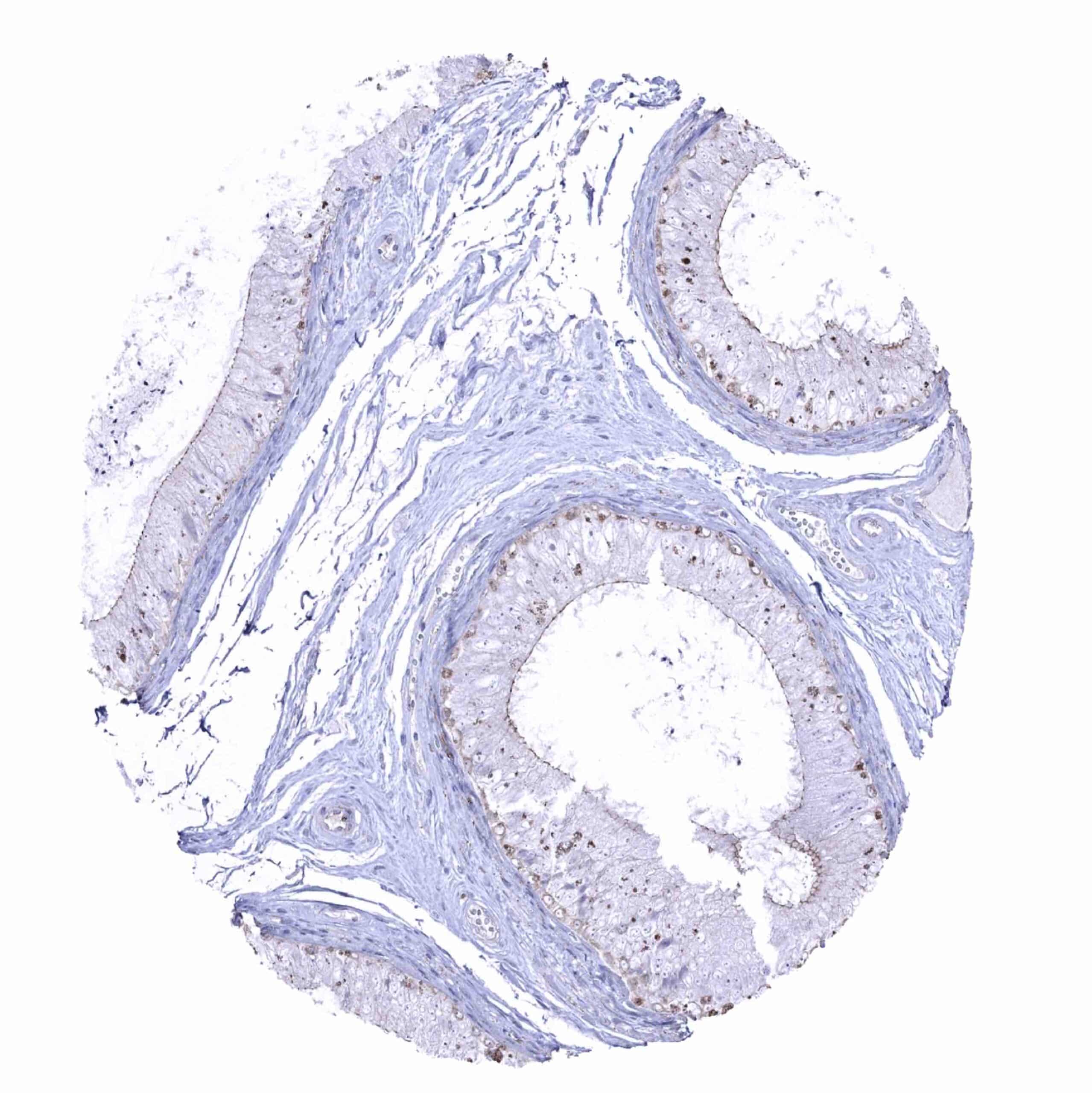
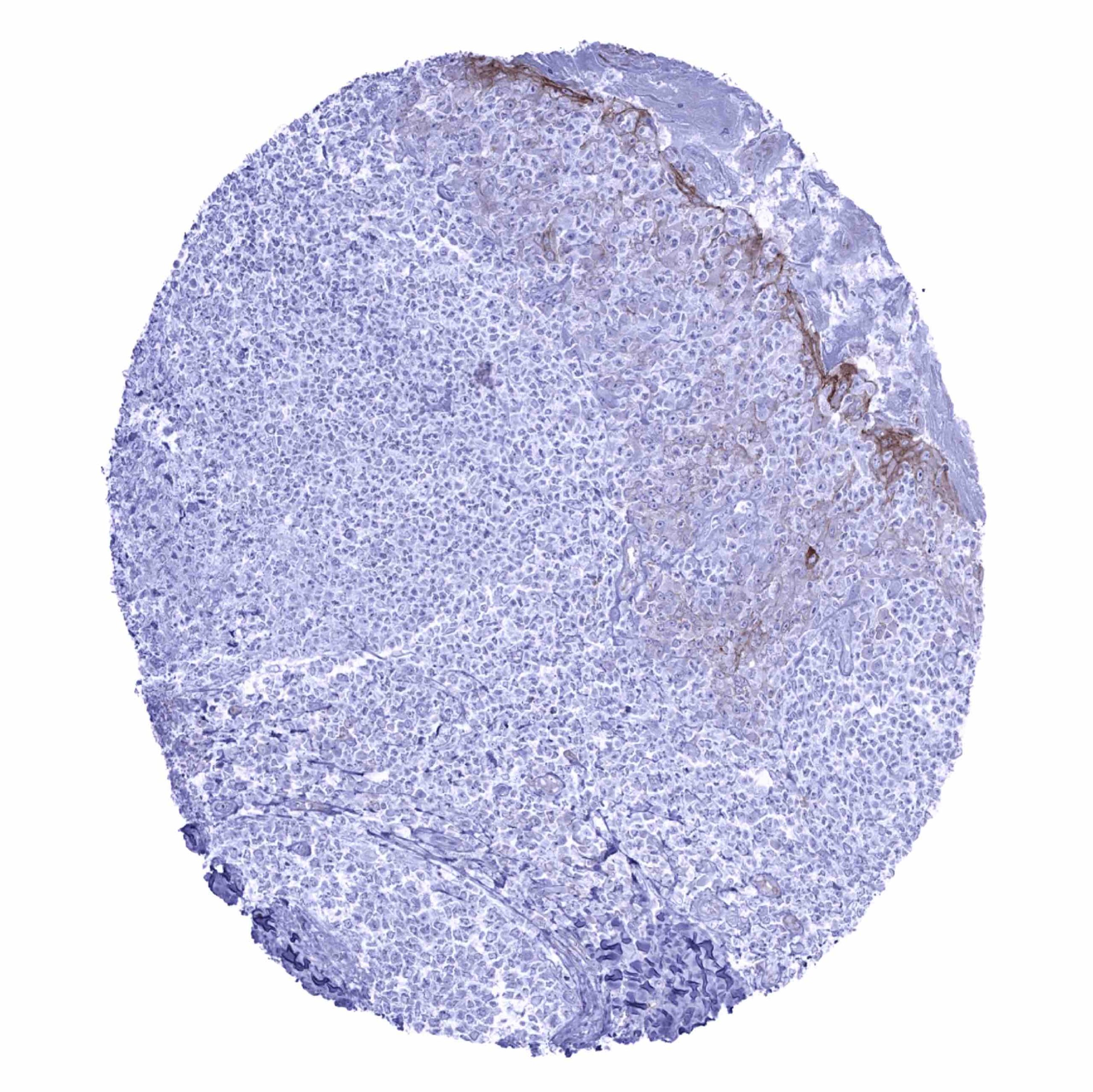
| Male genital | Prostate | Rather weak occludin staining of epithelial cells. |
| Seminal vesicles | Intense occludin staining of epithelial cells. | |
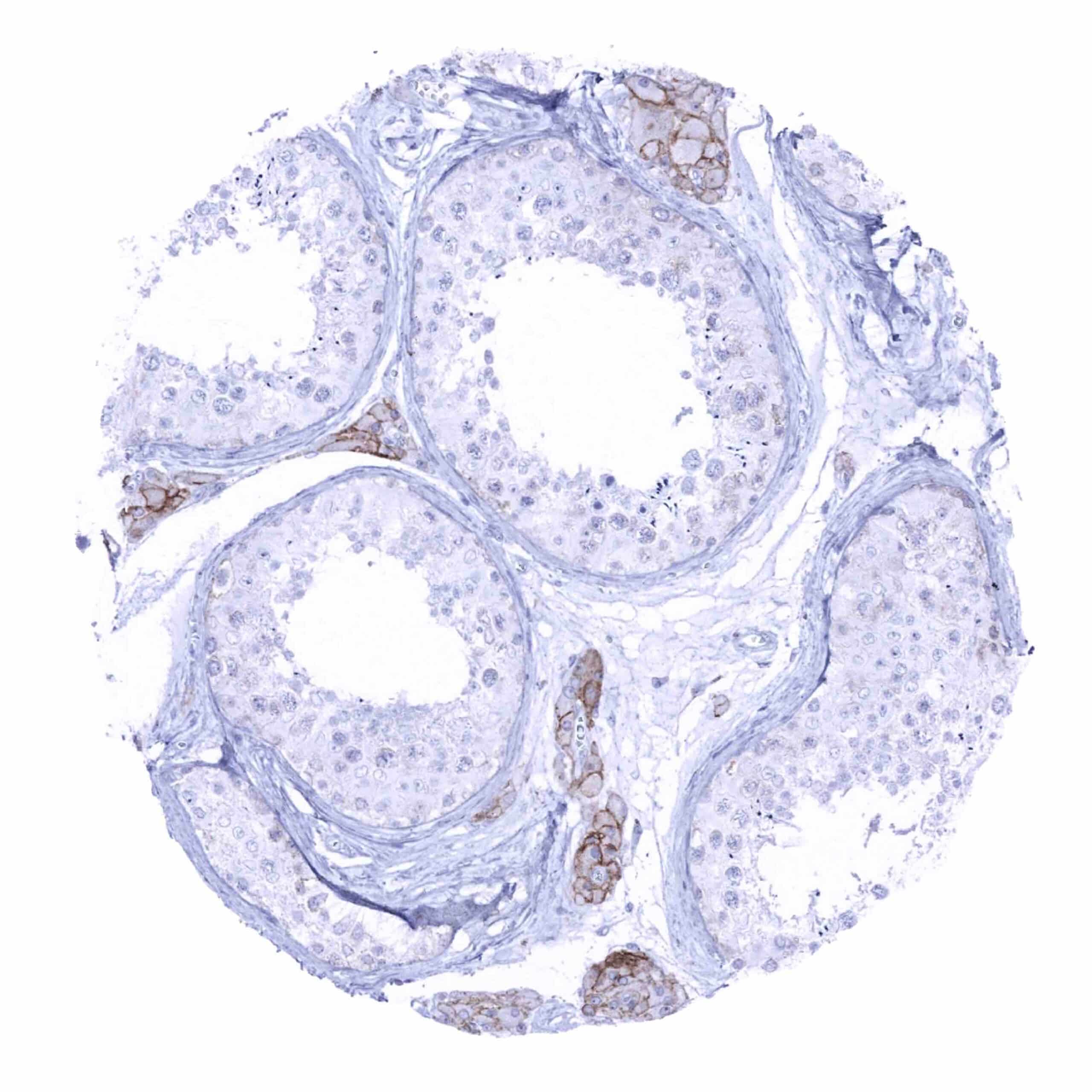
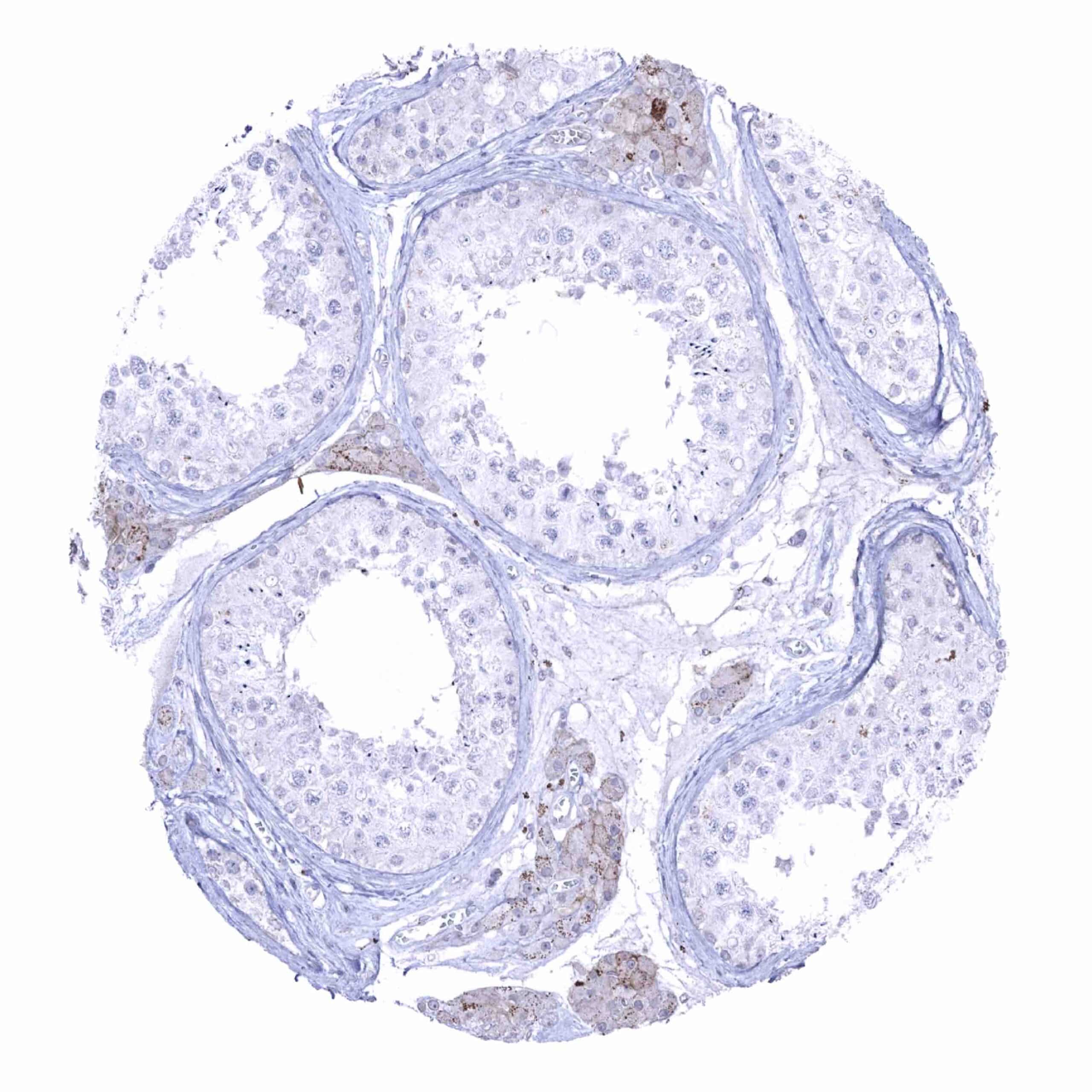
| Testis | Occludin staining is largely limited to Leydig cells. | |
| Epididymis | Rather faint membranous occludin staining, predominantly at the apical membranes of chief cells in the corpus. | |
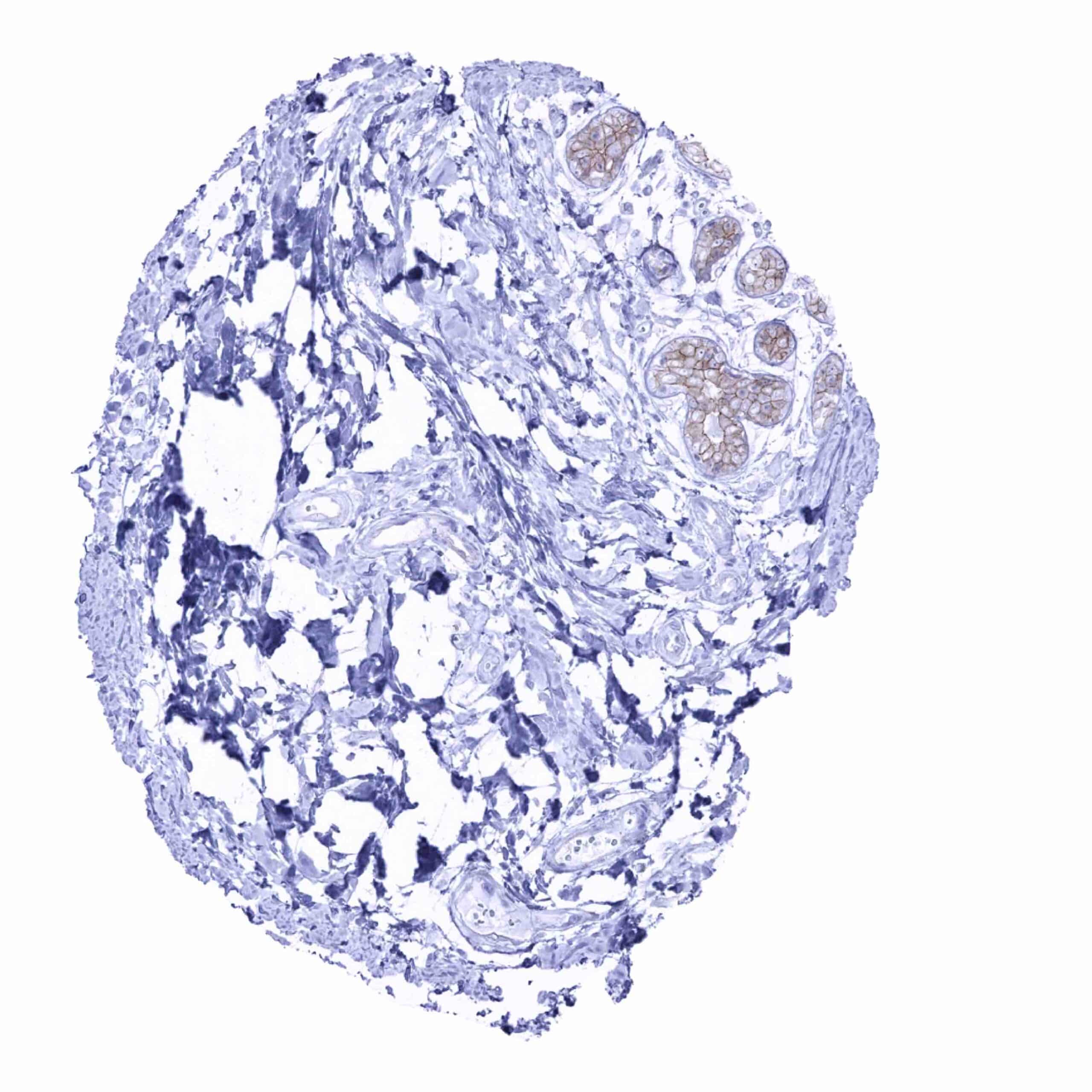
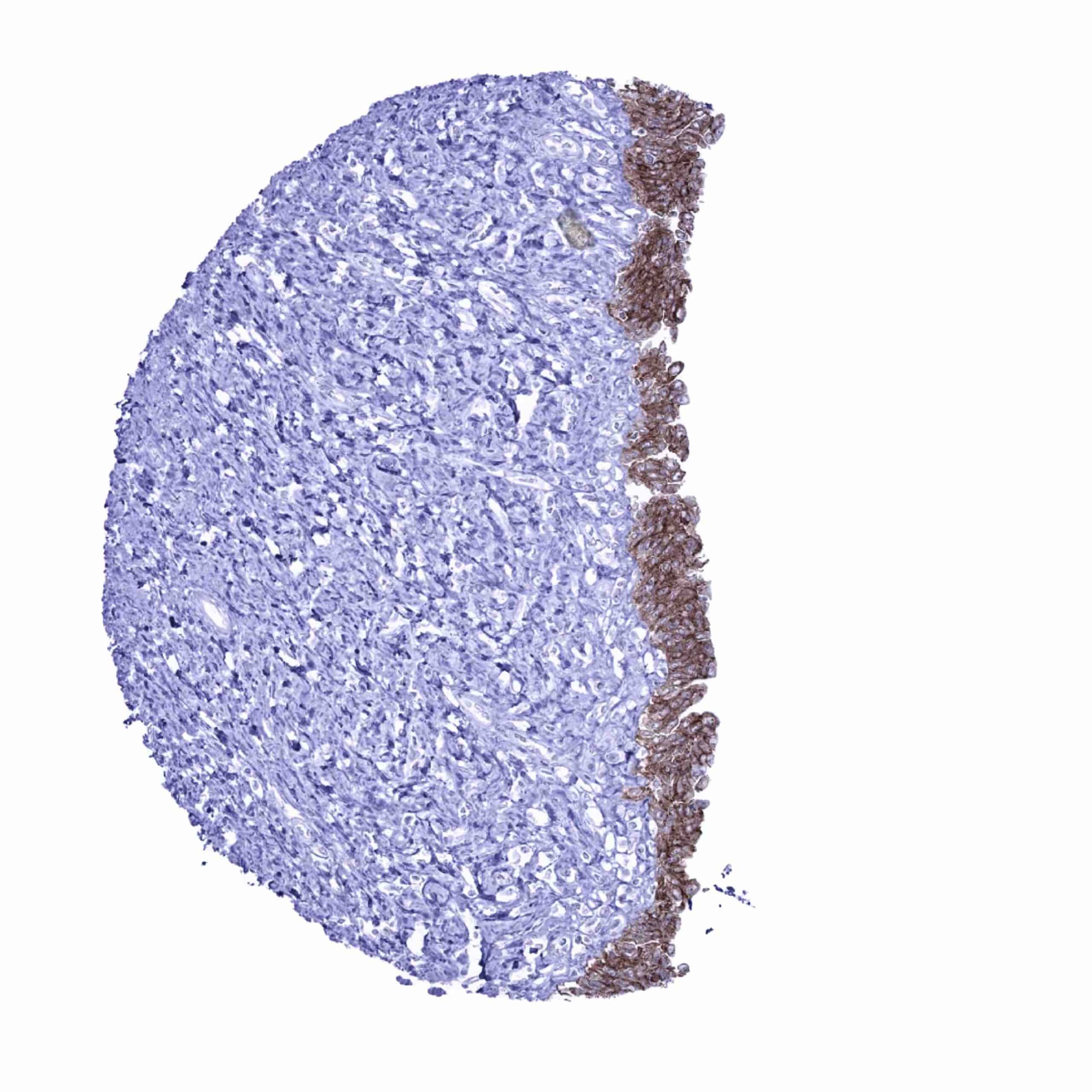
| Female genital | Breast | Luminal cells show a moderate membranous occludin staining while basal cells lack staining. |
| Uterus, myometrium | Occludin staining is limited to endothelial cells. | |
| Uterus, ectocervix | In the squamous epithelium, occludin staining predominates in intermediate cell layers while occludin positivity is either absent or reduced in basal and superficial cell layers. | |
| Uterus endocervix | Significant membranous occludin staining, most prominently in basal cell layers. | |
| Uterus, endometrium | Significant membranous occludin staining of epithelial cells. In the pregnant uterus, endometrial cell staining may be less intensive. | |
| Fallopian Tube | Strong diffuse membranous occludin positivity. | |
| Ovary | Moderate membranous occludin staining of granulosa cells. Faint membranous staining of corpus luteum cells. The stroma is largely occludin negative. | |
| Placenta early | Strong membranous occludin staining predominates in the cytotrophoblast and at the luminal membrane of the syncytiotrophoblast. | |
| Placenta mature | Strong membranous occludin staining predominates in the cytotrophoblast. | |
| Amnion | Occludin staining preferentially occurs at the apical membrane of amnion cells. | |
| Chorion | Negative. | |
| Skin | Epidermis | Occludin staining is either absent or reduced in basal and superficial cell layers while intermediate cell layers show significant positivity. |
| Sebaceous glands | Sebaceous glands are largely occludin negative but ekkrine glands show strong staining. | |
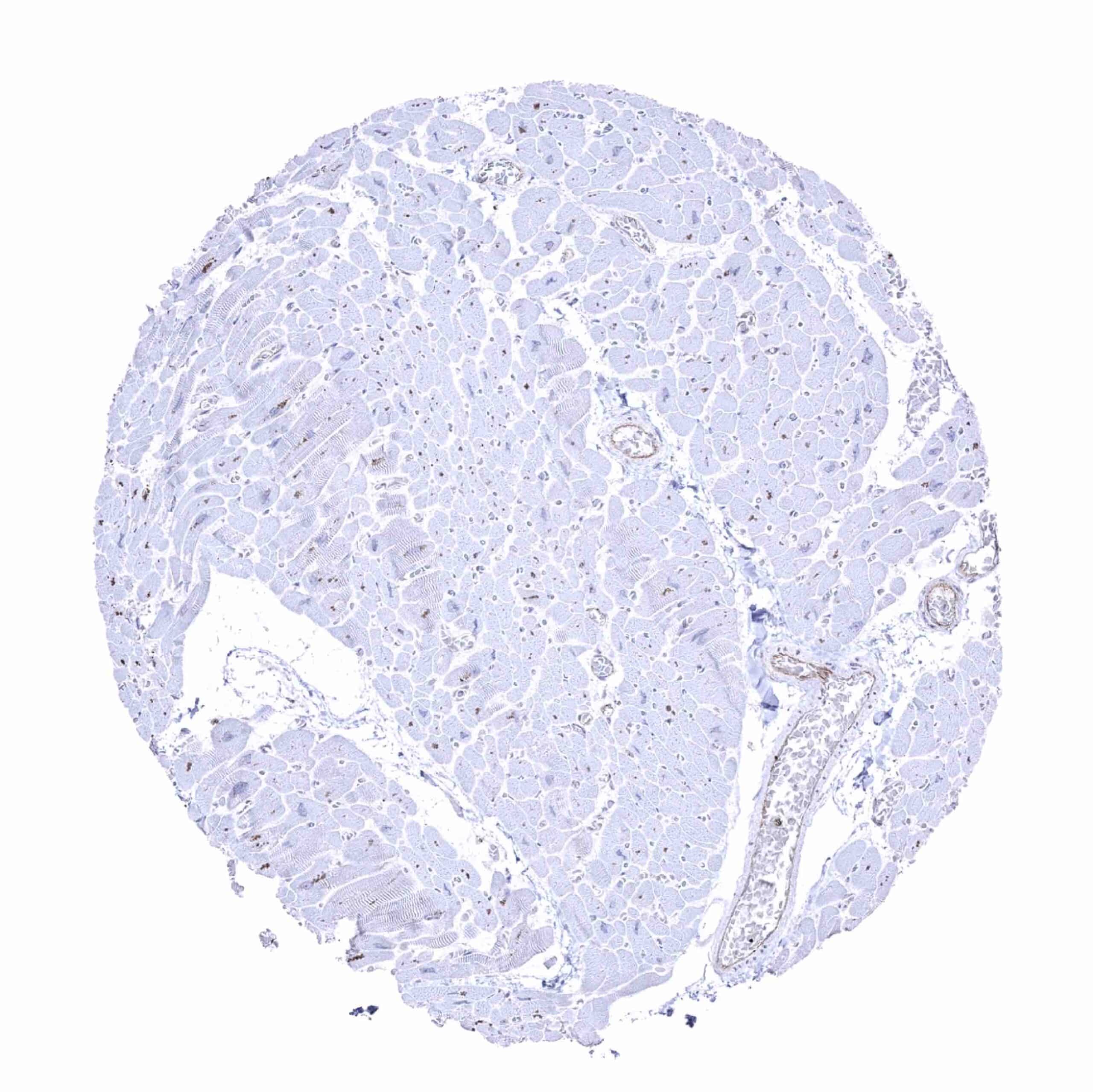
| Muscle/connective tissue | Heart muscle | Muscle cells are occludin negative. Occludin staining is limited to endothelial cells. |
| Skeletal muscle | Muscle cells are occludin negative. | |
| Smooth muscle | Muscle cells are occludin negative. | |
| Vessel walls | Muscle cells are occludin negative. | |
| Fat | Negative. | |
| Stroma | Negative. | |
| Endothelium | Occludin staining at variable levels in endothelial cells of all tissues. | |
| Bone marrow/lymphoid tissue | Bone marrow | Negative. |
| Lymph node | Significant occludin staining of endothelial cells. | |
| Spleen | Significant occludin staining of endothelial cells. | |
| Thymus | Strong occludin staining of a fraction of cells in corpuscles of Hassall‘s. | |
| Tonsil | In the surface epithelium, occludin predominates in intermediate cell layers while occludin positivity is either absent or reduced in basal and superficial cell layers. Squamous epithelium of the tonsil crypts show strongest occludin staining in superficial layers. | |
| Remarks | Occludin staining is almost exclusively membranous. |
The ubiquitous occludin immunostaining described above is largely consistent with the RNA and protein data described in the Human Protein Atlas (Tissue expression Occludin).
Positive control = Colon: A strong membranous occludin staining of epithelial cells should be seen with a particularly strong staining at the apical/luminal membranes.
Negative control = Colon: All non-epithelial and non-endothelial cells must not show any occludin staining.

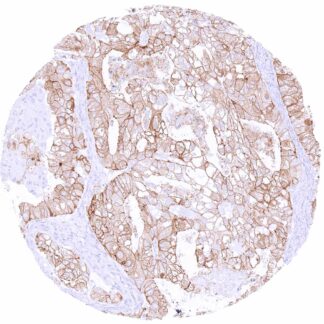
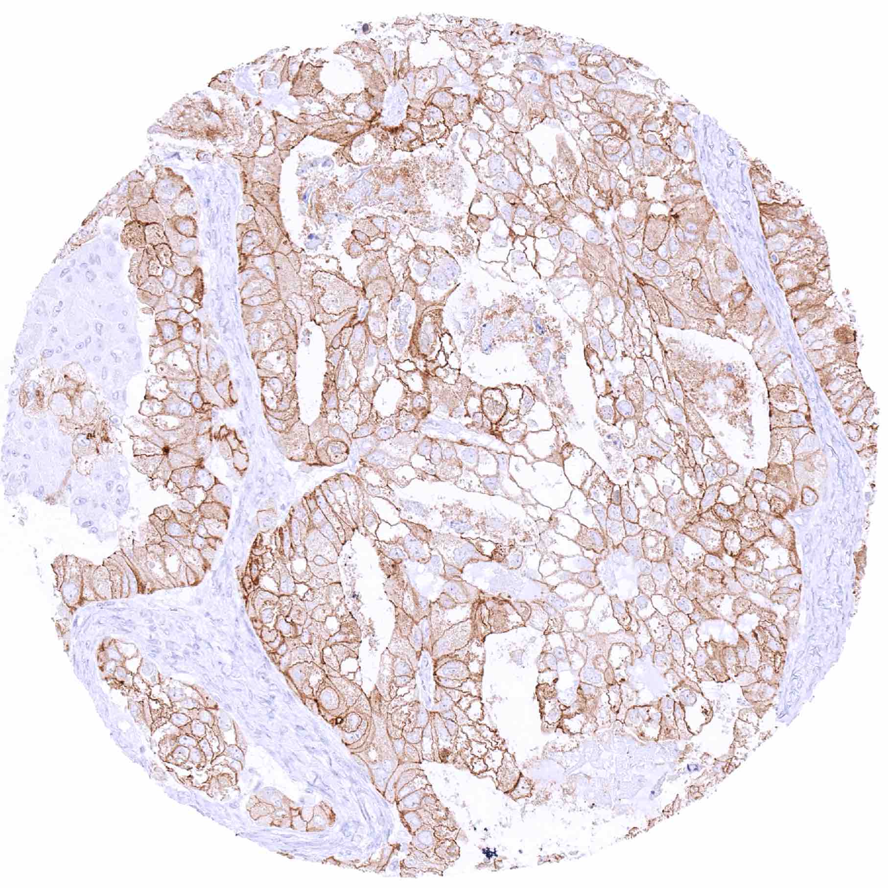
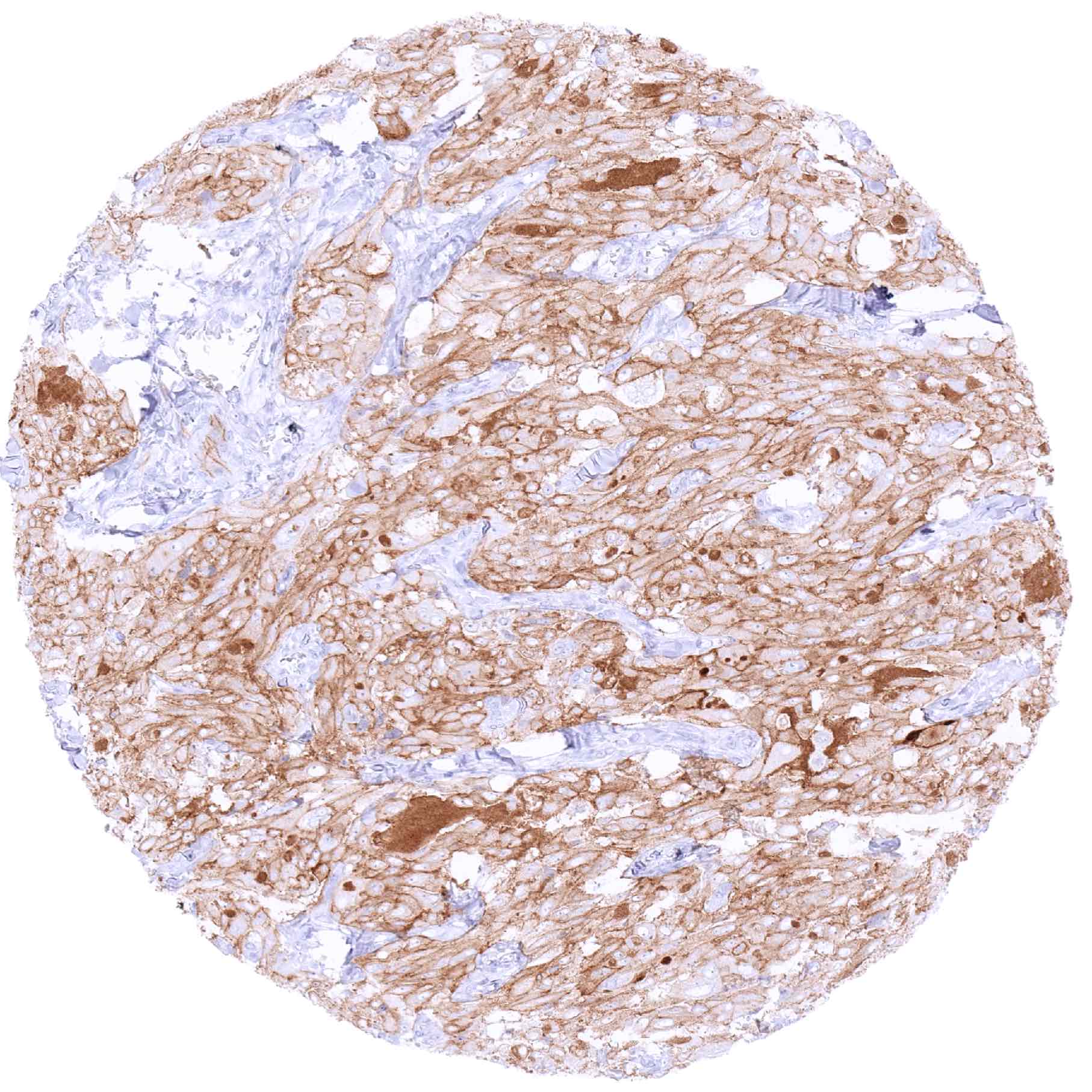
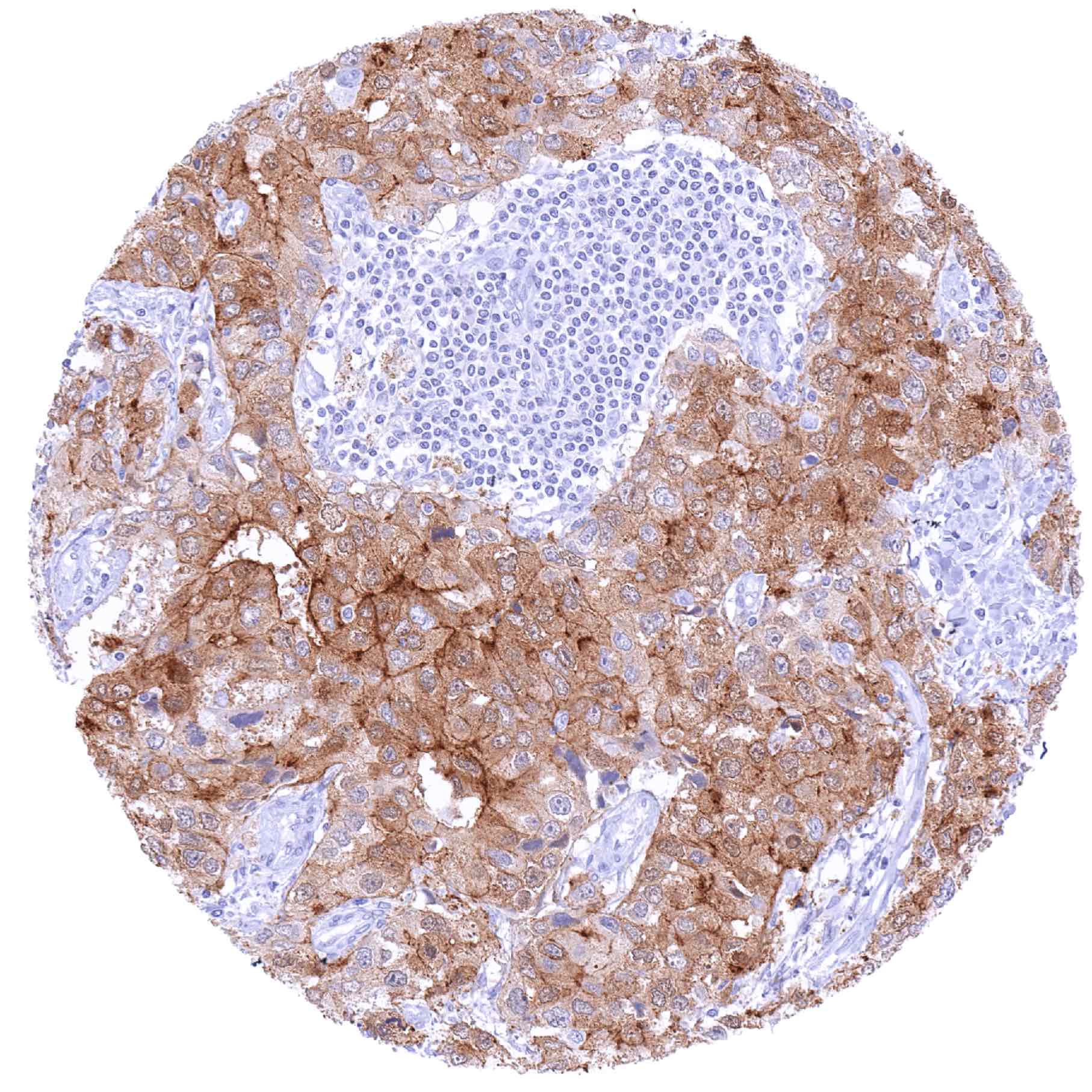
Staining Pattern in Relevant Tumor Types
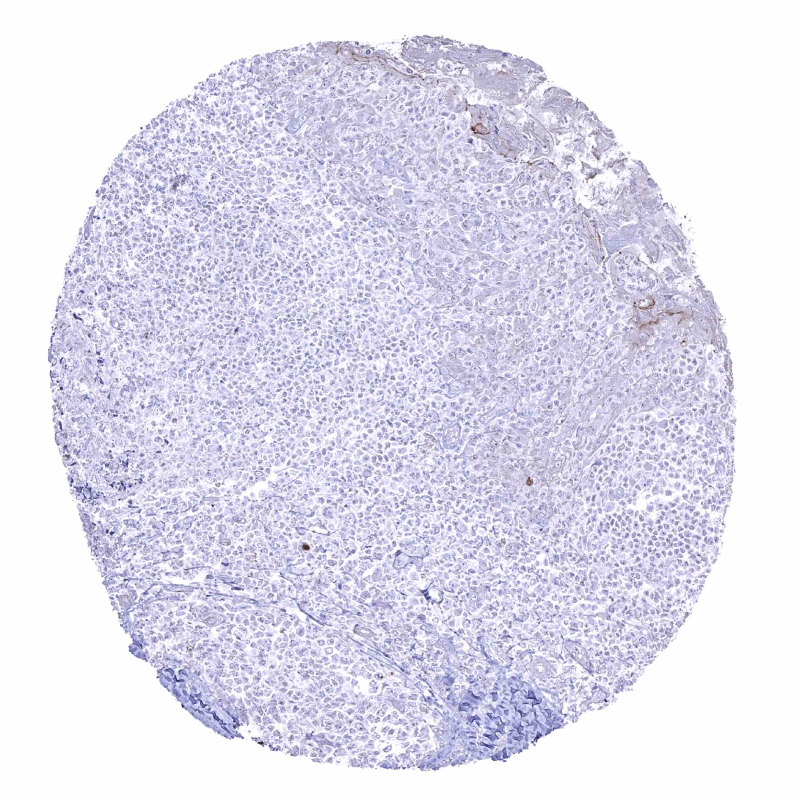
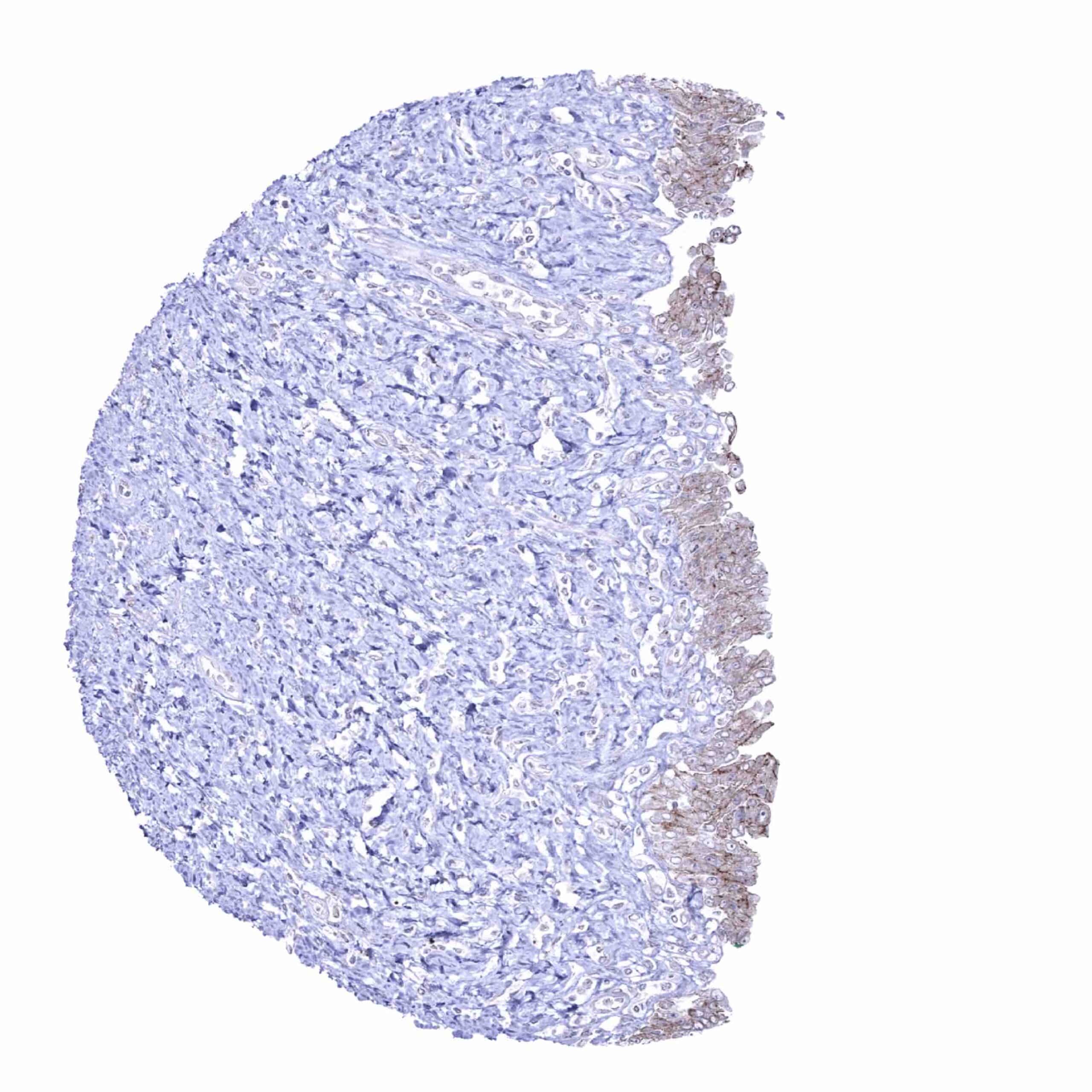
At variable levels, occludin immunostaining occurs in many different tumor entities. However, as compared to normal tissues, occludin expression may often be lower. Reduced expression of occludin in cancer has been linked to poor prognosis in studies.
The TCGA findings on Occludin RNA expression in different tumor categories have been summarized in the Human Protein Atlas.

Compatibility of Antibodies
No data available at the moment
Protocol Recommendations
IHC users have different preferences on how the stains should look like. Some prefer high staining intensity of the target stain and even accept some background. Others favor absolute specificity and lighter target stains. Factors that invariably lead to more intense staining include higher concentration of the antibody and visualization tools, longer incubation time, higher temperature during incubation, higher temperature and longer duration of the heat induced epitope retrieval (slide pretreatment). The impact of the pH during slide pretreatment has variable effects and depends on the antibody and the target protein.
All images and data shown here and in our image galleries are obtained by the manual protocol described below. Other protocols resulting in equivalent staining are described as well.
Manual protocol
Freshly cut sections should be used (less than 10 days between cutting and staining). Heat-induced antigen retrieval for 5 minutes in an autoclave at 121°C in pH 7,8 Target Retrieval Solution buffer. Apply MSVA-415M at a dilution of 1:150 at 37°C for 60 minutes. Visualization of bound antibody by the EnVision Kit (Dako, Agilent) according to the manufacturer’s directions.
Potential Research Applications
- The prognostic impact of occludin expression levels are unclear for many tumor types.
- Studies comparing occludin expression levels between different tumor entities are lacking.
Evidence for Antibody Specificity in IHC
There are two ways how the specificity of antibodies can be documented for immunohistochemistry on formalin fixed tissues. These are: 1. comparison with a second independent method for target expression measurement across a large number of different tissue types (orthogonal strategy), and 2. Comparison with one or several independent antibodies for the same target and showing that all positive staining results are also seen with other antibodies for the same target (independent antibody strategy).
Orthogonal validation: Orthogonal validation is not applicable for occludin antibodies because of the ubiquitous expression of the protein in blood vessels and epithelial cells in virtually all organs. The conspicuous absence of MSVA-415M staining seen in adrenal gland, muscle, and bone marrow as well as the low staining in prostate and the adenohypophysis is, however, consistent with data from three independent RNA screening studies, including the Human Protein Atlas (HPA) RNA-seq tissue dataset, the FANTOM5 project, and the Genotype-Tissue Expression (GTEx) project, which are all summarized in the Human Protein Atlas (Tissue expression Occludin).
Comparison of antibodies: True expression of occludin in all cell types identified as occludin positive by MSVA-415M is corroborated by comparison with a commercially available independent second antibody (termed “validation antibody”). A confirmation of all positive findings obtained by MSVA-415M by the “validation antibody” fully confirms the specificity of MSVA-415M.