
195,00 € – 695,00 €
Product details
Synonyms = Antigen LB39-AA, Antigen SK29-AA, Melanoma antigen recognized by T-cells 1, MLAN-A, MLANA
Antibody type = Recombinant Mouse monoclonal / IgG
Clone = MSVA-901M+
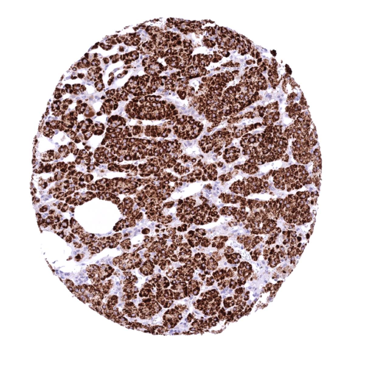
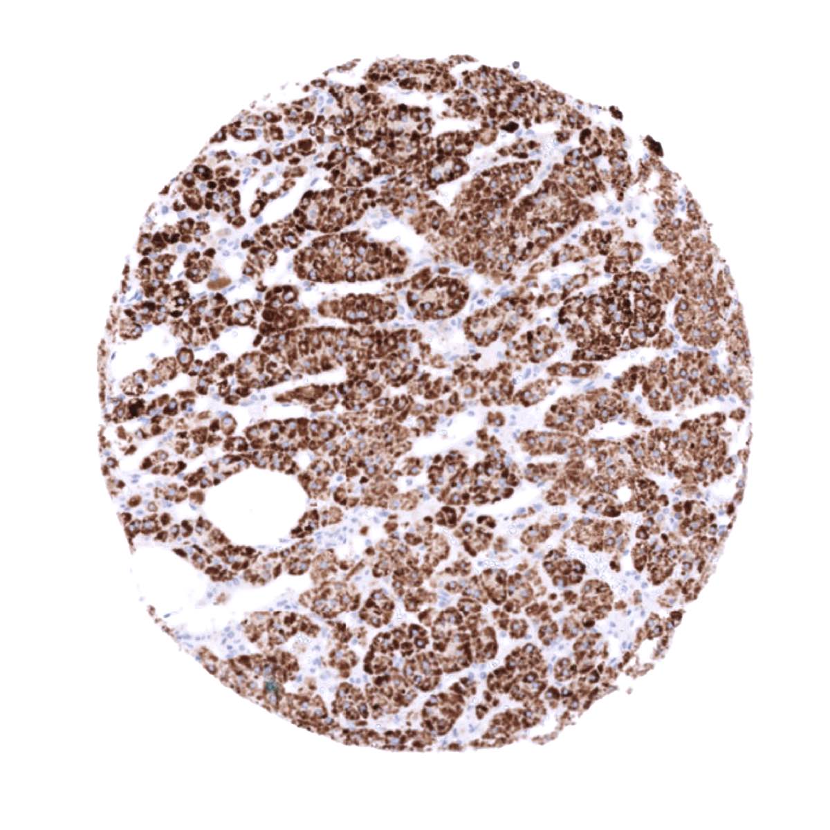
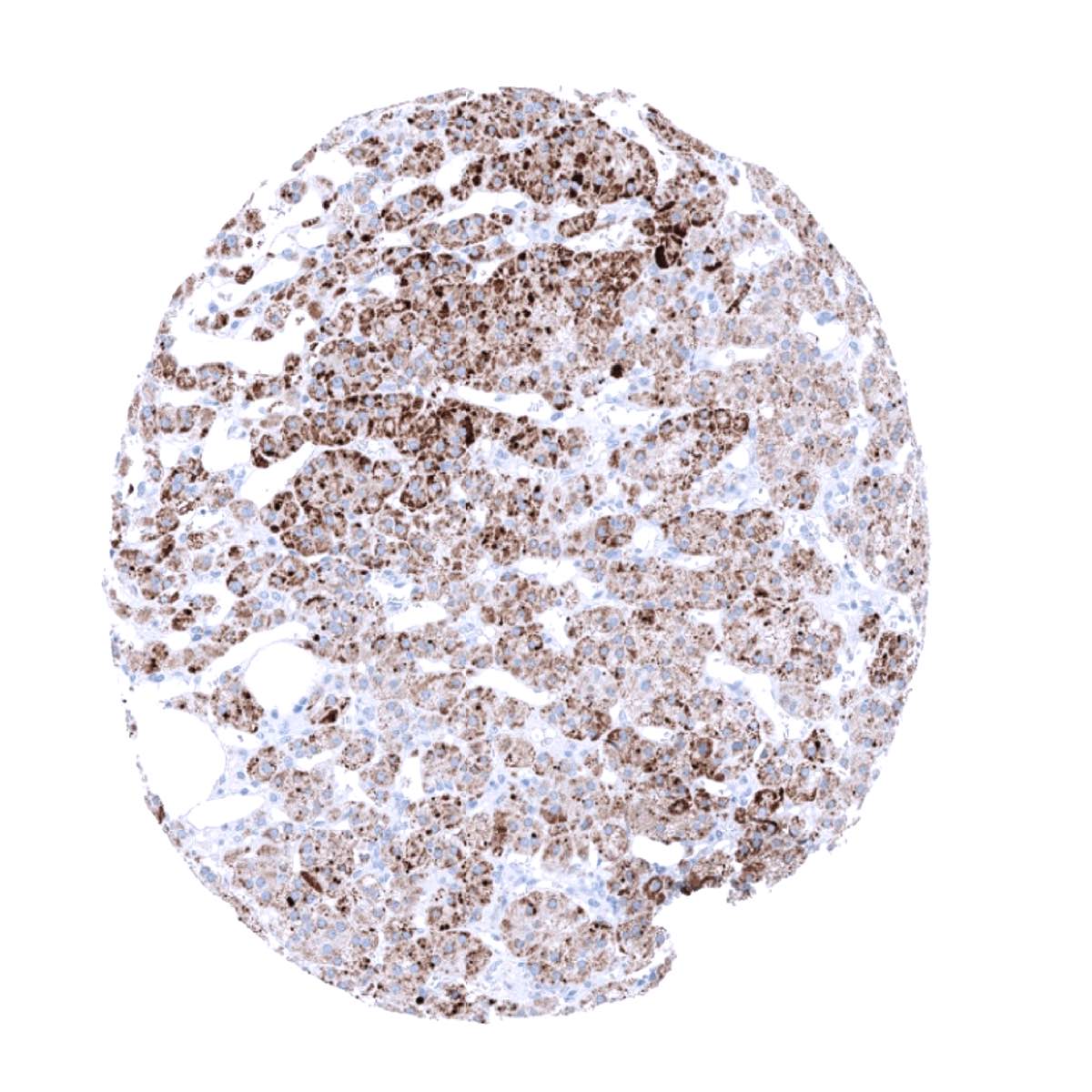
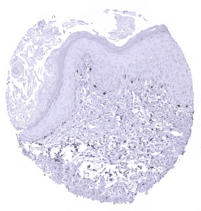
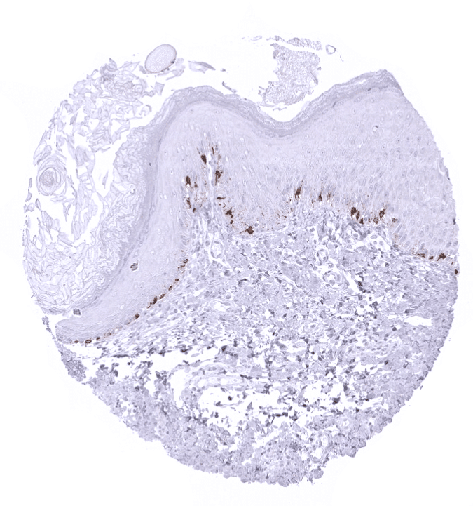
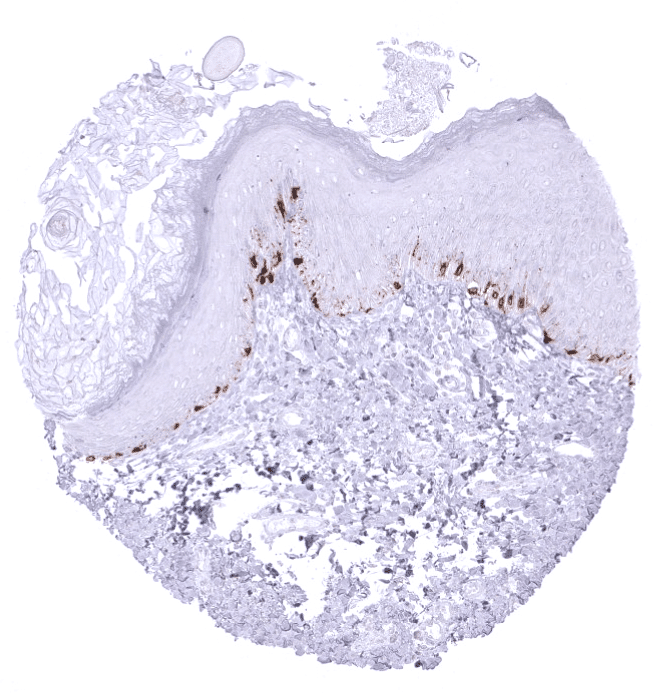
Positive control = Skin: Virtually all melanocytes should show a strong Melan A+ immunostaining including a weak to moderate staining in melanocytic dendrites. Adrenal gland: An at least moderate Melan A+ immunostaining should be seen in adrenocortical cells.
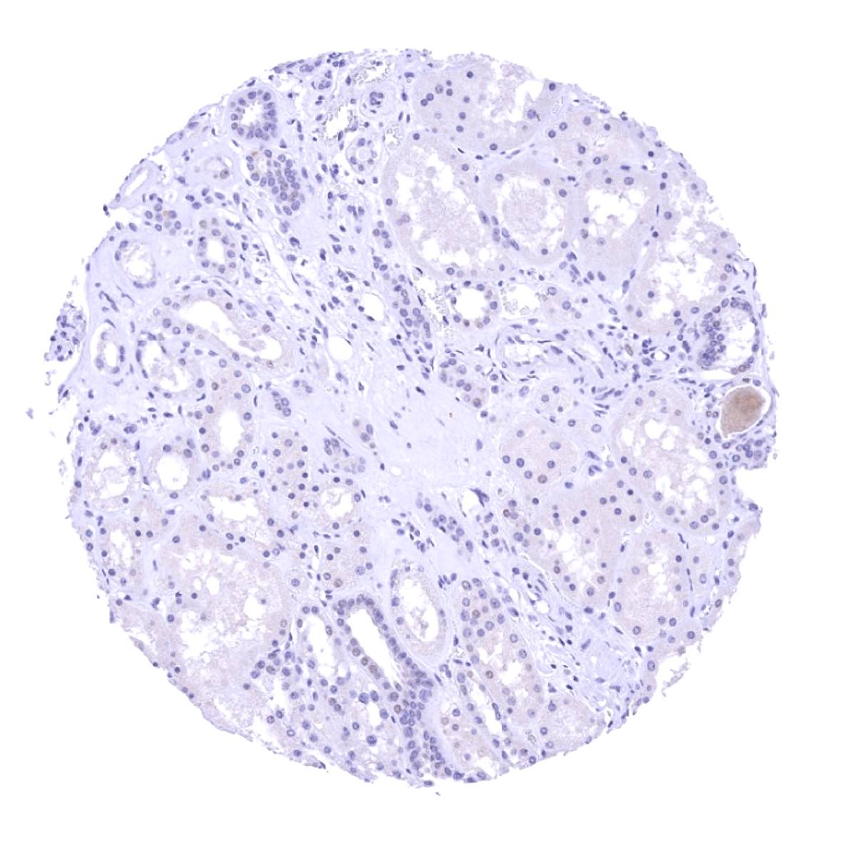
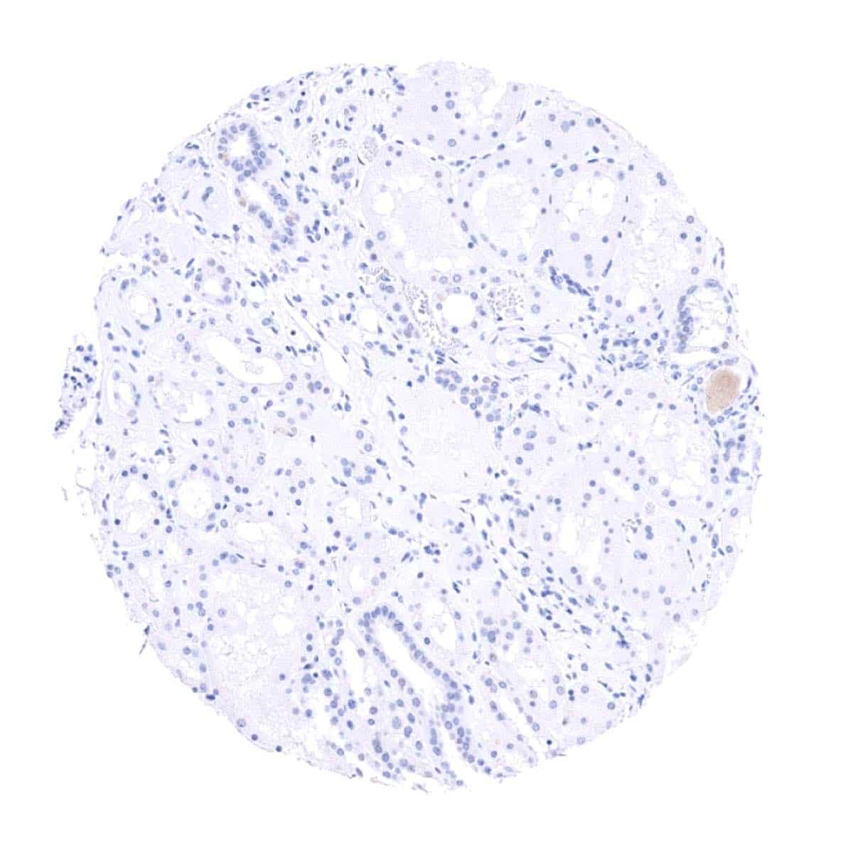
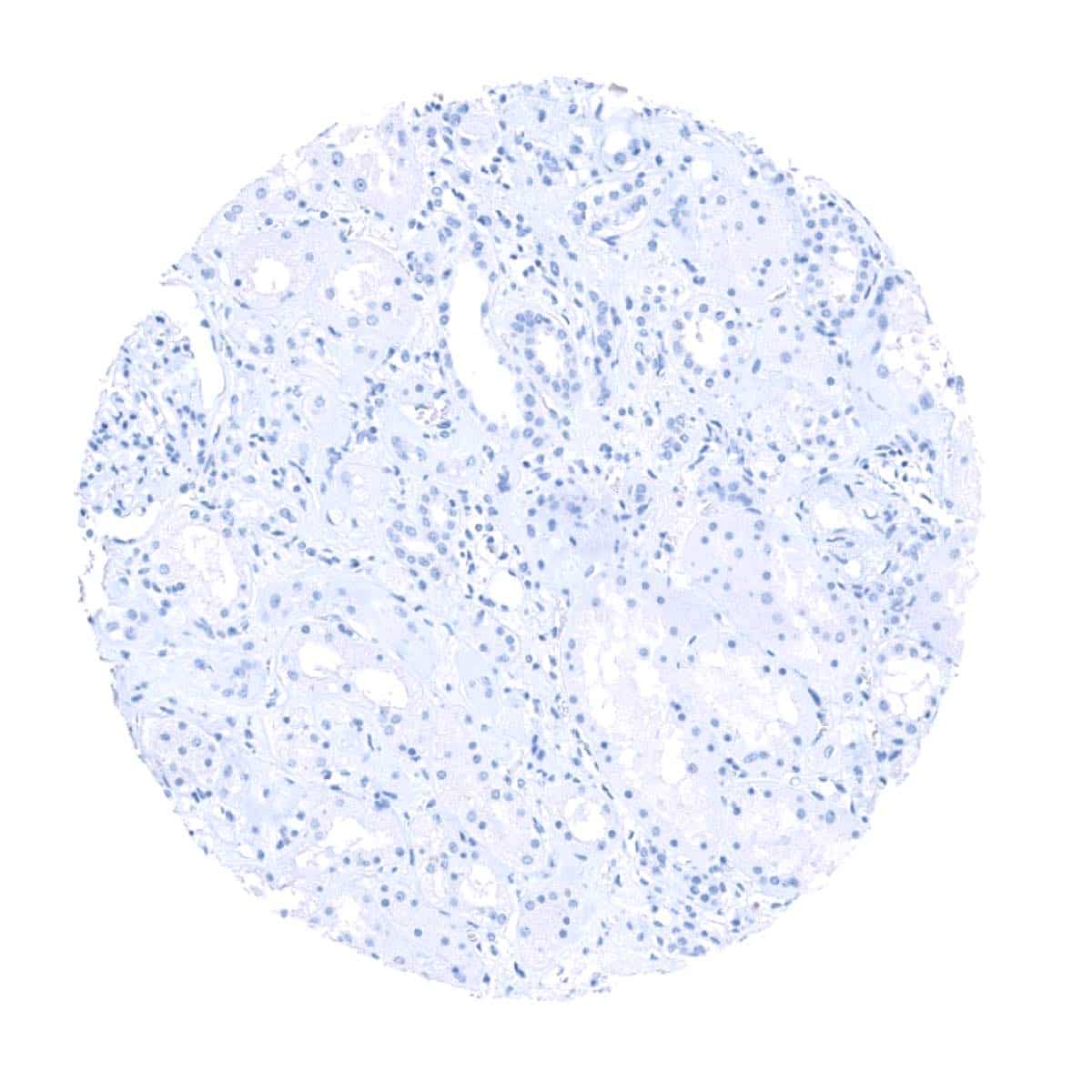
Negative control = Kidney: Melan A+ immunostaining should be absent in all cells. Adrenal gland: Melan A+ immunostaining should be absent in all medullary cells.
Cellular localization = Cytoplasmic
Reactivity = Human
Application = Immunohistochemistry
Dilution = 1:50 – 1:100
Intended Use = Research Use Only
Relevance of Antibody
Melan A+ is a marker for melanoma, adrenocortical and other tumors.
Biology Behind
The Melan A (melanocyte antigen) protein, also termed “melanoma antigen recognized by T cells 1” (MART-1) is coded by a gene on chromosome 9p24.1. The 18 kDa protein has a single transmembrane domain and consists of 118 amino acids. The function of the protein is unknown. A small fragment of the protein (about nine amino acids) is bound by MHC class I complexes and presented to cytotoxic T cells. The MART-1/melan A antigen is specific for the melanocyte lineage, found in normal skin, the retina, and melanocytes, but not in other normal tissues.
As few other Melan-A antibodies, MSVA-901M+ (Melan-A+) not only recognizes the Melan A protein but – as a result of a cross-reactivity – also an unknown structure related to corticosteroids. Therefore, Melan A+ also recognizes adrenocortical cells and a few other tissues. Melan A+ is thus useful as a marker for benign and malignant melanocytic tumors as well as for various steroid producing tissues.
Staining Pattern in Normal Tissues
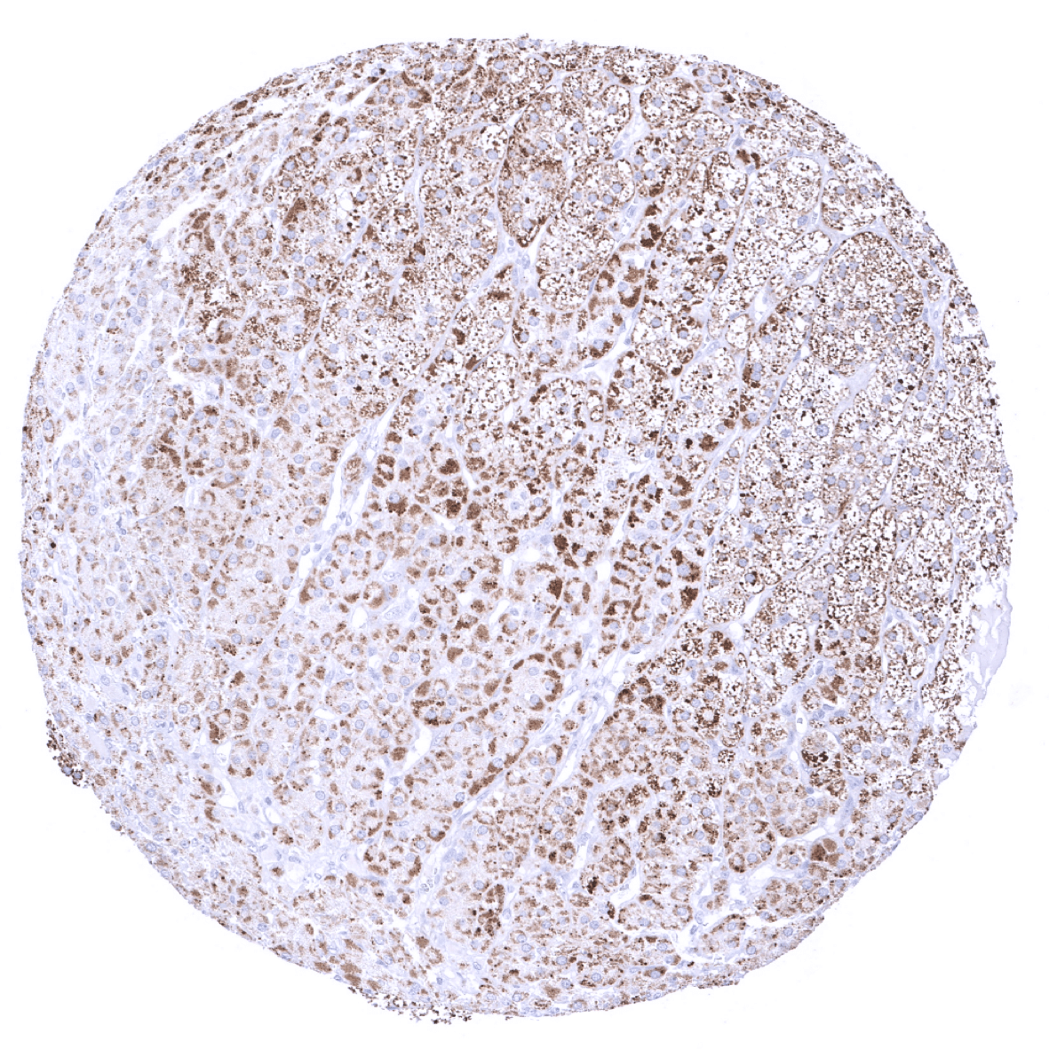
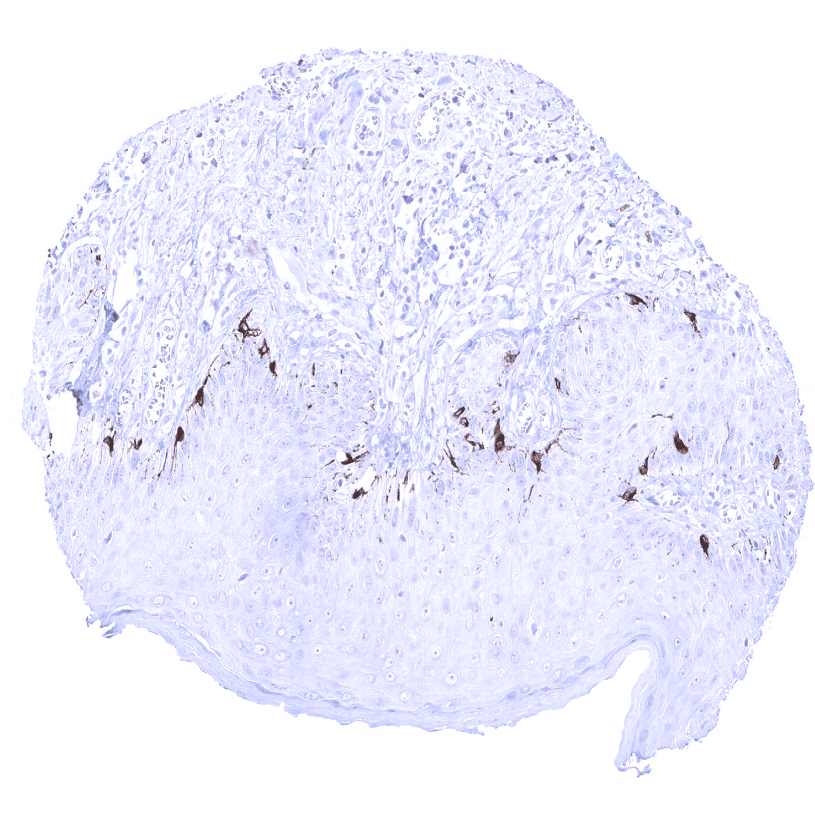
Using the antibody MSVA-901M+ (Melan A+), a strong staining can be observed in melanocytes in skin and non-keratinizing squamous epithelia from various sites. Adrenocortical cells, and a variable fraction of epithelial cells in the adenohypophysis also show a moderate to strong Melan A+ immunostaining. A moderate intensity staining is present in theca interna cells of ovary and in testicular Leydig cells. Moreover, a faint immunostaining is detectable at the apical membranes of ciliated cells of respiratory epithelium.
These findings are not fully comparable to the RNA and protein data summarized in the Human Protein Atlas (Tissue expression Melan A) but in part consistent with protein data of the protein atlas. It is of note, that the different antibodies developed within the protein atlas project contain antibodies of the Melan-A+ type showing strong immunostaining of adrenocortical cells (CAB000057) und others that do not stain adrenal tissue (HPA048662)
Suggested positive tissue control: Skin: Virtually all melanocytes should show a strong Melan A+ immunostaining including a weak to moderate staining in melanocytic dendrites. Adrenal gland: An at least moderate Melan A+ immunostaining should be seen in adrenocortical cells.
Suggested negative tissue control: Kidney: Melan A+ immunostaining should be absent in all cells. Adrenal gland: Melan A+ immunostaining should be absent in all medullary cells.

Staining Pattern in Relevant Tumor Types
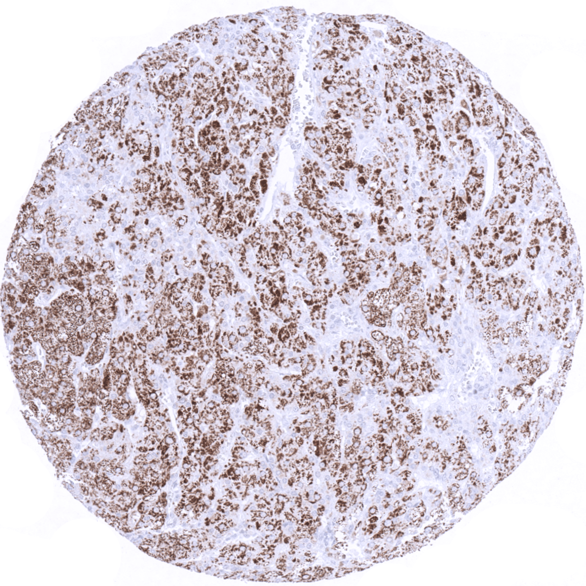
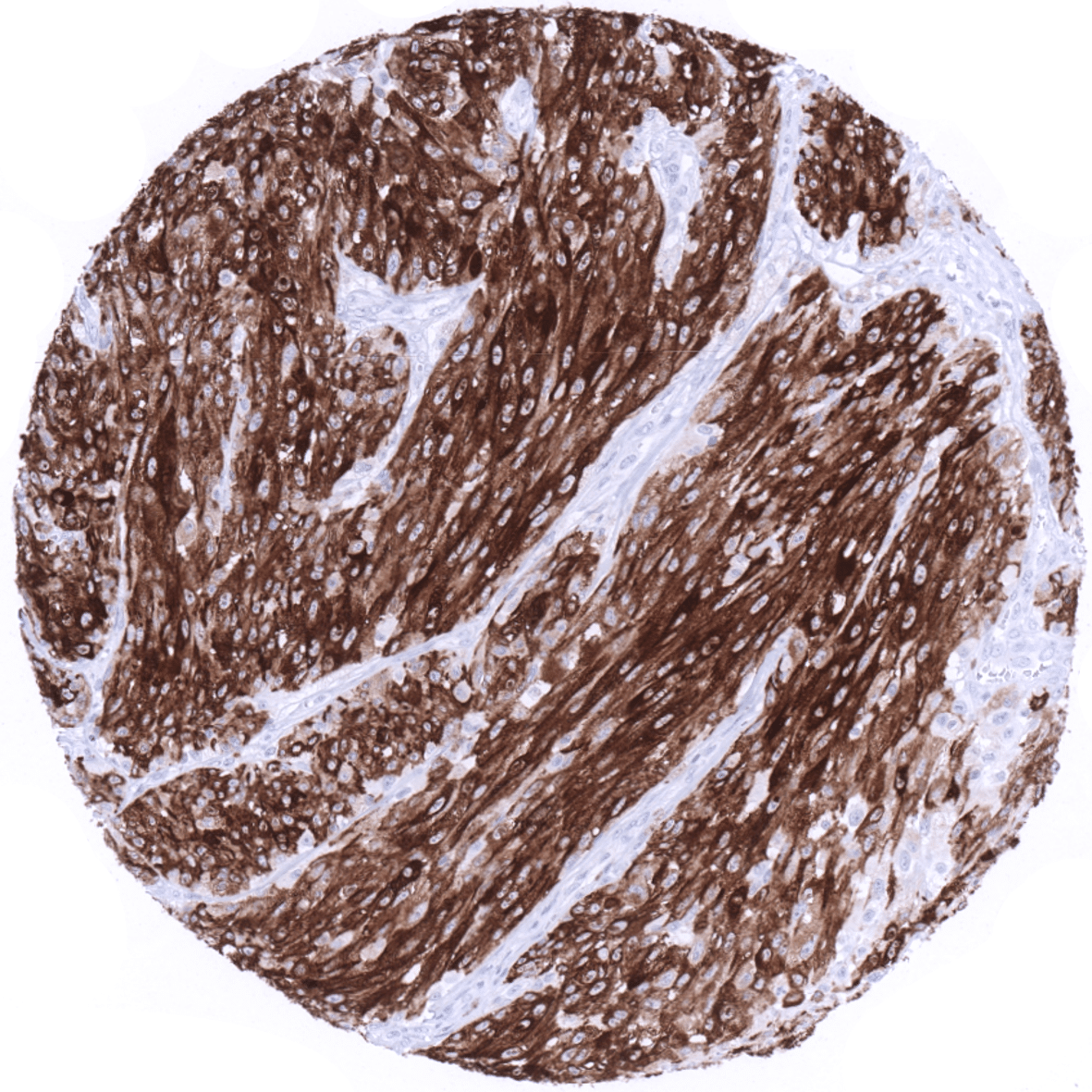
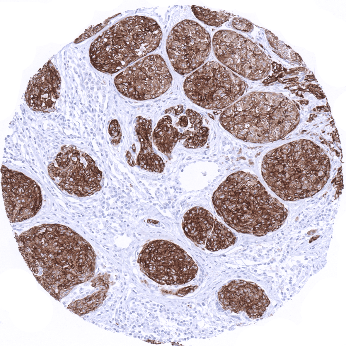
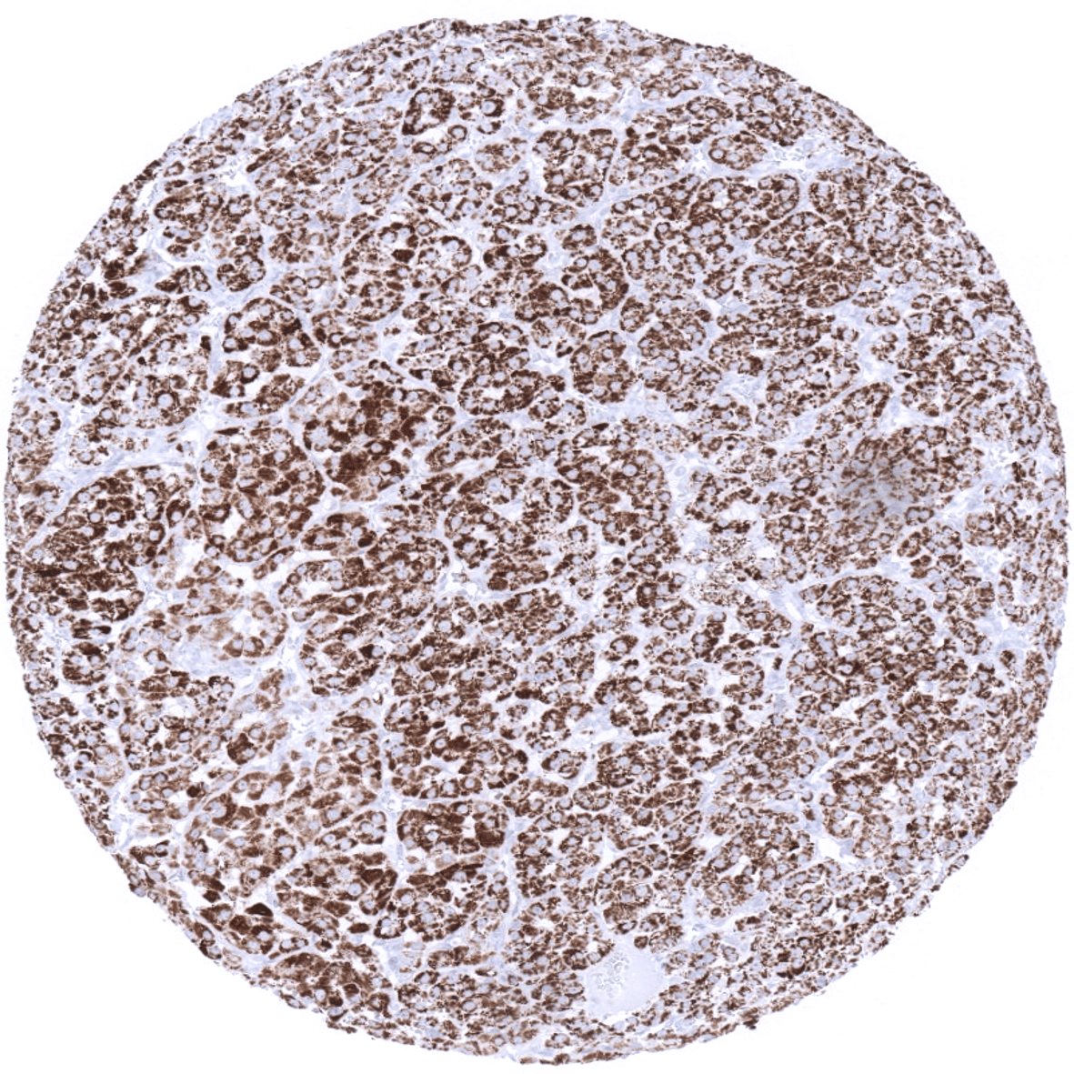
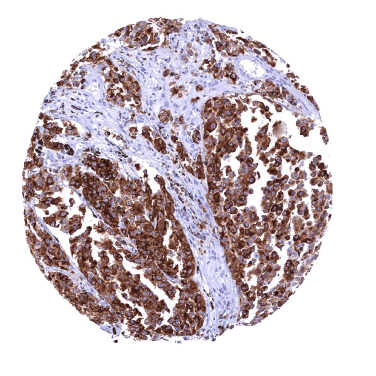
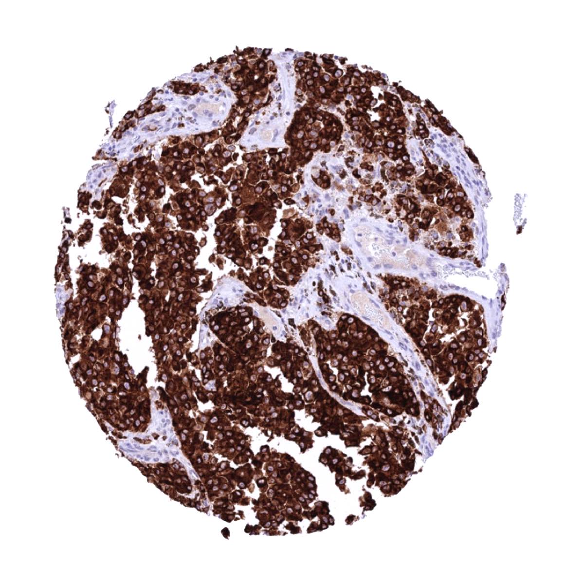
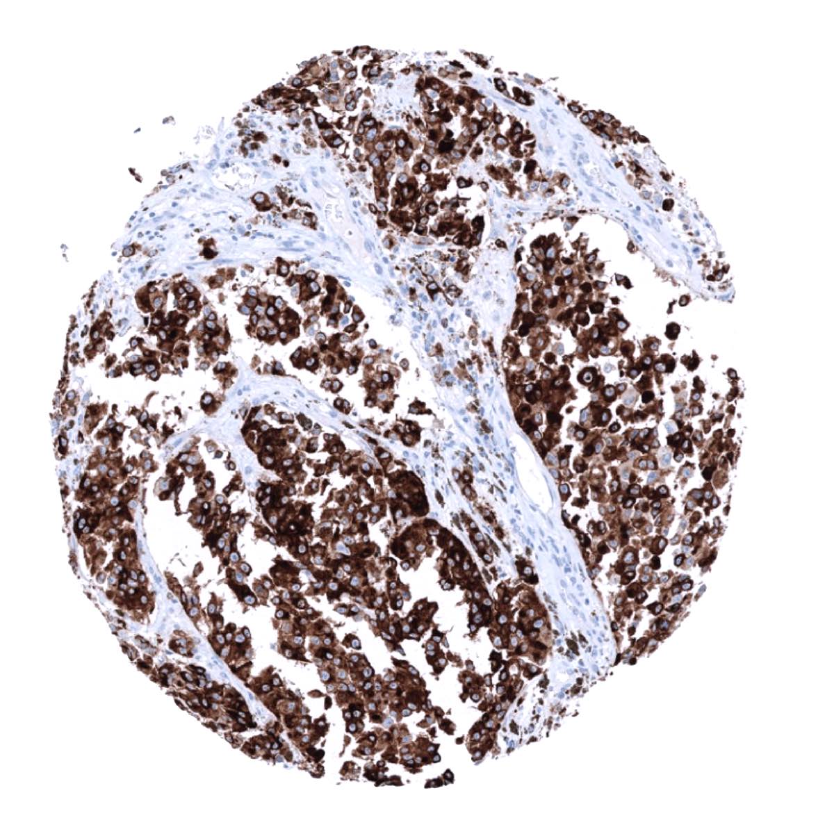
A Melan A+ staining is seen in the vast majority of primary malignant melanomas, all types of cutaneous naevi, and in other tumors of melanocytic differentiation, such as clear cell sarcoma, melanotic neurofibroma, melanotic schwannoma as well as PEComas (perivascular epitheloid cell tumor) including angiomyolipoma, lymphangioleiomyoma(-tosis), and pulmonary sugar tumor. Melan A+ staining is sometimes reduced and/or only patchy in nature in desmoplastic melanoma and in metastatic melanomas.
MSVA-900M+ (Melan A+) belongs to a subgroup of “Melan A” antibodies that share a diagnostically useful cross-reactivity with a structure linked to steroid hormones. This results in a strong Melan A+ immunostaining of adrenocortical adenomas and carcinomas, sex cord-stromal tumors of the ovary, Leydig cell tumor of the testis as well as individual tumors of various other entities. These tumor entities are, however, not recognized by several other – more specific -Melan-A antibodies.
The TCGA findings on Melan A RNA expression in different tumor categories have been summarized in the Human Protein Atlas.

Compatibility of Antibodies
No data available at the moment
Protocol Recommendations
IHC users have different preferences on how the stains should look like. Some prefer high staining intensity of the target stain and even accept some background. Others favor absolute specificity and lighter target stains. Factors that invariably lead to more intense staining include higher concentration of the antibody and visualization tools, longer incubation time, higher temperature during incubation, higher temperature and longer duration of the heat induced epitope retrieval (slide pretreatment). The impact of the pH during slide pretreatment has variable effects and depends on the antibody and the target protein. Accordingly, multiple different protocols can generate identical staining results.
All images and data shown here and in our image galleries are obtained by the manual protocol described below. Other protocols resulting in equivalent staining are described as well.
Manual protocol
Freshly cut sections should be used (less than 10 days between cutting and staining). Heat-induced antigen retrieval for 5 minutes in an autoclave at 121°C in pH9 Target Retrieval Solution buffer. Apply MSVA-901M+ (Melan A+) at a dilution of 1:75 at 37°C for 60 minutes. Visualization of bound antibody by the EnVision Kit (Dako, Agilent) according to the manufacturer’s directions.


Agilent / Dako – Autostainer Link 48
Pretreatment in PT-Link for 30 minutes at 95°C (pH high); FLEX peroxidase blocking for 5 minutes (room temperature), MSVA-901M+ 1:150 for 20 minutes (room temperature), FLEX+ mouse/rabbit (LINKER) for 15 minutes (room temperature), horseradish peroxidase (HRP) for 20 minutes (room temperature), FLEX DAB+Sub-Chromo for 10 minutes (room temperature), FLEX hematoxylin for 5 minutes (room temperature).
These images reflect stainings by the protocol described above. It is of note that a comparable staining result can also be obtained by different protocols. In general, a longer pretreatment, a longer incubation time of the primary antibody, a higher antibody concentration, and a longer incubation time of FLEX+LINKER result in stronger staining, potentially at the cost of more background staining. Modifications of the protocol with a strengthening effect on staining intensity in combination with changes of other parameters that result in lower staining intensity can result in a comparable result as shown above.
Leica – BOND RX
Dewax at 72°C for 30 seconds; Pretreatment in Bond Epitope Retrieval Solution (ER2 – EDTA pH9) for 20 minutes at 100°C; Peroxidase blocking for 5 minutes (room temperature), MSVA-901M+ 1:150 for 15 minutes (room temperature), Post primary (rabbit anti mouse) for 8 minutes (room temperature), Polymer (goat anti rabbit) for 8 minutes (room temperature), mixed DAB refine for 10 minutes (room temperature), hematoxylin for 5 minutes (room temperature).

These images reflect stainings by the protocol described above. It is of note that a comparable staining result can also be obtained by different protocols. In general, a longer pretreatment, a longer incubation time of the primary antibody, a higher antibody concentration, a higher temperature during incubation, and a longer incubation time of Post primary and or the Polymer result in stronger staining, potentially at the cost of more background staining. Modifications of the protocol with a strengthening effect on staining intensity in combination with changes of other parameters that result in lower staining intensity can result in a comparable result as shown above.
Roche – Ventana Discovery ULTRA
Pretreatment for 64 minutes at 100°C (pH 8,4); CM peroxidase blocking for 12 minutes (room temperature), MSVA-901M+ 1:50 for 20 minutes at 36°C, secondary antibody (anti-mouse HQ) for 12 minutes at 36°C, anti-HQ HRP for 12 minutes at room temperature, DAB at room temperature, hematoxylin II at room temperature for 8 minutes, bluing reagent at room temperature for 4 minutes.
These images depict staining results obtained by the protocol described above. It is of note, that the Ventana machines generally require higher antibody concentrations than other commonly used autostainers because the antibodies are automatically diluted during the procedure. Various other protocols can result in an identical result as shown above. A longer pretreatment, a longer incubation time of the primary antibody, a higher antibody concentration, a higher temperature during incubation, and a longer incubation time of secondary antibody and or the anti-HQ HRP result in stronger staining, potentially at the cost of more background staining.
Impact of pH
The strongest Melan A+ staining by MSVA-901M+ is obtained at a pH 9,0. However, pH 7,8 results in only a slight reduction of the staining intensity as compared to pH9. We thus consider pH7,8 as optimal for manual staining because of the better tissue preservation at pH7,8 than at pH 9,0.
-Potential pitfalls
Melan A antibodies with and without cross reactivity for steroid hormone producing cells can be confused resulting in false diagnosis of melanoma.
Potential Research Applications
- The exact function of Melan A is unknown.
- The utility of Melan A as a cancer vaccine target is under investigation.
Evidence for Antibody Specificity in IHC
Using the antibody MSVA-901M+ (Melan A+), a strong staining can be observed in melanocytes in skin and non-keratinizing squamous epithelia from various sites. Adrenocortical cells, and in a variable fraction of epithelial cells in the adenohypophysis. A moderate intensity staining is present in theca interna cells of ovary and in testicular Leydig cells. Moreover, a faint immunostaining is detectable at the apical membranes of ciliated cells of respiratory epithelium.
These findings are not fully comparable to the RNA and protein data summarized in the Human Protein Atlas (Tissue expression Melan A) but in part consistent with protein data of the protein atlas. It is of note, that the different antibodies developed within the protein atlas project contain antibodies of the Melan-A+ type showing strong immunostaining of adrenocortical cells (CAB000057) und others that do not stain adrenal tissue (HPA048662).
