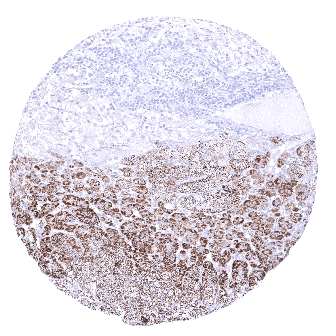
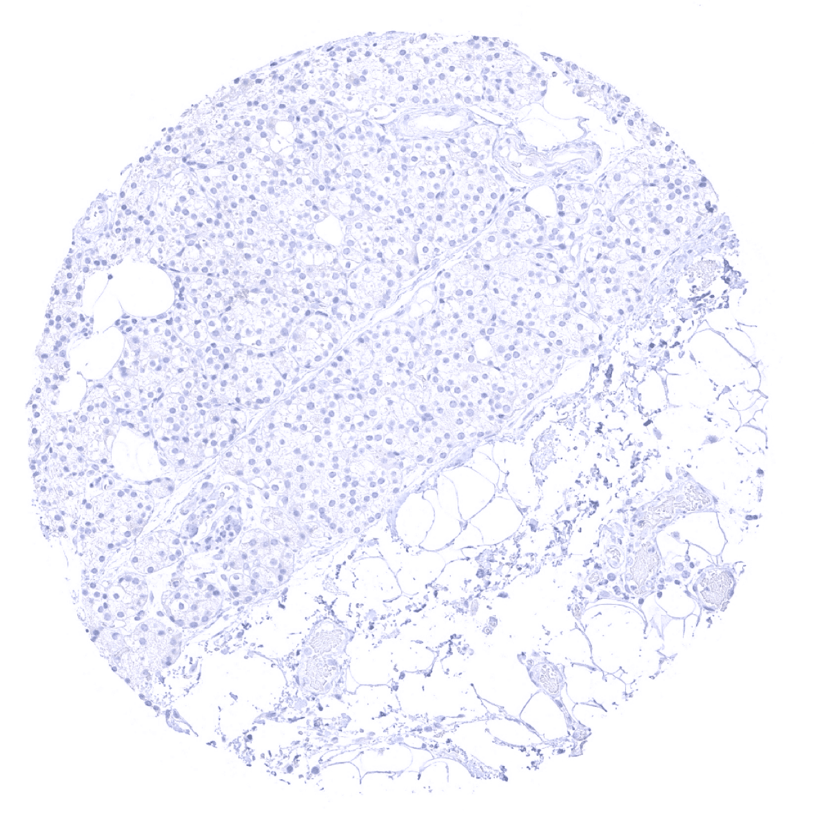
Adrenal gland - Cells of the adrenal gland show a moderate to strong Melan A+ immunostaining in the adrenal cortex but not in the medulla.
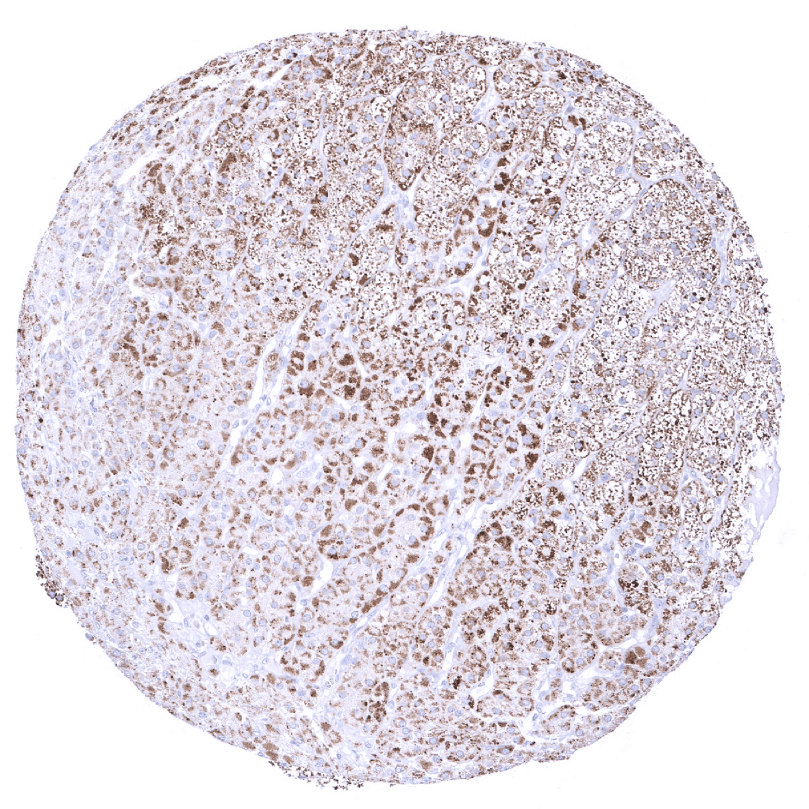
Adrenal gland - In the adrenal gland, a moderate to strong Melan-A+ immunostaining is seen in cortical but not medullary cells.
Anal canal, skin - A strong Melan-A immunostaining is seen in melanocytes of anal skin.
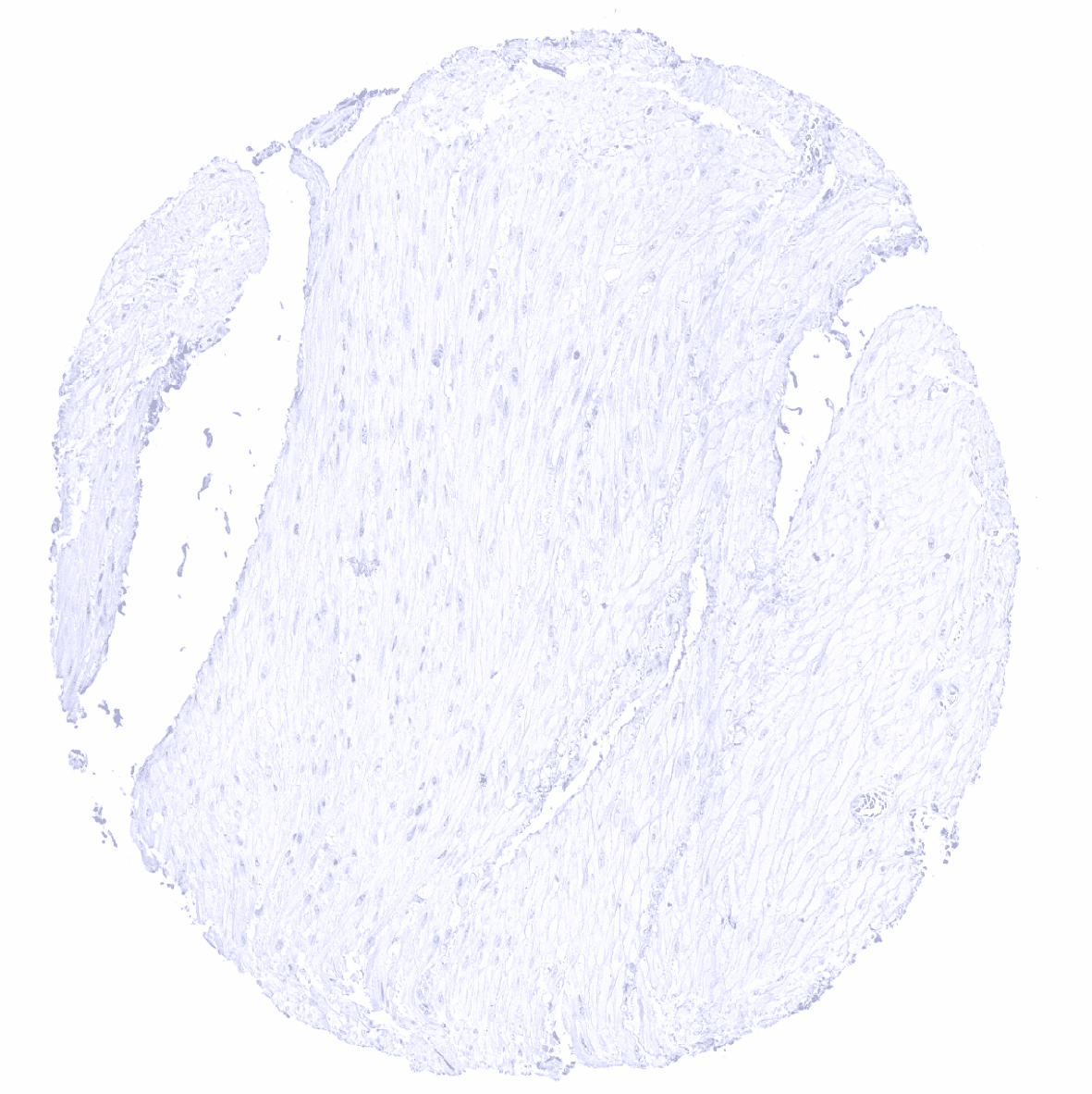
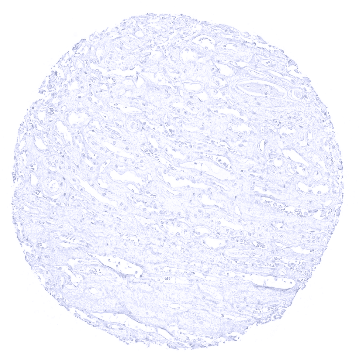
Aorta, media
Appendix, mucosa
Appendix, muscular wall

Bone marrow
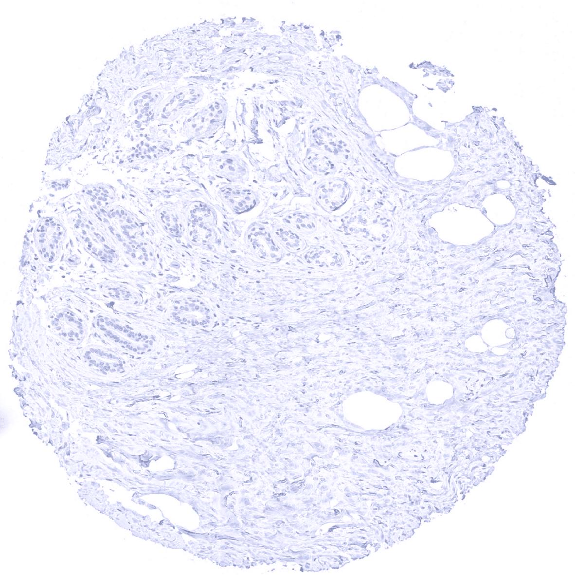
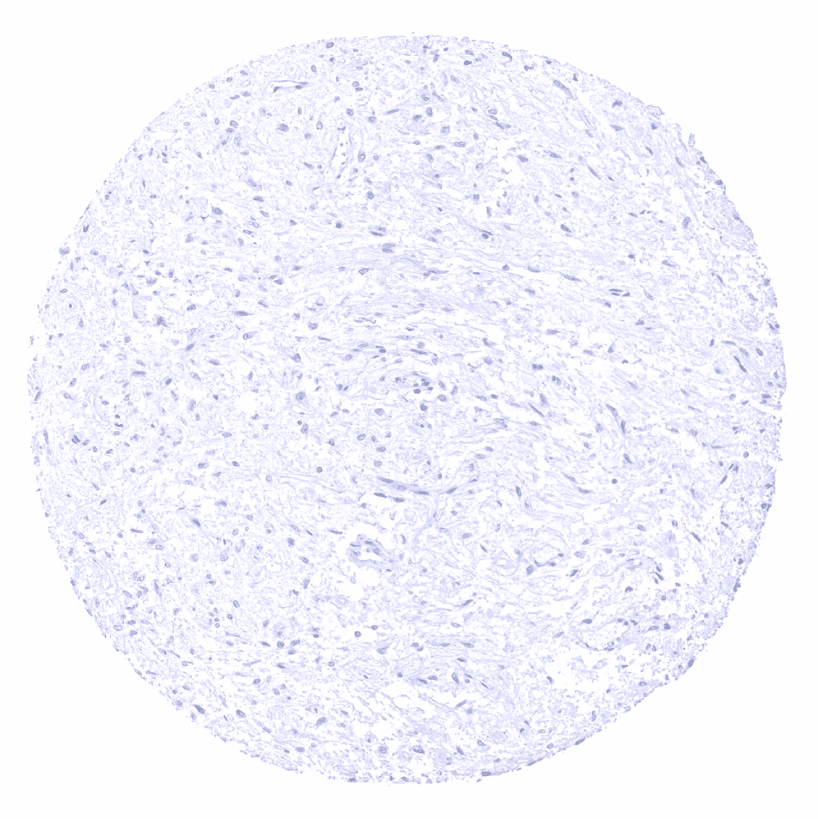
Breast

Bronchus, mucosa - A faint immunostaining can be seen at the apical membranes of a fraction of ciliated cells of respiratory epithelium (not in all samples).
Bronchus, mucosa
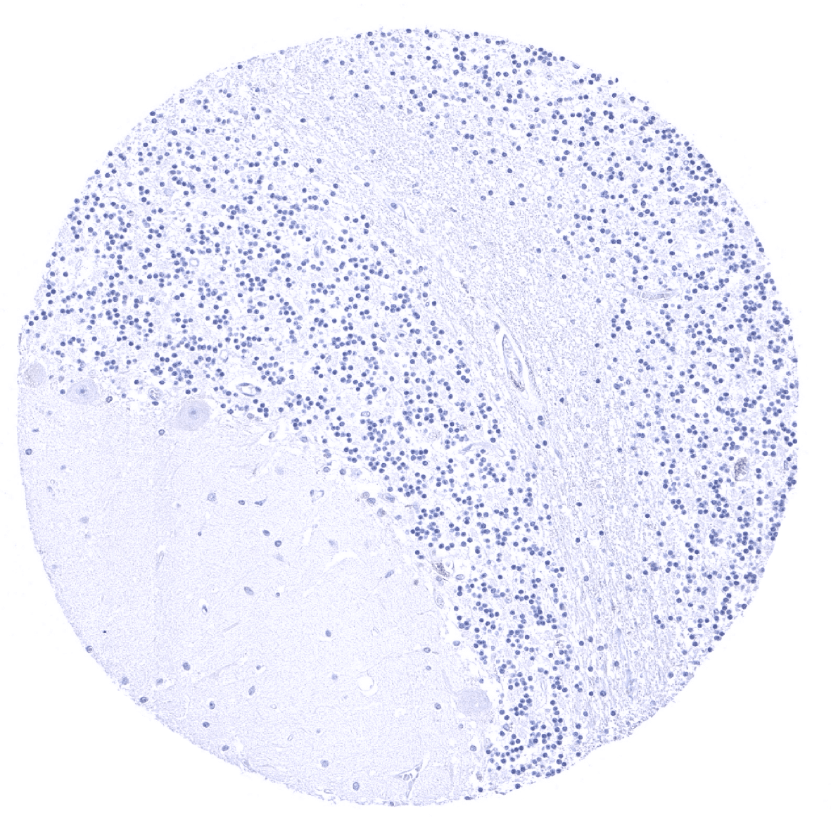
Cerebellum (molecular layer, Purkinje cell layer, granule cell layer, white matter)
Cerebellum (granule cell layer, white matter)
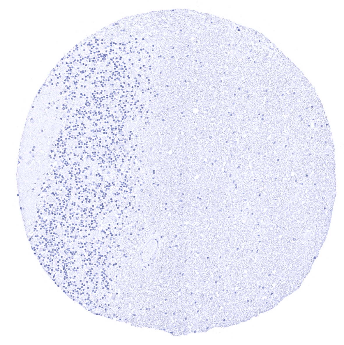
Cerebrum, grey matter

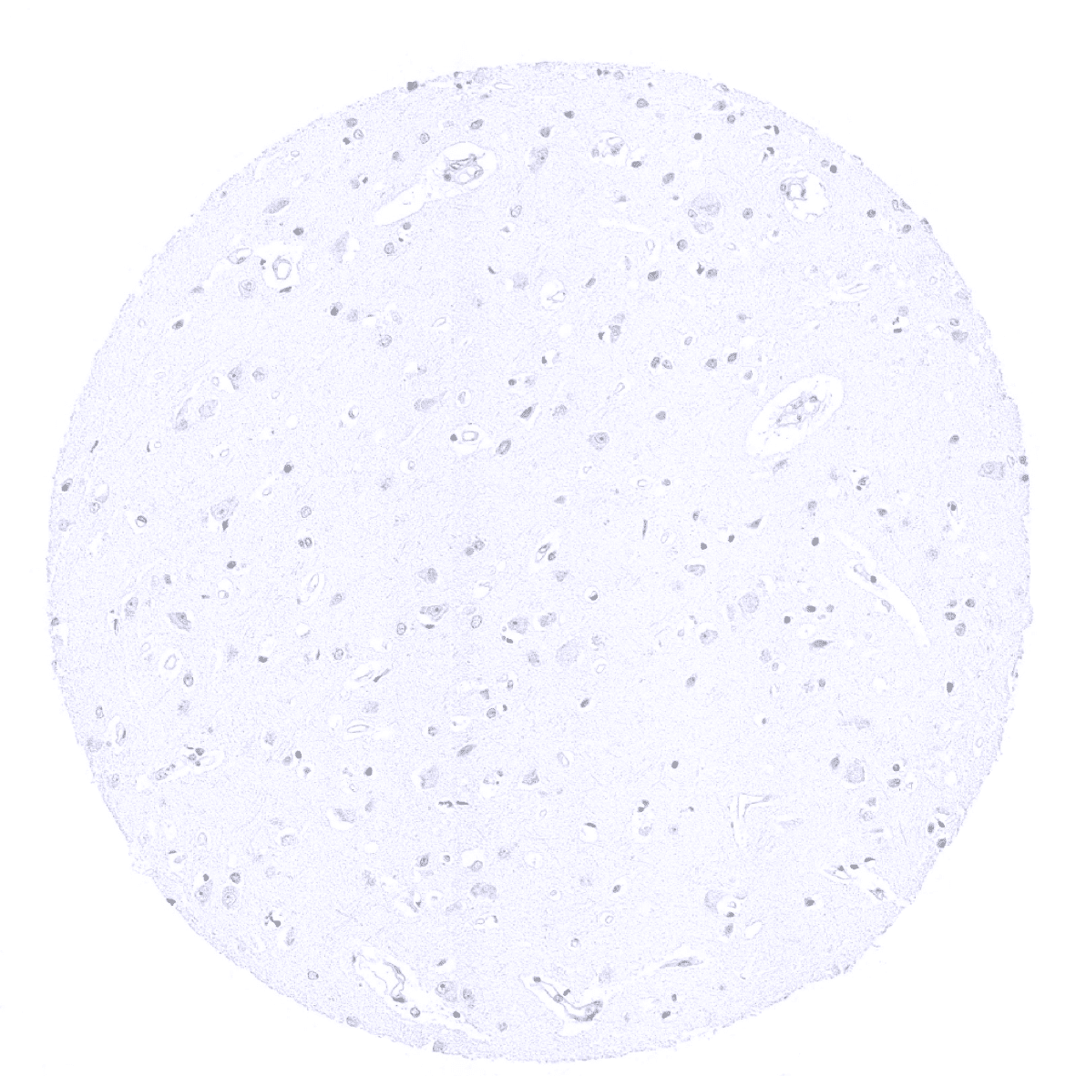
Cerebrum, white matter

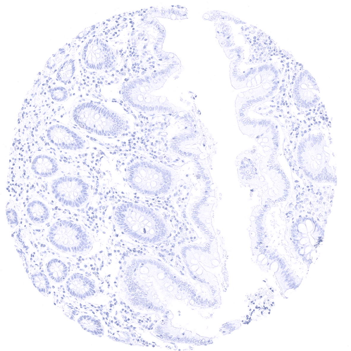
Colon descendens, mucosa
Colon descendens, muscular wall
Duodenum, Brunner gland
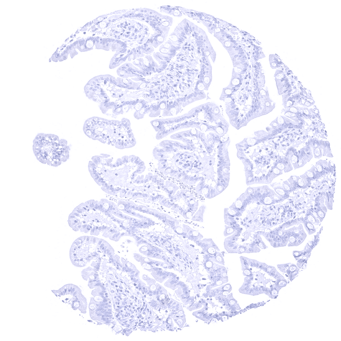
Duodenum, mucosa
Ectocervix
Endocervix
Endometrium, proliferation

Endometrium, secretion
Epididymis
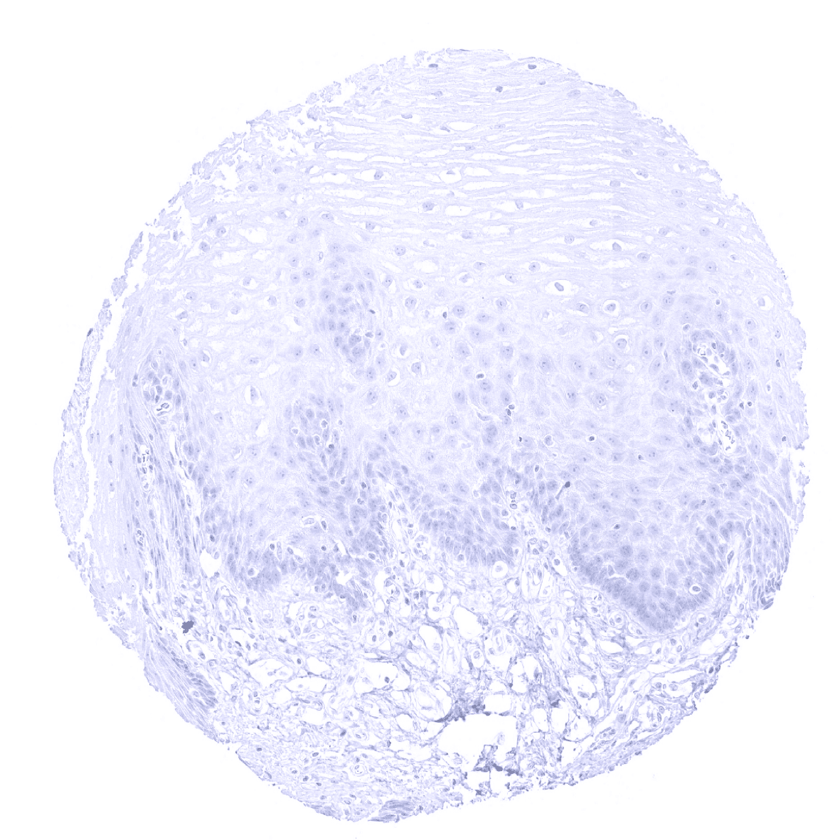
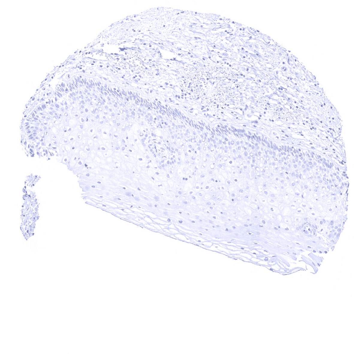
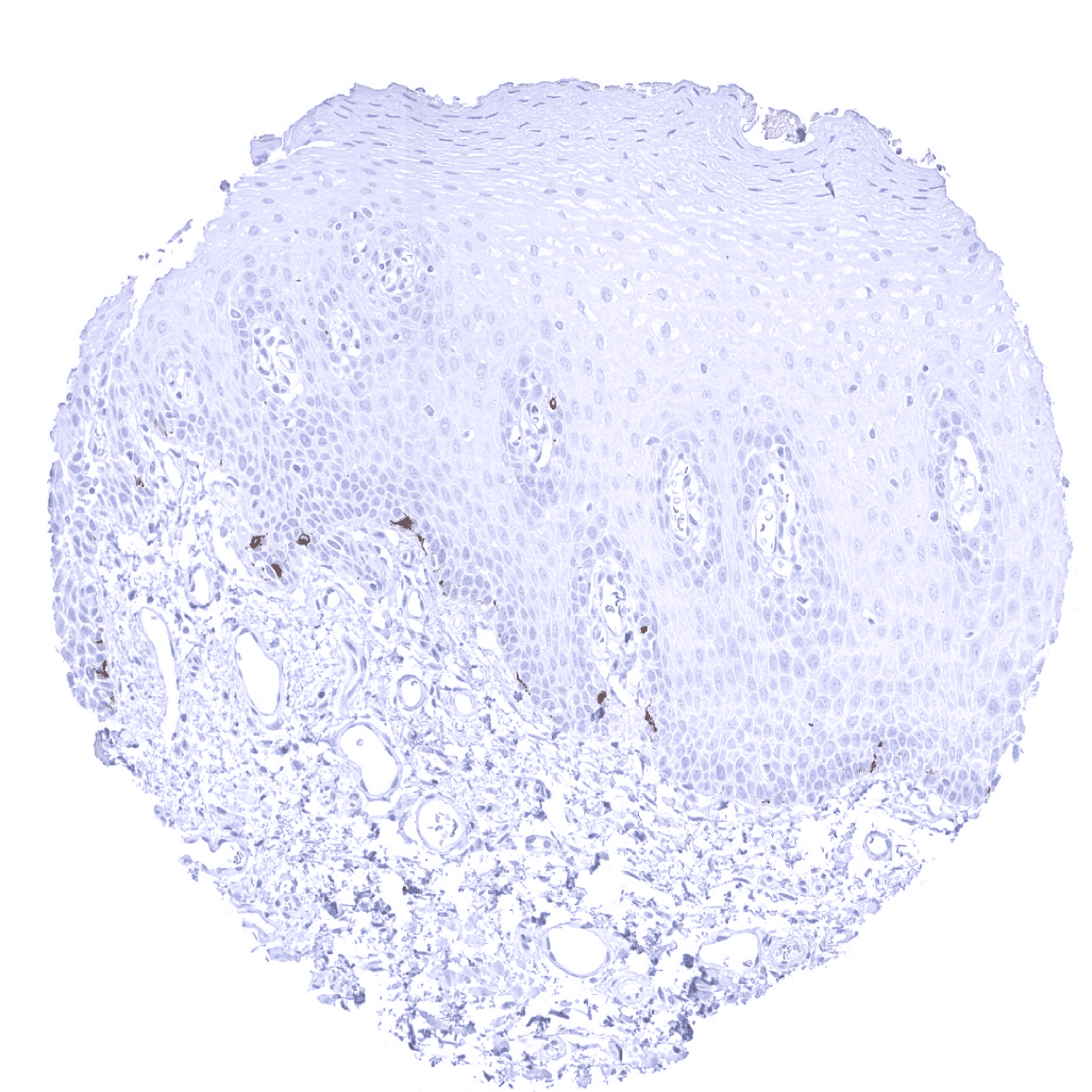
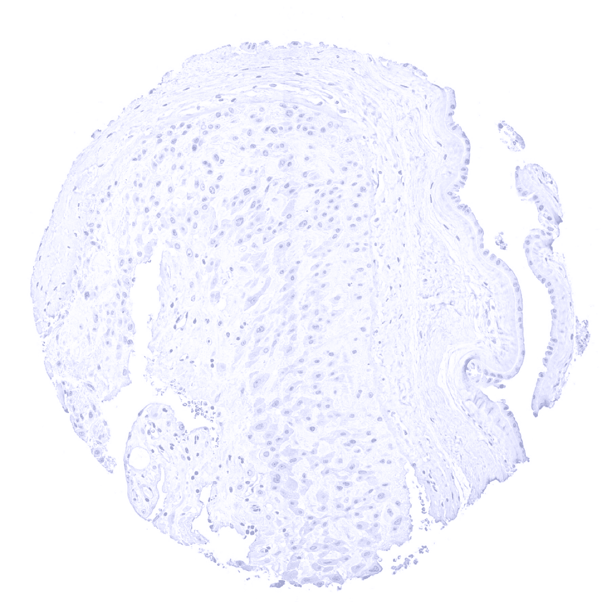
Esophagus, squamous epithelium

Fallopian tube, mucosa

Fat

Gallbladder, epithelium

Heart
Ileum, mucosa
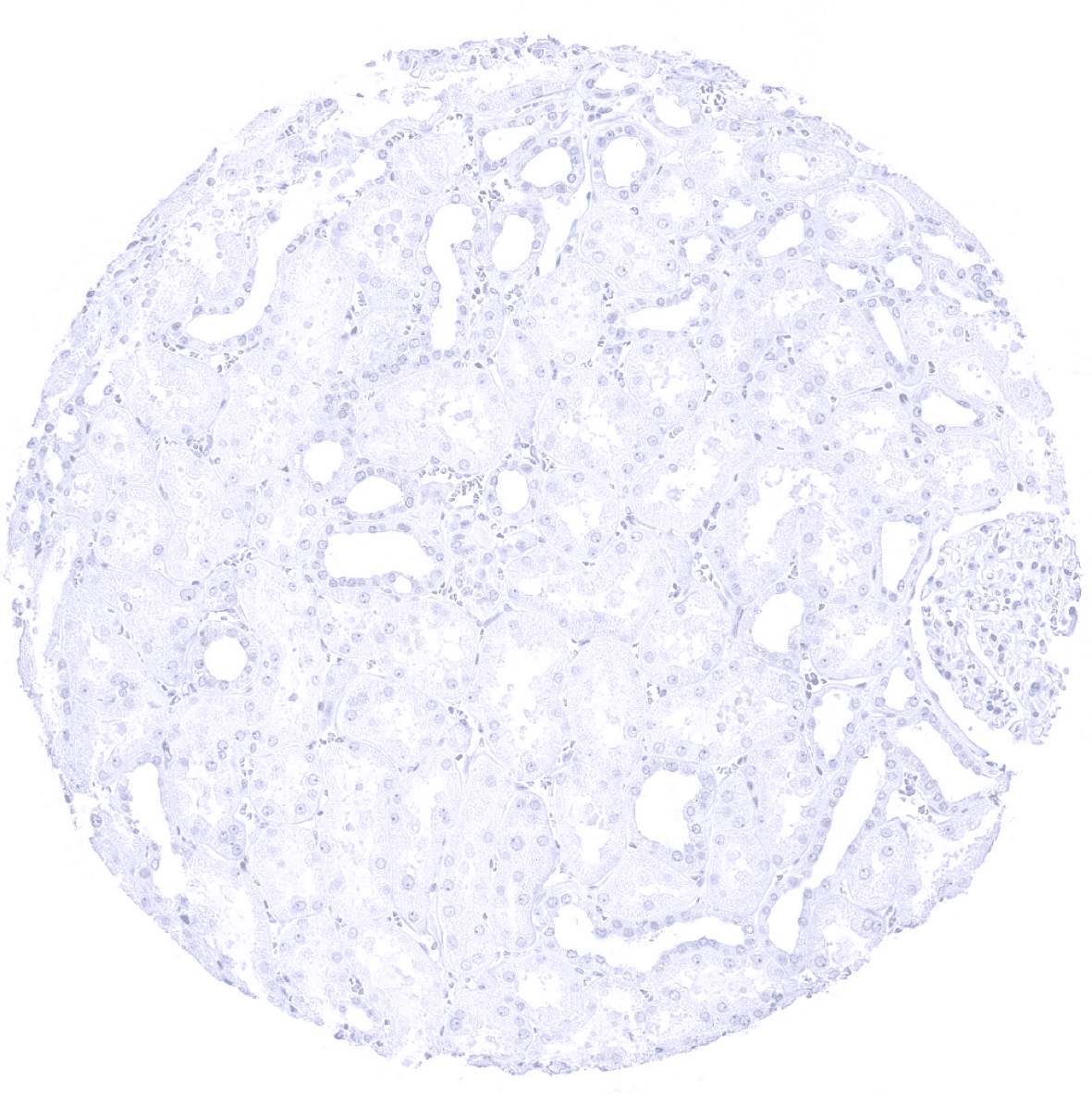
Kidney, cortex
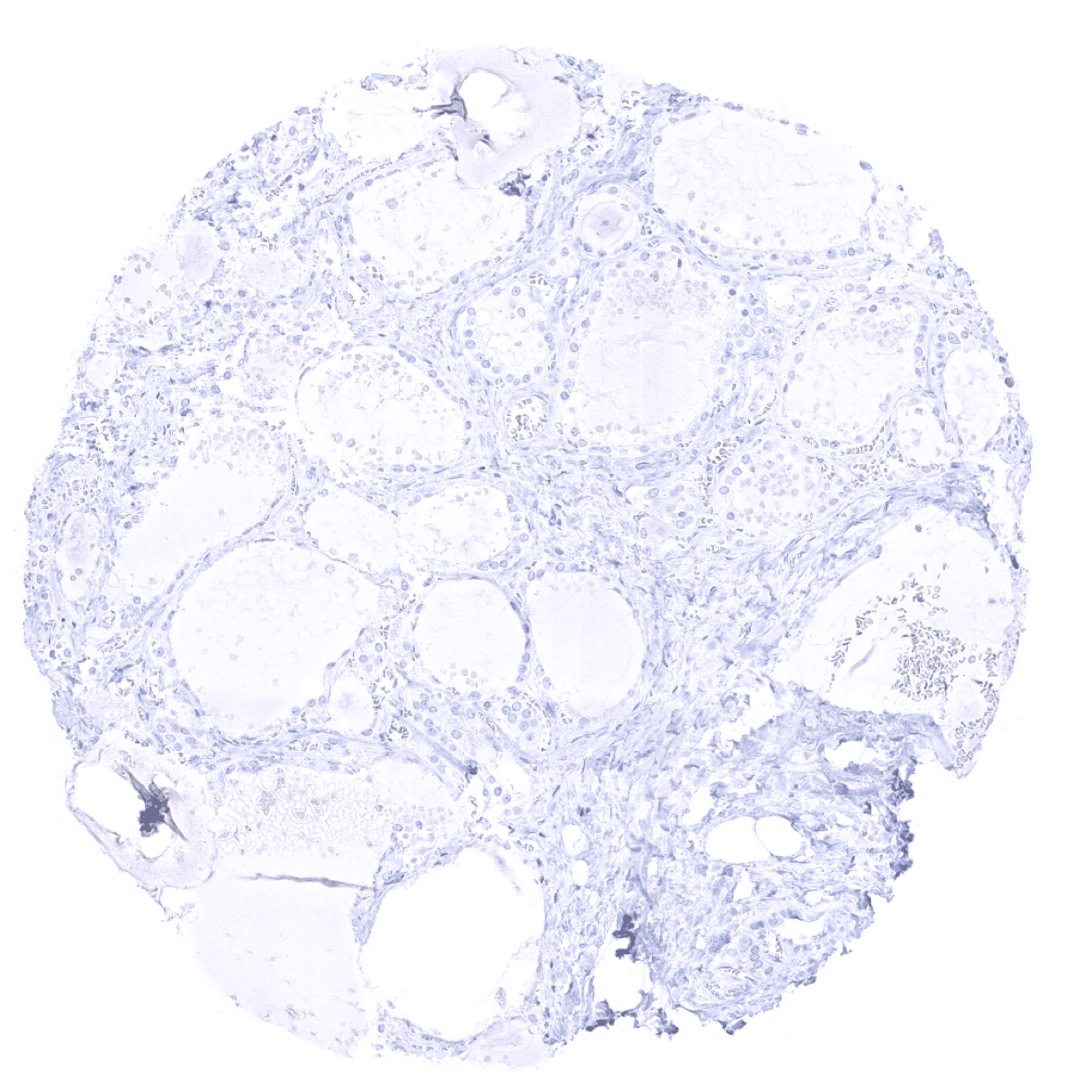
Kidney, medulla
Lip, oral mucosa - In the oral mucosa of the lip, a moderate to strong Melan-A immunostaining can be seen in few melanocytes.

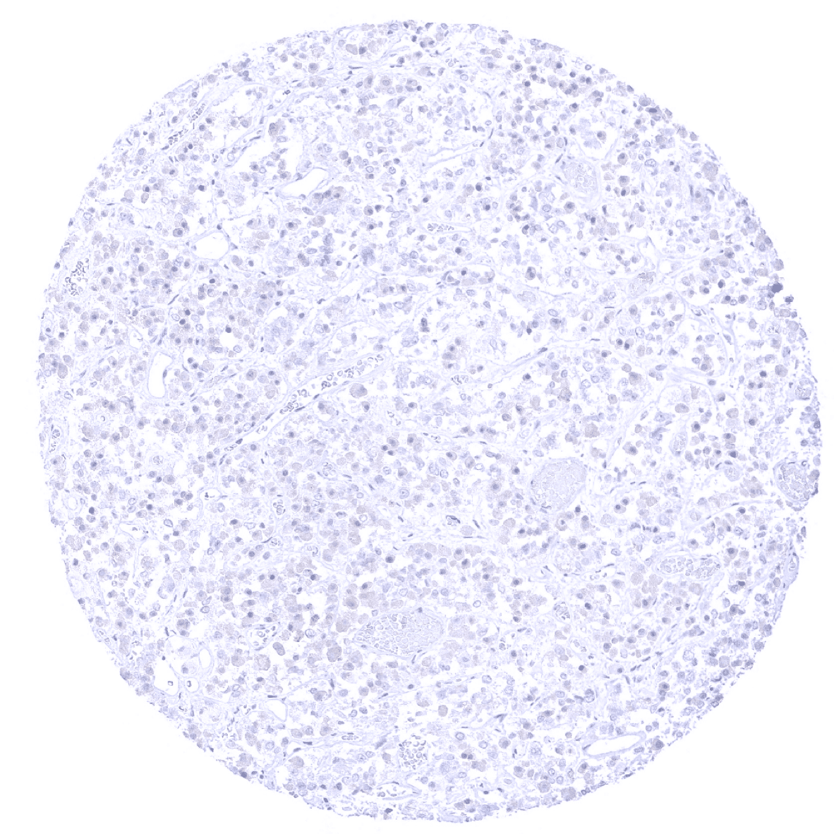
Liver
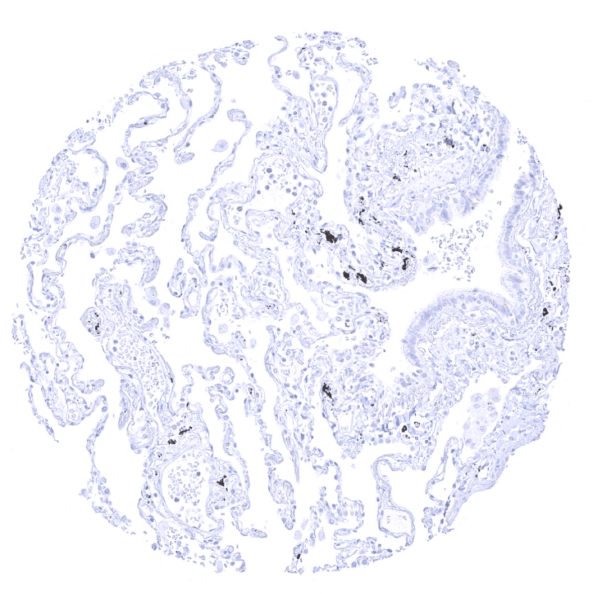
Lung
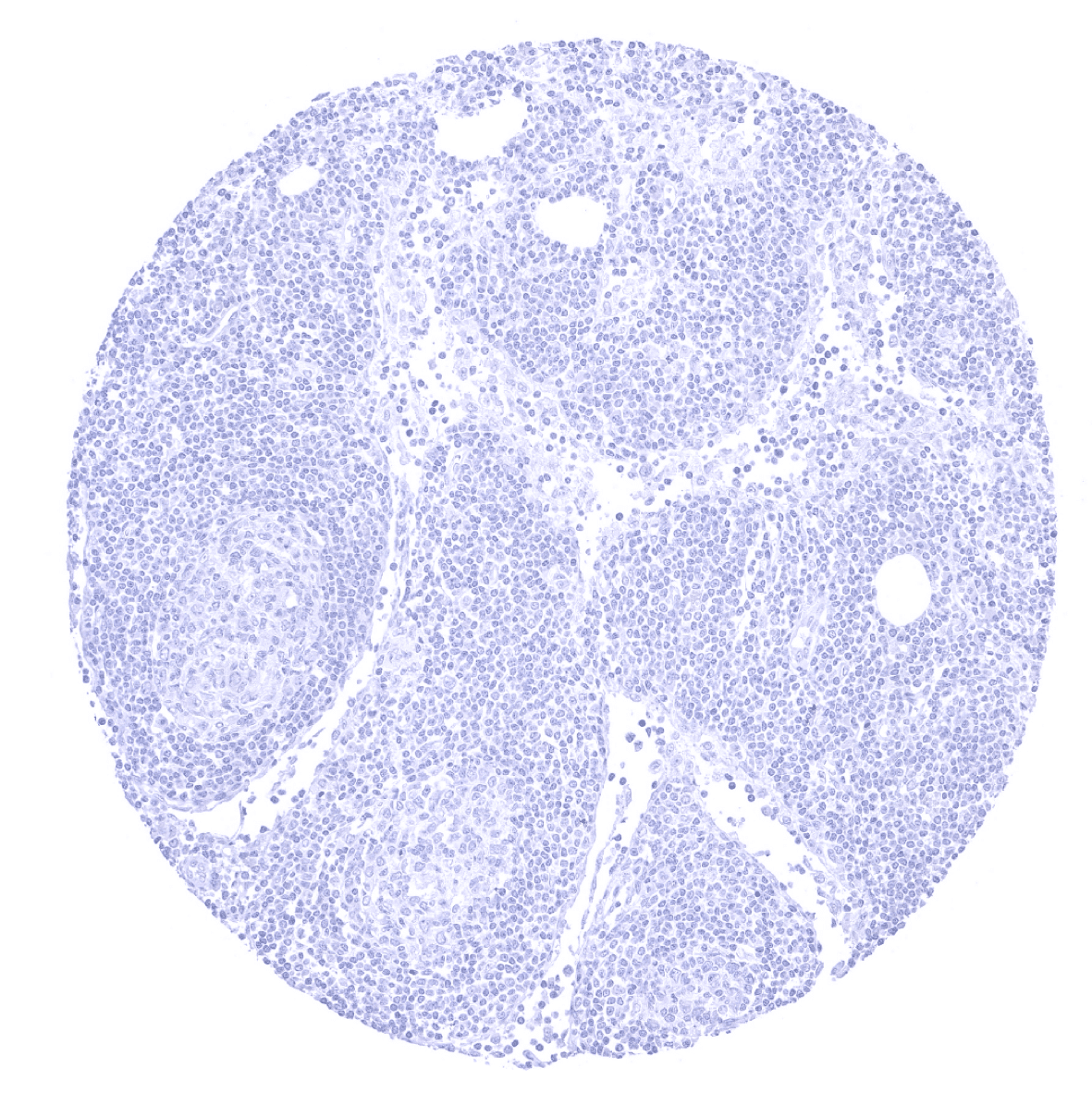
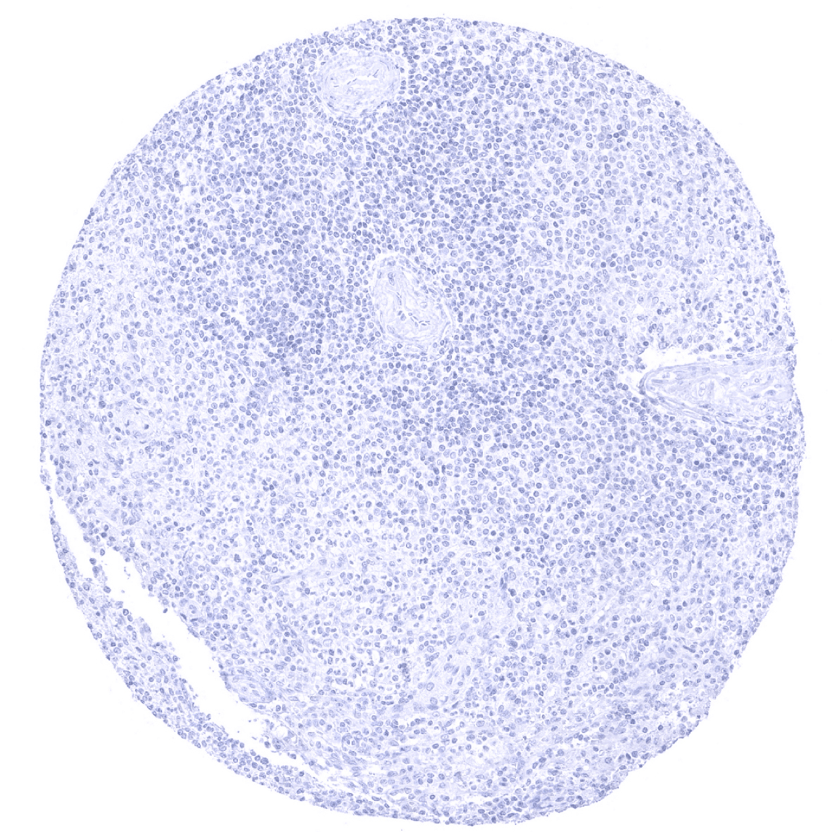
Lymph node

Ovary, stroma - In the ovary, a moderate intensity Melan A+ staining is seen in theca interna cells of ovary.
Ovary, stroma

Pancreas
Parathyroid
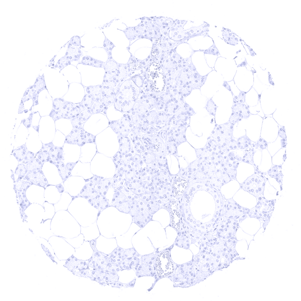
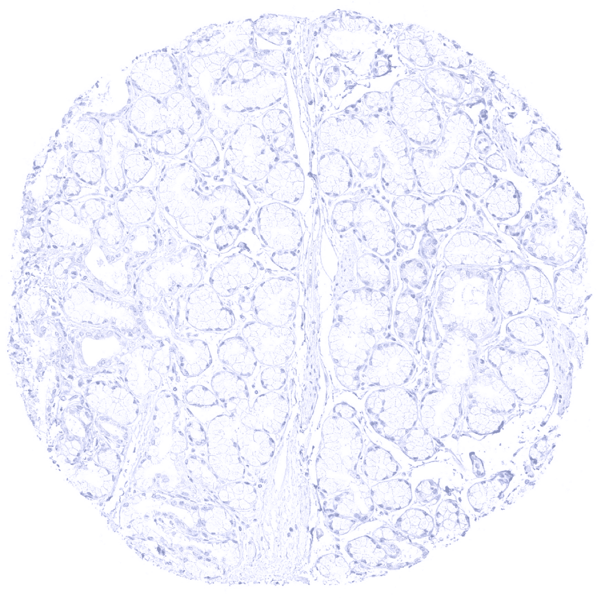
Parotid gland

Pituitary gland, anterior lobe - In the adenohypophysis, a moderate to strong Melan A+ immunostaining can be seen in scattered epithelial cells (not in all samples).
Pituitary gland, anterior lobe
Pituitary gland, posterior lobe

Pregnant uterus (decidua)
Placenta, early
Placenta (amnion and chorion)

Placenta, mature
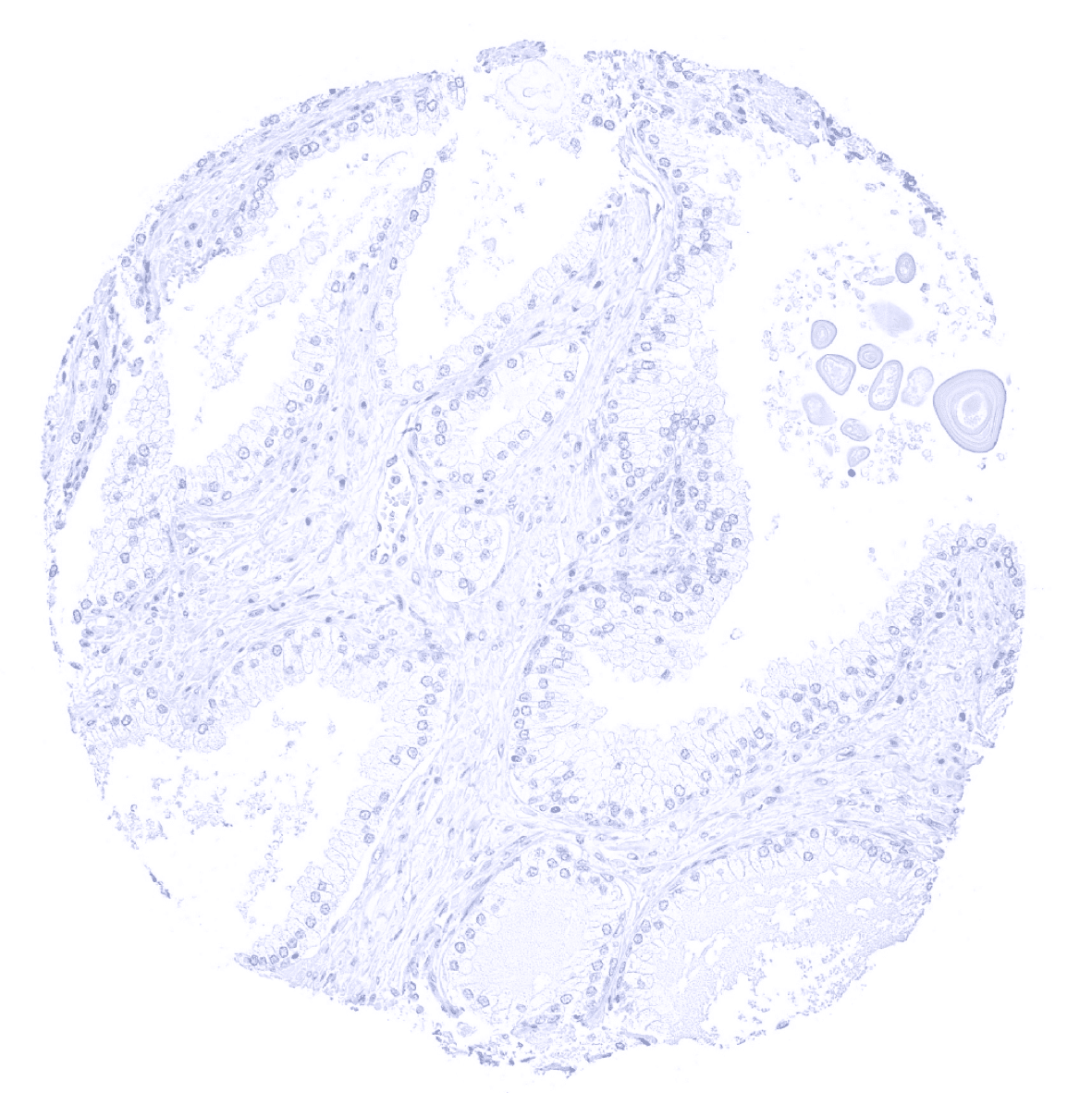
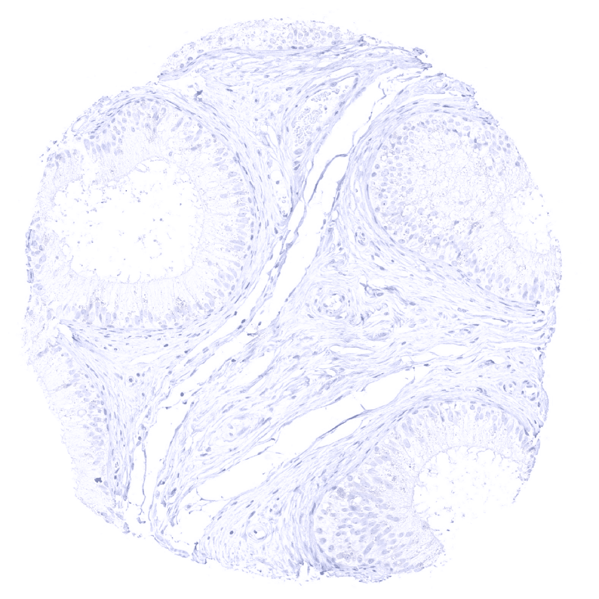
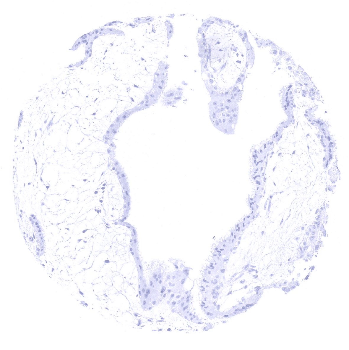
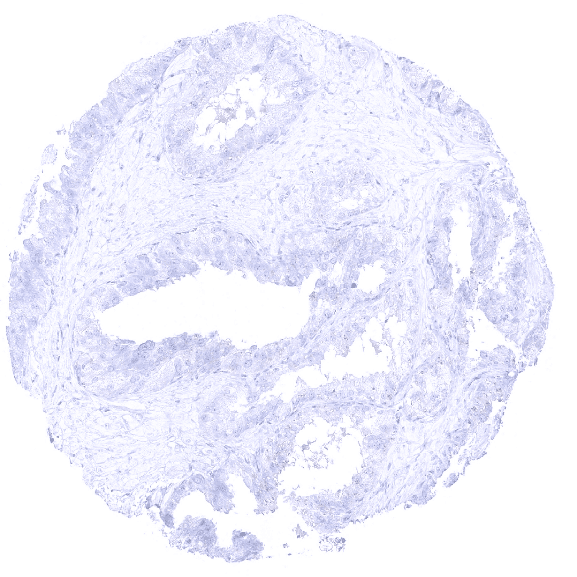
Prostate

Rectum, mucosa
Seminal vesicle
Sinus paranasales - A faint immunostaining can be seen at the apical membranes of a fraction of ciliated cells of respiratory epithelium (not in all samples).

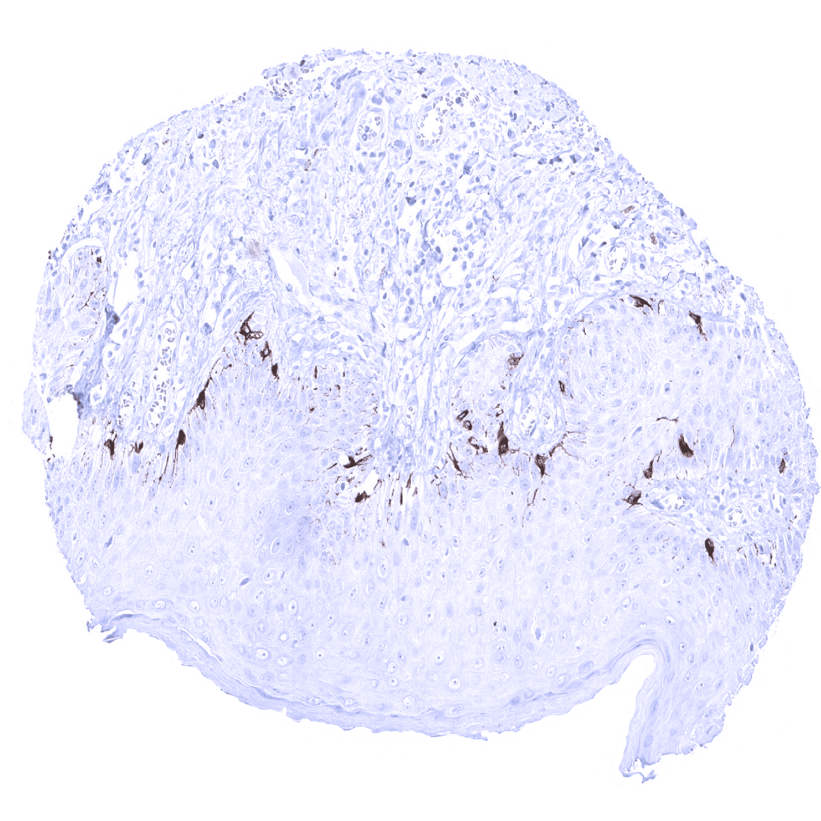
Skin - In the skin, a strong Melan-A immunostaining is seen in melanocytes.
Spleen

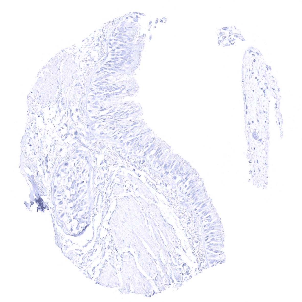
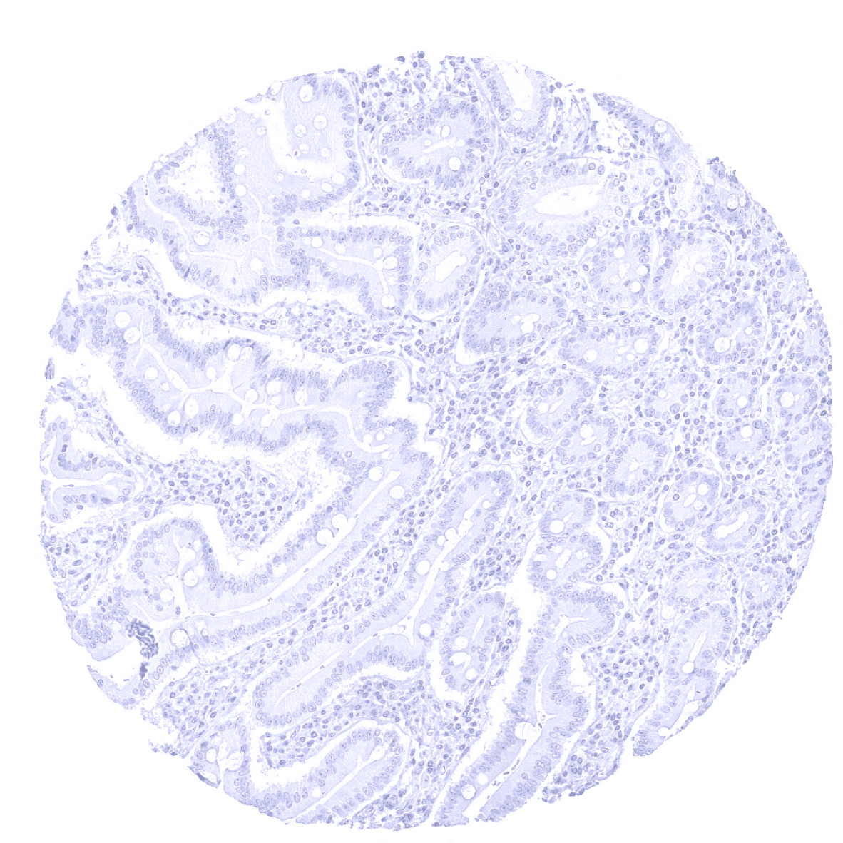
Stomach, antrum

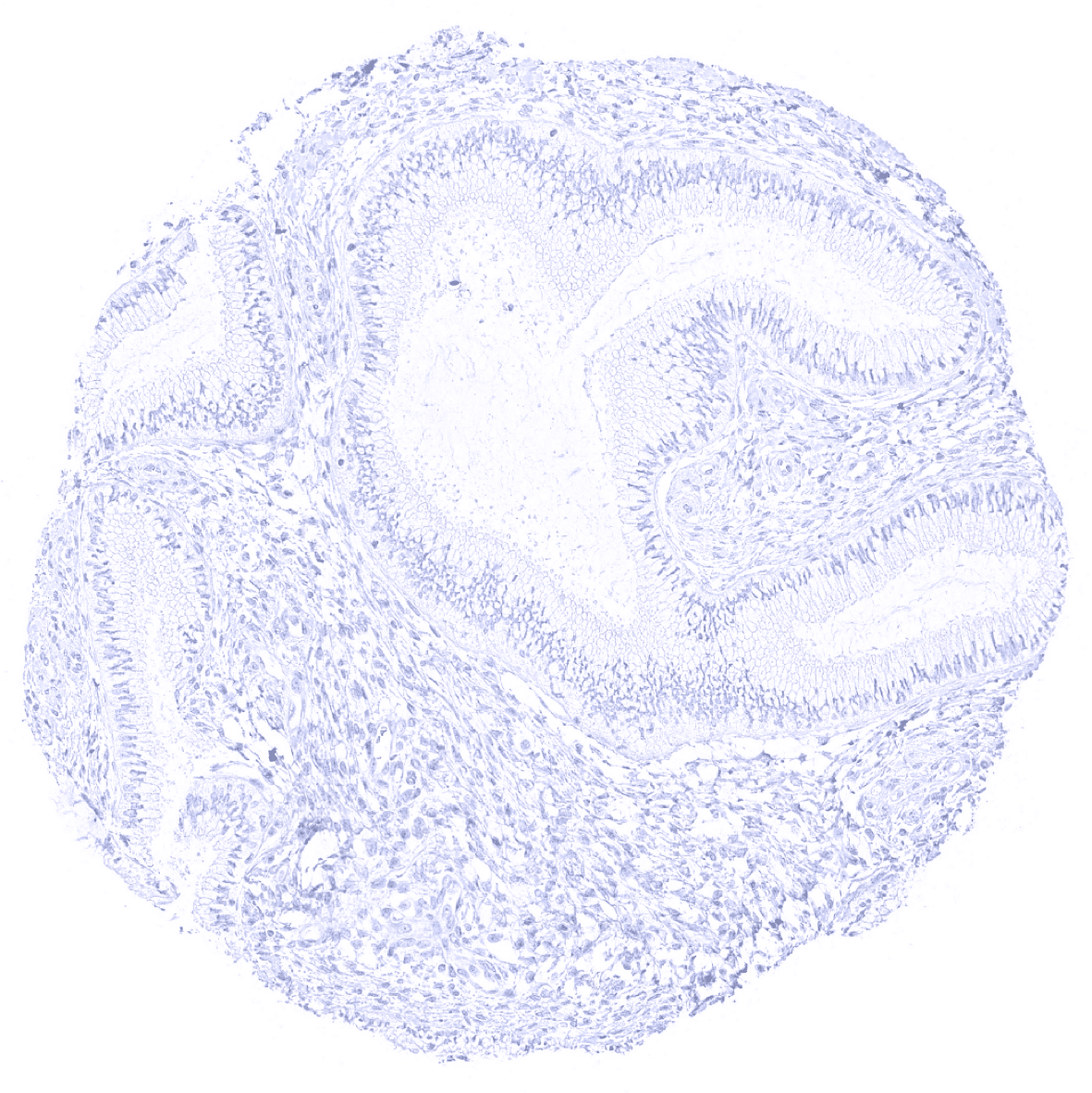
Stomach, corpus

Striated muscle
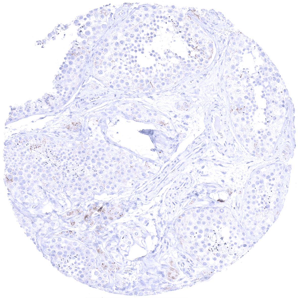
Testis - A weak to moderate Melan A+ immunostaining can be seen in a fraction of Leydig cells of the testis.
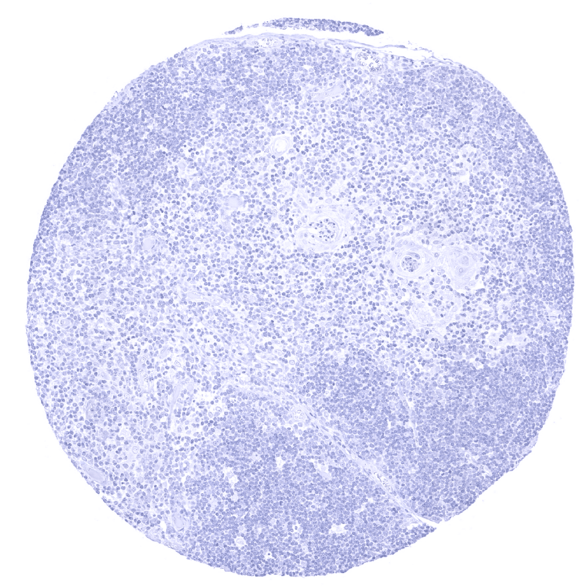
Thymus
Thyroid gland
Tonsil, surface epithelium
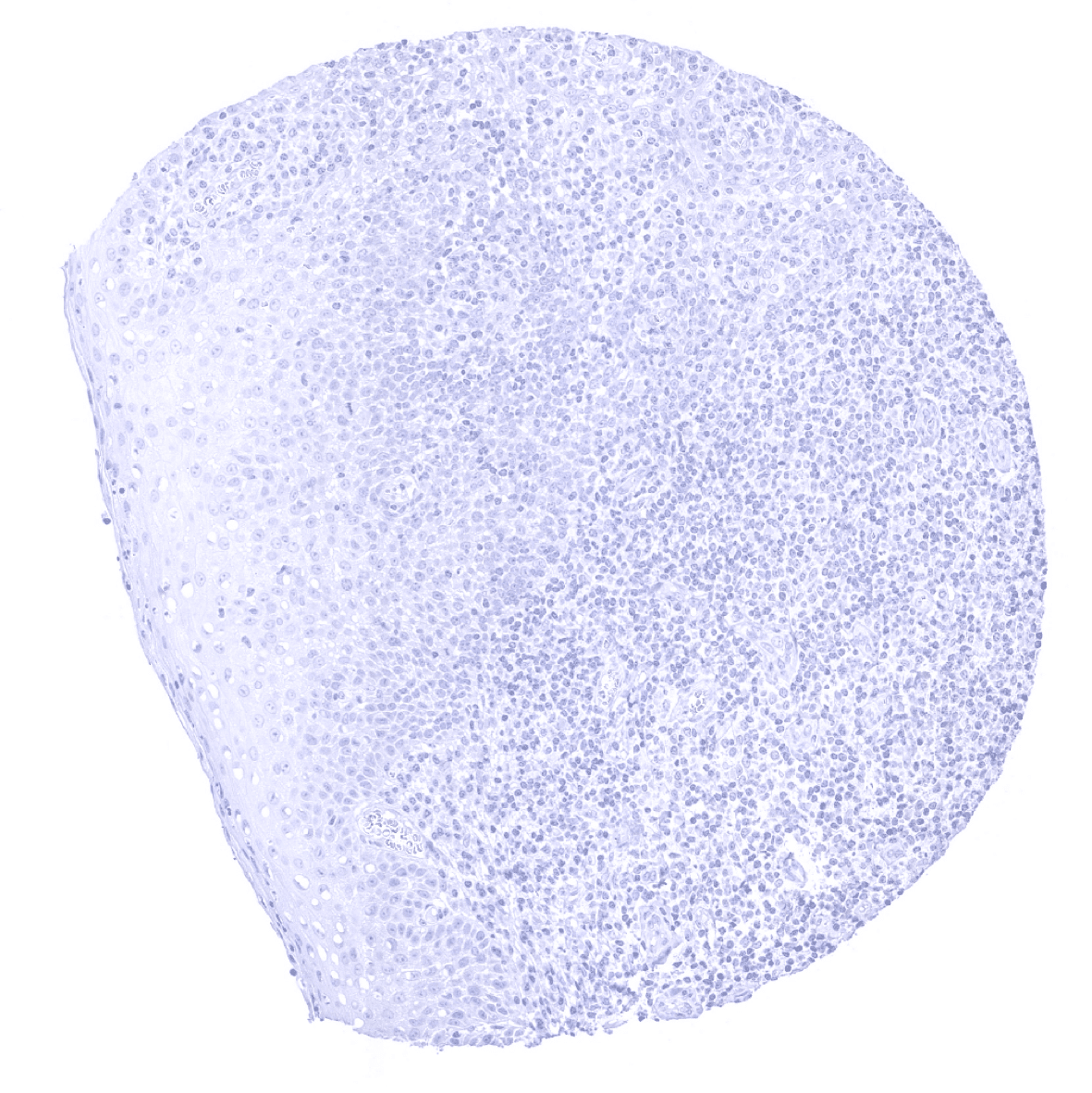
Tonsil
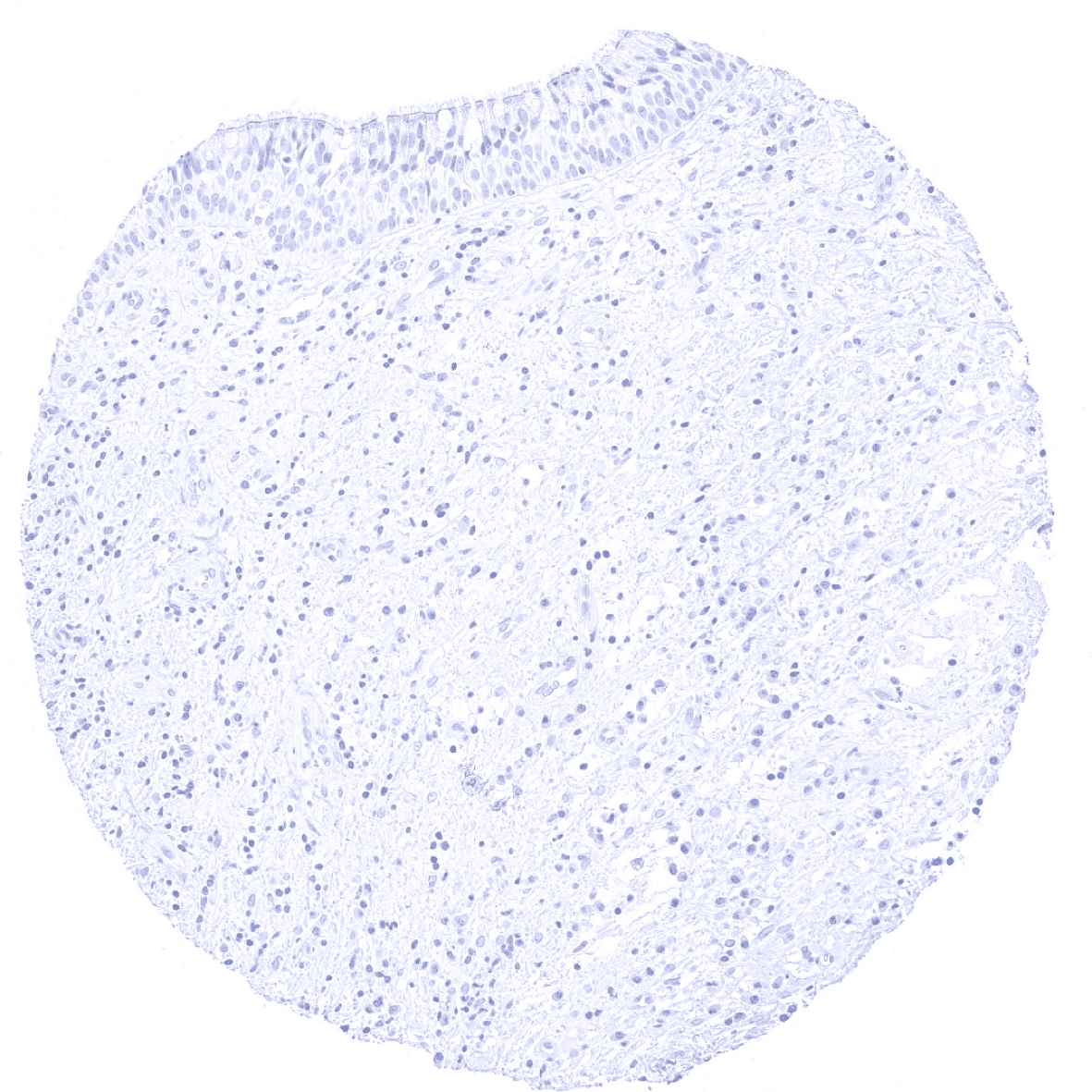
Urinary bladder, muscular wall
Urinary bladder, urothelium
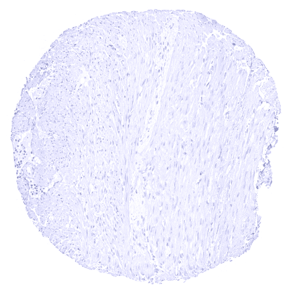
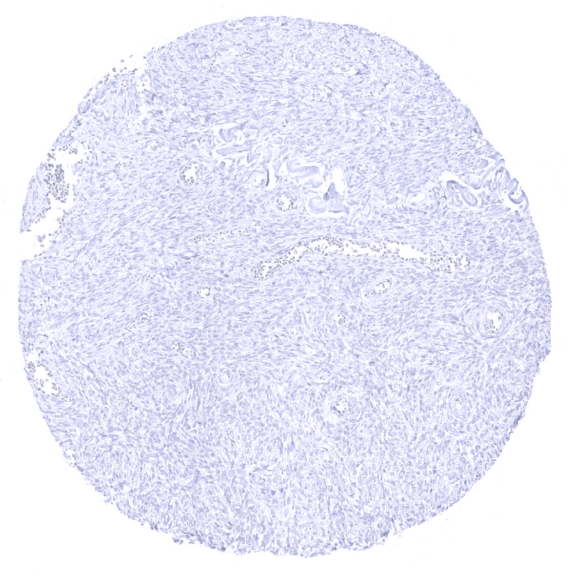
Uterus, myometrium