
295,00 € – 895,00 €
Product details
Synonyms = Hematopoietic Progenitor Cell Antigen, HPCA1, Mucosialin
Antibody type = Recombinant Rabbit monoclonal / IgG, Rabbit
Clone = MSVA-457R
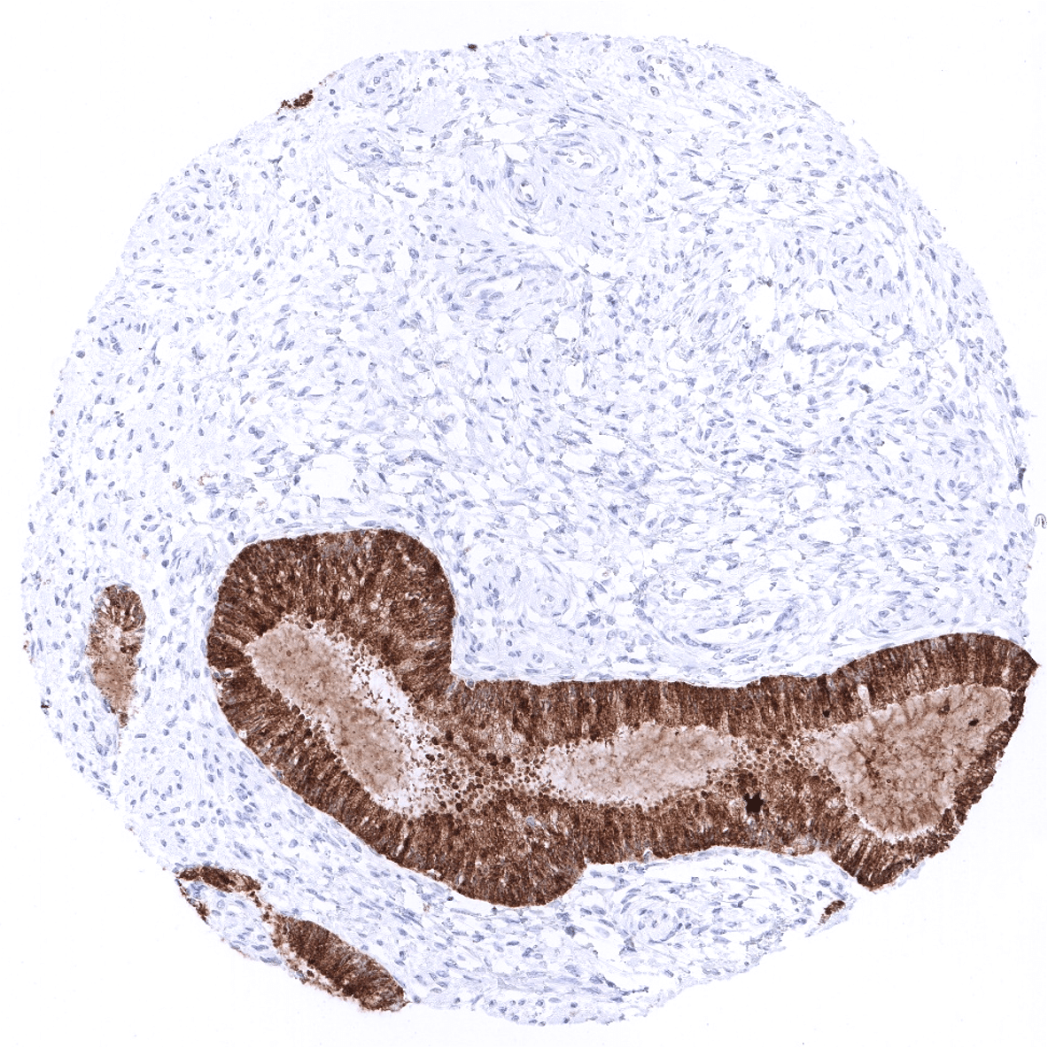
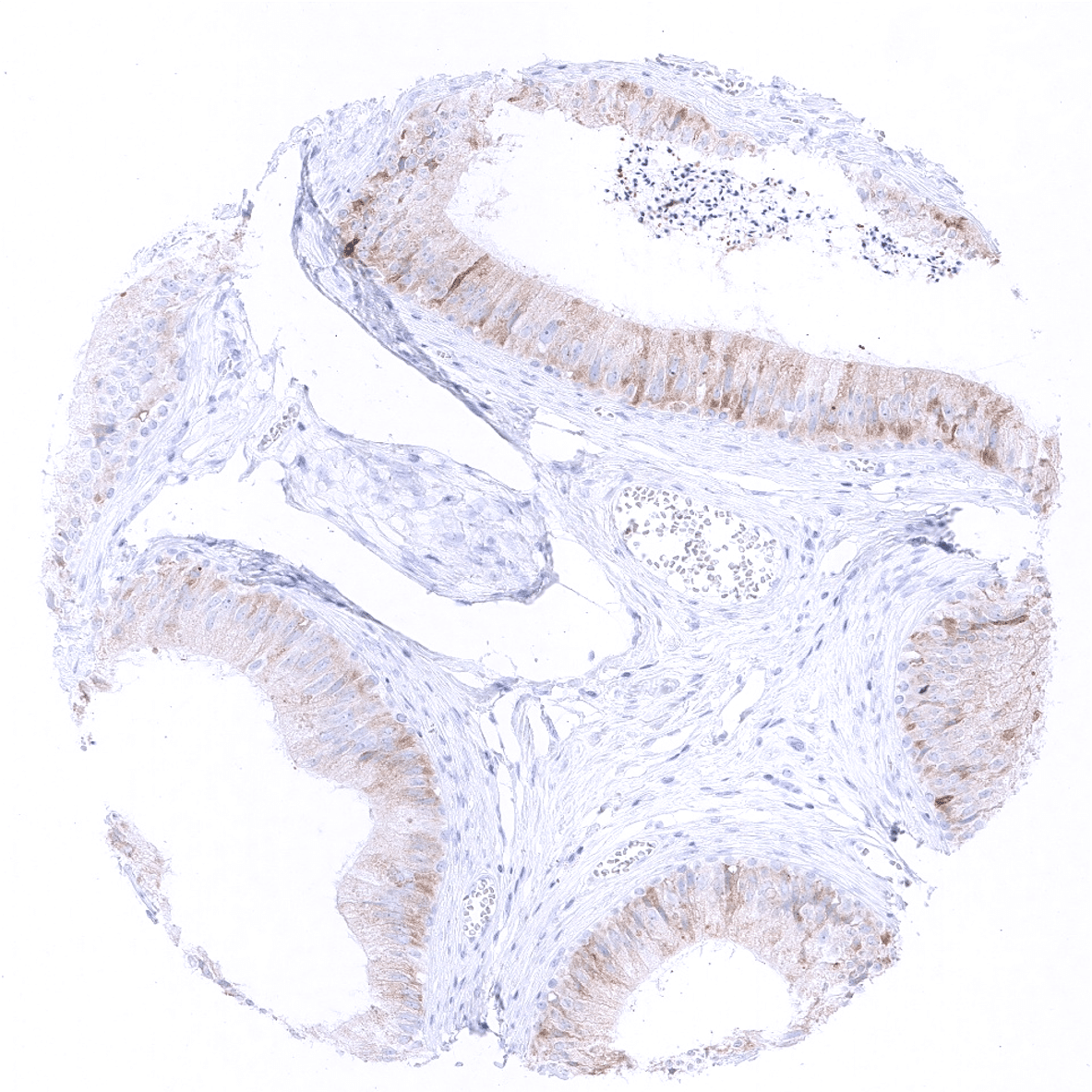
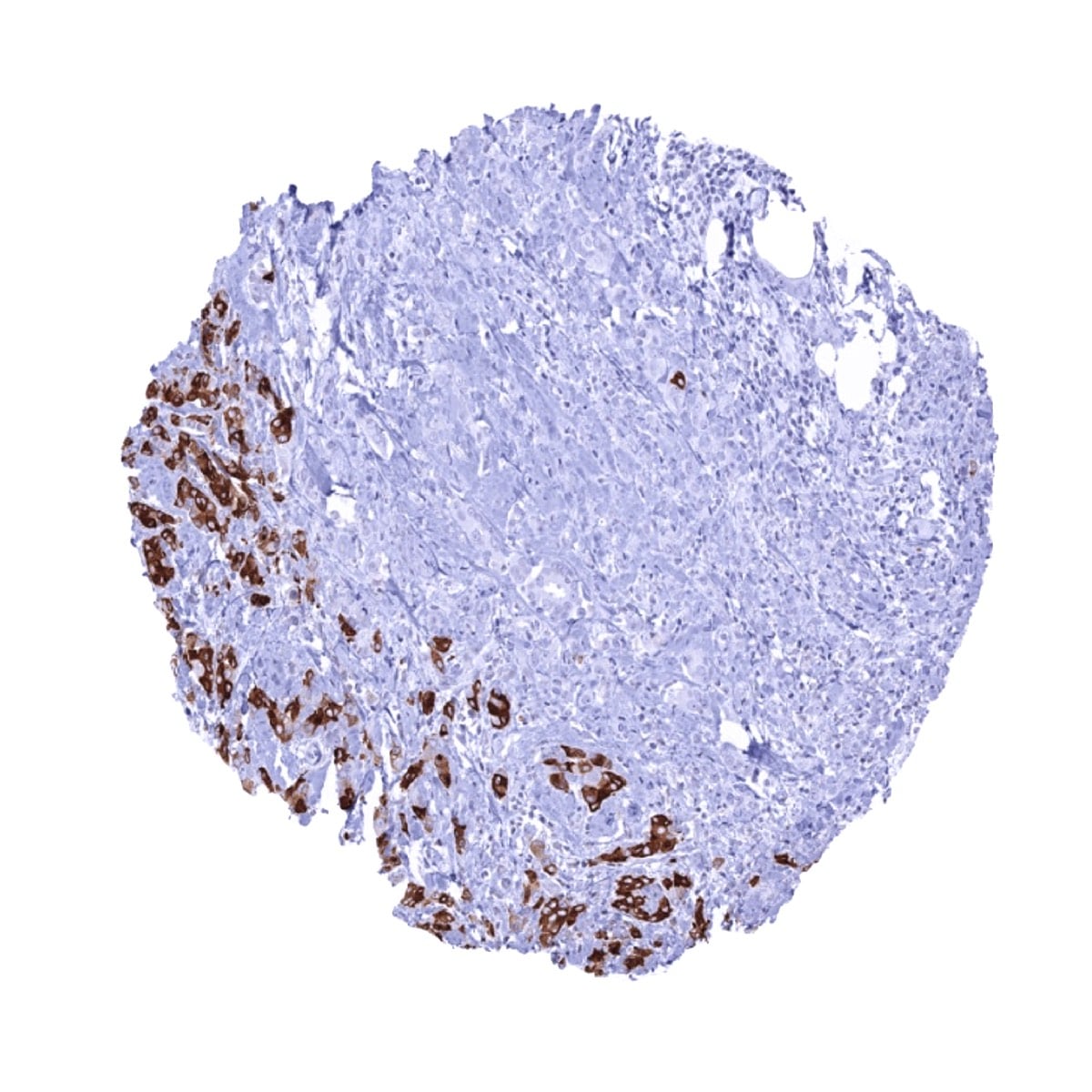
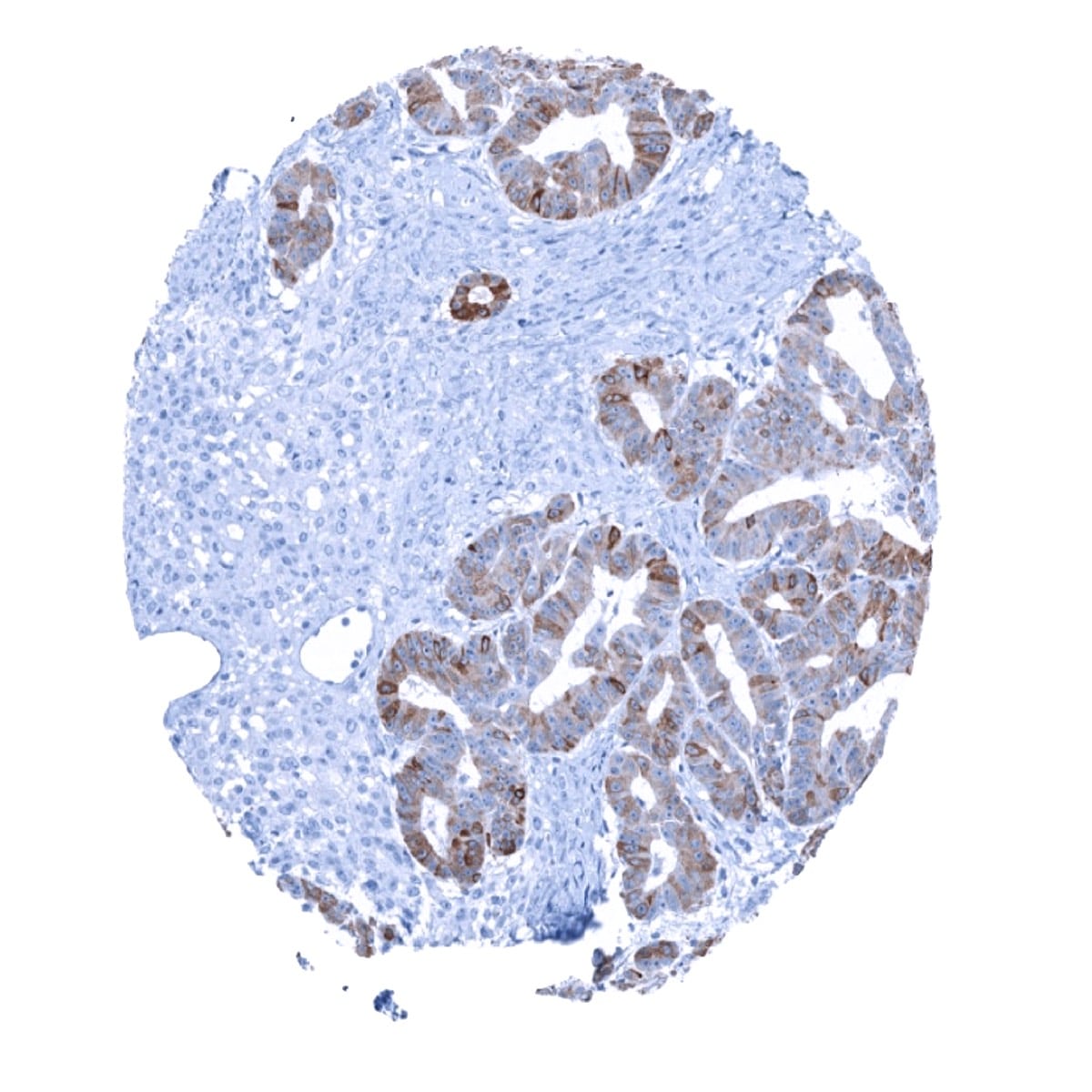
Positive control = Cervix uteri: a moderate to strong cytoplasmic staining of the endocervical glands should be seen while the squamous epithelium of the ektocervix remains negative.
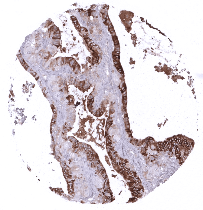
Negative control = Colon: All cells should not show any staining.
Cellular localization = Cytoplasmic
Reactivity = Human
Application = Immunohistochemistry
Dilution = 1:100 – 1:200
Intended Use = Research Use Only
Relevance of Antibody
Biology Behind
Human Mammaglobin-A (SCGB2A2) was described as one of more than 20 members of the uteroglobin/Clara cell protein family of small epithelial secretory proteins, termed secretoglobins. It is a 93-amino acid protein that occurs in the mammary tissue in two main forms with approximate molecular masses of 18 and 25 kDa. Although the secretoglobins are known to be regulated by steroid hormones, mammaglobin expression in the mammary gland is independent of steroid hormones. The function of mammaglobin protein is not known. Other related secretoglobins seem to be involved in cell signaling, immune response, and chemotaxis, and may also serve as transporters for steroid hormones in humans. Mammaglobin is upregulated in neoplastic breast epithelia as compared to normal cells but its overexpression does not influence tumor cell growth. The protein’s capability to bind steroid-like molecules suggests the existence of a hormonal transport or activation function.
Staining Pattern in Normal Tissues
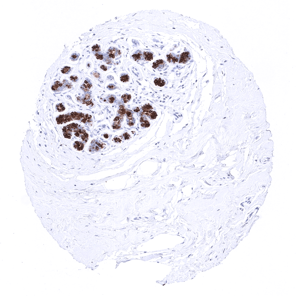
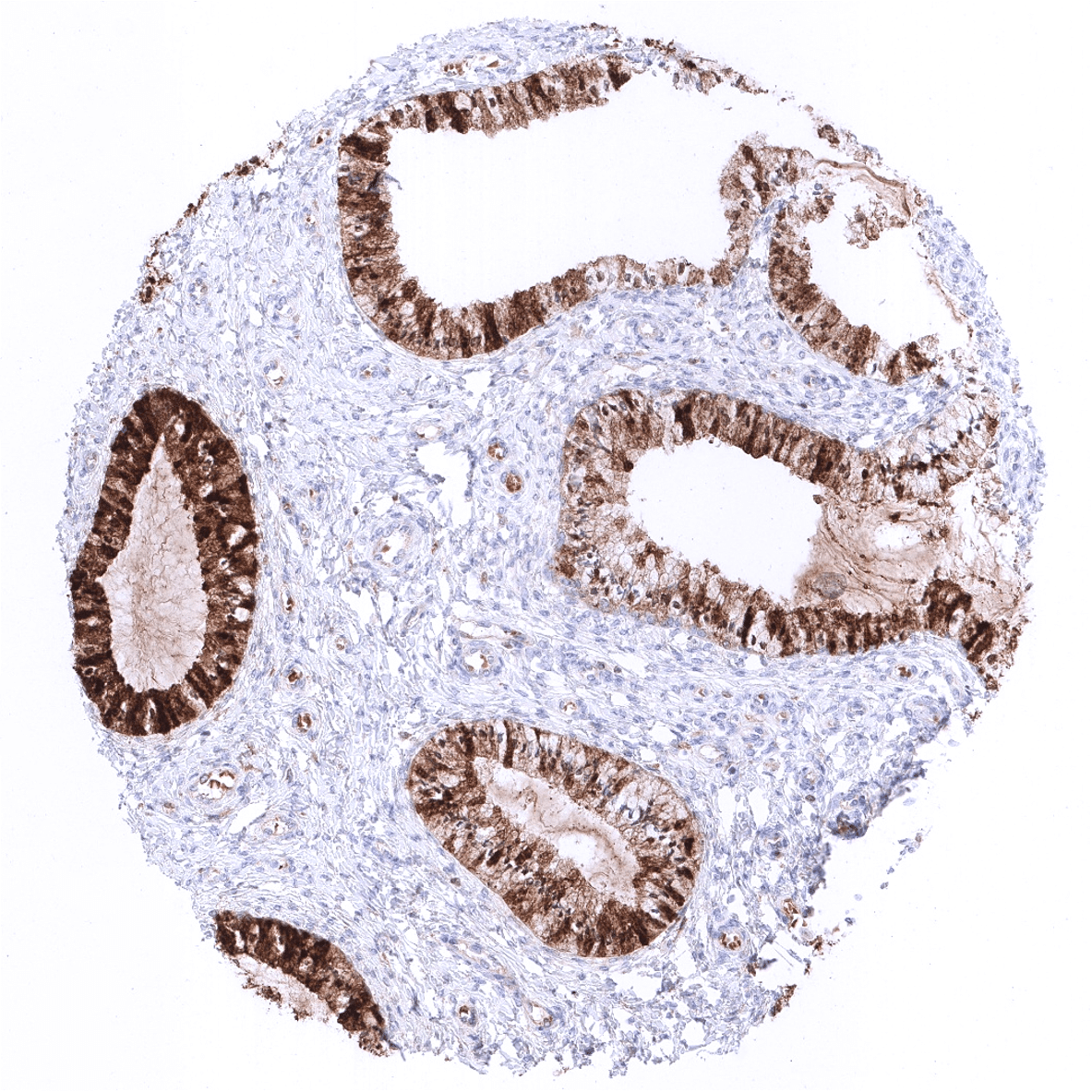
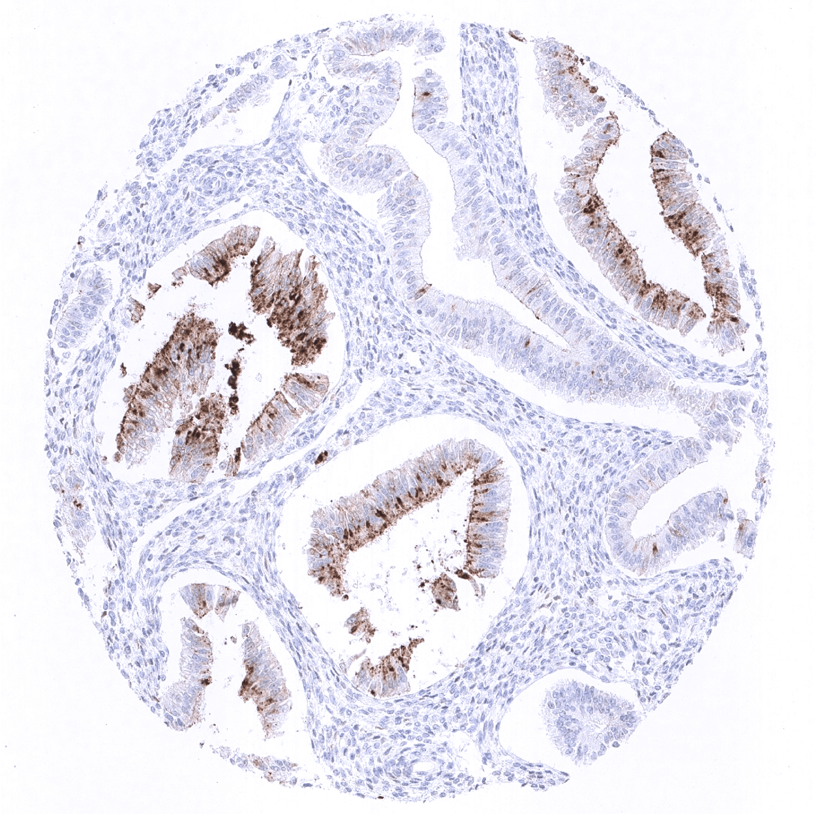
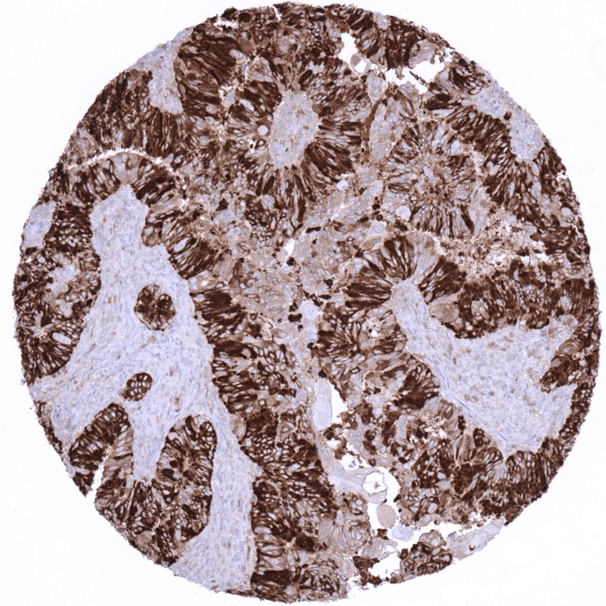
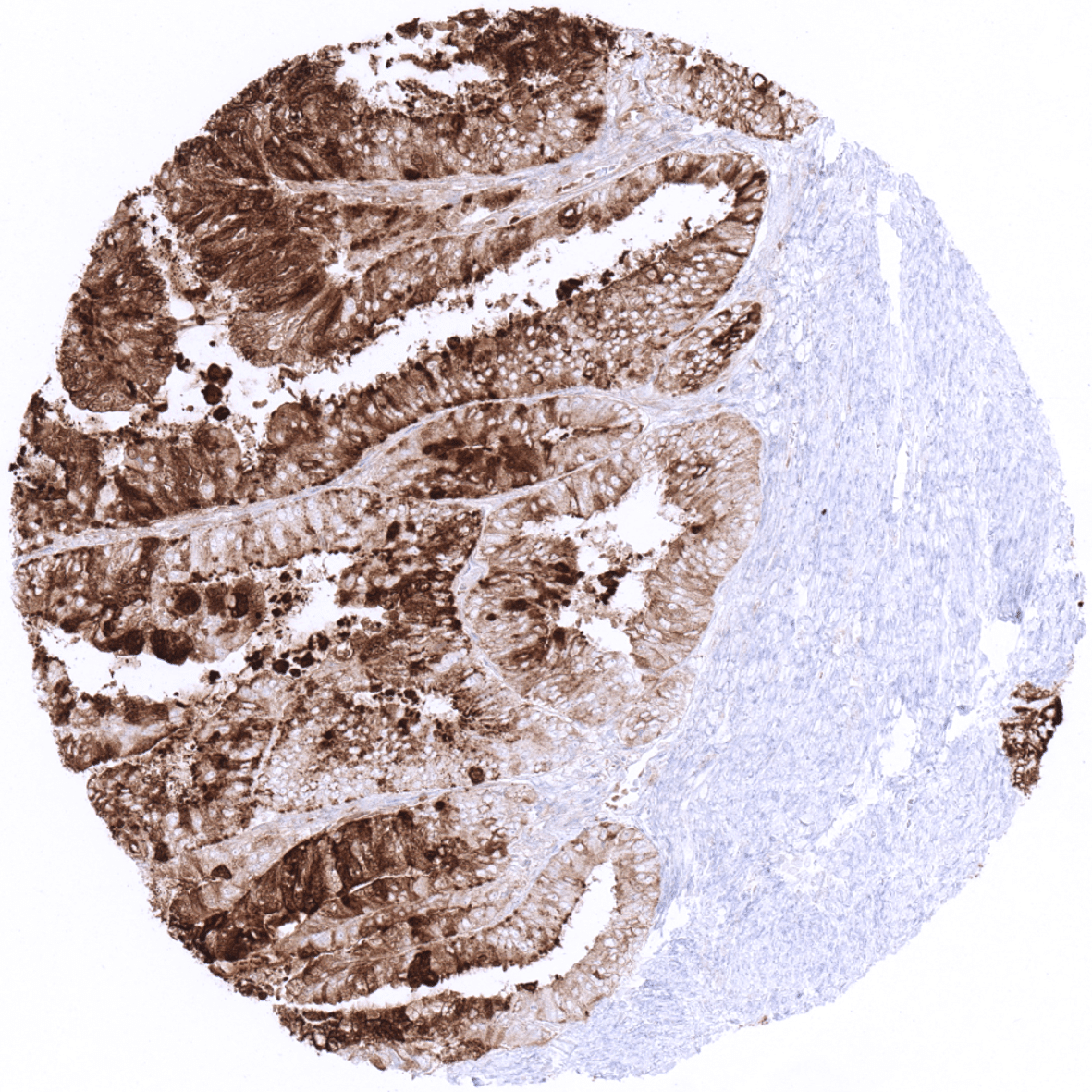
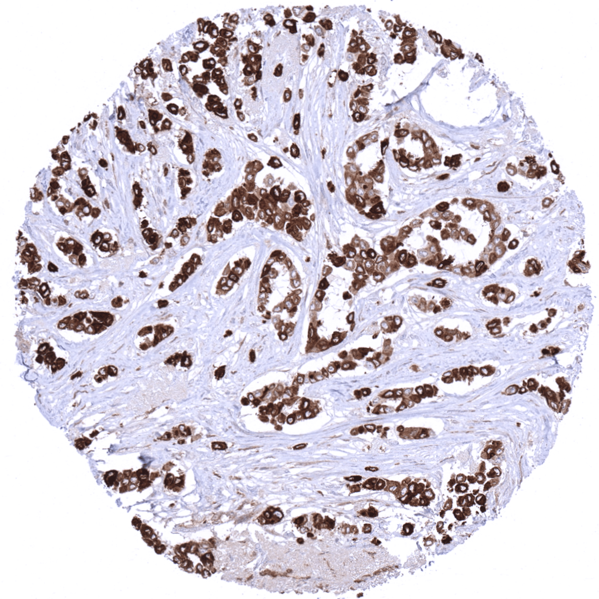
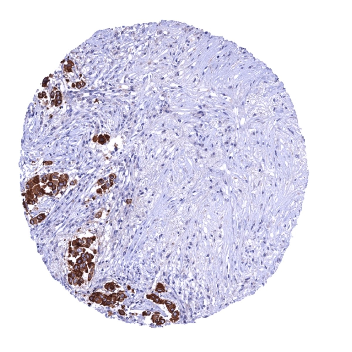
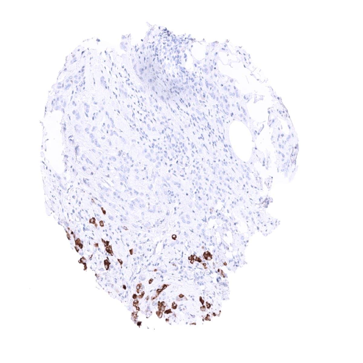
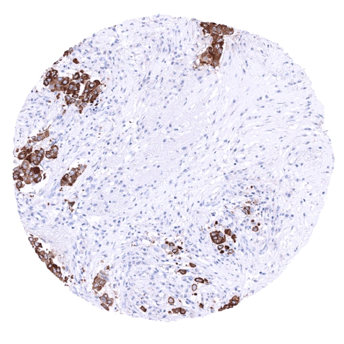
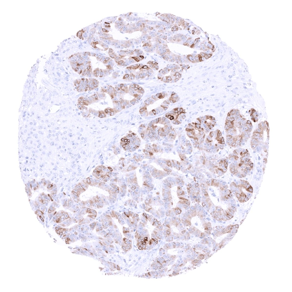
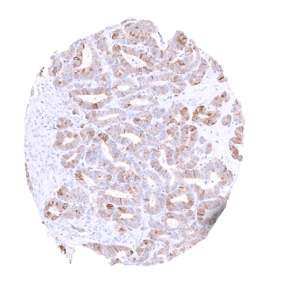
The mammaglobin protein is detectable in only a few normal tissue types including luminal cells of the breast (not always in all cells; + to +++), endocervical glands (mostly +++, but not all glands in all patients), endometrium (not always in all cells; + to +++), scattered epithelial cells in the fallopian tube (++), principal cells of the epididymis (+ to ++), and few scattered cells in salivary glands (+ – ++). Mammaglobin staining is most intense in endocervical and endometrial glands. In these tissues, the stroma cells are sometimes also positive (perhaps representing a diffusion artifact).
These findings are largely consistent with RNA and protein data described in the Human Protein Atlas (Tissue expression Mammaglobin). The protein atlas data are expanded by demonstration of scattered positive cells in the fallopian tube and in salivary glands. The latter is visible in one image depicted in the Human Protein Atlas but was not described.
Suggested positive tissue control: Cervix uteri: A moderate to strong cytoplasmic staining of the endocervical glands should be seen while the squamous epithelium of the ectocervix remains negative.
Suggested negative tissue control: Colon: All cells should not show any staining.
Staining Pattern in Relevant Tumor Types
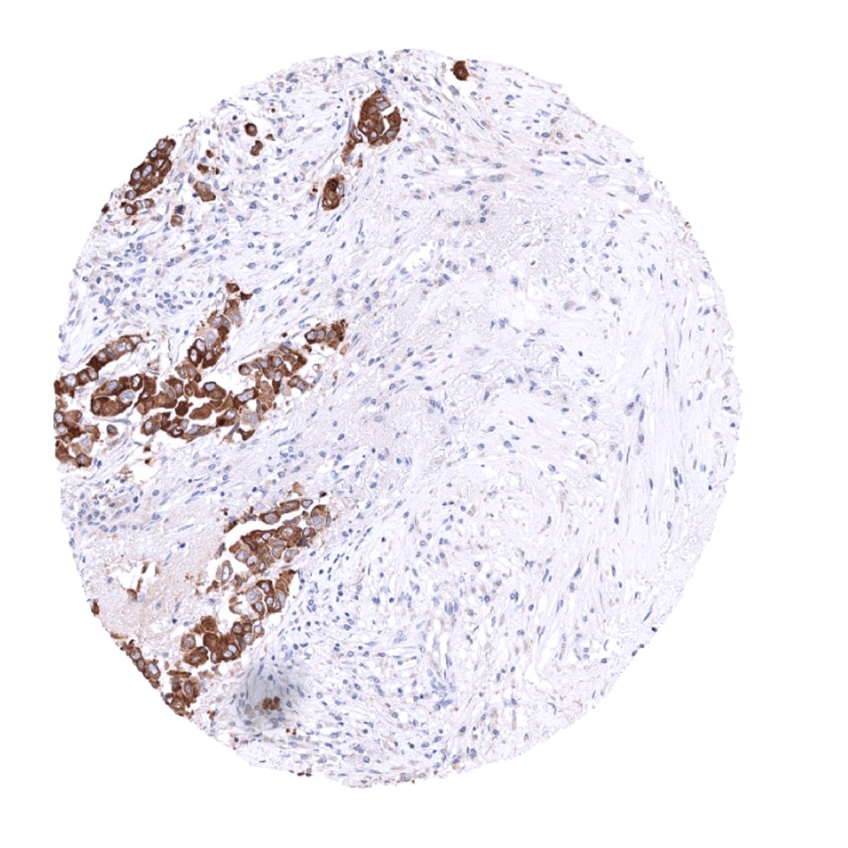
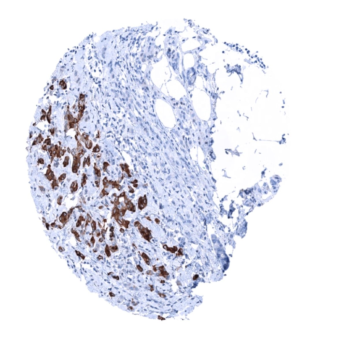
Mammaglobin occurs in about 80% of breast carcinomas. Among non–breast carcinomas, convincing mammaglobin expression is seen in endometrioid carcinomas (~40% cases) and sweat and salivary gland tumors. Up to 15% of other non breast carcinomas (such as stomach, lung, colon, hepatobiliary, thyroid, ovarian, and urothelial carcinomas) have been reported positive, but expression was usually only focally.
The TCGA findings on Mammaglobin RNA expression in different tumor categories have been summarized in the Human Protein Atlas.
Compatibility of Antibodies
Mammaglobin-A (MSVA-457R) publication summary
Relevant publication: Gorbokon et al. “Mammaglobin-A Expression Is Highly Specific for Tumors Derived from the Breast, the Female Genital Tract, and the Salivary Gland” Published in Diagnostics (Basel). 2023 Mar 22;13(6):1202. doi: 10.3390/diagnostics13061202. PMID: 36980510
A total of 14,232 tumors from 128 different tumor categories were successfully analyzed by using the following protocol: Heat-induced antigen retrieval for 5 minutes in an autoclave at 121°C in pH 9,0 Target Retrieval Solution buffer. MSVA-457R, at a dilution of 1:150 at 37°C for 60 minutes. Visualization of bound antibody by the EnVision Kit (Dako, Agilent). This protocol was also used for all stainings depicted in our tumor and normal tissue galleries.
Overall, 37 of 128 tumor categories showed detectable mammaglobin-A staining with 26 tumor categories showing at least one strongly positive case. 1437 of 1450 (99%) mammaglobin-A-positive tumors were derived from four organs, including the salivary glands, breast, endometrium, and the ovary. The distribution of positive staining results is shown in an “organ-systematic” (Figure 1) and in a “ranking order” figure (Figure 2) below (images based on data from Gorbokon et al). Data on associations with histopathological and clinical parameters are also summarized below (Figure 3; based on data described by Gorbokon et al).
Authors conclusions on diagnostic utility of Mammaglobin-A IHC with respect to the distinction of different tumor entities (Gorbokon et al):
- Mammaglobin-A immunostaining is a highly specific marker for tumors derived from either the breast, female genitals, or the salivary gland.
Authors conclusions on the prognostic role of Mammaglobin-A immunostaining results (Gorbokon et al):
- low mammaglobin-A staining was linked to high BRE grade (p=0.0011), loss of estrogen and progesterone receptor expression (p<0.0001 each), and triple-negative status (p<0.0001) in breast cancer NST.
- low mammaglobin-A staining was linked to an advanced tumor stage (p=0.0198) in endometrioid endometrium cancer.
Data from the publication: Gorbokon et al. “Mammaglobin-A Expression Is Highly Specific for Tumors Derived from the Breast, the Female Genital Tract, and the Salivary Gland” Published in Diagnostics (Basel). 2023 Mar 22;13(6):1202. doi: 10.3390/diagnostics13061202. PMID: 36980510
Summarized in own graphics:
Figure1. Mammaglobin staining in tumors (“organ-specific ; according to Gorbokon et al.”) with antibody MSVA-457R

Figure 2. Mammaglobin staining in tumors (“ranking-order ; according to Gorbokon et al.”) by positivity with antibody MSVA-457R.

Protocol Recommendations
IHC users have different preferences on how the stains should look like. Some prefer high staining intensity of the target stain and even accept some background. Others favor absolute specificity and lighter target stains. Factors that invariably lead to more intense staining include higher concentration of the antibody and visualization tools, longer incubation time, higher temperature during incubation, higher temperature and longer duration of the heat induced epitope retrieval (slide pretreatment). The impact of the pH during slide pretreatment has variable effects and depends on the antibody and the target protein. Accordingly, multiple different protocols can generate identical staining results.
All images and data shown here and in in our image galleries are obtained by the manual protocol described below. Other protocols resulting in equivalent staining are described as well.
Manual protocol
Freshly cut sections should be used (less than 10 days between cutting and staining). Heat-induced antigen retrieval for 5 minutes in an autoclave at 121°C in pH 9. Apply MSVA-457R at a dilution of 1:150 at 37°C for 60 minutes. Visualization of bound antibody by the EnVision Kit (Dako, Agilent) according to the manufacturer’s directions.

Agilent / Dako – Autostainer Link 48
Pretreatment in PT-Link for 30 minutes at 95°C (pH high); FLEX peroxidase blocking for 5 minutes (room temperature), MSVA-457R 1:150 for 20 minutes (room temperature), FLEX+ mouse/rabbit (LINKER) for 15 minutes (room temperature), horseradish peroxidase (HRP) for 20 minutes (room temperature), FLEX DAB+Sub-Chromo for 10 minutes (room temperature), FLEX hematoxylin for 5 minutes (room temperature).
These images reflect stainings by the protocol described above. It is of note that a comparable staining result can also be obtained by different protocols. In general, a longer pretreatment, a longer incubation time of the primary antibody, a higher antibody concentration, and a longer incubation time of FLEX+LINKER result in stronger staining, potentially at the cost of more background staining. Modifications of the protocol with a strengthening effect on staining intensity in combination with changes of other parameters that result in lower staining intensity can result in a comparable result as shown above.
Leica – BOND RX
Dewax at 72°C for 30 seconds; Pretreatment in Bond Epitope Retrieval Solution (ER2 – EDTA pH9) for 20 minutes at 100°C; Peroxidase blocking for 5 minutes (room temperature), MSVA-457R 1:150 for 15 minutes (room temperature), Post primary (rabbit anti mouse) for 8 minutes (room temperature), Polymer (goat anti rabbit) for 8 minutes (room temperature), mixed DAB refine for 10 minutes (room temperature), hematoxylin for 5 minutes (room temperature).

These images reflect stainings by the protocol described above. It is of note that a comparable staining result can also be obtained by different protocols. In general, a longer pretreatment, a longer incubation time of the primary antibody, a higher antibody concentration, a higher temperature during incubation, and a longer incubation time of Post primary and or the Polymer result in stronger staining, potentially at the cost of more background staining. Modifications of the protocol with a strengthening effect on staining intensity in combination with changes of other parameters that result in lower staining intensity can result in a comparable result as shown above.
Roche – Ventana Discovery ULTRA
Pretreatment for 64 minutes at 100°C (pH 8,4); CM peroxidase blocking for 12 minutes (room temperature), MSVA-457R 1:150 for 20 minutes at 36°C, secondary antibody (anti-rabbit HQ) for 12 minutes at 36°C, anti-HQ HRP for 12 minutes at room temperature, DAB at room temperature, hematoxylin II at room temperature for 8 minutes, bluing reagent at room temperature for 4 minutes.

These images depict staining results obtained by the protocol described above. It is of note, that the Ventana machines generally require higher antibody concentrations than other commonly used autostainers because the antibodies are automatically diluted during the procedure. Various other protocols can result in an identical result as shown above. A longer pretreatment, a longer incubation time of the primary antibody, a higher antibody concentration, a higher temperature during incubation, and a longer incubation time of secondary antibody and or the anti-HQ HRP result in stronger staining, potentially at the cost of more background staining.
Impact of pH


The strongest Mammaglobin staining by MSVA-457R is obtained at a pH 9,0. However, pH 7,8 results in only a slight reduction of the staining intensity as compared to pH9. We thus consider pH7,8 as optimal for manual staining because of the better tissue preservation at pH7,8 than at pH 9,0.
Potential Research Applications
- A comprehensive study analyzing mammaglobin-A in various different tumor entities would be helpful to better assess the diagnostic significance of mammaglobin IHC.
- The clinical significance of mammaglobin-A expression levels in breast cancer and other tumor types is not clear yet.
- The diagnostic accuracy of mammaglobin-A IHC in combination with other antibodies such as GATA3 and gcdfp-15 awaits further evaluation.
- The functional role of mammaglobin in normal and neoplastic cells is unknown.
Evidence for Antibody Specificity in IHC
Specificity and sensitivity of MSVA-457R is documented by strong positive staining in cell types that are well documented to express Mammaglobin such as breast, endometrium and endocervical glands in combination with the complete absence of staining in tissues that are not expected to express the protein (see normal tissue gallery). Immunostaining was also absent in tissues notorious for non-specific IHC background such as kidney, colonic mucosa, and epidermis.
