

295,00 € – 995,00 €
Product details
Synonyms = CK20; Cytokeratin-20; K20; KA20; Keratin 20; keratin 20, type I; Keratin type I cytoskeletal 20; Keratin-20; KRT20
Antibody type = Recombinant Rabbit monoclonal / IgG
Clone = MSVA-620R
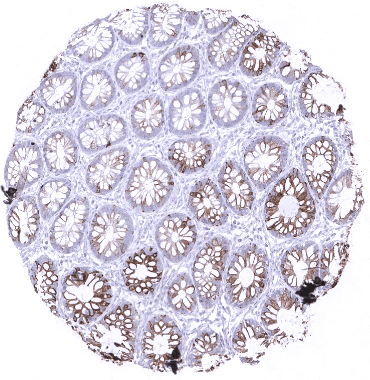
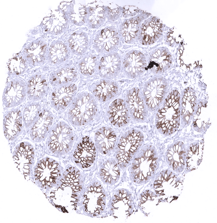
Positive control = Colon mucosa: Staining should be strong at the surface and at least moderate at the basis of krypts.
Negative control = Tonsil: All cells must stain negative.
Cellular localization = Cytoplasmic
Reactivity = Human
Application = Immunohistochemistry
Dilution = 1:100 – 1:200
Intended Use = Research Use Only
Relevance of Antibody
Biology Behind
Cytokeratin 20 (CK20) also termed keratin 20 (KRT20) is an acidic low molecular weight Type I cytokeratin encoded by the KRT20 gene located at 17q21.2. It forms intermediate filaments that primarily shape the cytoskeleton of specific epithelial cells, mainly of the gastrointestinal tract. CK20 expression is often retained in carcinomas derived from the gastrointestinal tract. CK20 immunohistochemistry can thus be used for defining the origin of cancer tissues. Because of a somewhat complementary staining pattern with CK7, CK20 IHC is often used together with CK7.
Staining Pattern in Normal Tissues
Cytokeratin 20 staining pattern in Normal Tissues with antibody MSVA-620R (images are shown in our “Normal Tissue Gallery”)
| Brain | Cerebrum | Negative. |
| Cerebellum | Negative. | |
| Endocrine Tissues | Thyroid | Negative. |
| Parathyroid | Negative. | |
| Adrenal gland | Negative. | |
| Pituitary gland | Negative. | |
| Respiratory system | Respiratory epithelium | Negative. |
| Lung | Negative. | |
| Gastrointestinal Tract | Salivary glands | Negative. |
| Esophagus | Negative. | |
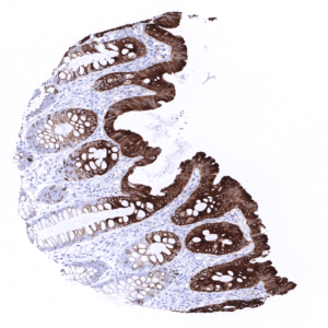
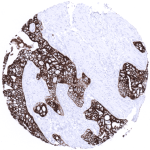
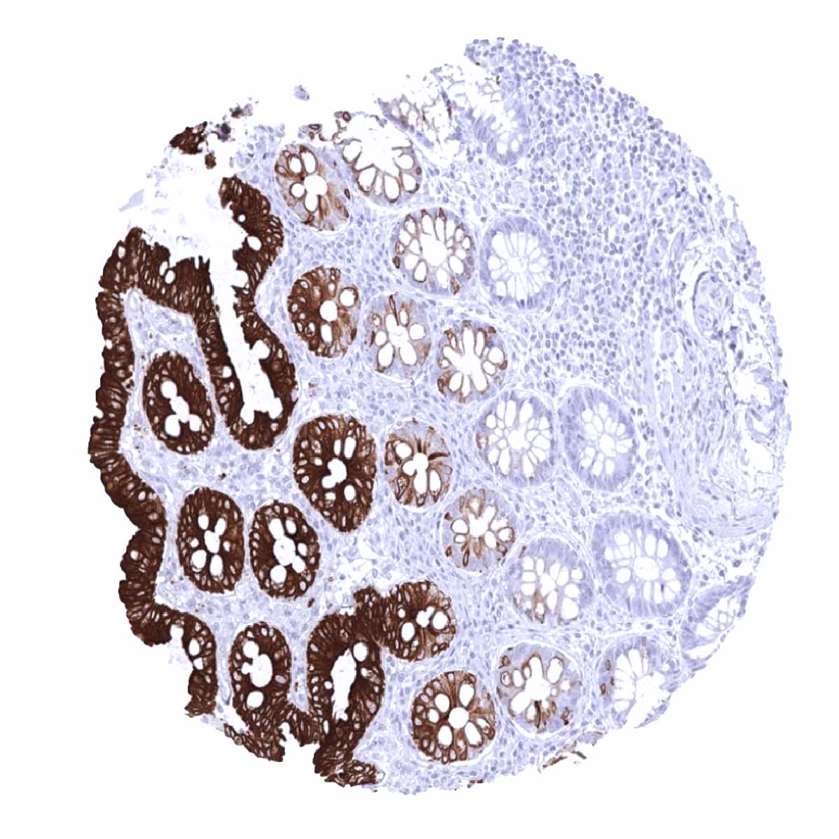
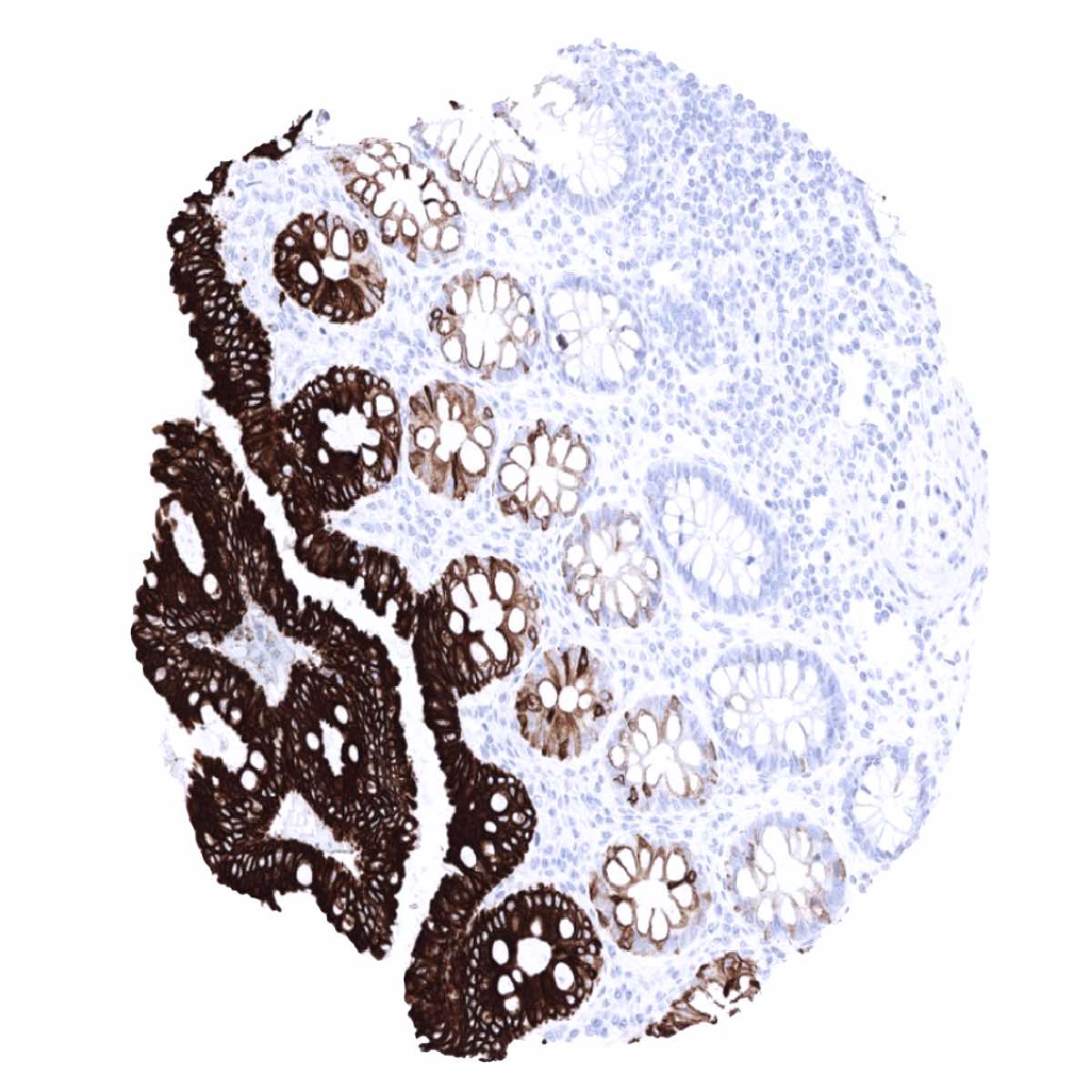
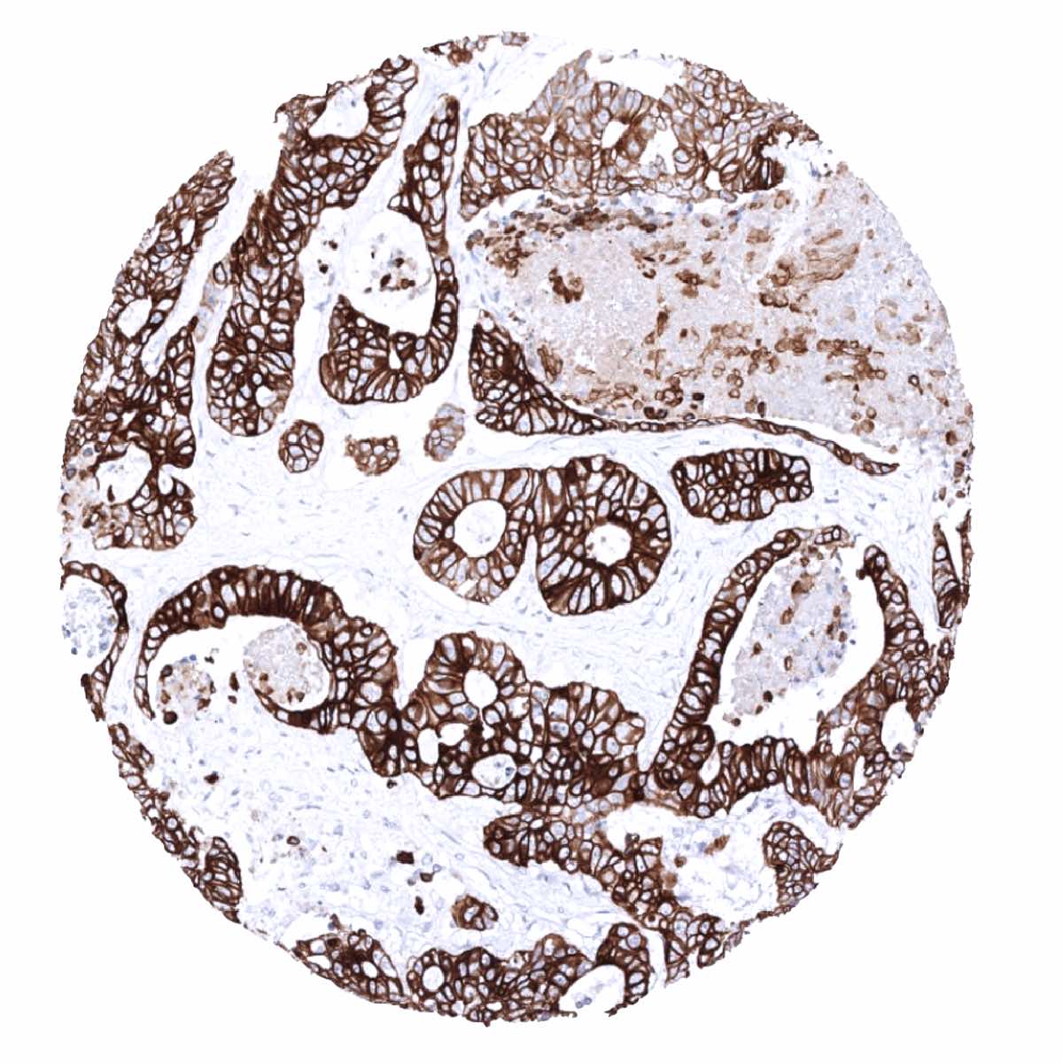
| Stomach | Strong CK20 positivity in surface epithelial cells.
CK20 staining is absent in the deep glands. |
|
| Duodenum | Strong CK20 staining of surface epithelial cells. The fraction of positive cells and the staining intensity decreases towards the base of glands/crypts. | |
| Small intestine | Strong CK20 staining of surface epithelial cells. The fraction of positive cells and the staining intensity decreases towards the base of glands/crypts. | |
| Appendix | Strong CK20 staining of surface epithelial cells. The fraction of positive cells and the staining intensity decreases towards the base of crypts. | |
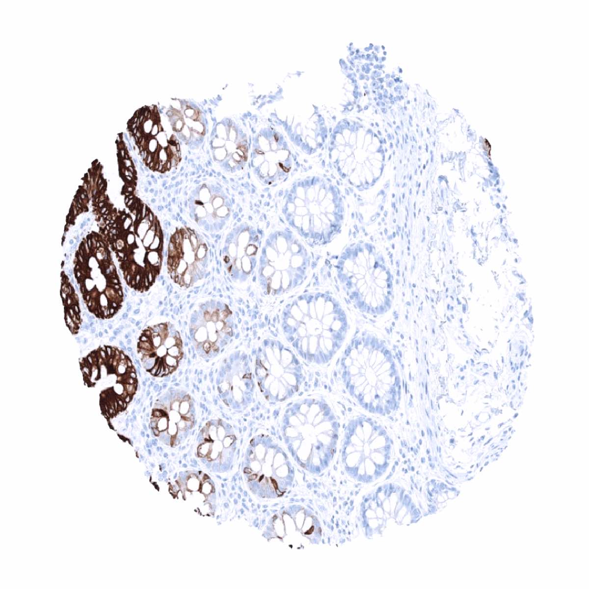
| Colon | Strong CK20 staining of surface epithelial cells. The fraction of positive cells and the staining intensity decreases towards the base of crypts. | |
| Rectum | Strong CK20 staining of surface epithelial cells. The fraction of positive cells and the staining intensity decreases towards the base of crypts. | |
| Liver | Negative. | |
| Gallbladder | Negative. | |
| Pancreas | Negative. | |
| Genitourinary | Kidney | Negative. |
| Urothelium | Strong CK20 staining of umbrella cells. | |
| Male genital | Prostate | Negative. |
| Seminal vesicles | Negative. | |
| Testis | Negative. | |
| Epididymis | Negative. | |
| Female genital | Breast | Negative. |
| Uterus, myometrium | Negative. | |
| Uterus, ectocervix | Negative. | |
| Uterus endocervix | Negative. | |
| Uterus, endometrium | Negative. | |
| Fallopian Tube | Negative. | |
| Ovary | Negative. | |
| Placenta early | Negative. | |
| Placenta mature | Negative. | |
| Amnion | Negative. | |
| Chorion | Negative. | |
| Skin | Epidermis | Negative. |
| Sebaceous glands | Negative. | |
| Muscle/connective tissue | Heart muscle | Negative. |
| Skeletal muscle | Negative. | |
| Smooth muscle | Negative. | |
| Vessel walls | Negative. | |
| Fat | Negative. | |
| Stroma | Negative. | |
| Endothelium | Negative. | |
| Bone marrow/ lymphoid tissue | Bone marrow | Negative. |
| Lymph node | Negative. | |
| Spleen | Negative. | |
| Thymus | Negative. | |
| Tonsil | Negative. | |
| Remarks | CK20 staining is largely limited to gastrointestinal epithelium and surface epithelial cells of the urothelium. |
These findings are largely consistent with the RNA and protein data described in the Human Protein Atlas (Tissue expression Cytokeratin 20)
Suggested positive tissue control: Colon mucosa: Staining should be strong at the surface and at least moderate at the basis of krypts.
Suggested negative tissue control: Tonsil: All cells must stain negative.

Staining Pattern in Relevant Tumor Types
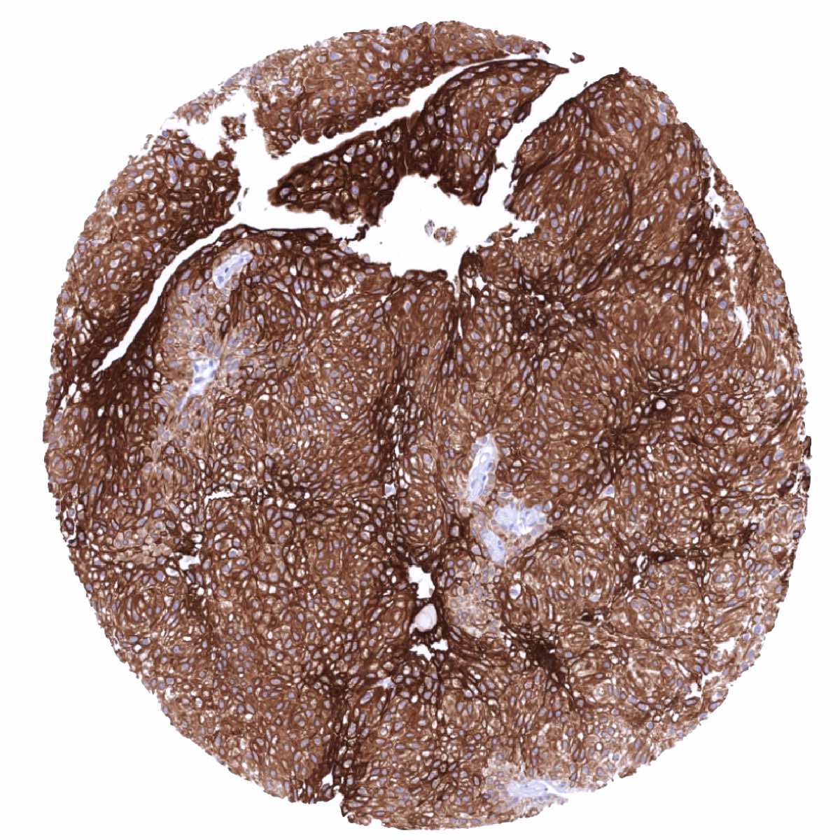
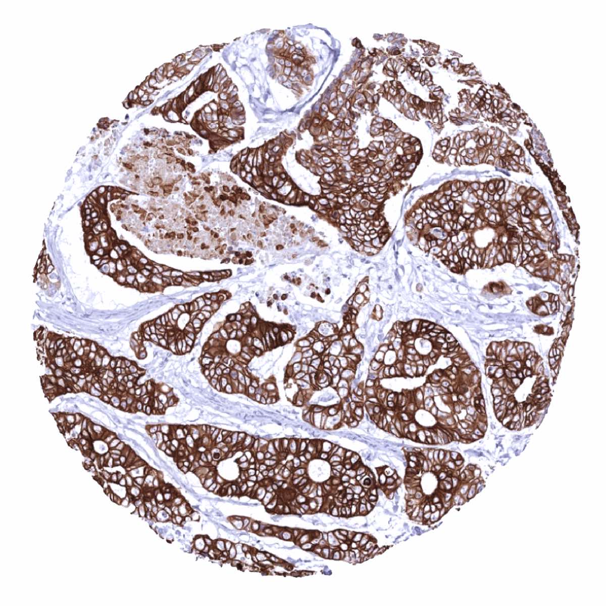
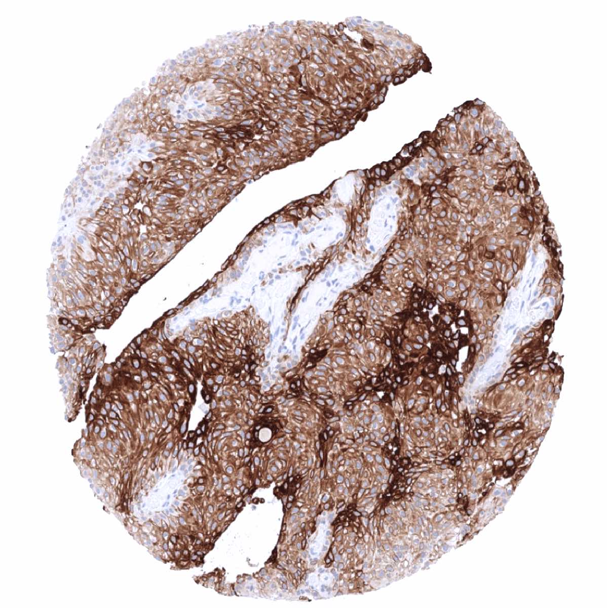
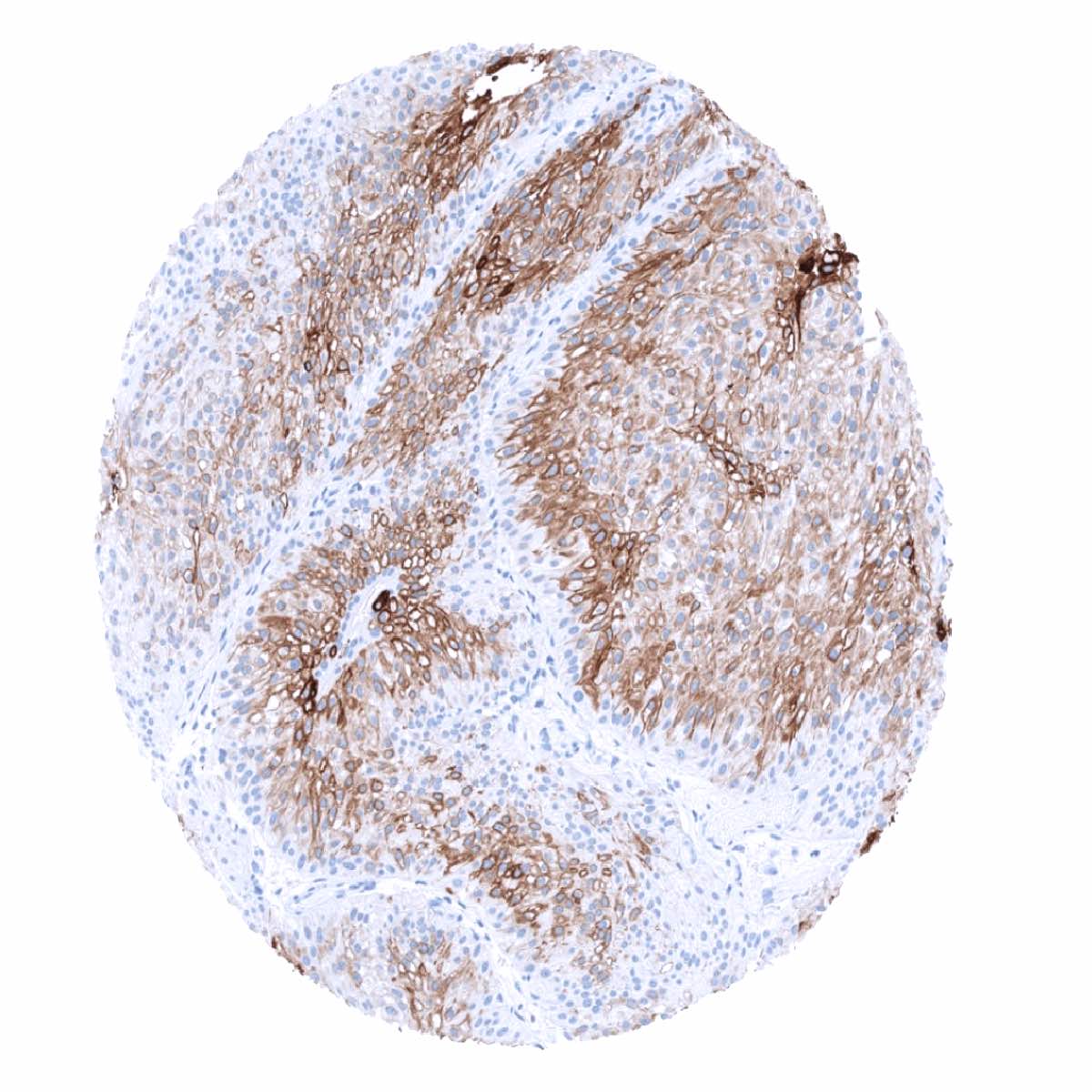
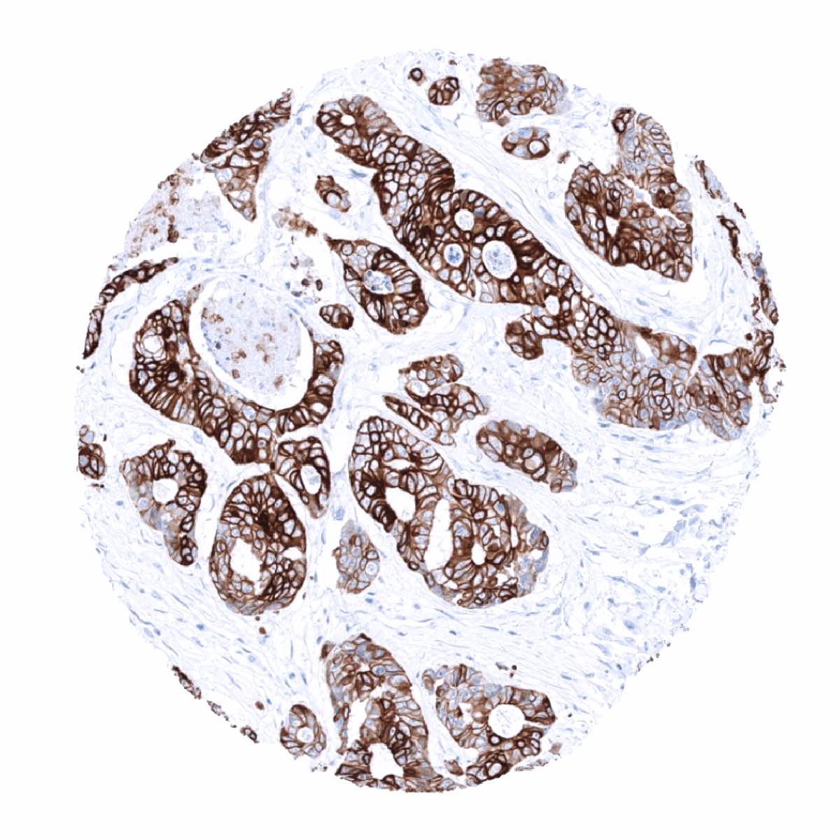
CK20 is commonly expressed in colorectal adenocarcinoma (95%), Merkel cell carcinoma, and mucinous ovarian cancer (60-70%) In Merkel cell carcinoma a characteristic dot like staining pattern is typically seen. In urothelial carcinoma, CK20 staining is more commonly seen in dysplasia, carcinoma in situ and non-invasive papillary cancer than in muscle invasive disease. CK20 staining occurs in 20-40% of adenocarcinomas of stomach, pancreas, and esophagus. At lower frequency CK20 expression can be seen in various other tumors especially adenocarcinomas.
The TCGA findings on Cytokeratin 20 RNA expression in different tumor categories have been summarized in the Human Protein Atlas.

Compatibility of Antibodies
Cytokeratin 20 (MSVA-620R) publication summary
Relevant publication: Dum et al.: “Cytokeratin 7 and cytokeratin 20 expression in cancer: A tissue microarray study on 15,424 cancers” Exp Mol Pathol 2022 Apr 4; 126:104762 Online ahead of print
A total of 14’110 tumors from 120 different tumor categories were successfully analyzed for CK 20 by using the following protocol: Heat-induced antigen retrieval for 5 minutes in an autoclave at 121°C in pH 9 Target Retrieval Solution buffer. MSVA-620R at a dilution of 1:150 at 37°C for 60 minutes. Visualization of bound antibody by the EnVision Kit (Dako, Agilent). This protocol was also used for all CK20 immunostainings depicted in our tumor and normal tissue galleries.
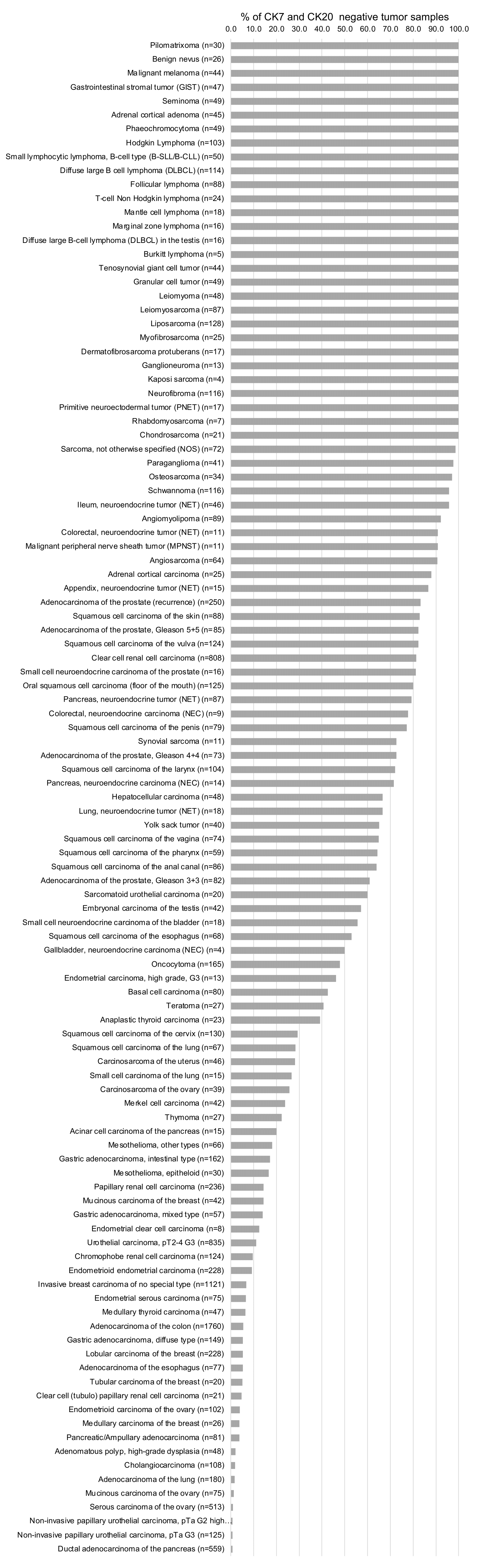
At least one case with a positive cytokeratin 20 staining was seen in 43 (35.8%) and at least one case with a strong cytokeratin 20 positivity was seen in 31 (25.8%) of 120 tumor categories. The distribution of positive staining results is shown in an “organ-systematic” (Figure 1) and in a “ranking order” figure (Figure 2) below (images based on data from Dum et al). Data on associations with histopathological and clinical parameters of tumor aggressiveness in several cancer types are also summarized below (Figure 3; based on data described by Dum et al).
Because CK20 is often used in combination with CK7, additional figures show ranking orders of tumors according to their CK20 positivity rate among CK7 negative (Figure 4) and among CK7 positive tumors (Figure 5). For completeness, a figure showing a ranking order of tumor entities according to their rate of CK7 and CK20 double-negativity is also given (Figure 6).
Authors conclusions on diagnostic utility with respect to the distinction of benign versus malignant (Dum et al):
- A positive CK20 staining is a feature of malignant transformation in flat urothelium and argues for urothelial dysplasia (in case of mild or moderate dysplasia) or carcinoma in situ (in case of severe atypia).
Authors conclusions on diagnostic utility with respect to the distinction of different tumor entities (Dum et al):
- A significant CK20 immunostaining is most commonly seen in gastrointestinal adenocarcinoma (This mirrors the situation in normal tissues, where detectable CK20 expression is largely limited to gastrointestinal mucosa).
- Urothelial tumors, Merkel cell carcinomas, mucinous ovarian cancer, and pancreatic cancers represent further tumor groups with frequent and high level CK20 expression.
- That CK20 upregulation occurs – in a minor fraction of cases – in more than 20 additional tumor entities is likely to reflect the general property of tumor cells to – occasionally – develop aberrant expression of alternative cytokeratins or other intermediate filaments during tumor dedifferentiation
Authors conclusions on prognostic/predictive role of CK20 expression (Dum et al.):
- Increased levels of CK20 are linked to high grade in non-invasive urothelial cancers and to nodal metastasis in muscle-invasive urothelial cancer.
- Reduced CK20 expression is tightly linked to MSI in colorectal and gastric cancer but unrelated to prognostic features such as pT, pN, L- status.
Because CK20 is often used in combination with CK7, additional figures show ranking orders of tumors according to their CK20 positivity rate among CK7 negative (Figure 4) and among CK7 positive tumors (Figure 5). For completeness, a figure showing a ranking order of tumor entities according to their rate of CK7 and CK20 “double-negativity” is also given (Figure 6). Dum et al. summarize these findings as follows:
- CK20+/CK7- is rather rare and predominates in colorectal adenocarcinomas and Merkel cell carcinomas but also occurs at relevant frequency in other tumors, most of which are derived from the GIT.
- CK20+/CK7+ is most commonly seen in mucinous ovarian cancer (63%) and in various types of non-invasive and invasive urothelial carcinomas (>45%) but also occurs at significant frequency in several other – mostly gastrointestinal – tumors.
- CK20-/CK7+ predominates in non-mucinous ovarian, breast, thyroid, kidney, endometrium, and pancreatic cancer, adenocarcinoma of the lung, and in mesotheliomas but also occurs in >50% of non-colorectal GIT adenocarcinomas.
- CK20-/ CK7- is frequent in prostate cancer, seminoma, clear cell renal cell carcinoma, neuroendocrine tumors, adrenal tumors, squamous cell carcinomas as well as in mesenchymal and hematopoietic neoplasms.
Data from the publication: “Cytokeratin 7 and cytokeratin 20 expression in cancer: A tissue microarray study on 15,424 cancers”. Published by Dum et al. Summarized in own graphics:
Figure 1. CK 20 staining in cancer (“organ-systematic”; according to Dum et al.)
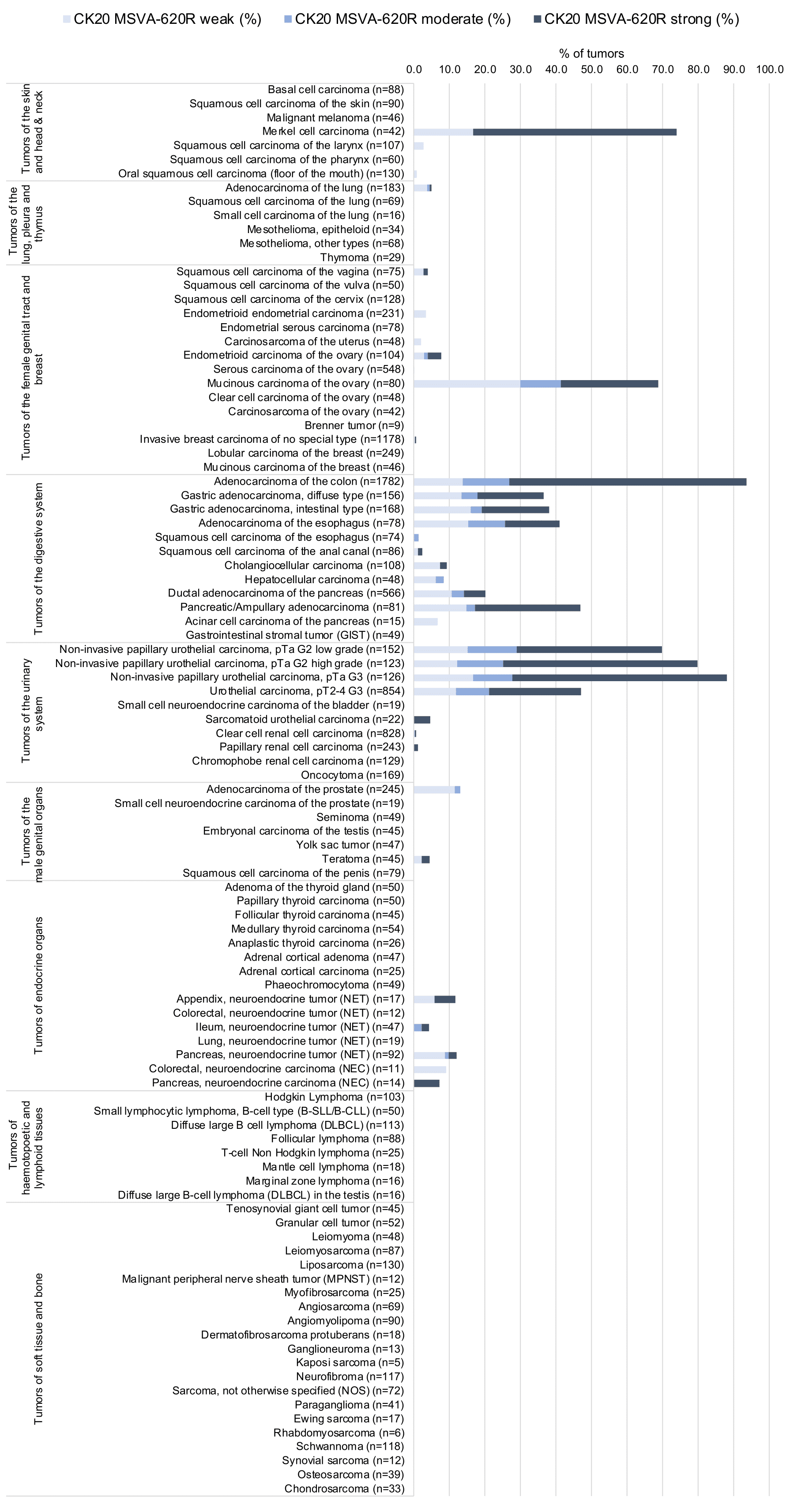
Figure 2. CK 20 staining in cancer (“ranking list”; according to Dum et al.)
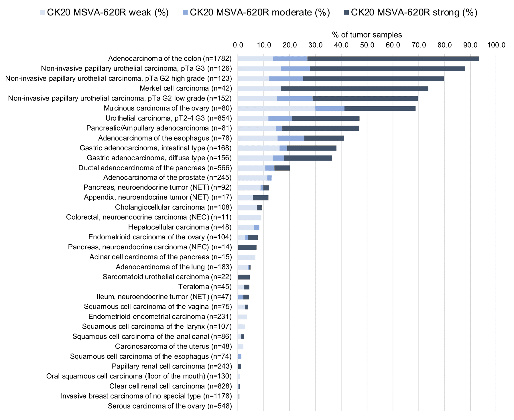
Figure 3. CK20 staining and tumor phenotype (according to Dum et al.)
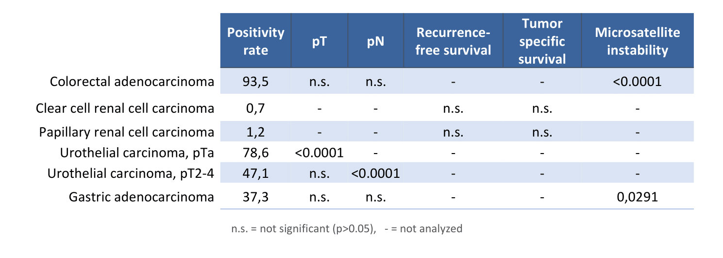
Figure 4. CK 20 staining in CK7 negative cancers (“ranking list”; according to Dum et al.)
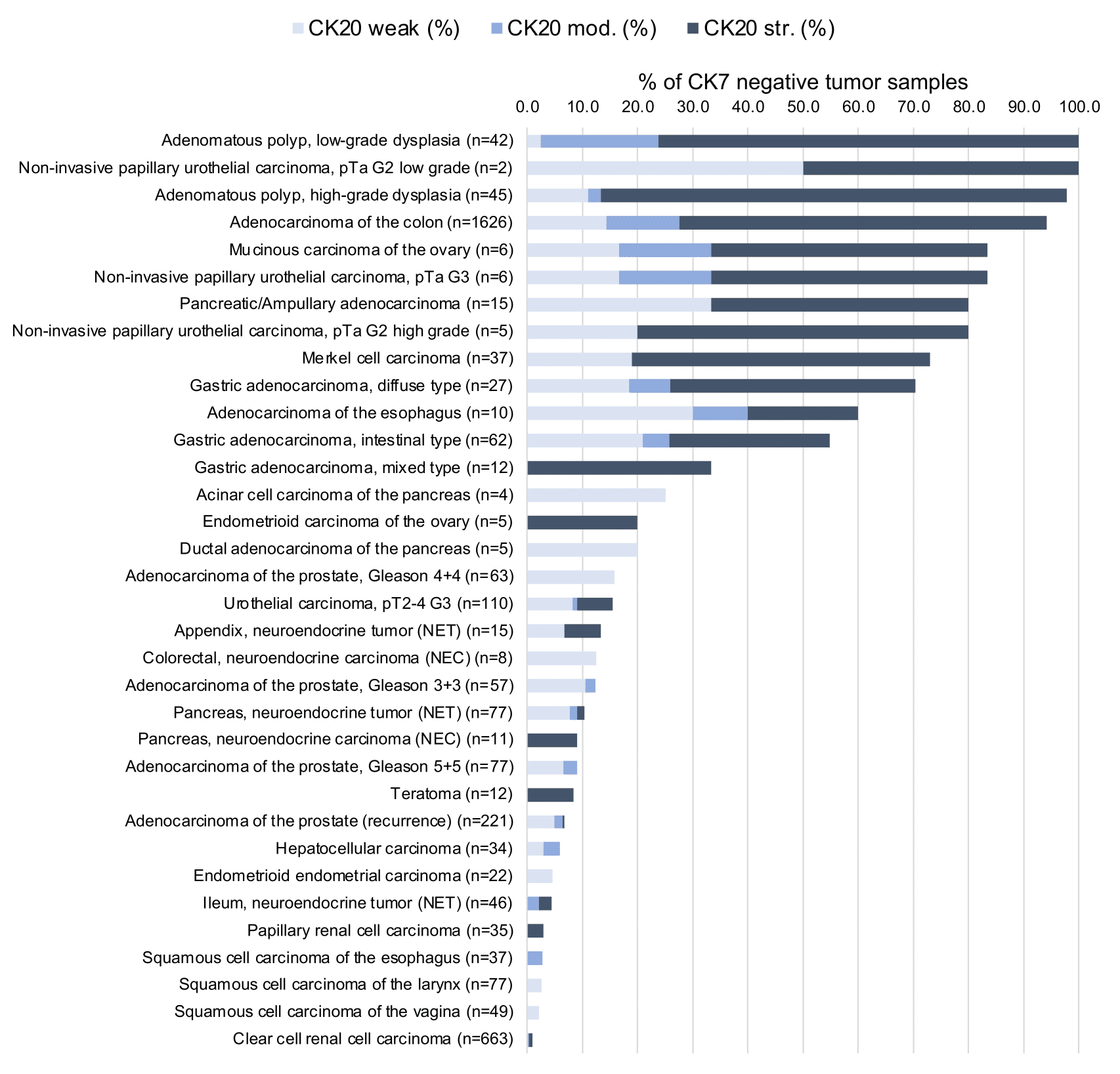
Figure 5. CK 20 staining in CK7 positive cancers (“ranking list”; according to Dum et al.)
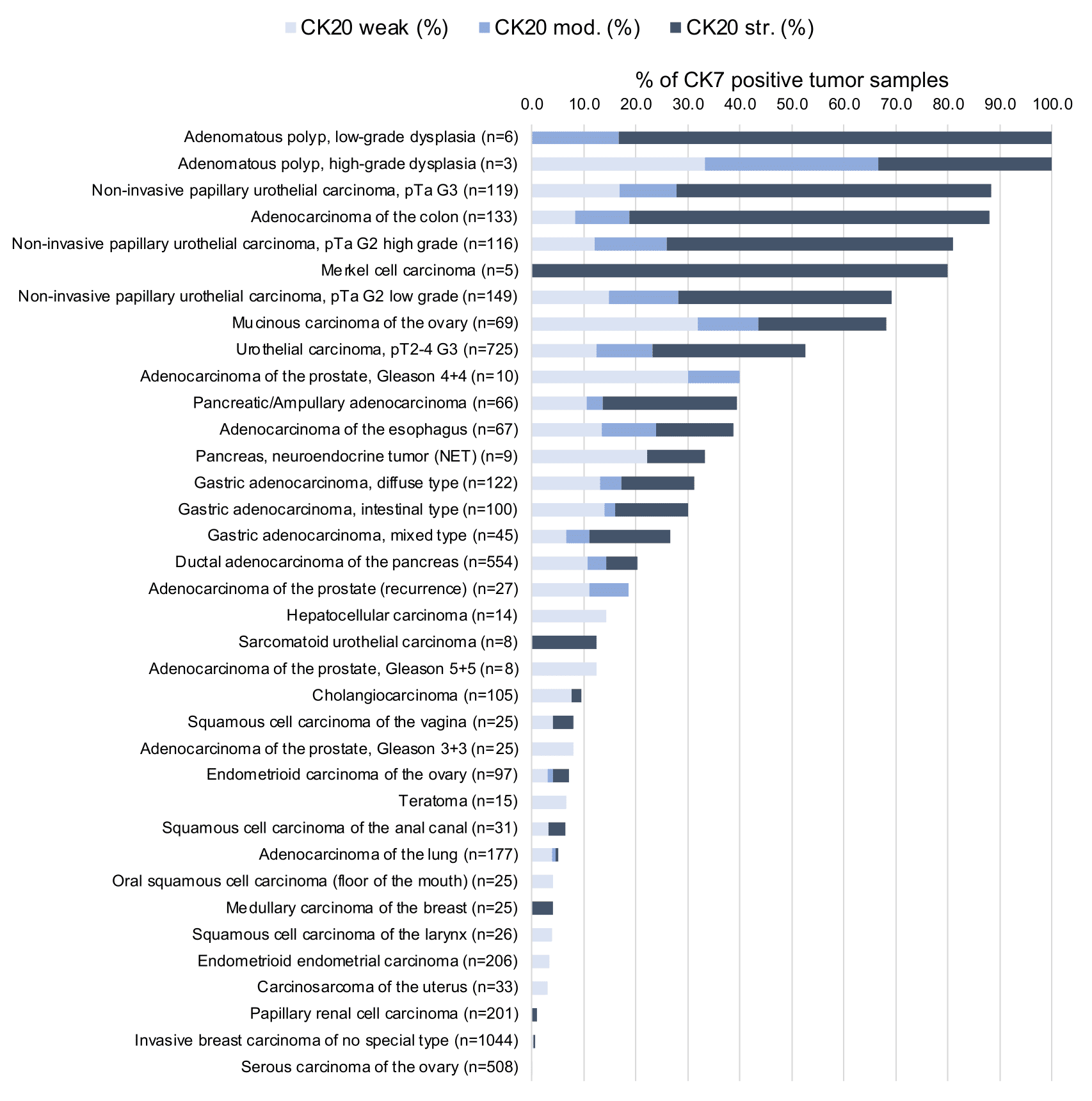
Figure 6. CK 20 negative and CK7 negative cancers (“ranking list”; according to Dum et al.)
Protocol Recommendations
IHC users have different preferences on how the stains should look like. Some prefer high staining intensity of the target stain and even accept some background. Others favor absolute specificity and lighter target stains. Factors that invariably lead to more intense staining include higher concentration of the antibody and visualization tools, longer incubation time, higher temperature during incubation, higher temperature and longer duration of the heat induced epitope retrieval (slide pretreatment). The impact of the pH during slide pretreatment has variable effects and depends on the antibody and the target protein.
All images and data shown here and in our image galleries are obtained by the manual protocol described below. Other protocols resulting in equivalent staining are described as well.
Manual protocol
Freshly cut sections should be used (less than 10 days between cutting and staining). Heat-induced antigen retrieval for 5 minutes in an autoclave at 121°C in pH 9 Target Retrieval Solution buffer. Apply MSVA-620R at a dilution of 1:150 at 37°C for 60 minutes. Visualization of bound antibody by the EnVision Kit (Dako, Agilent) according to the manufacturer’s directions.
Agilent / Dako – Autostainer Link 48
Pretreatment in PT-Link for 30 minutes at 95°C (pH high); FLEX peroxidase blocking for 5 minutes (room temperature), MSVA-620R 1:150 for 20 minutes (room temperature), FLEX+ mouse/rabbit (LINKER) for 15 minutes (room temperature), horseradish peroxidase (HRP) for 20 minutes (room temperature), FLEX DAB+Sub-Chromo for 10 minutes (room temperature), FLEX hematoxylin for 5 minutes (room temperature).


These images reflect stainings by the protocol described above. It is of note that a comparable staining result can also be obtained by different protocols. In general, a longer pretreatment, a longer incubation time of the primary antibody, a higher antibody concentration, and a longer incubation time of FLEX+LINKER result in stronger staining, potentially at the cost of more background staining. Modifications of the protocol with a strengthening effect on staining intensity in combination with changes of other parameters that result in lower staining intensity can result in a comparable result as shown above.
Leica – BOND RX
Dewax at 72°C for 30 seconds; Pretreatment in Bond Epitope Retrieval Solution (ER2 – EDTA pH9) for 20 minutes at 100°C; Peroxidase blocking for 5 minutes (room temperature), MSVA-620R 1:150 for 15 minutes (room temperature), Post primary (rabbit anti mouse) for 8 minutes (room temperature), Polymer (goat anti rabbit) for 8 minutes (room temperature), mixed DAB refine for 10 minutes (room temperature), hematoxylin for 5 minutes (room temperature).

These images reflect stainings by the protocol described above. It is of note that a comparable staining result can also be obtained by different protocols. In general, a longer pretreatment, a longer incubation time of the primary antibody, a higher antibody concentration, a higher temperature during incubation, and a longer incubation time of Post primary and or the Polymer result in stronger staining, potentially at the cost of more background staining. Modifications of the protocol with a strengthening effect on staining intensity in combination with changes of other parameters that result in lower staining intensity can result in a comparable result as shown above.
Roche – Ventana Discovery ULTRA
Pretreatment for 64 minutes at 100°C (pH 8,4); CM peroxidase blocking for 12 minutes (room temperature), MSVA-620R 1:150 for 20 minutes at 36°C, secondary antibody (anti-rabbit HQ) for 12 minutes at 36°C, anti-HQ HRP for 12 minutes at room temperature, DAB at room temperature, hematoxylin II at room temperature for 8 minutes, bluing reagent at room temperature for 4 minutes.
These images depict staining results obtained by the protocol described above. It is of note, that the Ventana machines generally require higher antibody concentrations than other commonly used autostainers because the antibodies are automatically diluted during the procedure. Various other protocols can result in an identical result as shown above. A longer pretreatment, a longer incubation time of the primary antibody, a higher antibody concentration, a higher temperature during incubation, and a longer incubation time of secondary antibody and or the anti-HQ HRP result in stronger staining, potentially at the cost of more background staining.
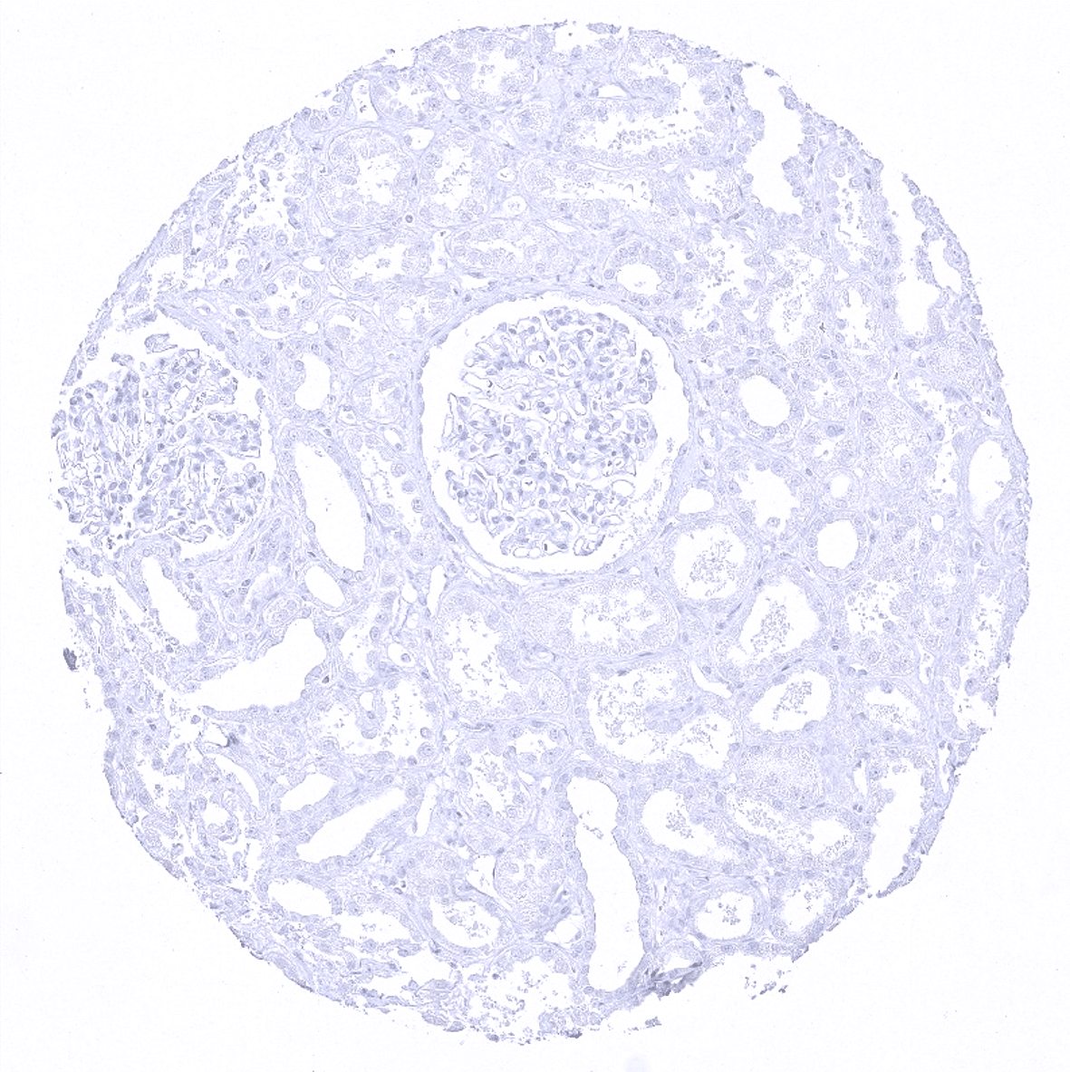
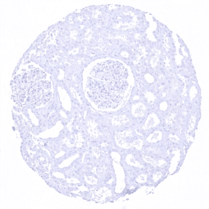
Impact of pH

MSVA-620R results in strongest staining if pH7,8 is used for slide pretreatment. pH9 is acceptable but lower pH results in a significant deterioration of sensitivity. An example of the impact of pH is shown below for normal kidney tissue.
Potential Research Applications
- As the literature is partly confusing, the diagnostic utility of KRT20 expression analysis (in combination with KRT7 analysis) should be investigated in a large cohort of tumors from different entities.
- The biologic/clinical significance of aberrant KRT20 expression in cancers should be evaluated (for example: what are the specific properties of KRT20 negative colorectal cancers or of CK20 positive prostate cancers?)
Evidence for Antibody Specificity in IHC
Specificity of MSVA-620R is documented by strong positive staining in cell types that are well documented to express CK20 such as surface epithelium of the stomach and colorectal mucosa and absence of staining in all tissues known to not express CK20 including tissues notorious for non-specific IHC background such as kidney and brain.
