195,00 € – 995,00 €
Product details
Synonyms = APY; ATOPY; B-lymphocyte cell-surface antigen B1; Bp35; Fc epsilon receptor I beta chain; Fc Fragment of IgE high affinity I receptor for beta polypeptide; FCER1B; High affinity immunoglobulin epsilon receptor subunit beta; IgE Fc receptor subunit beta; IGEL; IGER; IGHER; LEU16; Leukocyte surface antigen Leu-16; Ly44; MS4A1; MS4A2
Antibody type = Recombinant Rabbit monoclonal / IgG
Clone = MSVA-020R
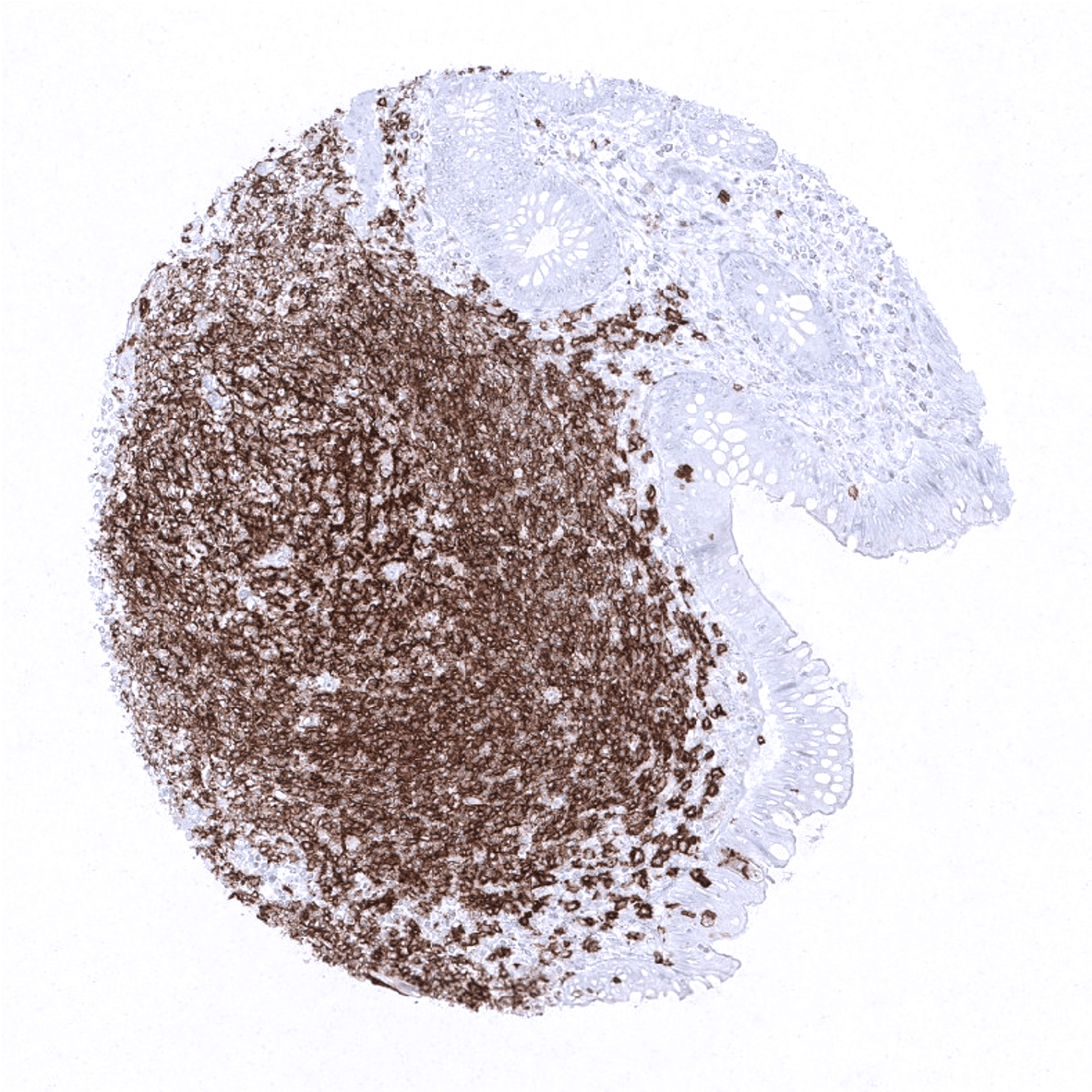
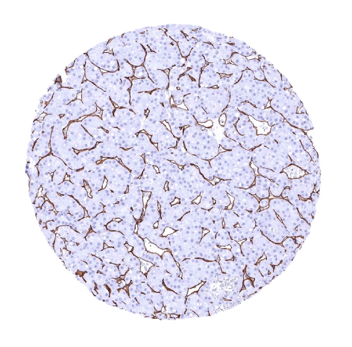
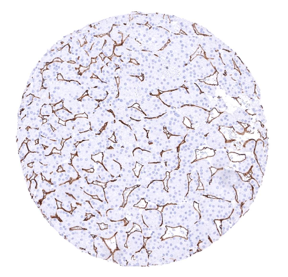
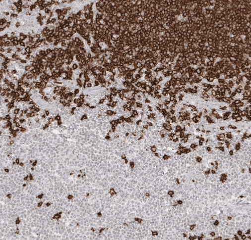
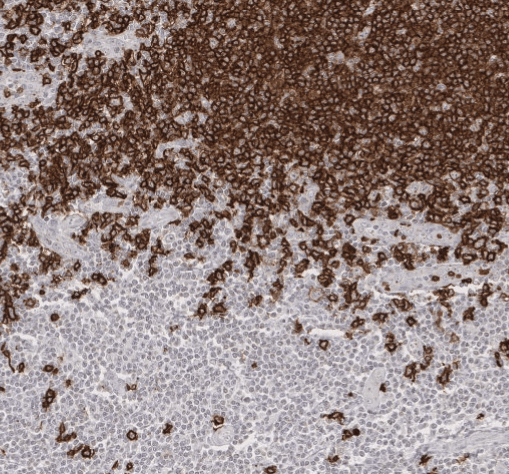
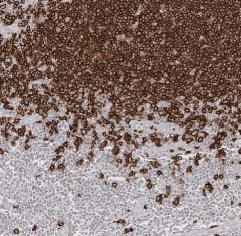
Positive control = Tonsil or appendix: A strong, predominantly membranous staining of the mantle zone B-cells, the germinal centre B- cells and the interfollicular B-cells is required, while all other cells should stain negative.
Negative control = Tonsil or appendix: A large fraction of the interfollicular lymphocytes and all epithelial cells must not show any staining.
Cellular localization = Cell Surface
Reactivity = Human
Application = Immunohistochemistry
Dilution = 1:100 – 1:200
Intended Use = Research Use Only
Relevance of Antibody
Biology Behind
The expression of the B-lymphocyte antigen CD20 starts between the time of light chain rearrangement and expression of intact surface immunoglobulin and increases progressively in concentration until B-cell maturity. CD20 is no longer expressed on plasmablasts and plasma cells. The protein has no known natural ligand and it is suspected to act as a calcium channel in the cell membrane. CD20 enables optimal B-cell immune response, specifically against T-independent antigens. CD20 is the target of several therapeutic monoclonal antibodies such as rituximab, obinutuzumab and others for the treatment of all B cell neoplasias and B cell-mediated autoimmune diseases.
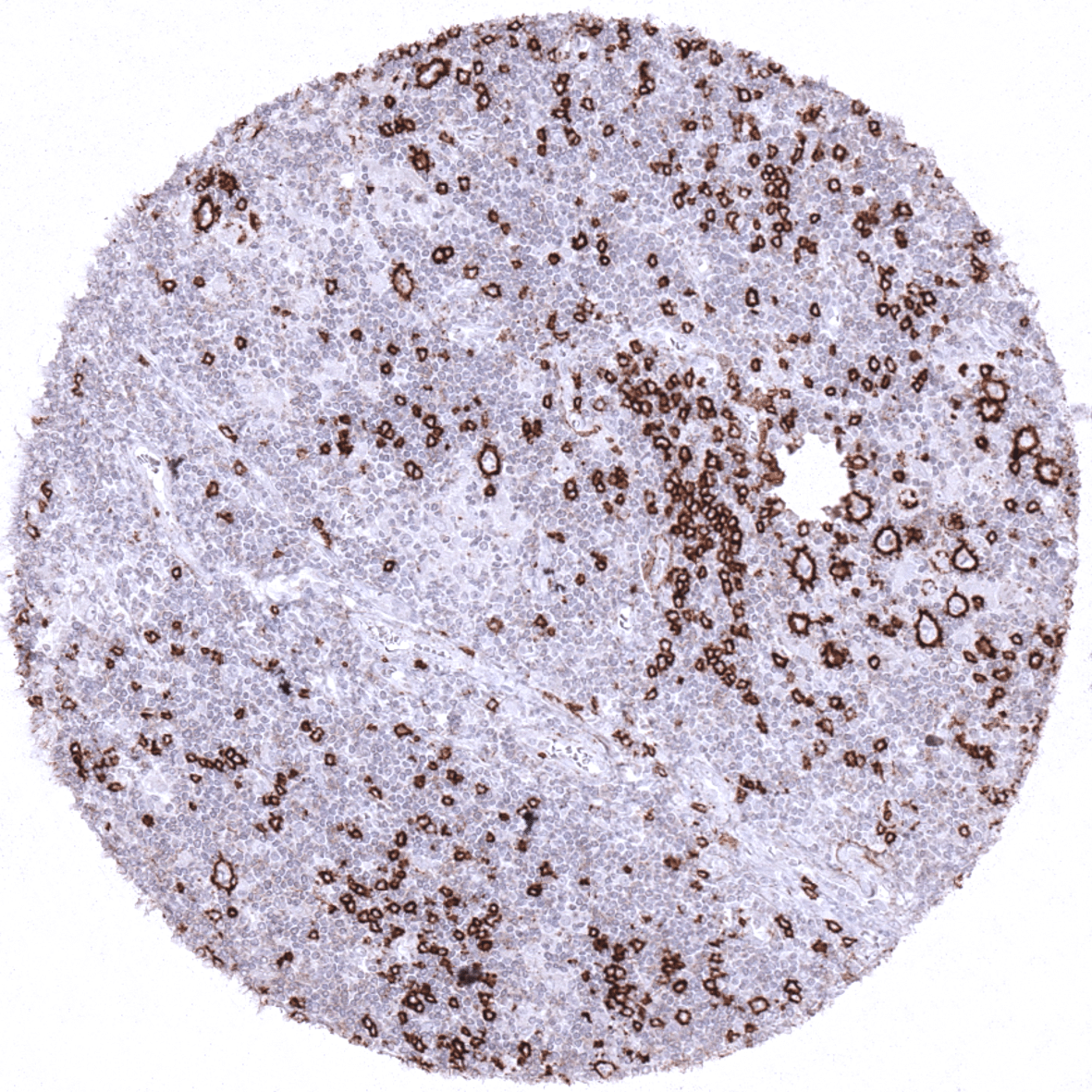
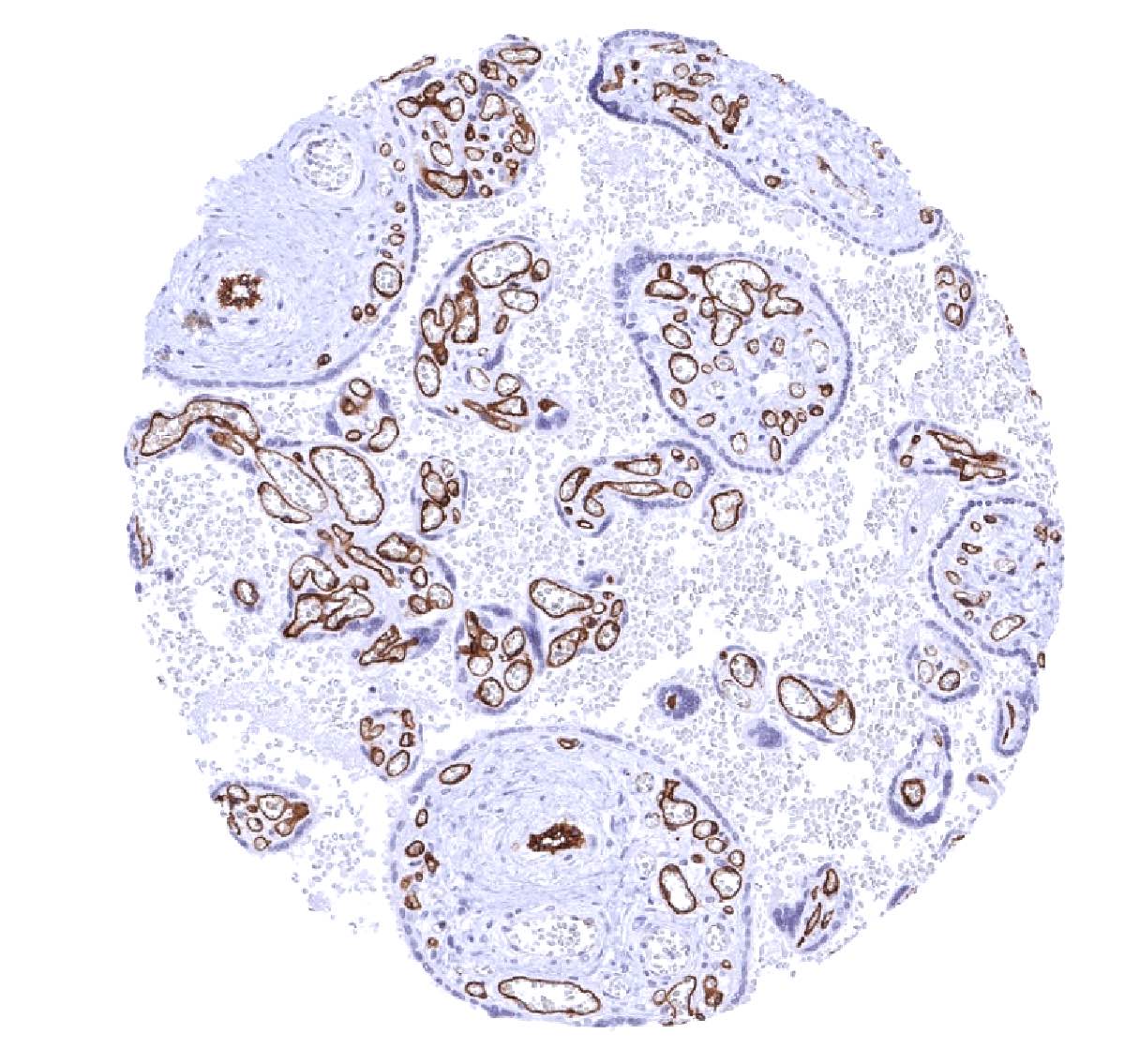
Staining Pattern in Normal Tissues
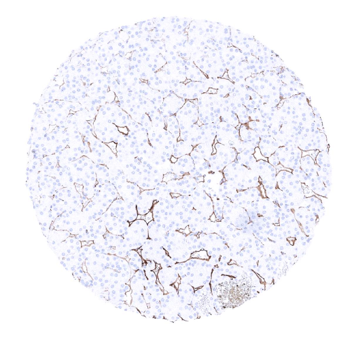
CD20 is expressed on the vast majority of B-lymphocytes (but not on plasma cells). CD20 positive cells are thus preferably seen in lymphatic organs especially in germinal follicles and mantle zones but some scattered positive lymphocytes can be found in virtually every organ.
These findings are largely consistent with the RNA and protein data described in the Human Protein Atlas (Tissue expression CD20)
Suggested positive tissue control: Tonsil or appendix: A strong, predominantly membranous staining of the mantle zone B-cells, the germinal centre B- cells and the interfollicular B-cells is required, while all other cells should stain negative.
Suggested negative tissue control: Tonsil or appendix: A large fraction of the interfollicular lymphocytes and all epithelial cells must not show any staining.
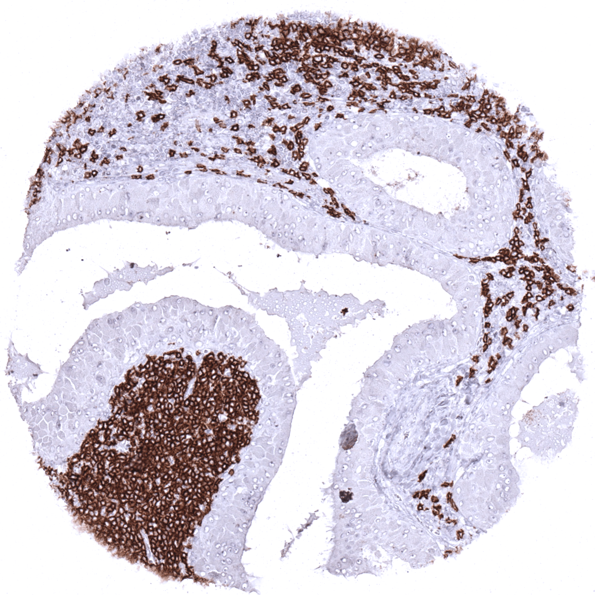
Staining Pattern in Relevant Tumor Types
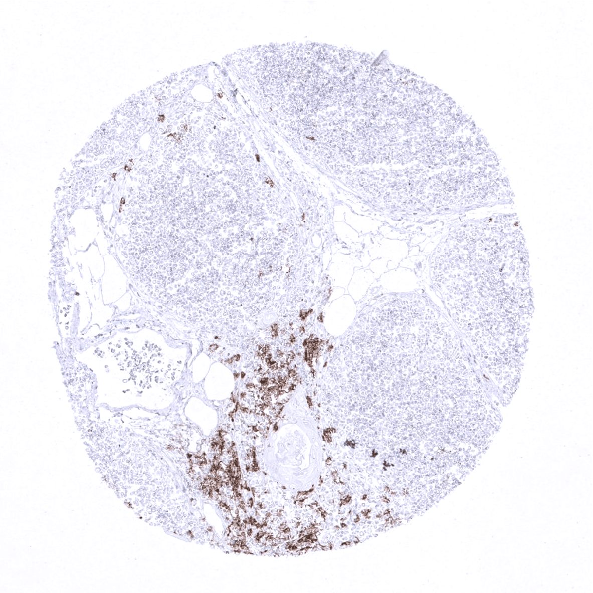
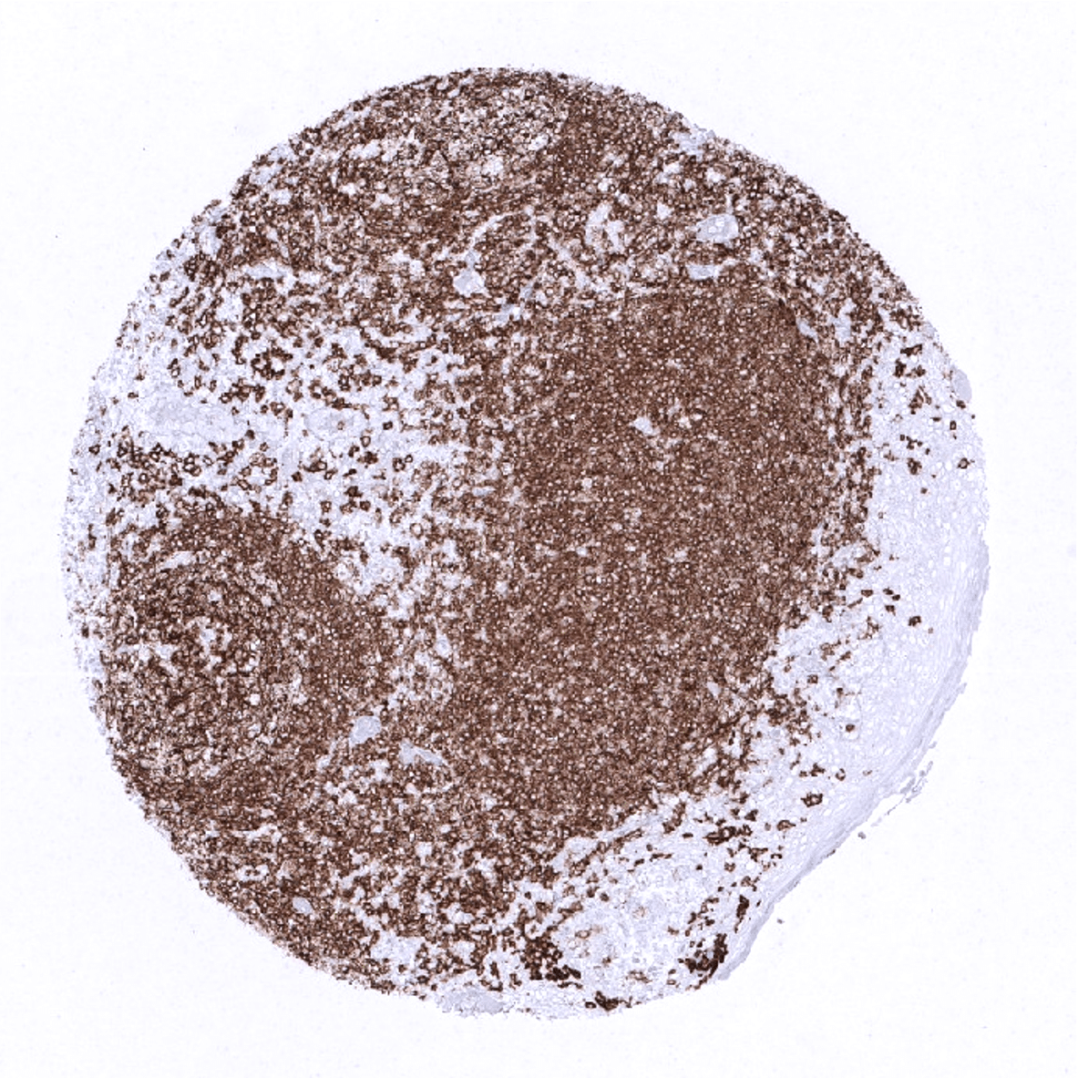
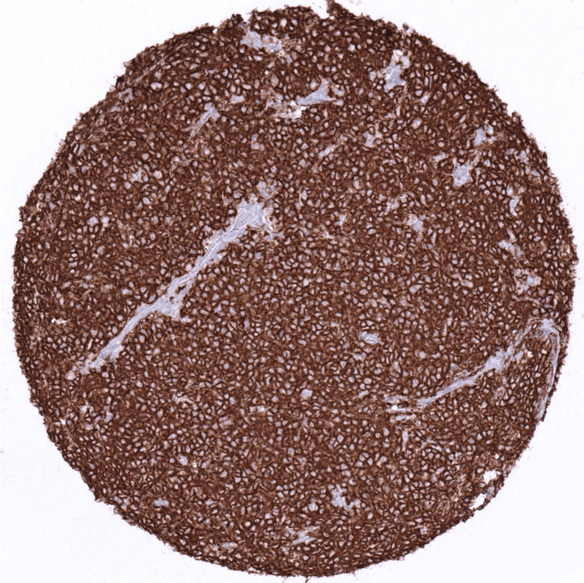
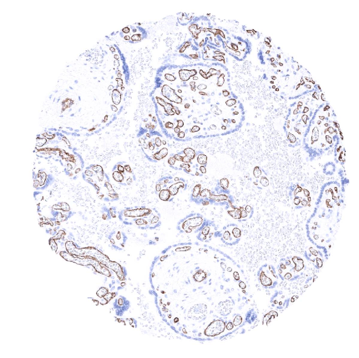
CD20 is expressed in neoplastic cells of most mature B-cell lymphomas but CD20 staining is negative in most B-cell neoplasms derived from immature precursor cells and in plasmocytoma. CD20 is also expressed in neoplastic cells of 80% of nodular lymphocyte predominant Hodgkin lymphoma and in 20% of classic Hodgkin lymphoma. Occasionally, CD20 is aberrantly expressed in T-cell lymphomas. In all other tumor types, variable fractions of CD20 positive B-lymphocytes are detectable as a component of the tumor’s microenvironment.
The TCGA findings on CD20 RNA expression in different tumor categories have been summarized in the Human Protein Atlas.
Compatibility of Antibodies
No data available at the moment
Protocol Recommendations
IHC users have different preferences on how the stains should look like. Some prefer high staining intensity of the target stain and even accept some background. Others favor absolute specificity and lighter target stains. Factors that invariably lead to more intense staining include higher concentration of the antibody and visualization tools, longer incubation time, higher temperature during incubation, higher temperature and longer duration of the heat induced epitope retrieval (slide pretreatment). The impact of the pH during slide pretreatment has variable effects and depends on the antibody and the target protein.
All images and data shown here and in our image galleries are obtained by the manual protocol described below. Protocols resulting in equivalent staining by using an automated immunostainer are described as well. Visualization of bound antibody by the EnVision Kit (Dako, Agilent) according to the manufacturer’s directions.
Manual protocol
Freshly cut sections should be used (less than 10 days between cutting and staining). Heat-induced antigen retrieval for 5 minutes in an autoclave at 121°C in pH 7,8 Target Retrieval Solution buffer. Apply MSVA-020R at a dilution of 1:150 at 37°C for 60 minutes. Visualization of bound antibody by the EnVision Kit (Dako, Agilent) according to the manufacturer’s directions.
Agilent / Dako – Autostainer Link 48
Pretreatment in PT-Link for 30 minutes at 95°C (pH high); FLEX peroxidase blocking for 5 minutes (room temperature), MSVA-020R 1:150 for 20 minutes (room temperature), FLEX+ mouse/rabbit (LINKER) for 15 minutes (room temperature), horseradish peroxidase (HRP) for 20 minutes (room temperature), FLEX DAB+Sub-Chromo for 10 minutes (room temperature), FLEX hematoxylin for 5 minutes (room temperature).
These images reflect stainings by the protocol described above. It is of note that a comparable staining result can also be obtained by different protocols. In general, a longer pretreatment, a longer incubation time of the primary antibody, a higher antibody concentration, and a longer incubation time of FLEX+LINKER result in stronger staining, potentially at the cost of more background staining. Modifications of the protocol with a strengthening effect on staining intensity in combination with changes of other parameters that result in lower staining intensity can result in a comparable result as shown above.
Leica – BOND RX
Dewax at 72°C for 30 seconds; Pretreatment in Bond Epitope Retrieval Solution (ER2 – EDTA pH9) for 20 minutes at 100°C; Peroxidase blocking for 5 minutes (room temperature), MSVA-020R 1:150 for 15 minutes (room temperature), Post primary (rabbit anti mouse) for 8 minutes (room temperature), Polymer (goat anti rabbit) for 8 minutes (room temperature), mixed DAB refine for 10 minutes (room temperature), hematoxylin for 5 minutes (room temperature).


These images reflect stainings by the protocol described above. It is of note that a comparable staining result can also be obtained by different protocols. In general, a longer pretreatment, a longer incubation time of the primary antibody, a higher antibody concentration, a higher temperature during incubation, and a longer incubation time of Post primary and or the Polymer result in stronger staining, potentially at the cost of more background staining. Modifications of the protocol with a strengthening effect on staining intensity in combination with changes of other parameters that result in lower staining intensity can result in a comparable result as shown above.
Impact of pH
pH9 is optimal but pH7,8 results in only slightly weaker stainings. At pH6, CD1a immunostaining is markedly reduced.
Potential Research Applications
- The clinical significance of the number of intratumoral CD20 positive lymphocytes is under intensive research.
- CD20 is a key component of multicolor assays analyzing the role of lymphocyte subsets in cancers and other diseases.
- The prevalence of a positive CD20 immunostaining in hematological and non-hematological neoplasms should be further evaluated.
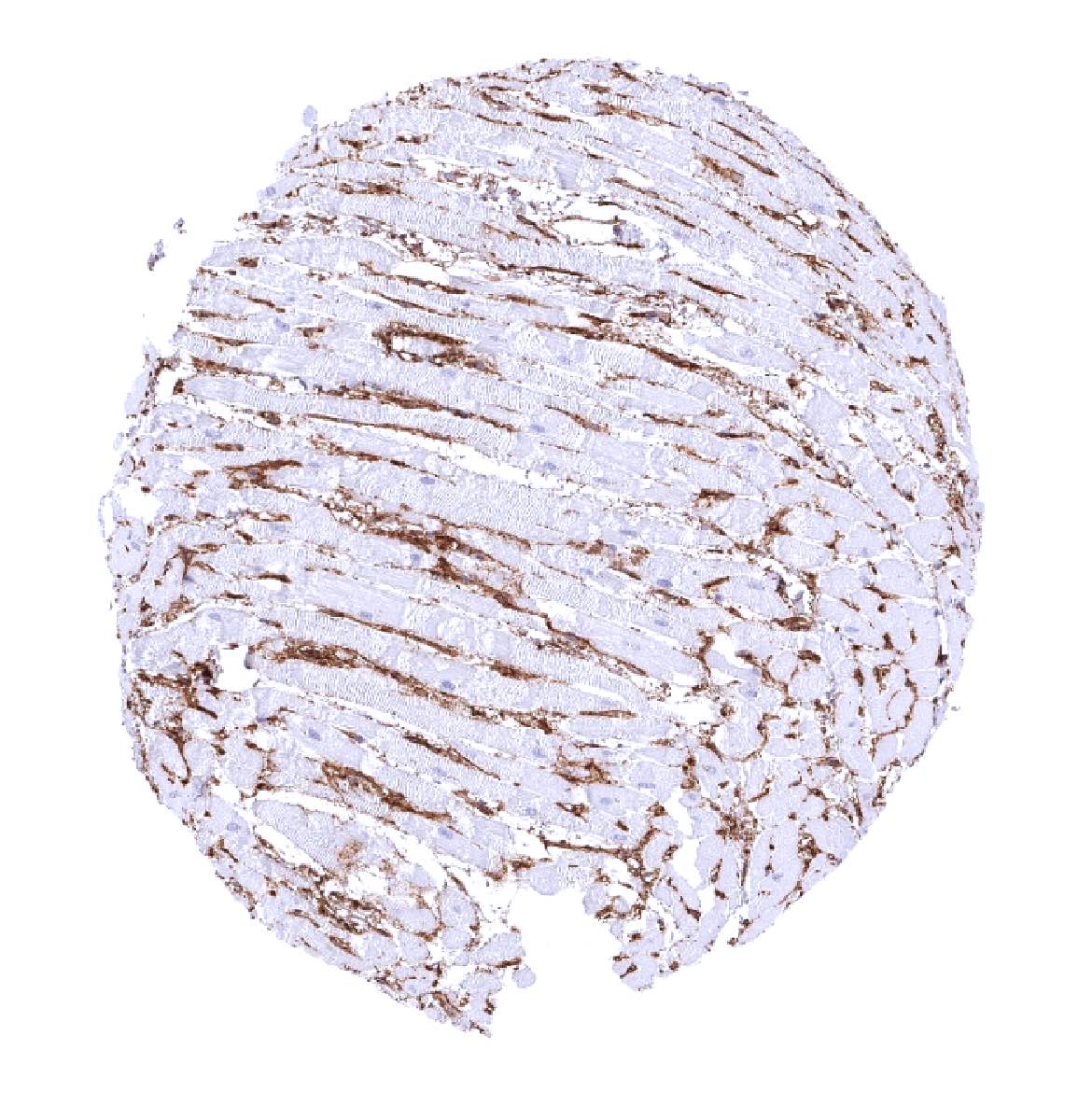
Evidence for Antibody Specificity in IHC
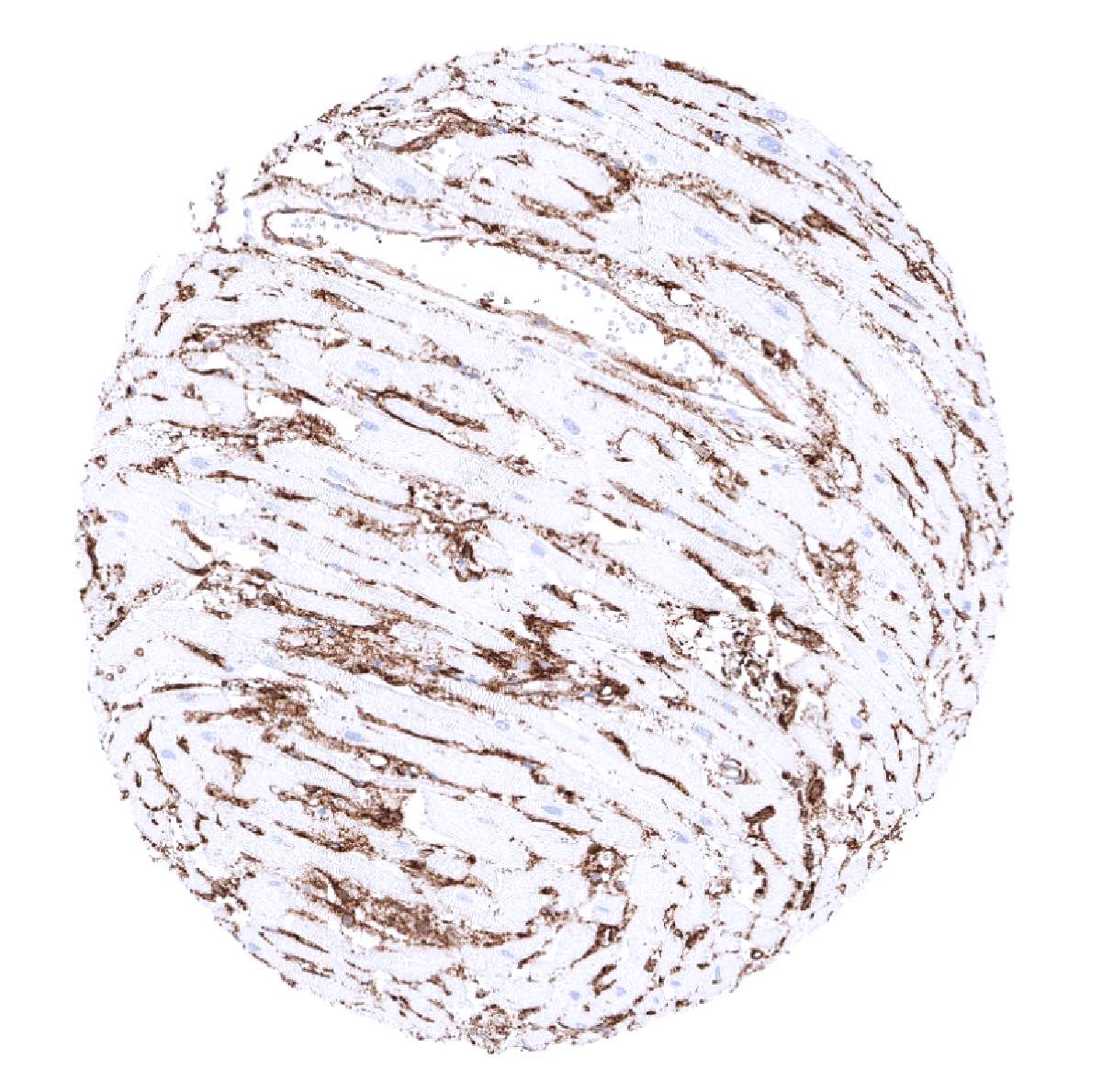
Specificity of MSVA-020R is documented by strong positive staining in B-cells types in lymphatic organs and absence of staining in all tissues known to not express CD20 including tissues notorious for non-specific IHC background such as kidney, colonic mucosa, and epidermis.
