295,00 € – 995,00 €
Product details
Synonyms = Ig delta chain C region; IGHD; Immunoglobulin heavy constant delta
Antibody type = Rabbit monoclonal / IgG
Clone = MSVA-701R
Positive control = Tonsil: A strong IgD staining should be seen in lymphocytes of the mantle zone.
Negative control = Tonsil: Epithelial cells and most “non-mantle zone” lymphocytes should not stain for IgD.
Cellular localization = Cytoplasmic
Reactivity = Human
Application = Immunohistochemistry
Dilution = 1:100 – 1:200
Intended Use = Research Use Only
Relevance of Antibody
IgD is a pivotal molecule in the immune response.
Biology Behind
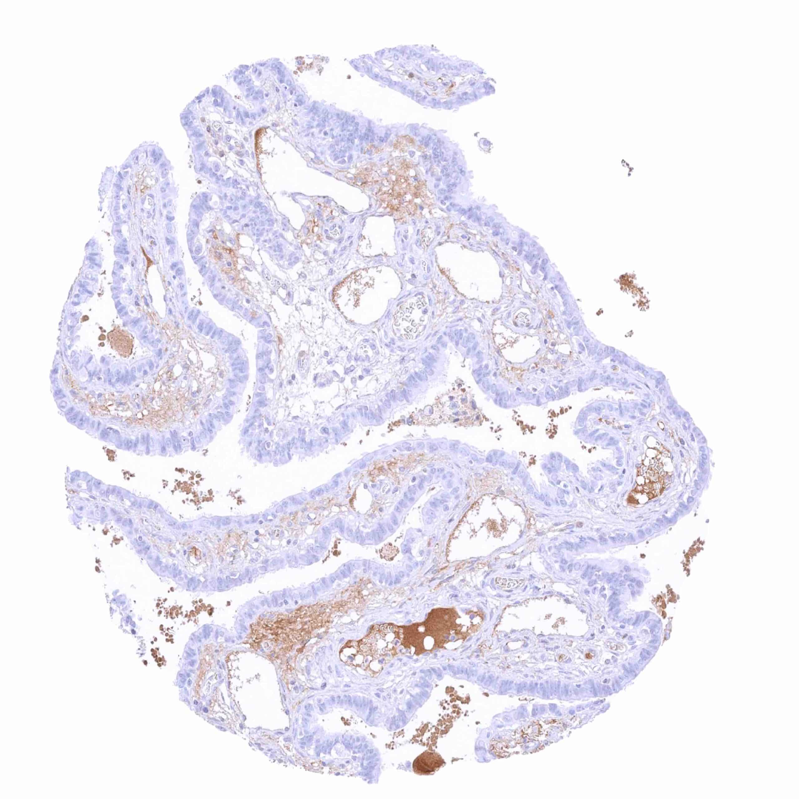
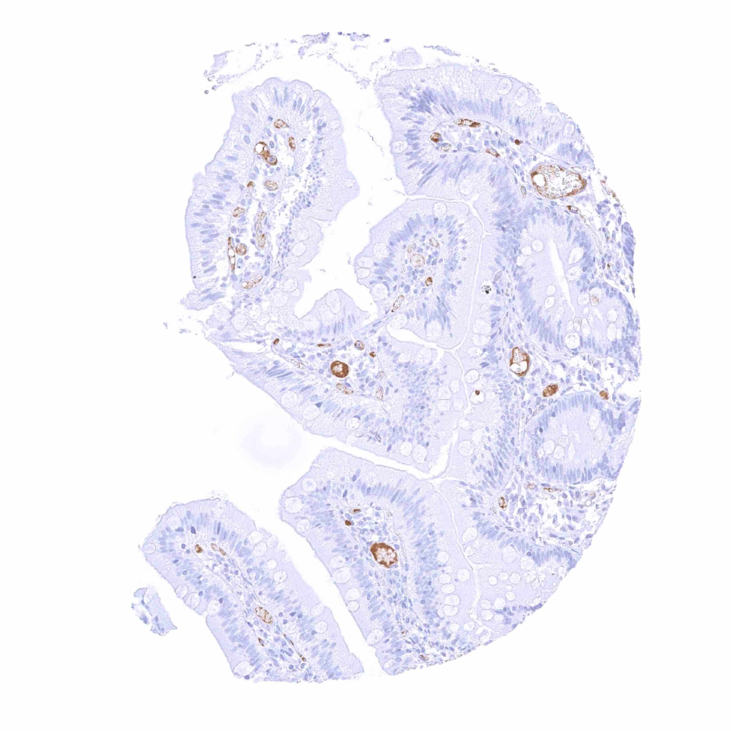
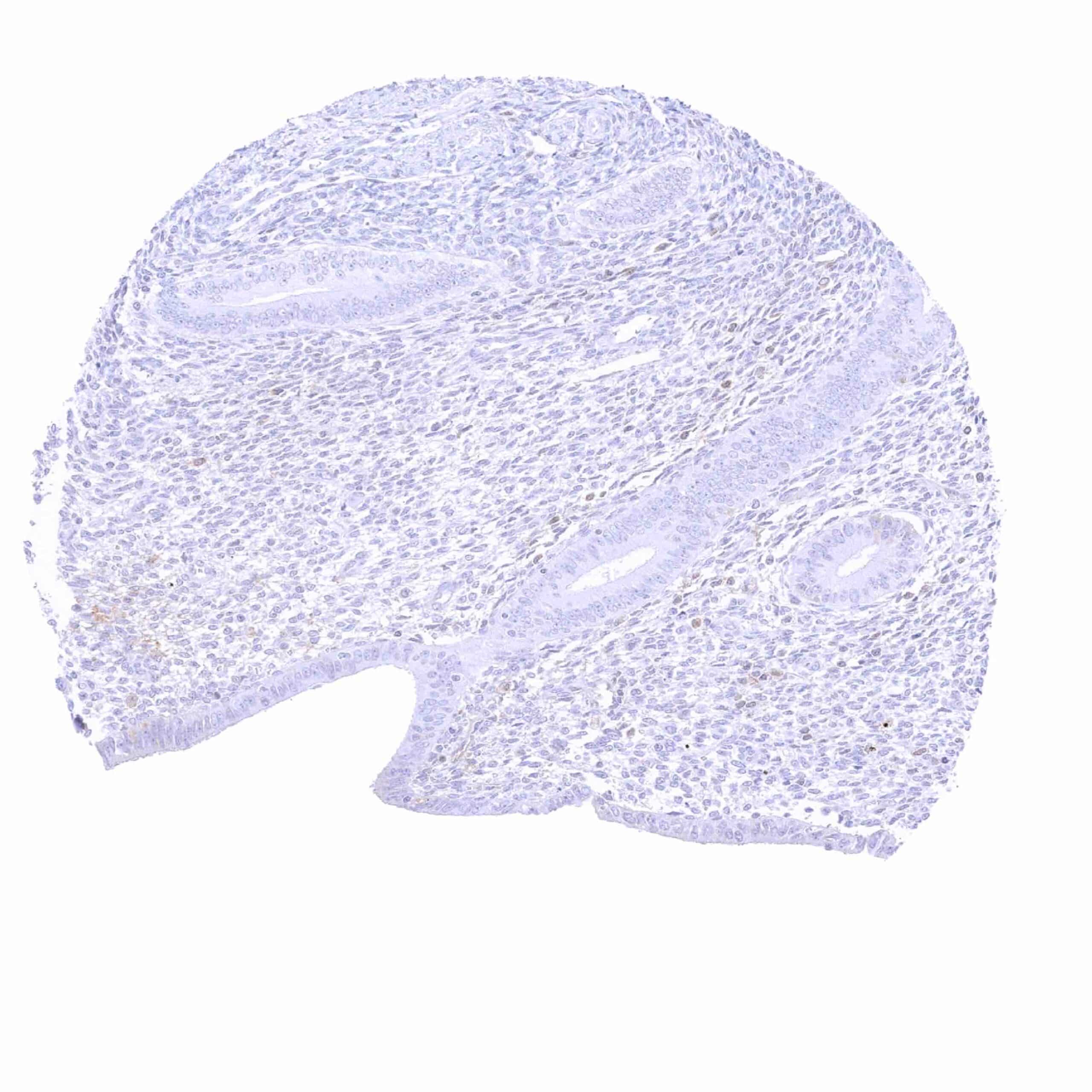
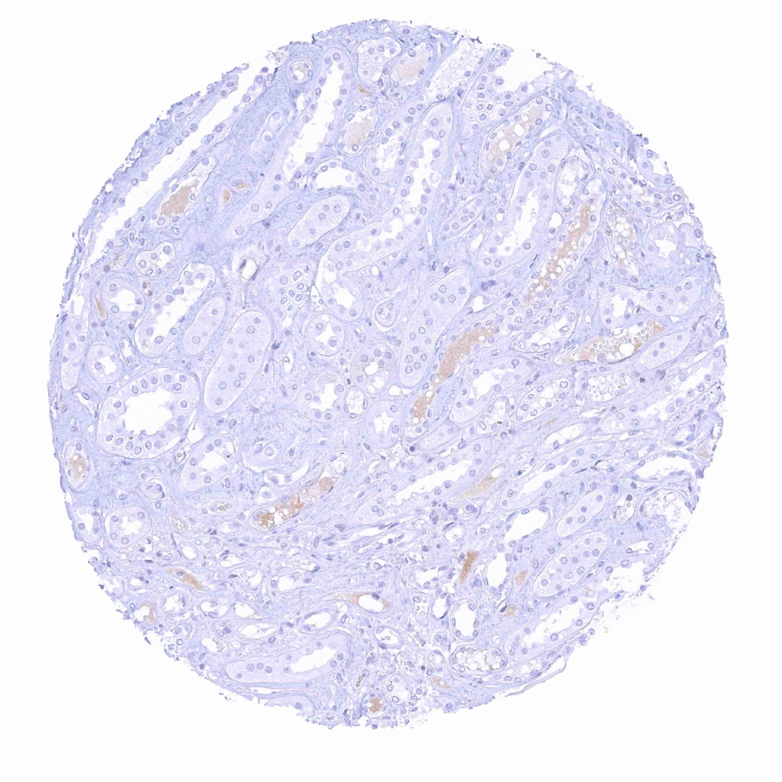
Immunoglobulin D (IgD) is an antibody isotype with a relative molecular mass of 185 kDa. During B cell differentiation, IgD starts to be expressed when B cells exit the bone marrow to populate peripheral lymphoid tissues. When a B cell reaches its mature state, it co-expresses both IgM and IgD. Only a few plasma cells express IgD and the level of IgD in the serum is therefore low. IgD represents only 0.25% of immunoglobulins in blood serum. The function of IgD is unclear. Target specific IgD antibodies are not produced, even after immunization. IgD plays a role in B-cell signaling and activation and it binds to basophils and mast cells and activates these cells to produce antimicrobial factors. However, IgD knockout mice do not appear to have major intrinsic B cell defects. The preservation of IgD throughout the evolution implies important functions of IgD that confer a specific survival advantage to the host. In normal lymphatic tissues, IgD is primarily expressed in B-lymphocytes of the mantle zone. Sporadic IgD positive lymphocytes can be seen in most tissue types. IgD positive plasma cells are rare and most commonly seen in the upper airways. In addition, a diffuse IgD staining is sometimes seen in mucus, blood vessels or in the stroma of areas of inflammation. This staining pattern represents soluble IgD derived from serum or body fluids. In tumors, IgD positivity is most commonly seen in mantle zone lymphomas. A positive IgD immunostaining can occasionally also be seen in other lymphomas and in a very small fraction of plasmacytomas.
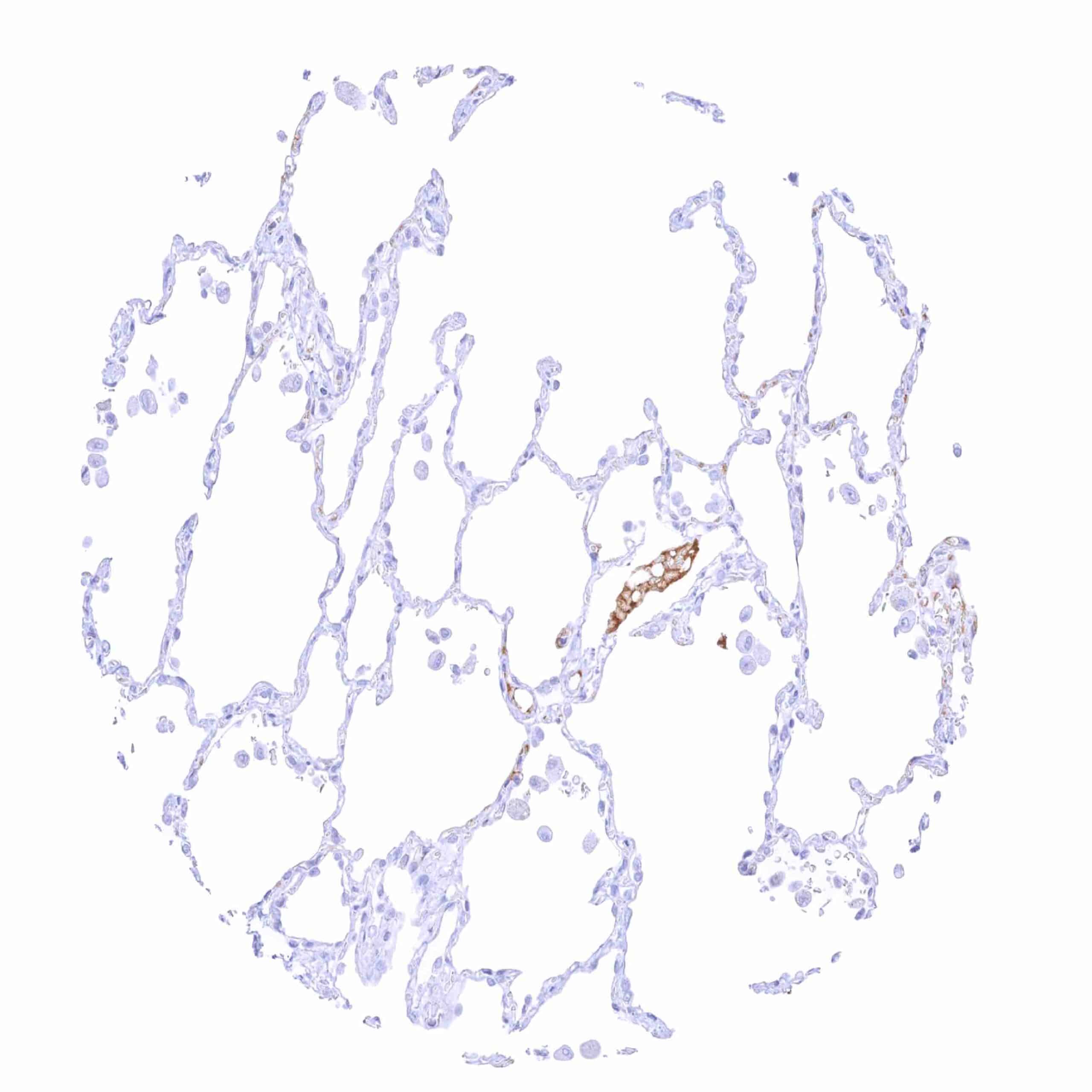
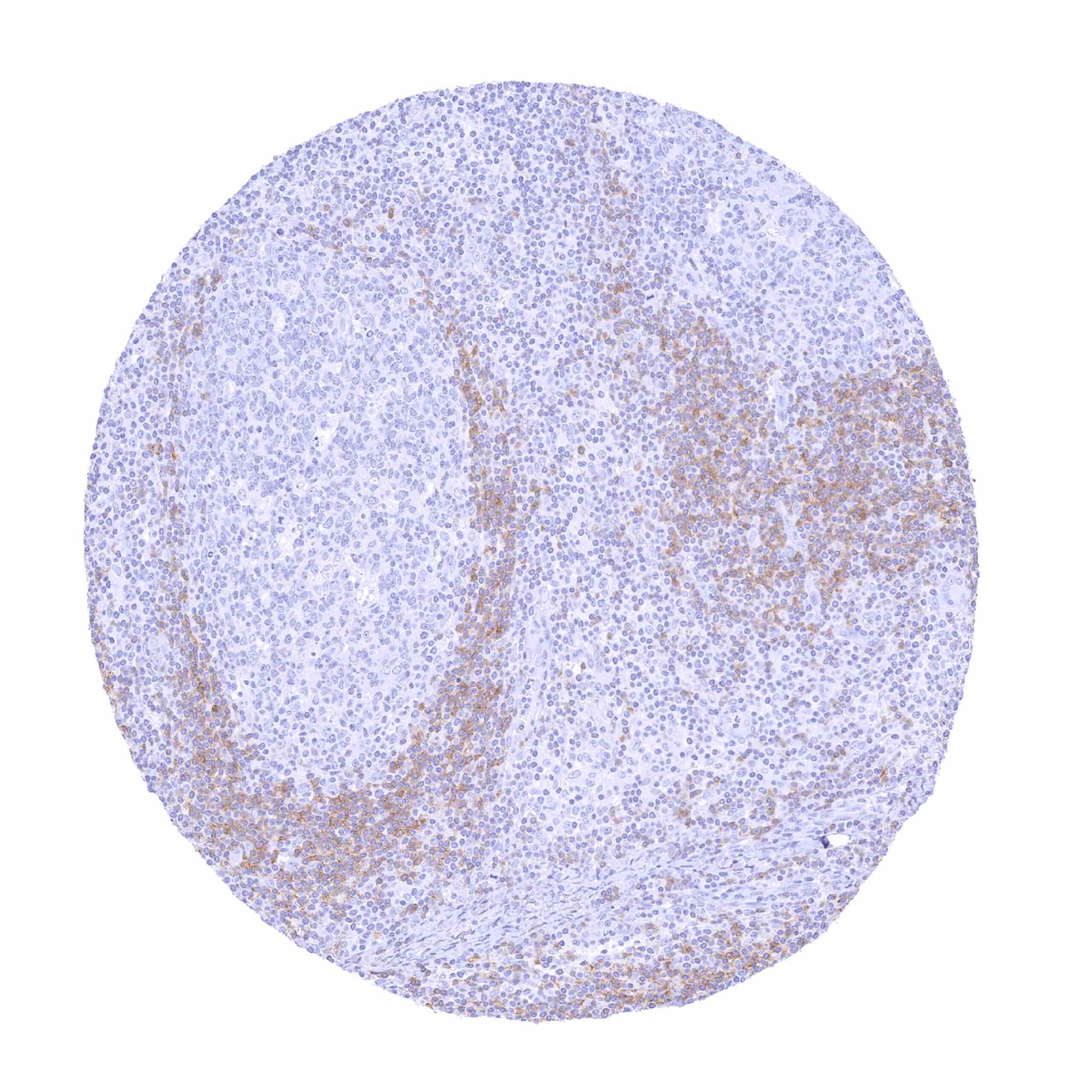
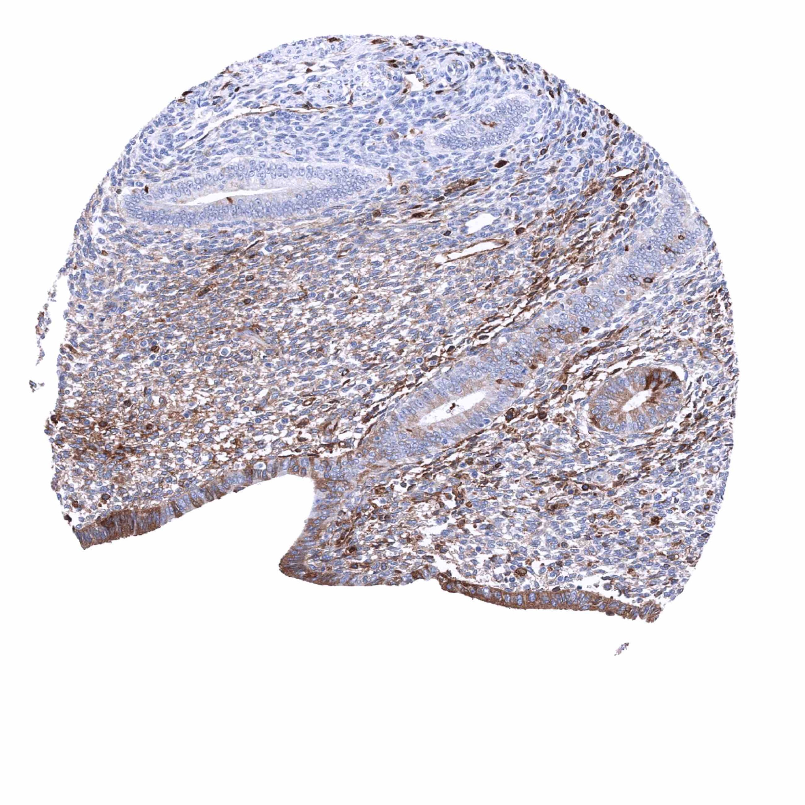
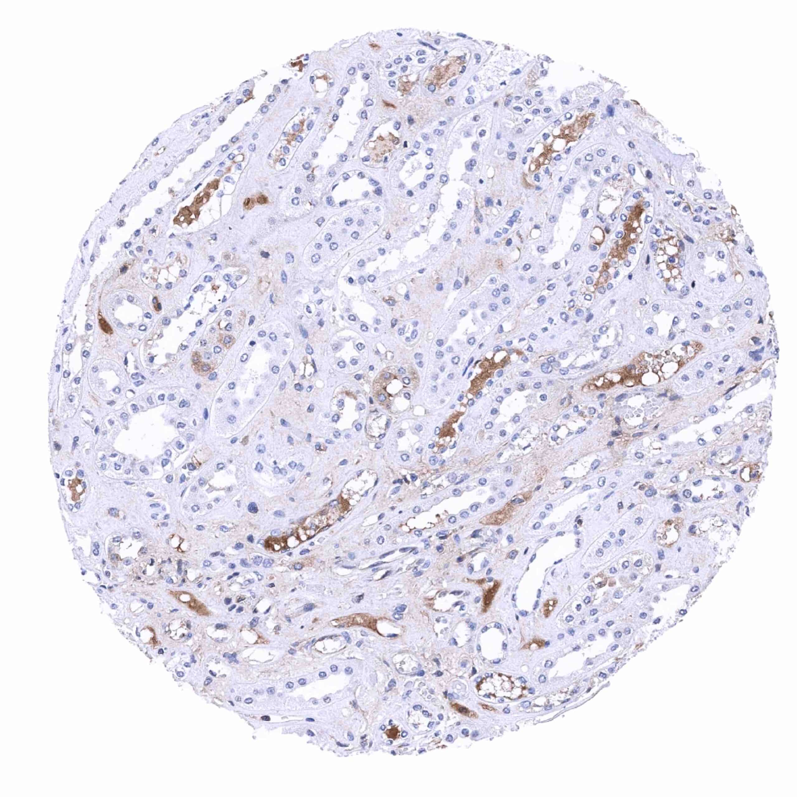
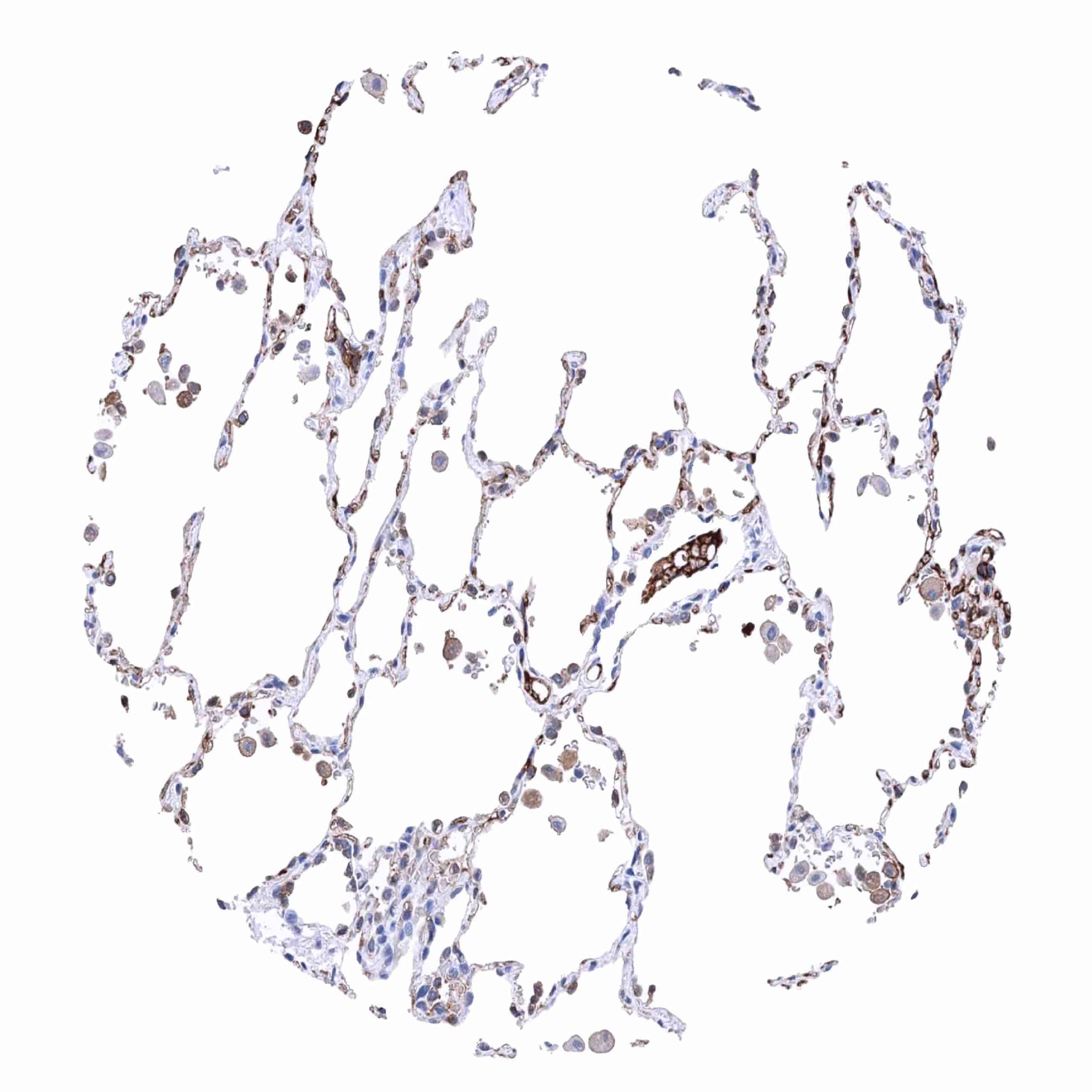
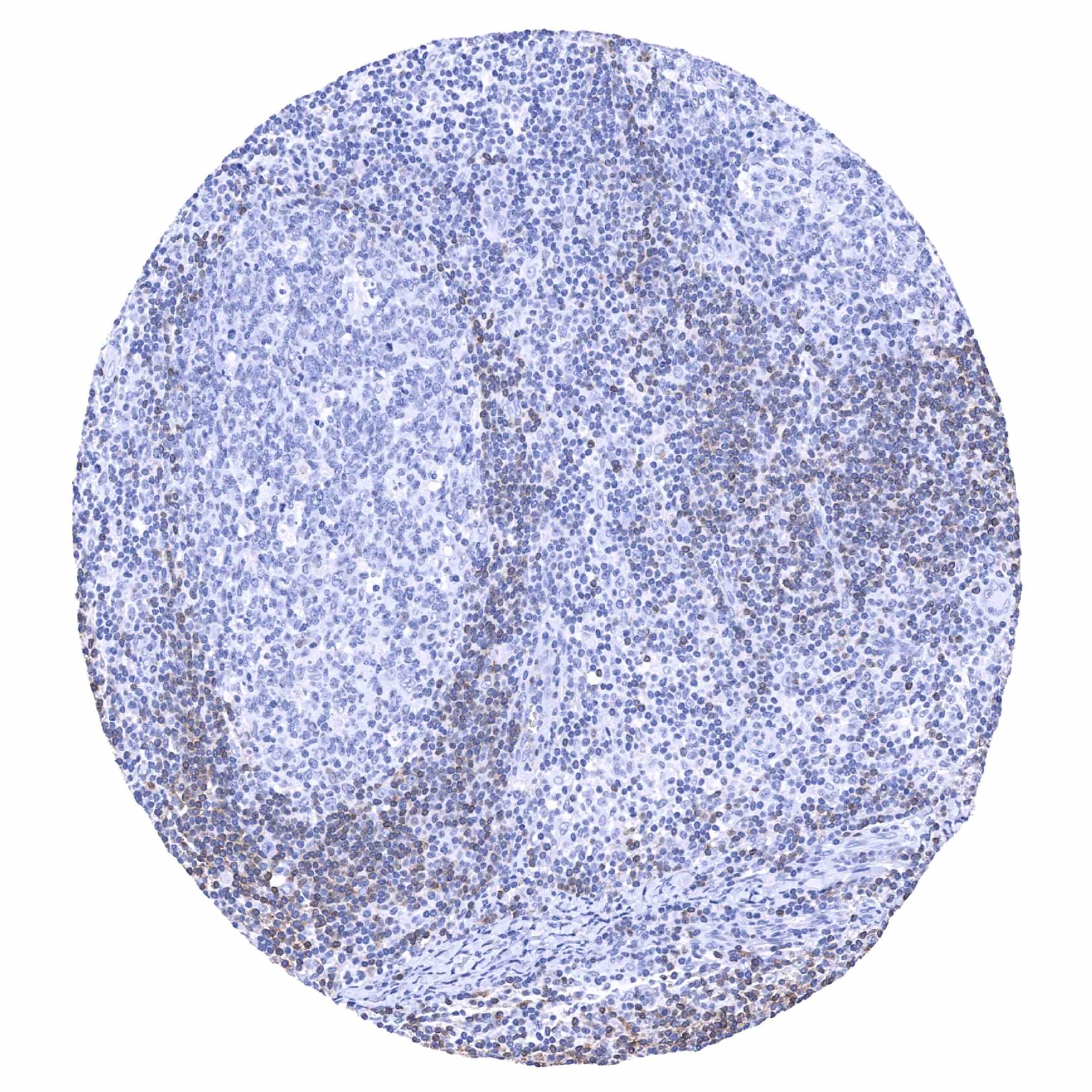
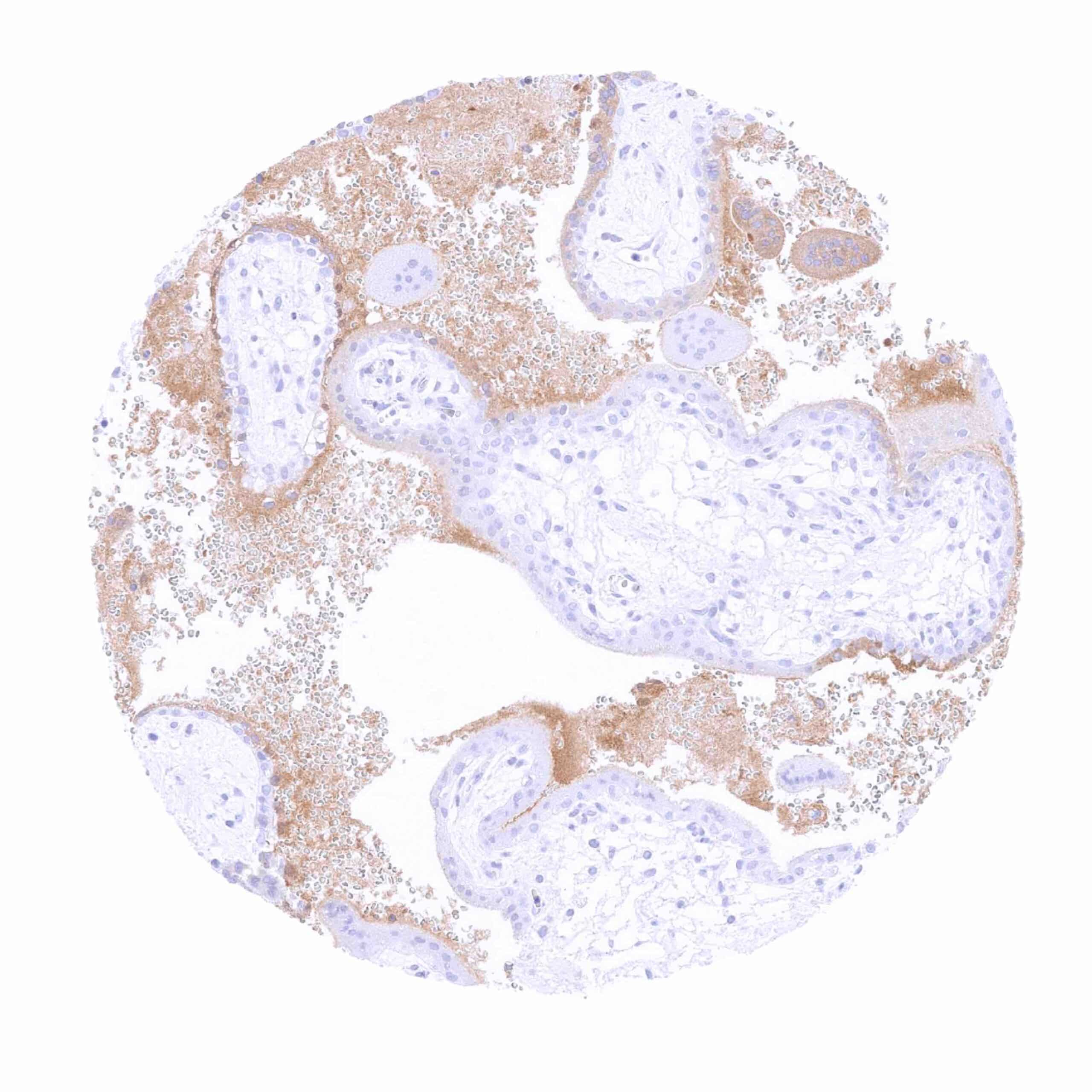
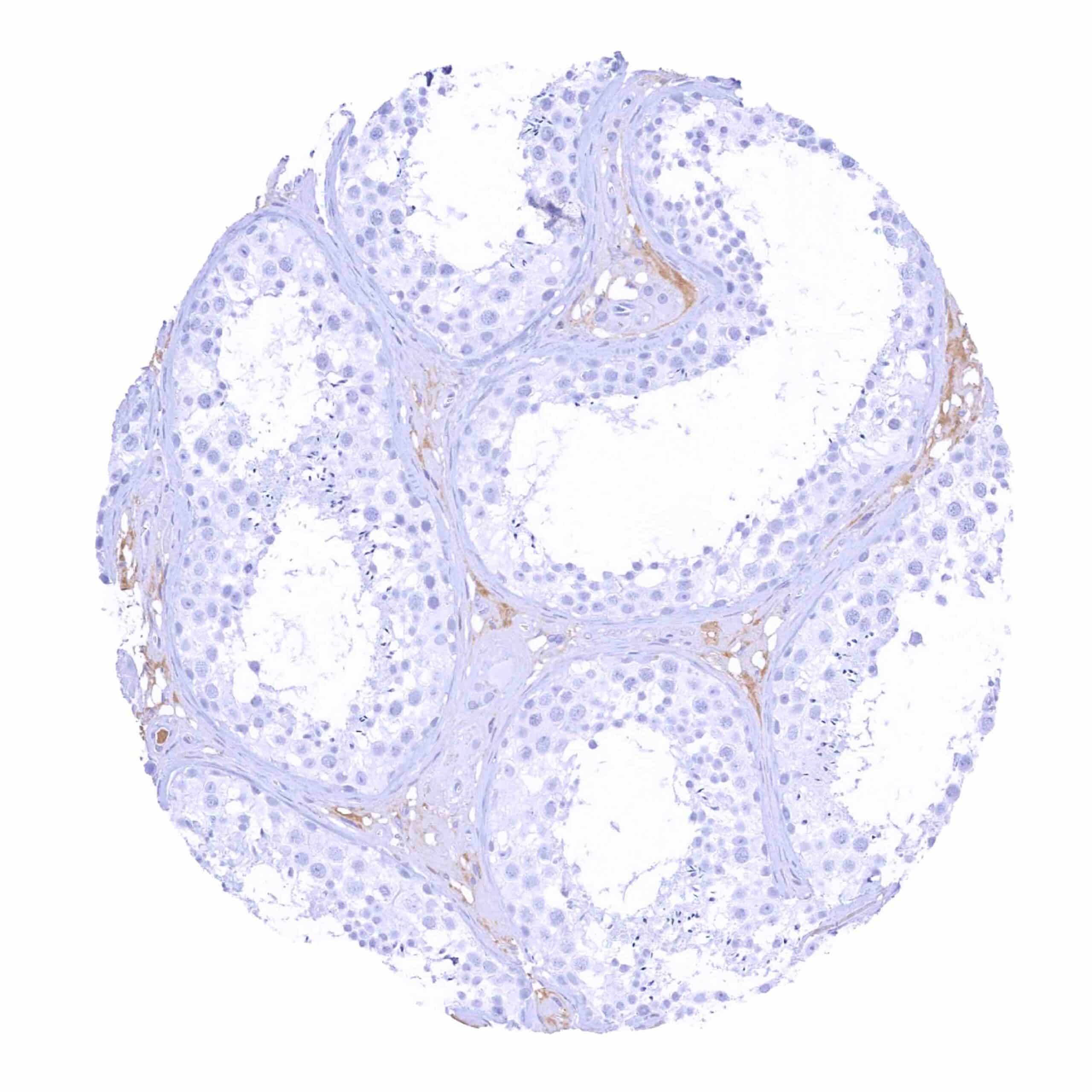
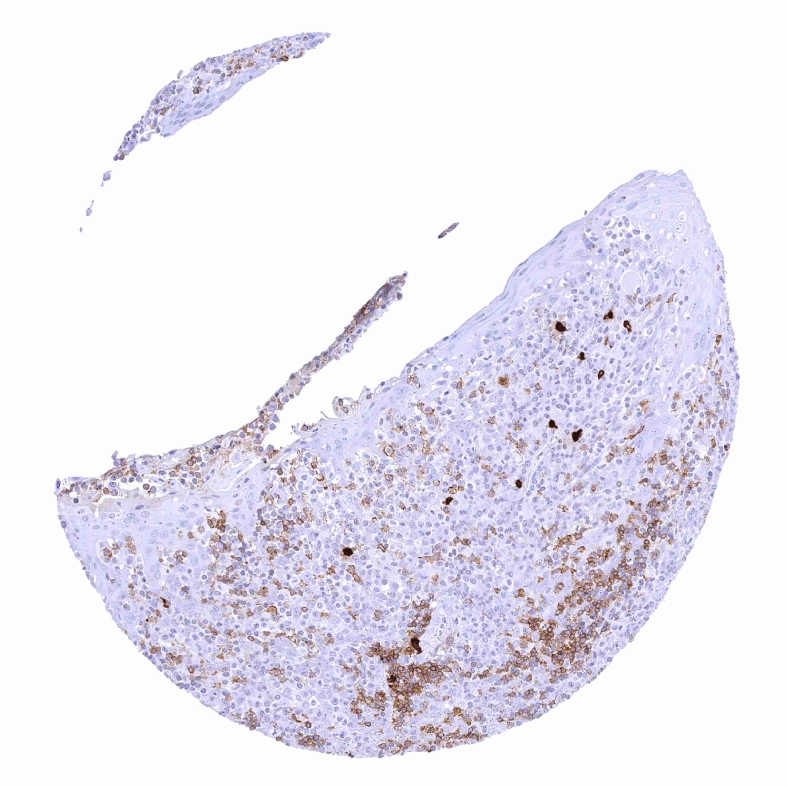
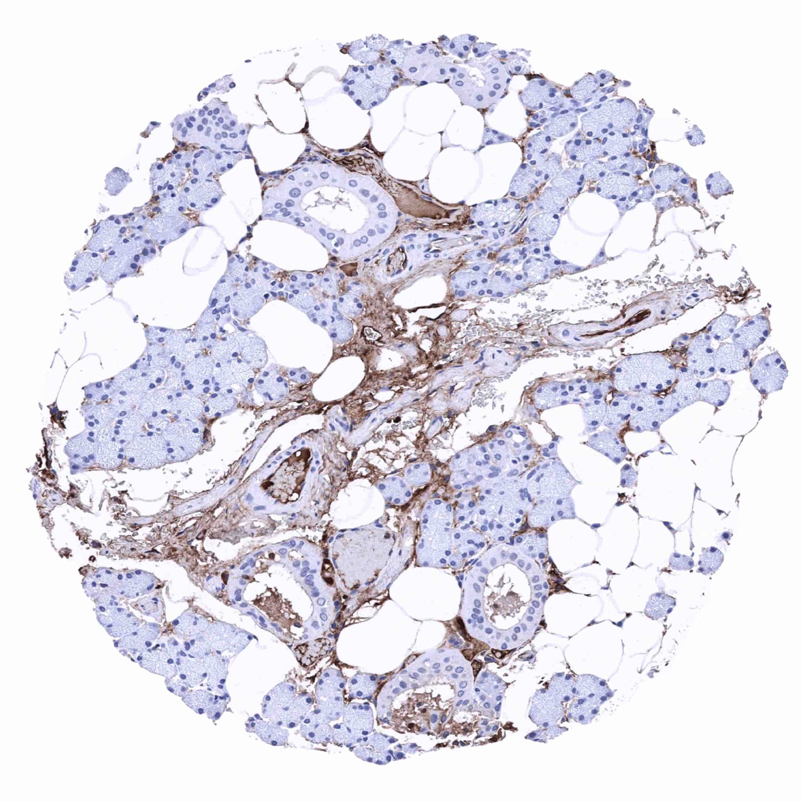
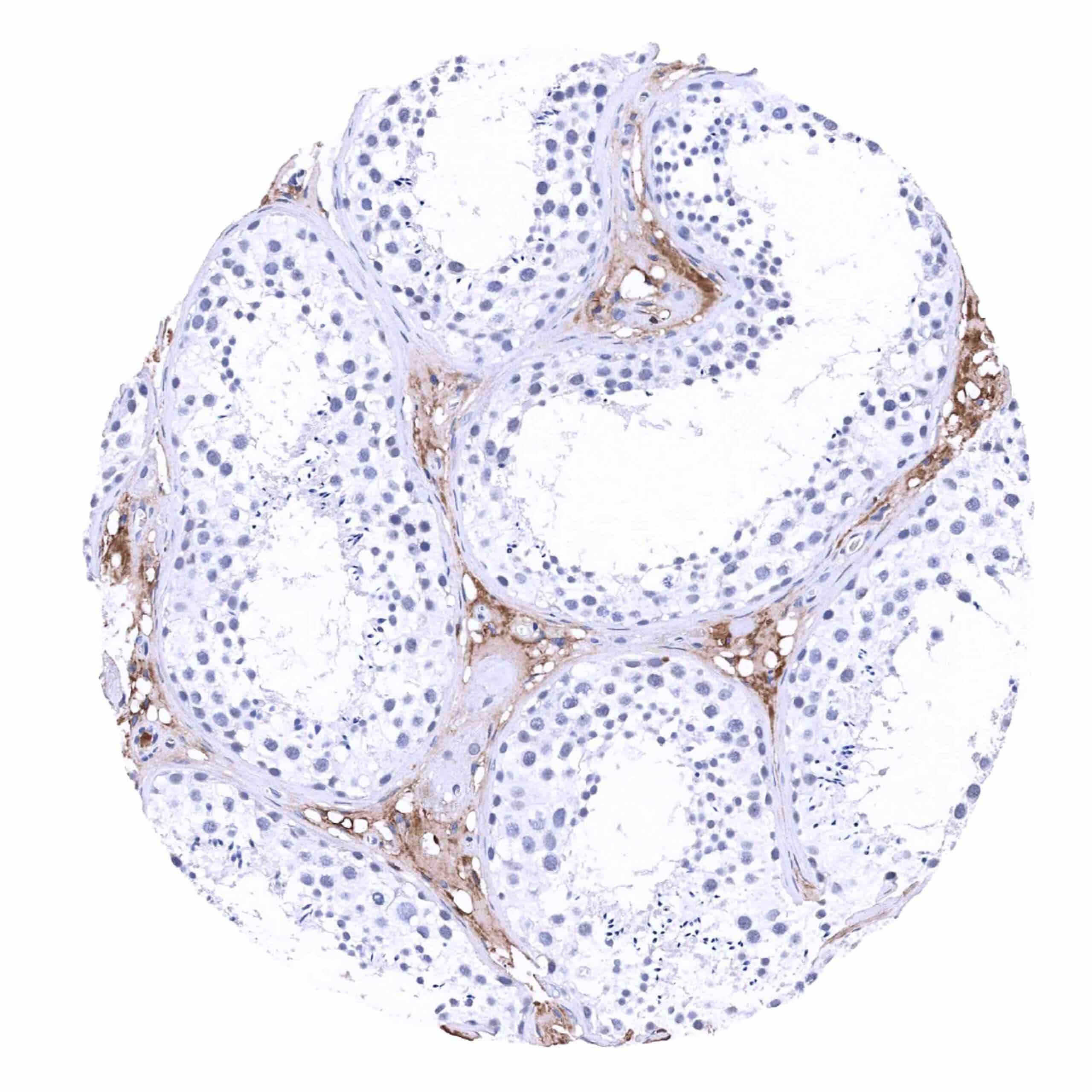
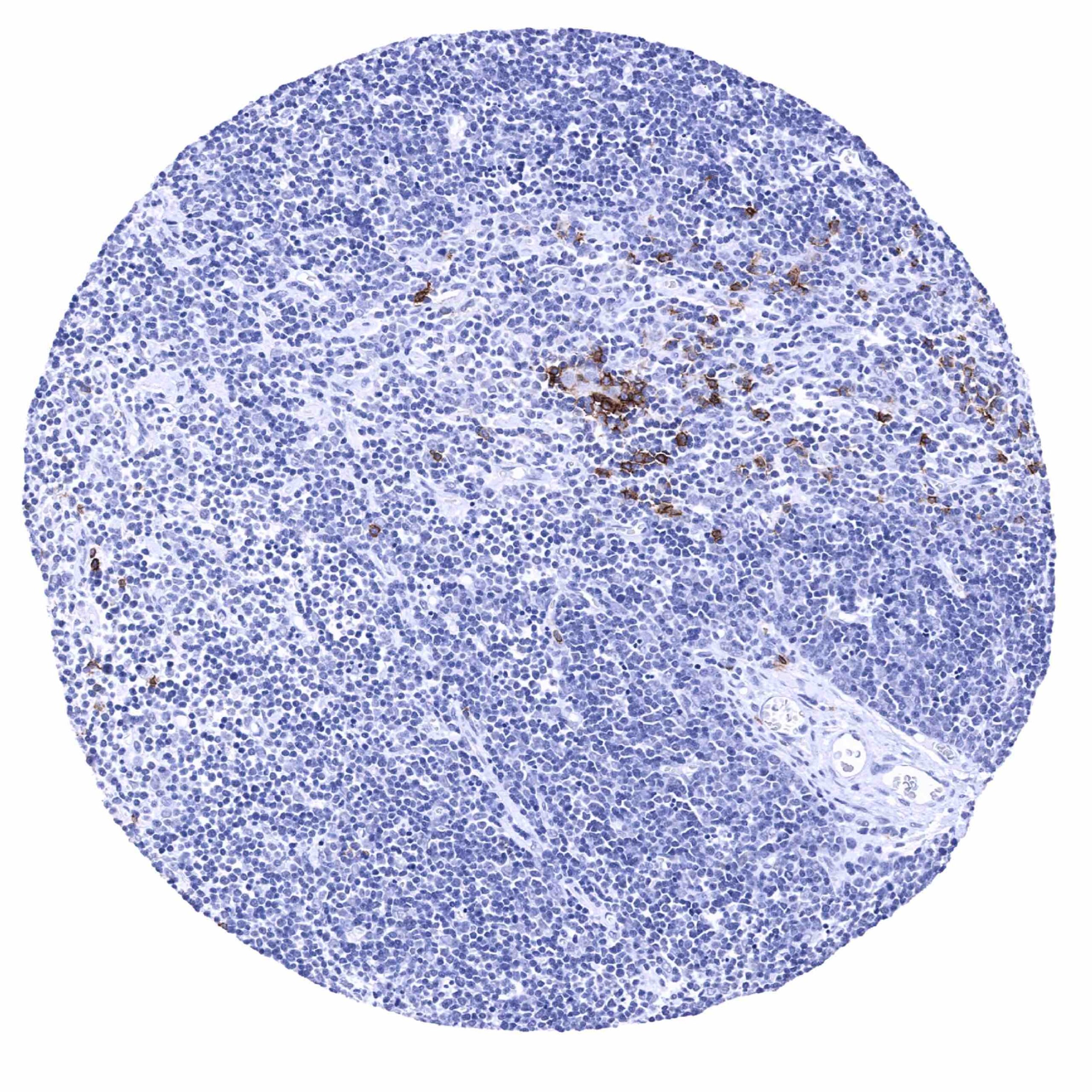
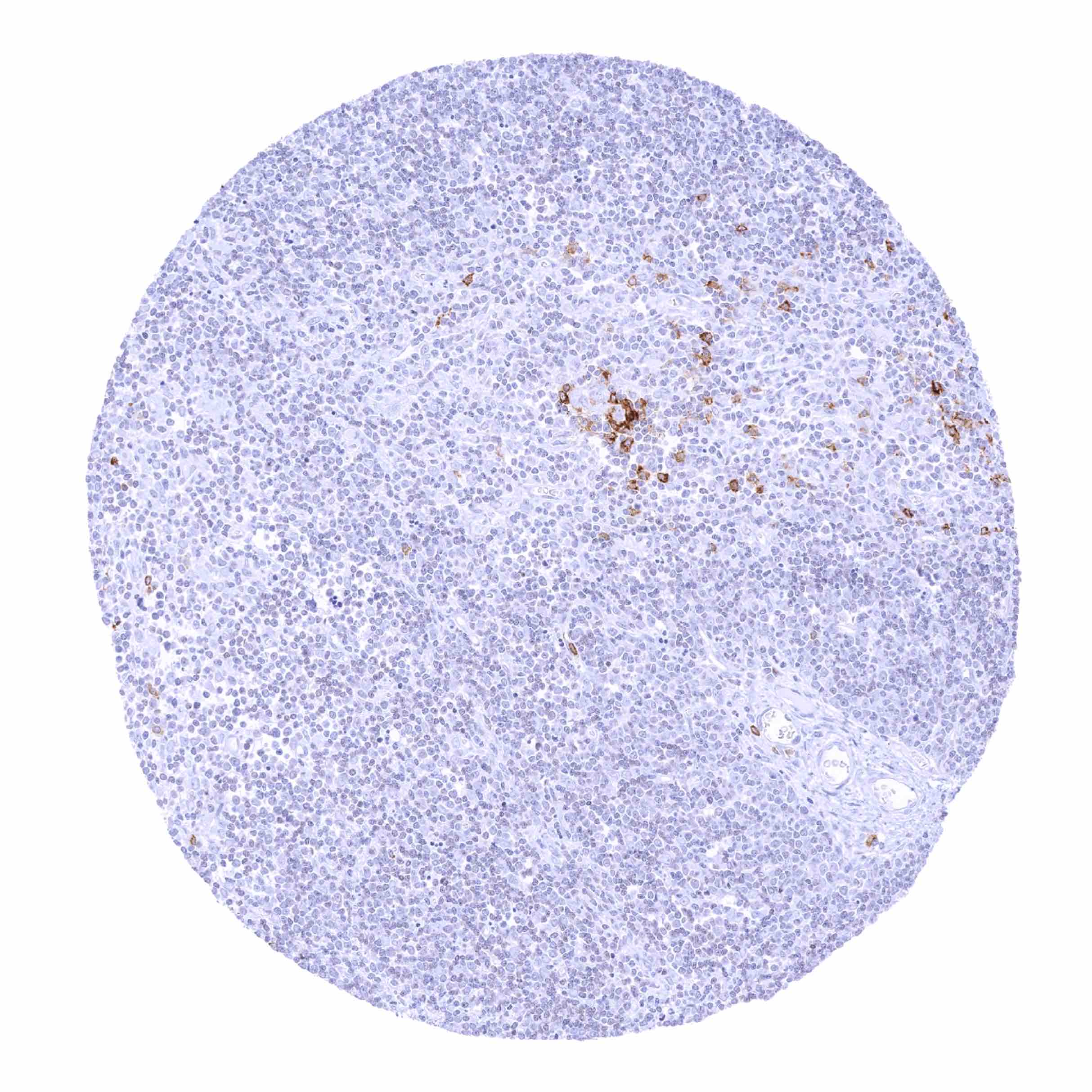
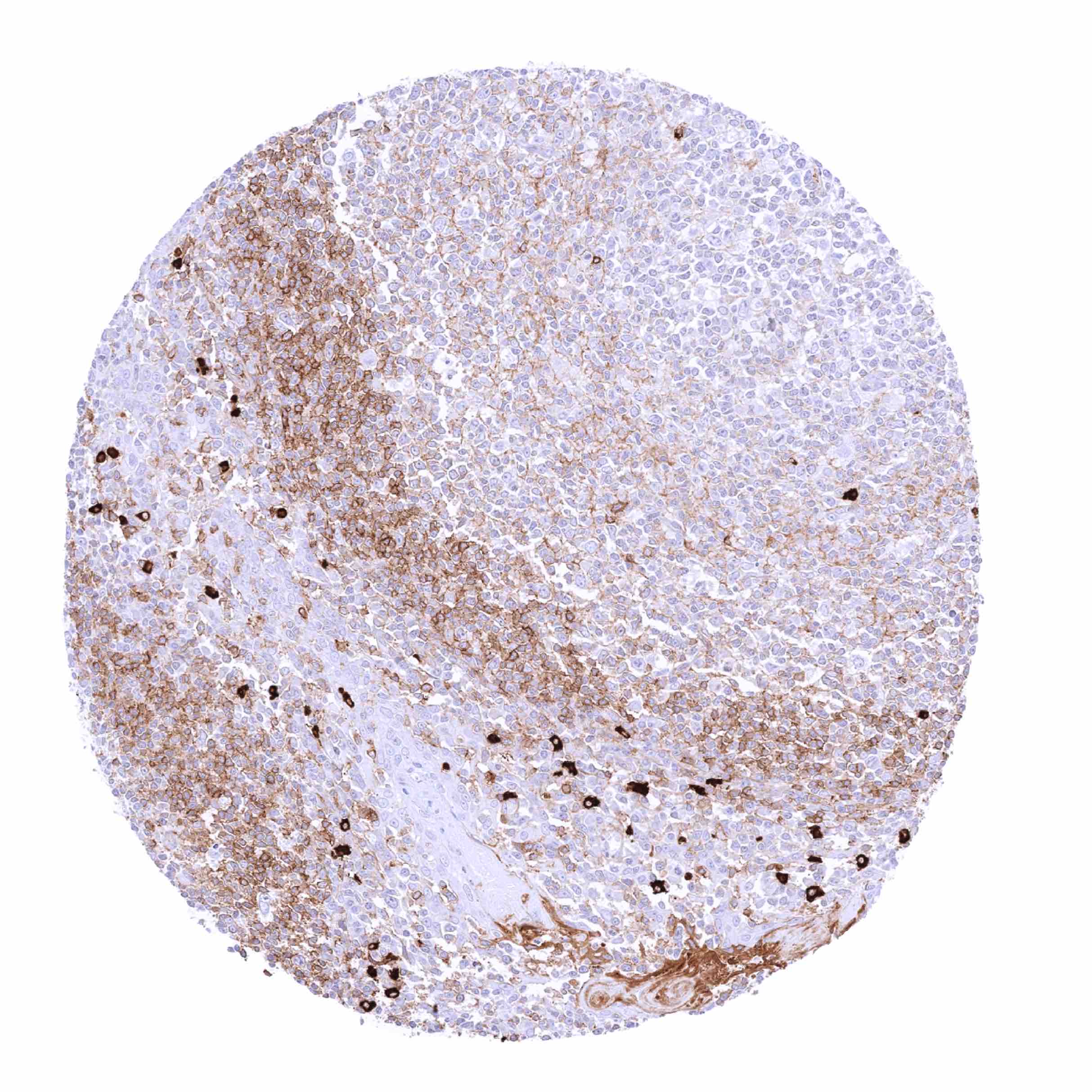
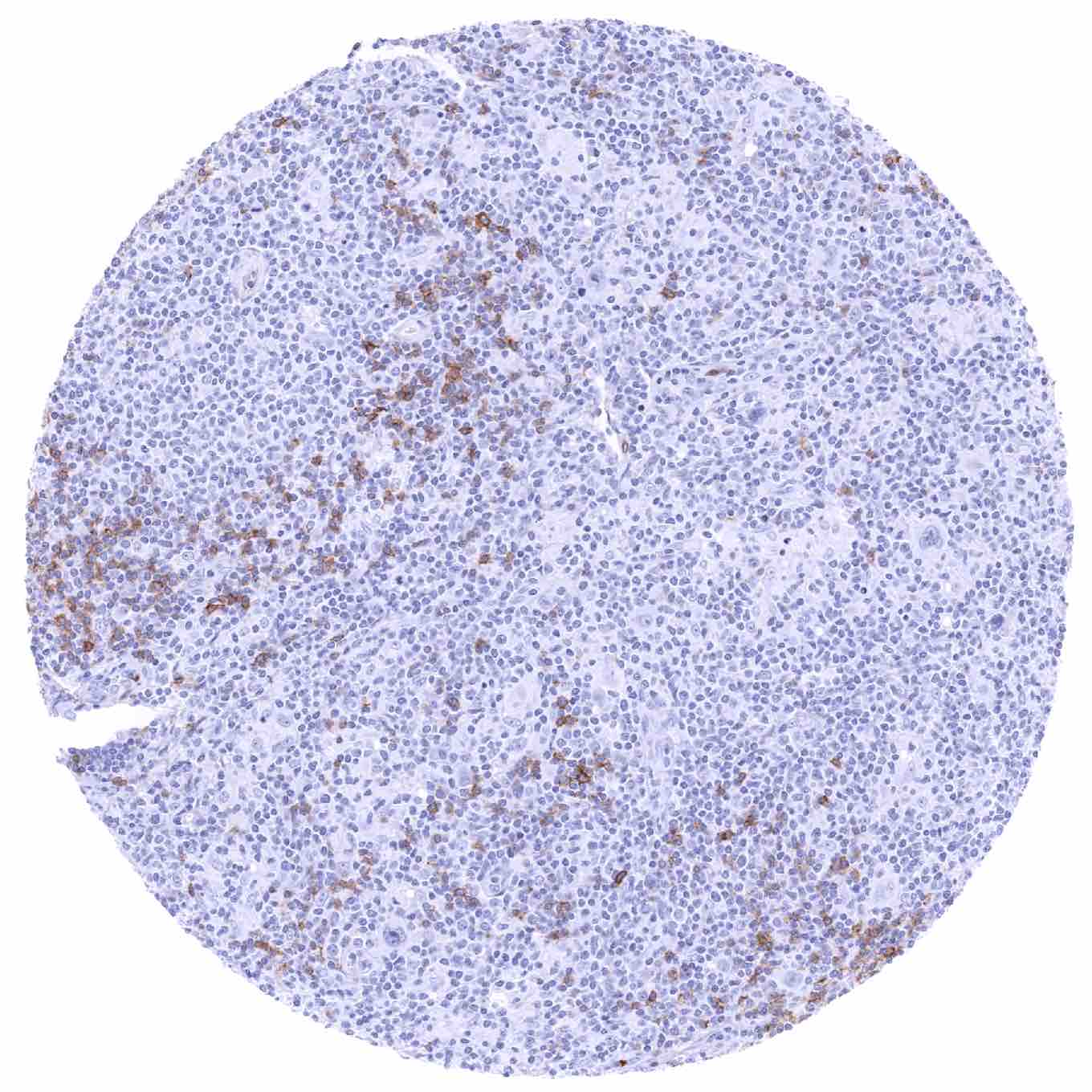
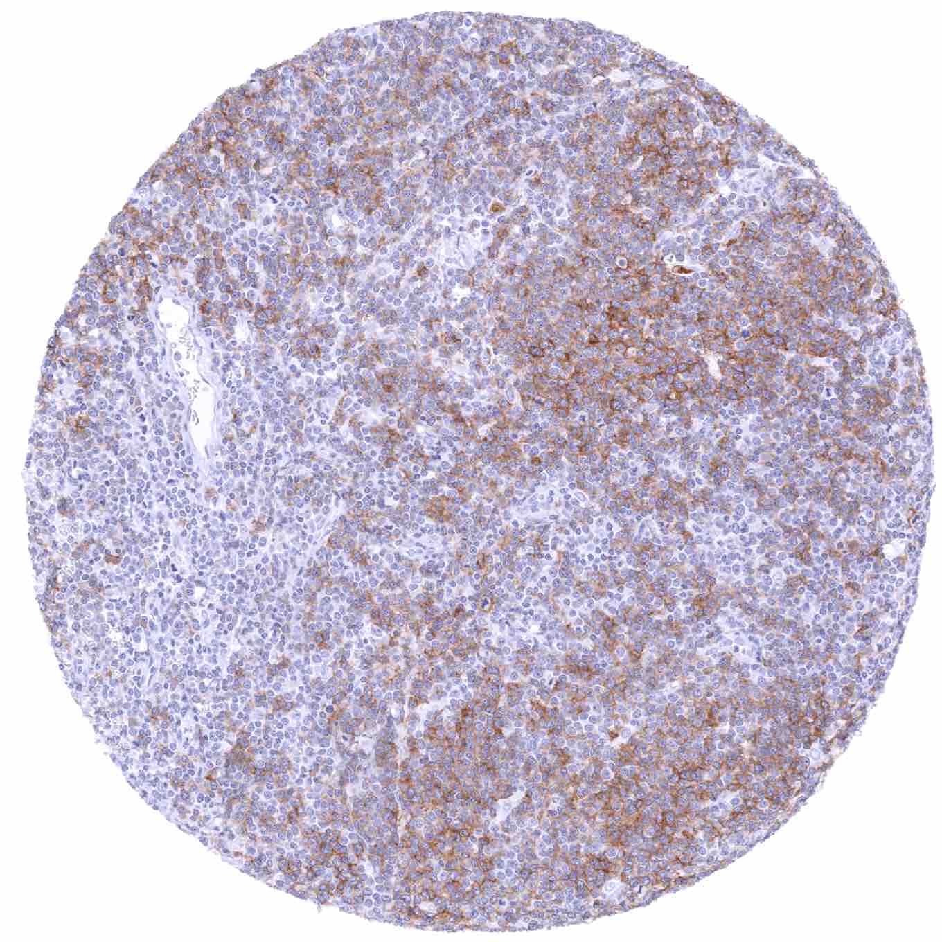
Staining Pattern in Normal Tissues
IgD is expressed on the surfaces of most peripheral B cells. In lymphatic tissues, IgD is primarily expressed in B-lymphocytes of the mantle zone. Sporadic IgD positive lymphocytes can be seen in most tissue types. IgD positive plasma cells are rare and most commonly seen in the upper airways. In addition, a diffuse IgD staining is sometimes seen in mucus, blood vessels or in the stroma of areas of inflammation. This staining pattern represents soluble IgD derived from serum or body fluids.
Images describing the IgD staining pattern in normal tissues obtained by the antibody MSVA-701R are shown in our “Normal Tissue Gallery”.
Positive control = Tonsil: A strong IgD staining should be seen in lymphocytes of the mantle zone.
Negative control = Tonsil: Epithelial cells and most “non-mantle zone” lymphocytes should not stain for IgD.


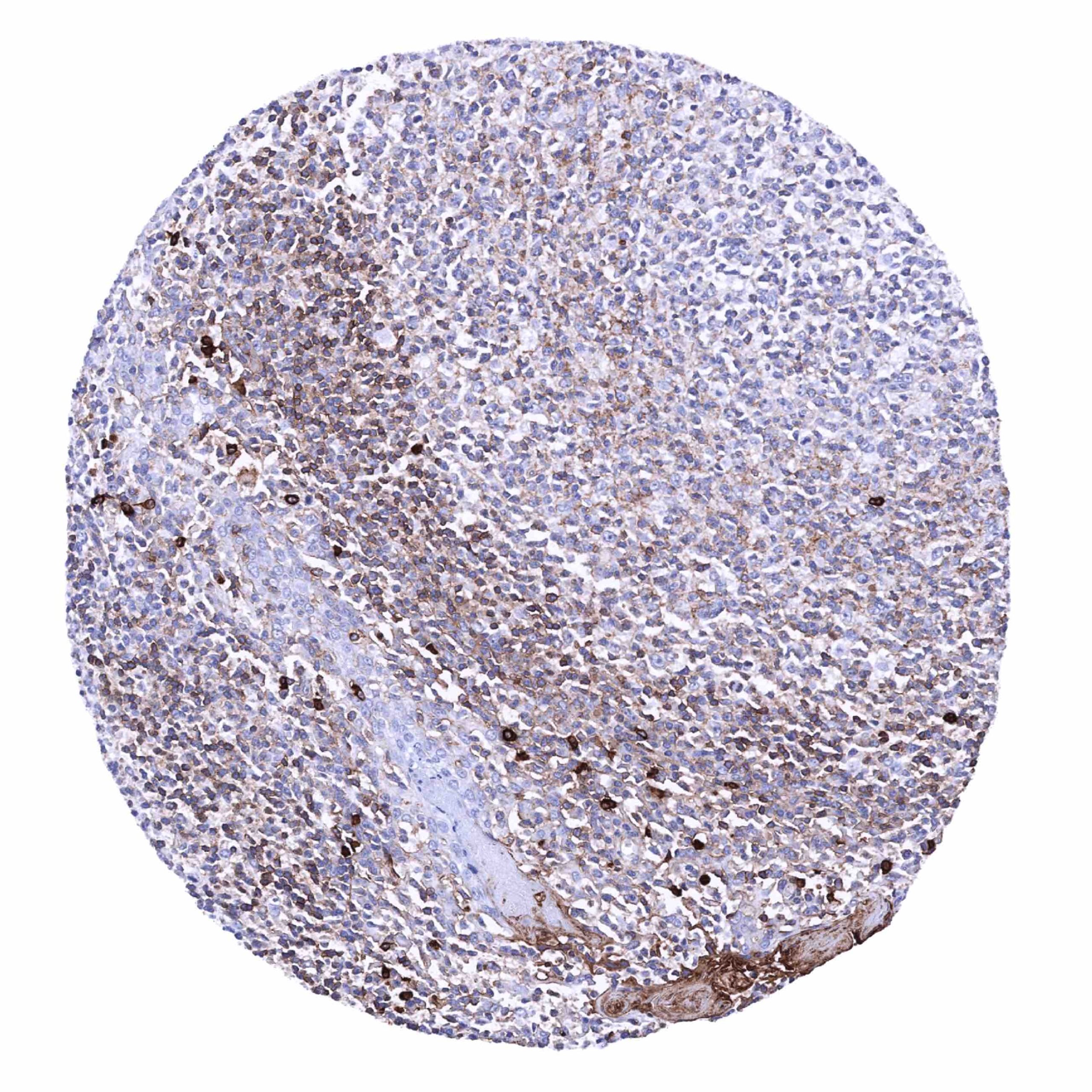
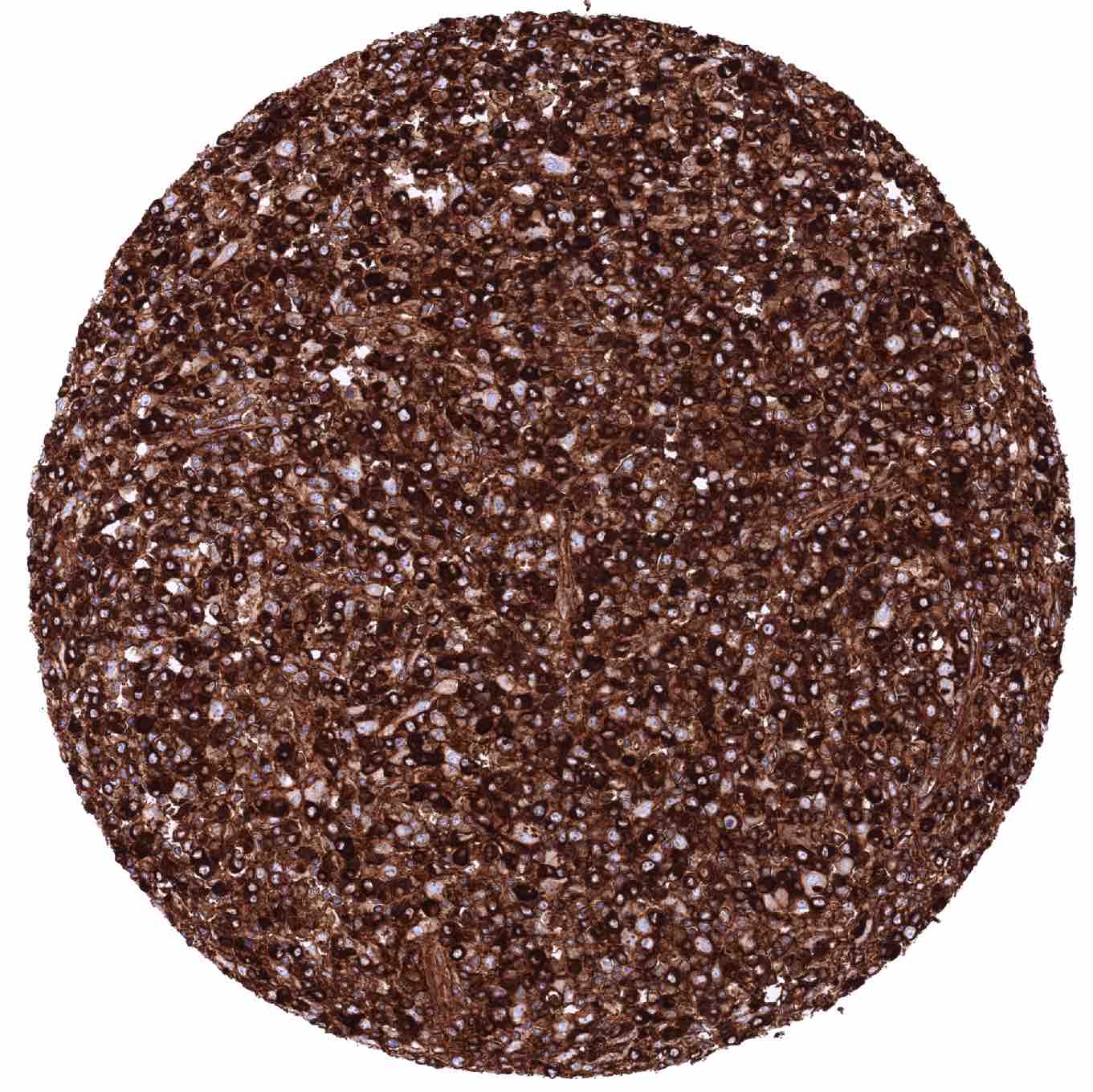
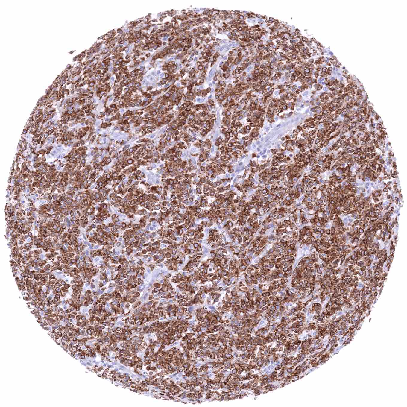
Staining Pattern in Relevant Tumor Types
IgD positivity is most commonly seen in mantle zone lymphomas. A positive IgD immunostaining can occasionally also be seen in other lymphomas and in a very small fraction of plasmacytomas.
Compatibility of Antibodies
No data available at the moment
Protocol Recommendations
IHC users have different preferences on how the stains should look like. Some prefer high staining intensity of the target stain and even accept some background. Others favor absolute specificity and lighter target stains. Factors that invariably lead to more intense staining include higher concentration of the antibody and visualization tools, longer incubation time, higher temperature during incubation, higher temperature and longer duration of the heat induced epitope retrieval (slide pretreatment). The impact of the pH during slide pretreatment has variable effects and depends on the antibody and the target protein.
All images and data shown here and in our image galleries are obtained by the manual protocol described below. Other protocols resulting in equivalent staining are described as well.
Manual protocol
Freshly cut sections should be used (less than 10 days between cutting and staining). Heat-induced antigen retrieval for 5 minutes in an autoclave at 121°C in pH 7,8 Target Retrieval Solution buffer. Apply MSVA-701R at a dilution of 1:150 at 37°C for 60 minutes. Visualization of bound antibody by the EnVision Kit (Dako, Agilent) according to the manufacturer’s directions.
Potential Research Applications
- The role and function of IgD is largely unknown and requires further research.
Evidence for Antibody Specificity in IHC
There are two ways how the specificity of antibodies can be documented for immunohistochemistry on formalin fixed tissues. These are: 1. Comparison with a second independent method for target expression measurement across a large number of different tissue types (orthogonal strategy), and 2. Comparison with one or several independent antibodies for the same target and showing that all positive staining results are also seen with other antibodies for the same target (independent antibody strategy).
Orthogonal validation is not well applicable for IgD antibodies because of the ubiquitous presence of IgD expressing lymphocytes in virtually all organs. Moreover, IgD expression data in normal tissues are unavailable in public databases.
Comparison of antibodies: A specific IgD staining of MSVA-701R in a subset of lymphocytes and plasma cells, in mucus, blood vessels or in the stroma of areas of inflammation is corroborated by comparison with a commercially available independent second antibody (termed “validation antibody”). In this comparison, all stainings obtained by MSVA-701R were matched by the “validation antibody”. It is of note that the “validation antibody” – an IVDR RTU – shows an oversensitive staining in this analysis.