
295,00 € – 995,00 €
Product details
Synonyms = GAD65
Antibody type = Mouse monoclonal / IgG1, Kappa
Clone = MSVA-602M
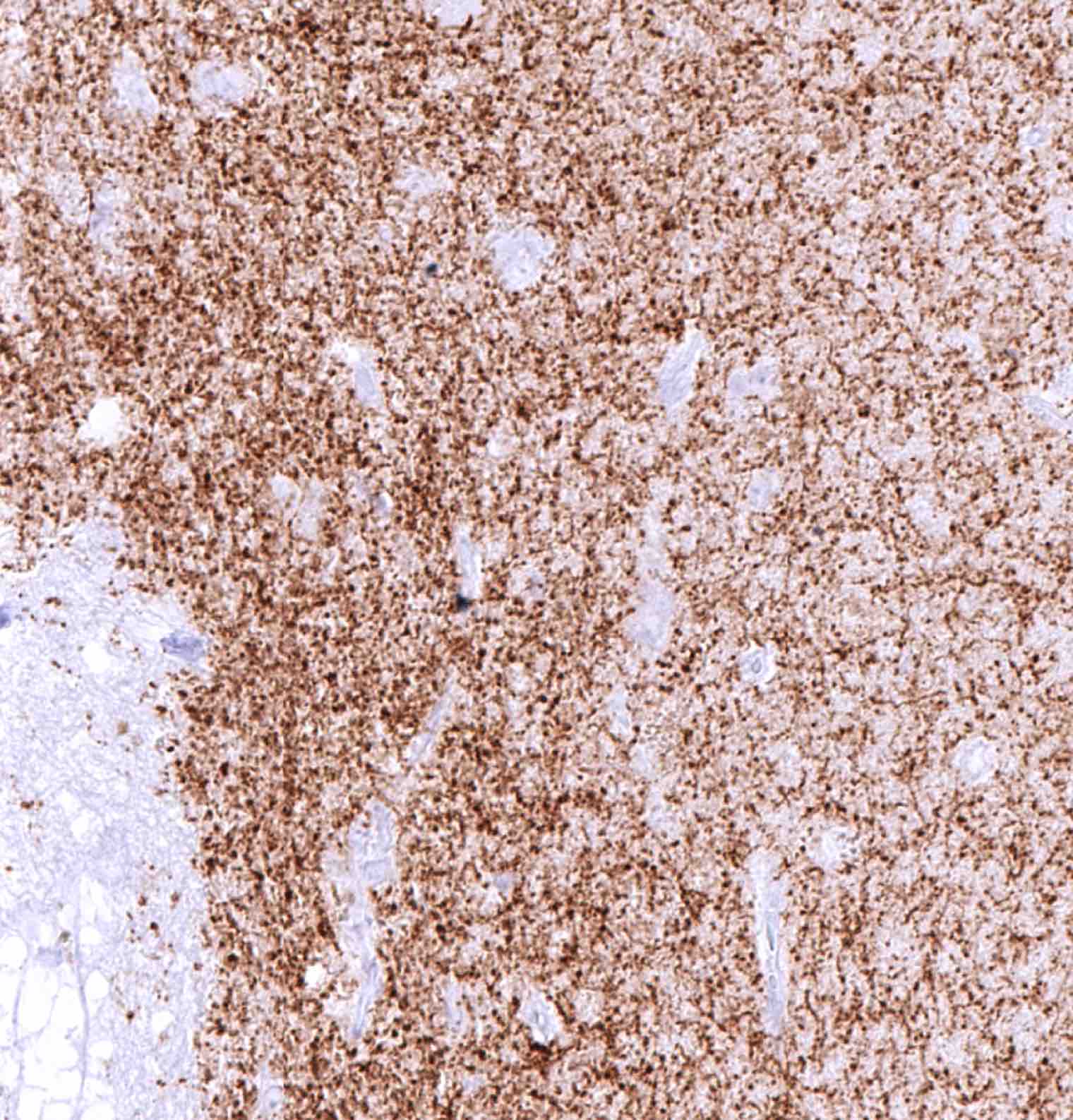
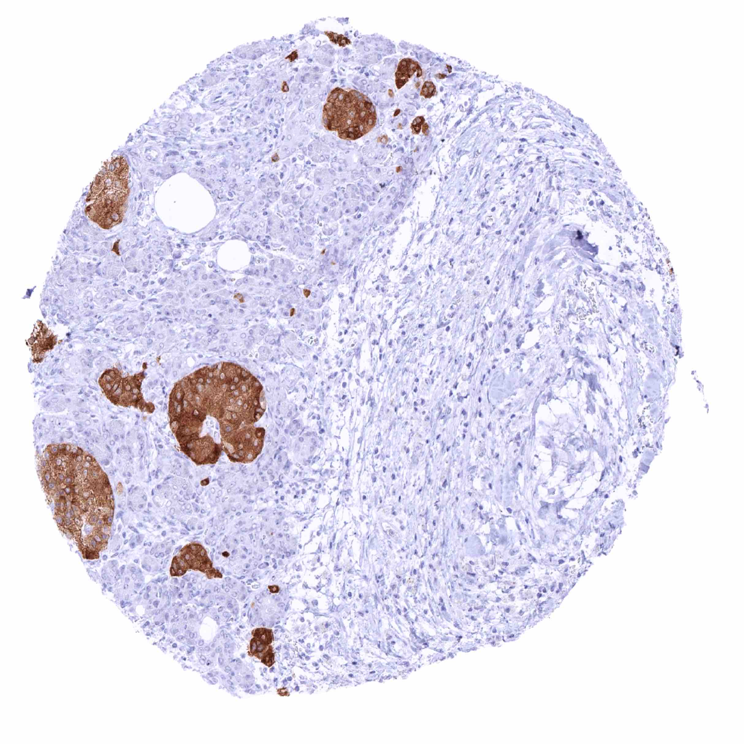
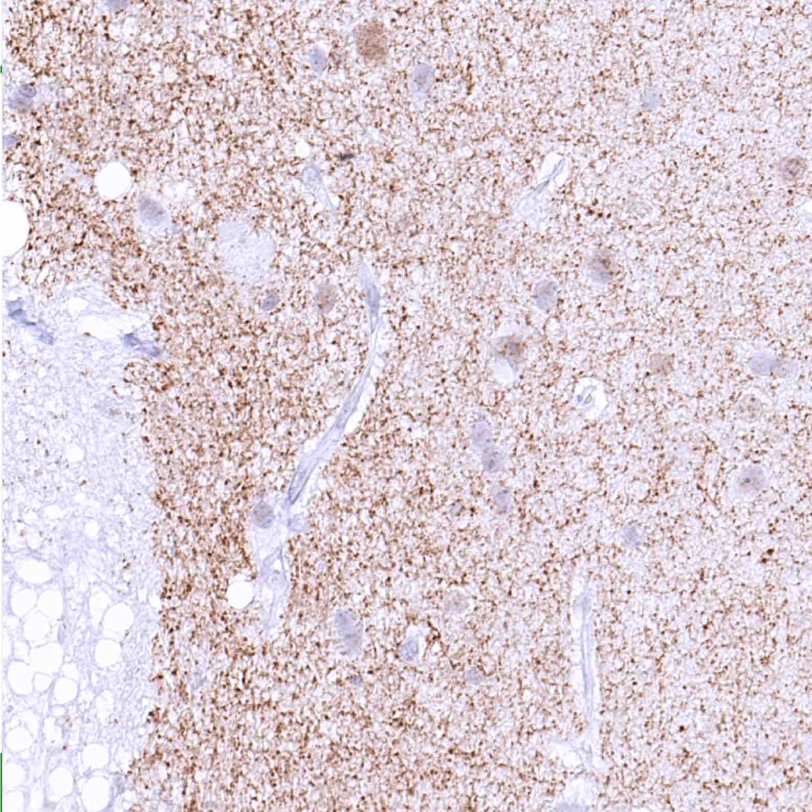
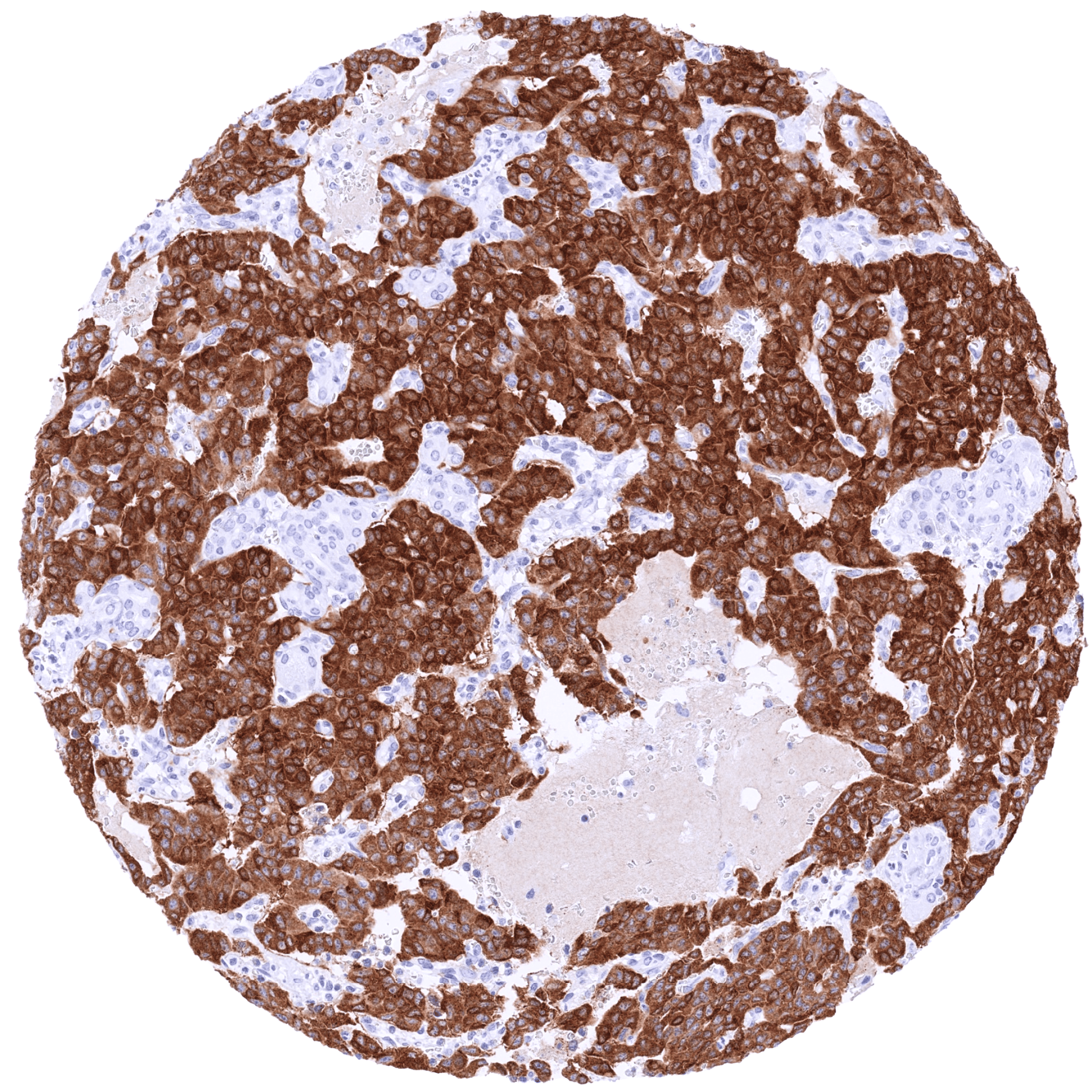
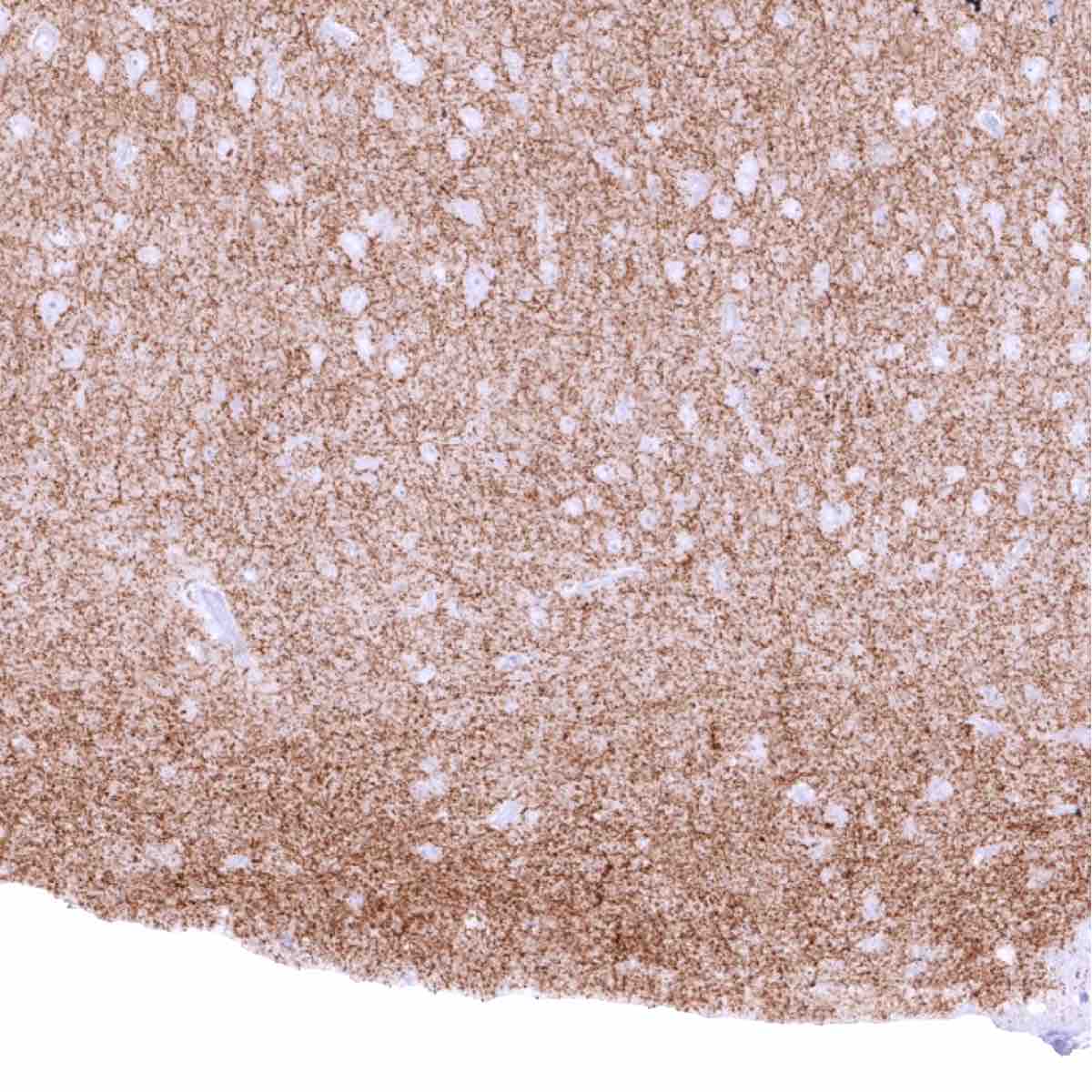
Positive control = Pancreas: A strong GAD2 staining should be seen in pancreatic islet cells.
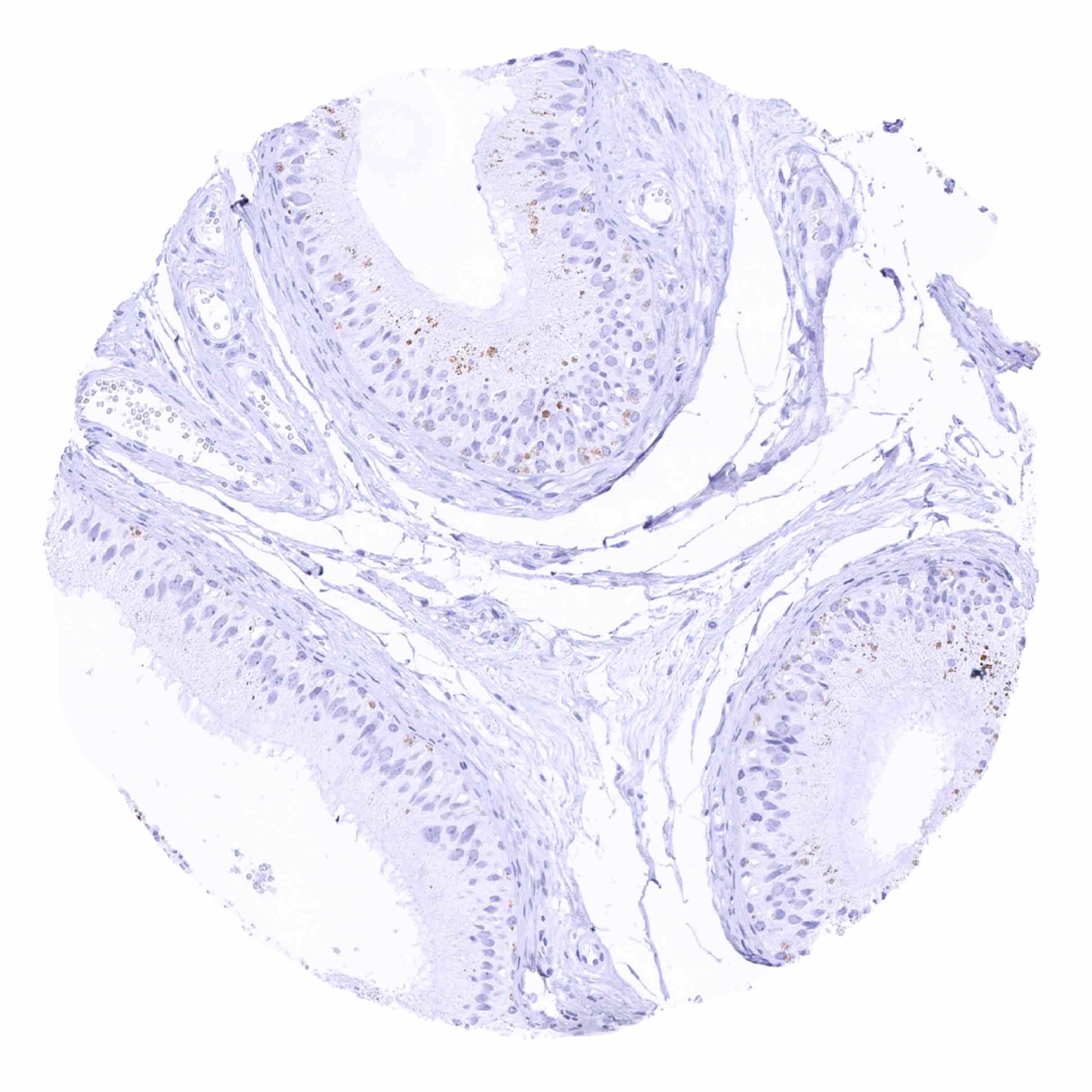
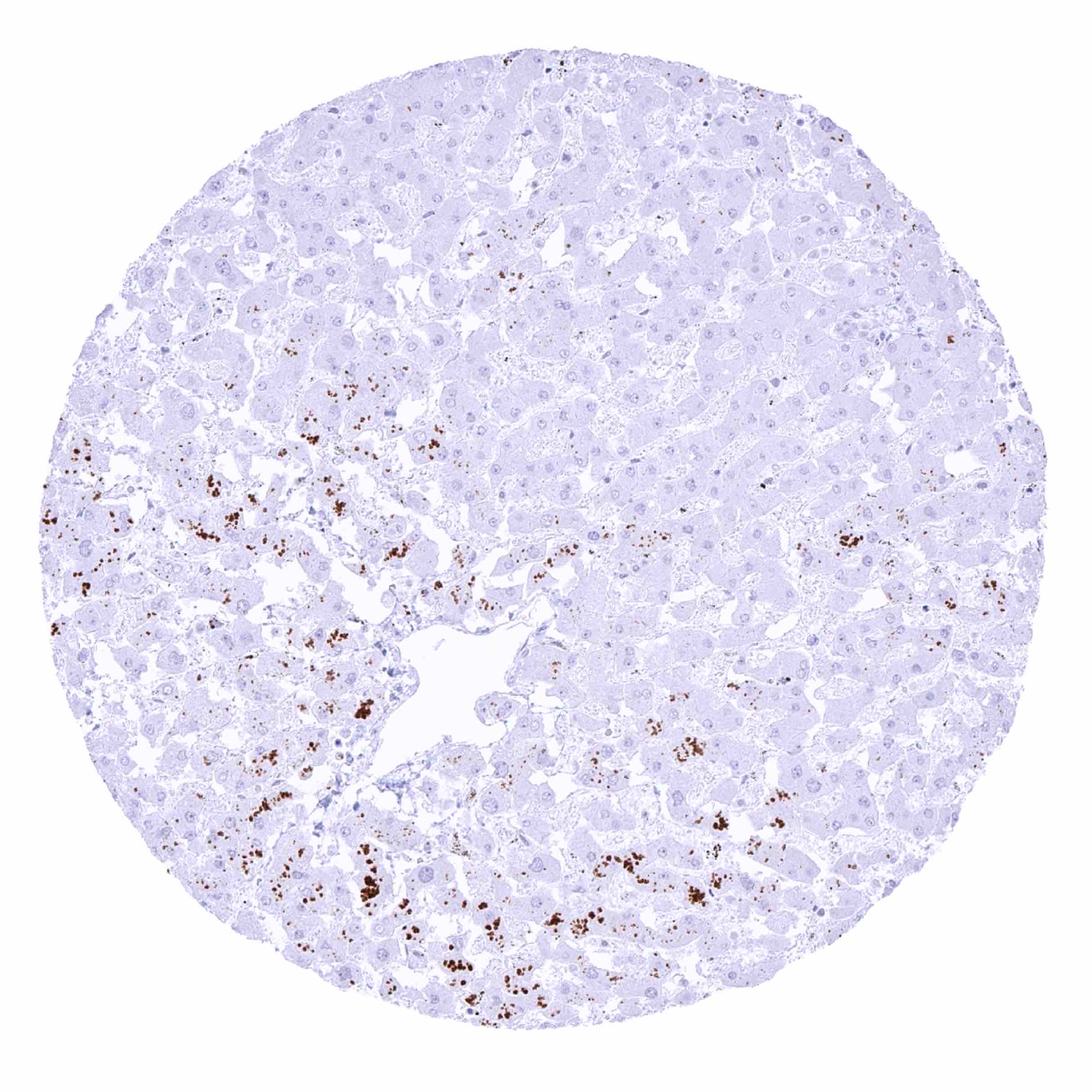
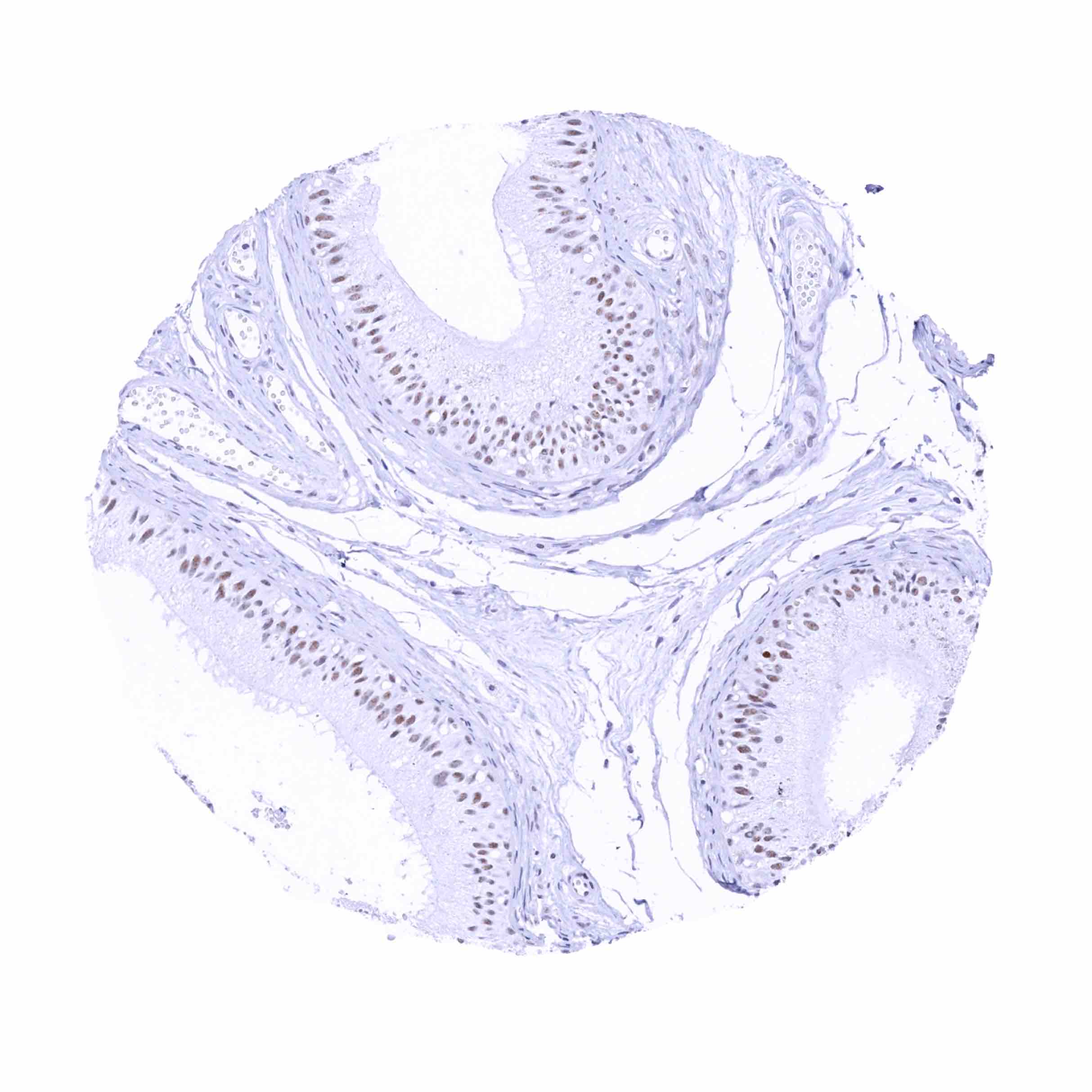
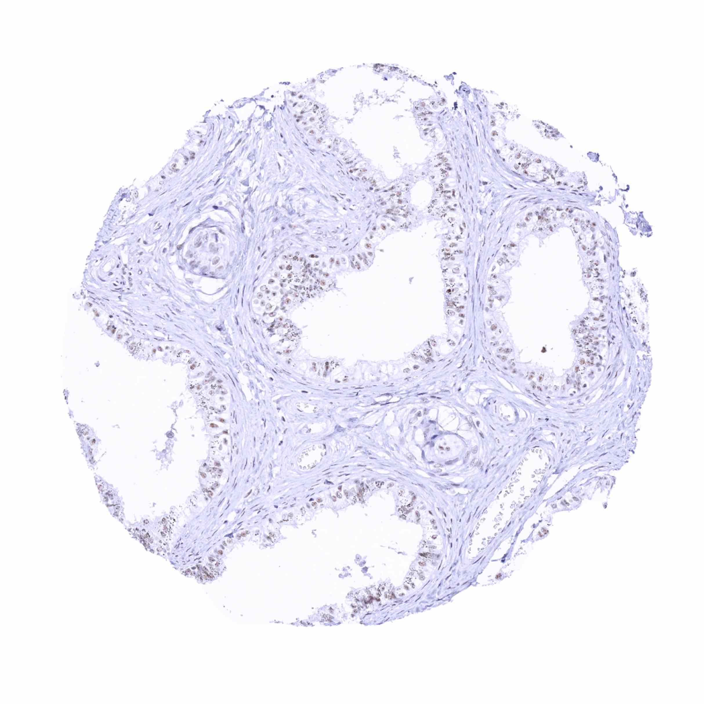
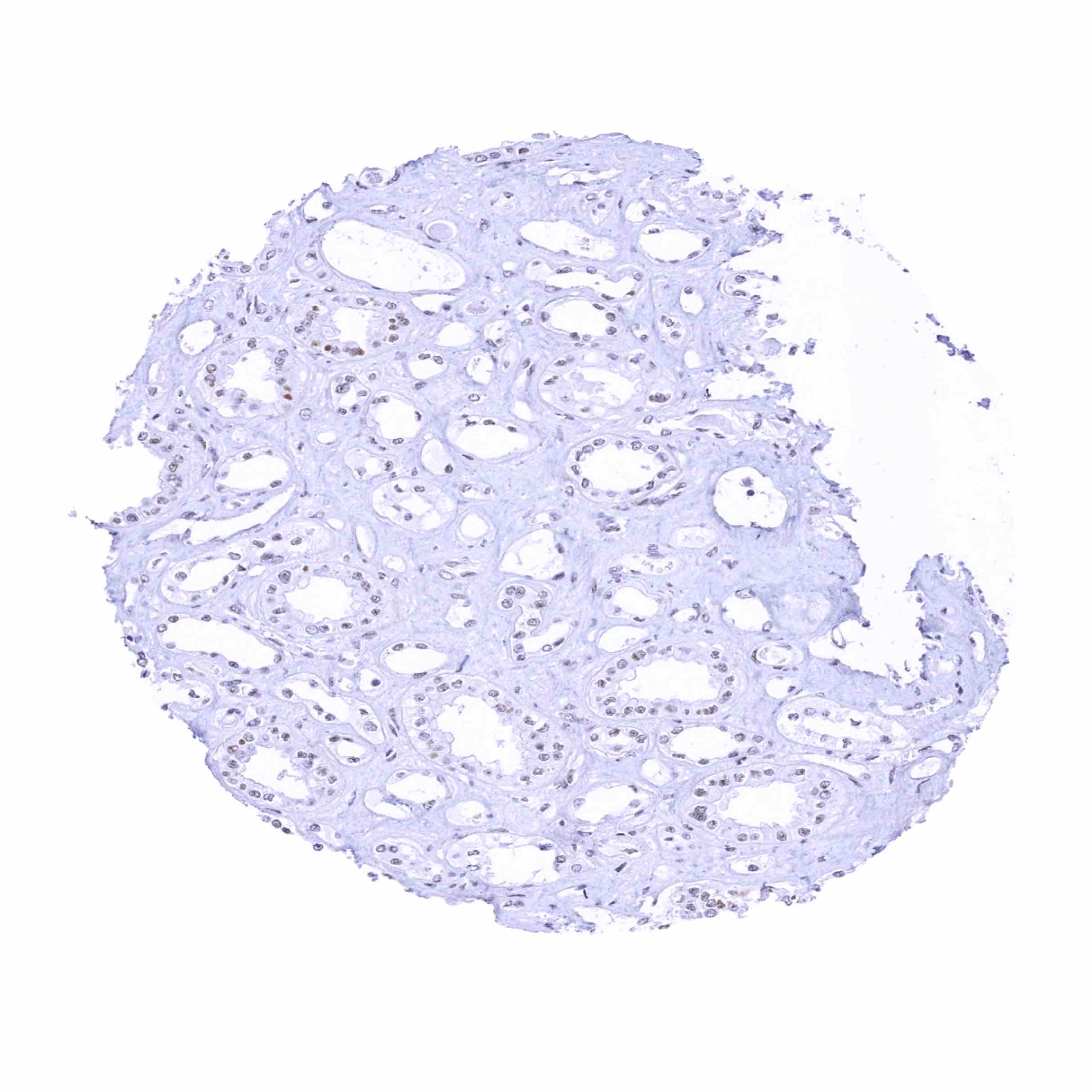
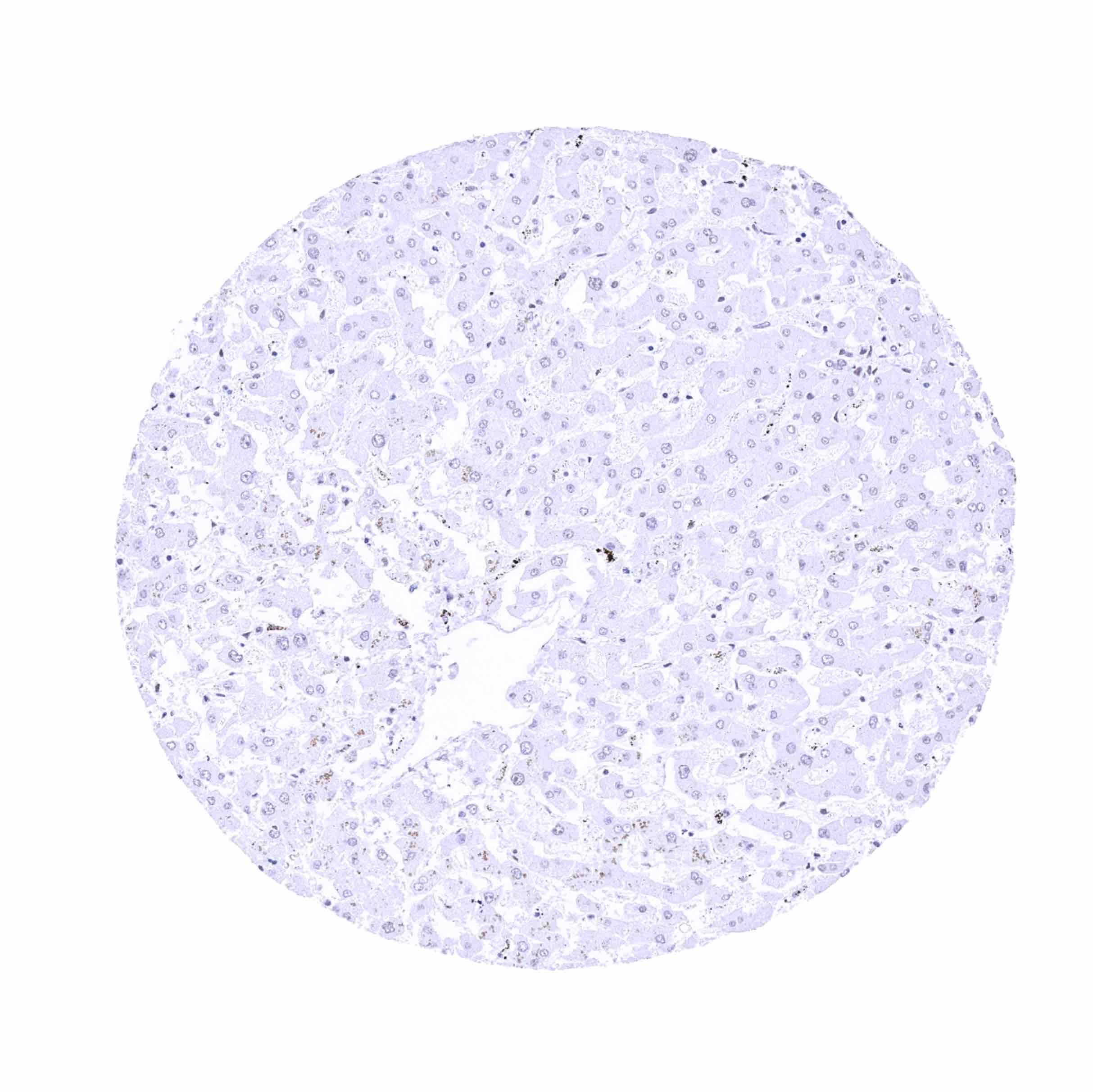
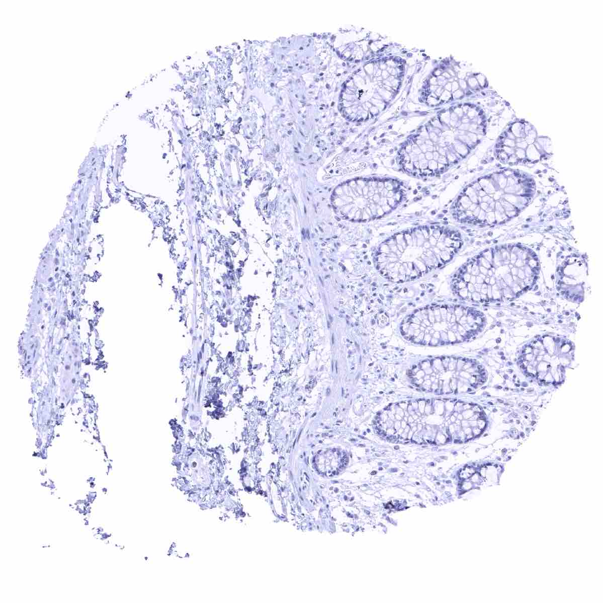
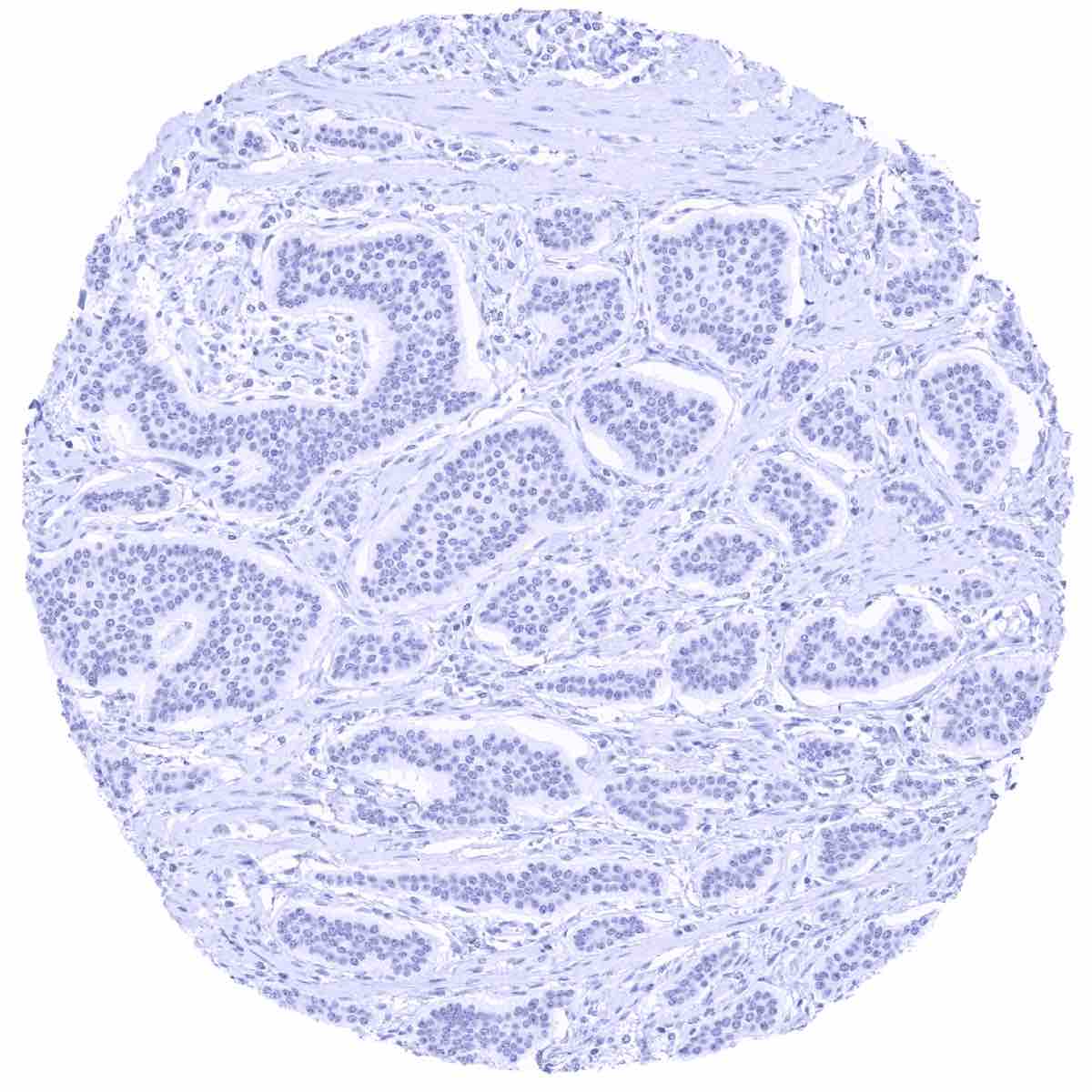
Negative control = Colon: GAD2 immunostaining should be absent in all cell types
Cellular localization = Intracellular
Reactivity = Human
Application = Immunohistochemistry
Dilution = 1:100 – 1:200
Intended Use = Research Use Only
Relevance of Antibody
GAD2 is highly expressed in pancreatic islet cells.
Biology Behind
Glutamate decarboxylase 2 (GAD2) is coded by the GAD2 gene at chromosome 10p12. It is one out of two glutamate decarboxylases that catalyze the decarboxylation of glutamate to GABA and CO2. GABA (gamma-Aminobutyric acid) is the most relevant inhibitory neurotransmitter in the central nervous system which serves the purpose of reducing neuronal excitability. In the brain, GAD2 is therefore required at nerve terminals and synapses. In the pancreas, GAD2 plays a role in insulin-producing β-cells of pancreatic islets. GAD2 is involved in several different disease types. It acts as a target for autoantibodies in people who later develop type 1 diabetes. Downregulation of GAD2 occurs in autism.
Staining Pattern in Normal Tissues
Images describing the GAD2 staining pattern in normal tissues obtained by the antibody MSVA-602M are shown in our “Normal Tissue Gallery”.
These findings are fully consistent with the RNA data described in the Human Protein Atlas (Tissue expression GAD2). Both organs with documented Gad2 RNA expression (brain, pancreas) are IHC positive for MSVA-602M. Moreover, immunostaining by MSVA-602M is limited to these organs.
Positive control = Pancreas: A strong GAD2 staining should be seen in pancreatic islet cells.
Negative control = Colon: GAD2 immunostaining should be absent in all cell types

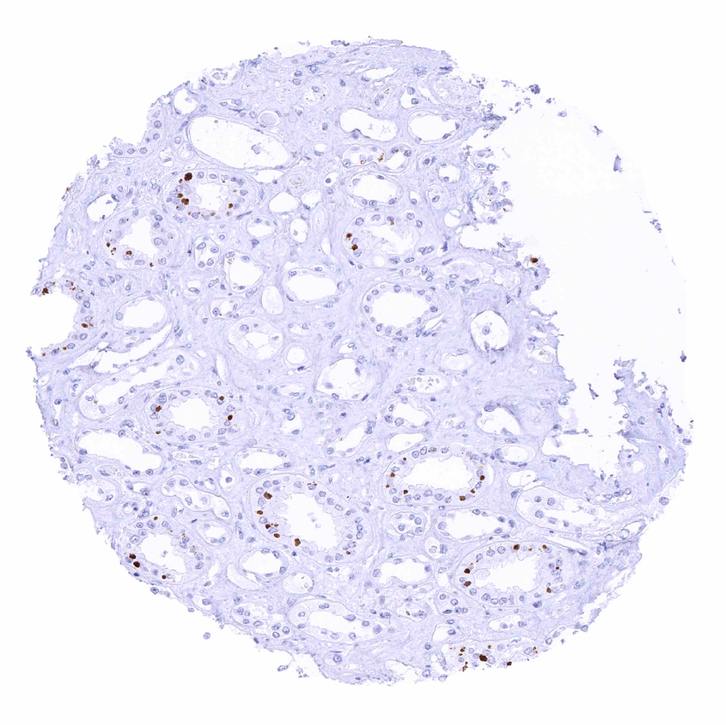
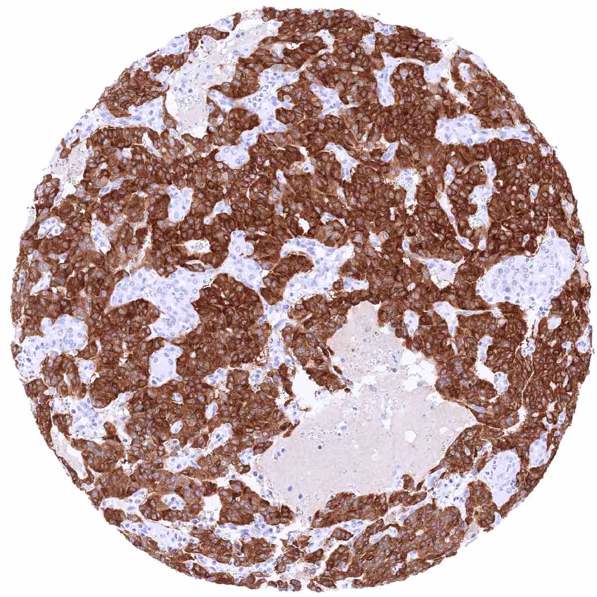
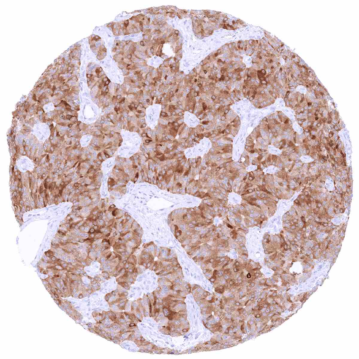
Staining Pattern in Relevant Tumor Types
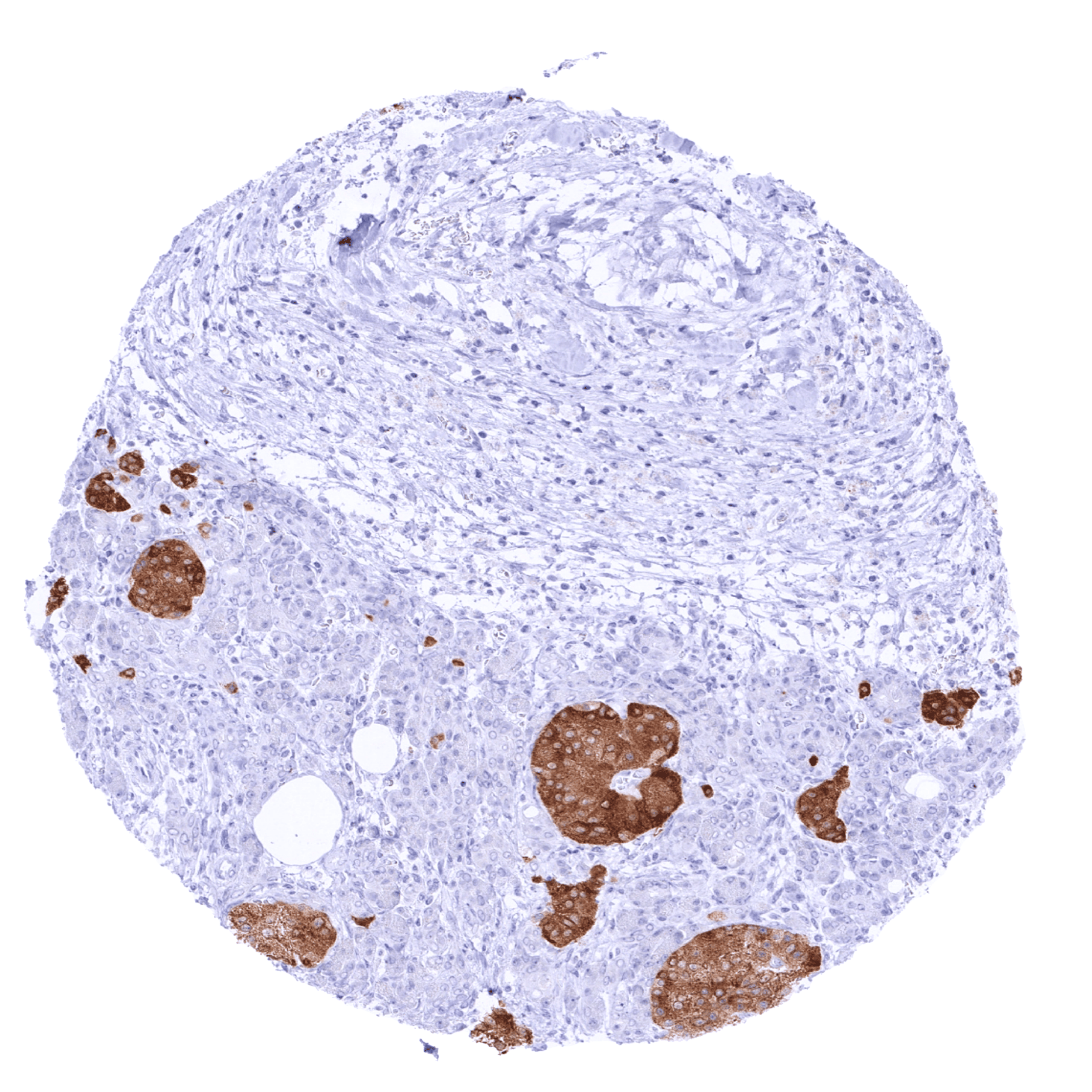
A positive GAD2 immunostaining is often seen in neuroendocrine tumors of the pancreas and – much less commonly – also in various other tumor entities.
The TCGA findings on GAD2 RNA expression in different tumor categories have been summarized in the GAD2 RNA expression in different tumor categories.
Compatibility of Antibodies
GAD2 (MSVA-602M) publication summary
Relevant publication: Lennartz et al: “GAD2 Is a Highly Specific Marker for Neuroendocrine Neoplasms of the Pancreas” published in Am J Surg Pathol. 2024 Jan 25. doi: 10.1097/PAS.0000000000002186. Epub ahead of print. PMID: 38271200.
A total of 17507 tumors were analyzed from 152 different tumor categories by using the following protocol: Heat-induced antigen retrieval for 5 minutes in an autoclave at 121°C in pH 9,0 Target Retrieval Solution buffer. MSVA-602M at a dilution of 1:150 at 37°C for 60 minutes. Visualization of bound antibody by the EnVision Kit (Dako, Agilent). This protocol was also used for all stainings depicted in our tumor and normal tissue galleries.
The analysis revealed that GAD2 expression was strictly limited to the brain and islet cells of the pancreas in normal tissues. Accordingly, GAD2 expression in (extracranial) tumors severely predominated in (neuroendocrine) neoplasms derived from the pancreatic islets.
While 20 of 152 tumor categories contained at least one case with weak GAD2 expression there were only 5 tumor categories includingat least 1 case with strong GAD2 positivity. GAD2 immunostaining was most commonly seen in neuroendocrine tumors (63.2% of 87) and neuroendocrine carcinomas (58.3% of 12) of the pancreas, followed by granular cell tumors (37.0%) and neuroendocrine tumors of the lung (11.1%). The distribution of positive staining results is shown in “organ-systematic” and in “ranking order” figures below (images based on data from Lennartz et al).
Authors conclusions on diagnostic utility of GAD2 IHC with respect to the distinction of different tumor entities (Lennartz et al.):
- GAD2 was proposed to represent a useful marker for the distinction of neuroendocrine neoplasms of the pancreas from other neuroendocrine neoplasms (For this application, Lennartz et al proposed a sensitivity of 64,2%, a specificity of 96,3%, and a positive predictive value of 87,7%).
- The authors also assessed the diagnostic value of GAD2 immunostaining in combination of previous data on PR expression obtained by MSVA-570R and suggested a >99% specificity for neuroendocrine neoplasms with positivity for both GAD2 and PR to be of pancreatic origin.
Figure 1. GAD2 staining in cancer (“organ-systematic”; according to Lennartz et al.)

Figure 2. GAD2 staining in cancer (“ranking list”; according to Lennartz et al.)

Protocol Recommendations
IHC users have different preferences on how the stains should look like. Some prefer high staining intensity of the target stain and even accept some background. Others favor absolute specificity and lighter target stains. Factors that invariably lead to more intense staining include higher concentration of the antibody and visualization tools, longer incubation time, higher temperature during incubation, higher temperature and longer duration of the heat induced epitope retrieval (slide pretreatment). The impact of the pH during slide pretreatment has variable effects and depends on the antibody and the target protein.
All images and data shown here and in our image galleries are obtained by the manual protocol described below. Other protocols resulting in equivalent staining are described as well.
Manual protocol
Freshly cut sections should be used (less than 10 days between cutting and staining). Heat-induced antigen retrieval for 5 minutes in an autoclave at 121°C in pH 7,8 Target Retrieval Solution buffer. Apply MSVA-602M at a dilution of 1:150 at 37°C for 60 minutes. Visualization of bound antibody by the EnVision Kit (Dako, Agilent) according to the manufacturer’s directions.
Potential Research Applications
- The function of GAD2 in neurological and behavioral disorders deserves further investigation.
- The diagnostic utility of GAD2 immunohistochemistry has not been evaluated.
Evidence for Antibody Specificity in IHC
There are two ways how the specificity of antibodies can be documented for immunohistochemistry on formalin fixed tissues. These are: 1. Comparison with a second independent method for target expression measurement across a large number of different tissue types (orthogonal strategy), and 2. Comparison with one or several independent antibodies for the same target and showing that all positive staining results are also seen with other antibodies for the same target (independent antibody strategy).
Orthogonal validation: For the antibody MSVA-602M, specificity is suggested by the complete concordance of its immunostaining results with data from three independent RNA screening studies, including the Human Protein Atlas (HPA) RNA-seq tissue dataset, the FANTOM5 project, and the Genotype-Tissue Expression (GTEx) project, which are all summarized in the Human Protein Atlas (Tissue expression GAD2). Immunostaining by MSVA-602M was only seen in the pancreas and the brain. These are the only two evaluated normal tissue types for which RNA expression has been described.
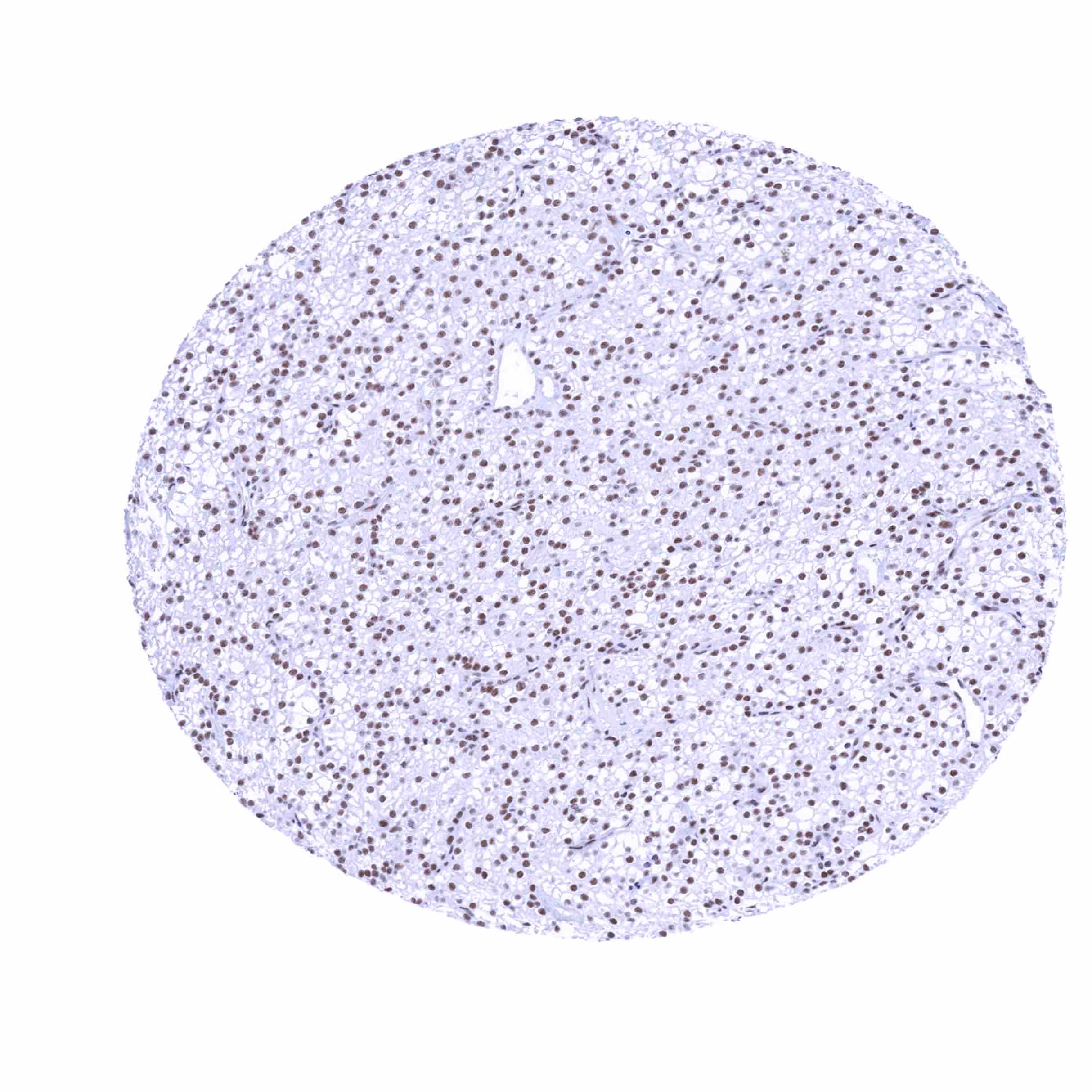
Comparison of antibodies: Specific GAD2 staining by MSVA-602M was also suggested by a confirmation of all MSVA-602M stainings by an independent – commercially available – second antibody termed “validation antibody”. Independence of the two antibodies MSVA-602M and “validation antibody” is documented by additional nuclear staining seen by validation antibody in numerous tissues. This nuclear staining is considered a cross-reactivity of the “validation antibody”. Note: Staining of physiologic pigments (epididymis, liver, kidney) appears to be stronger in case of MSVA-602M than seen for the validation antibody.