
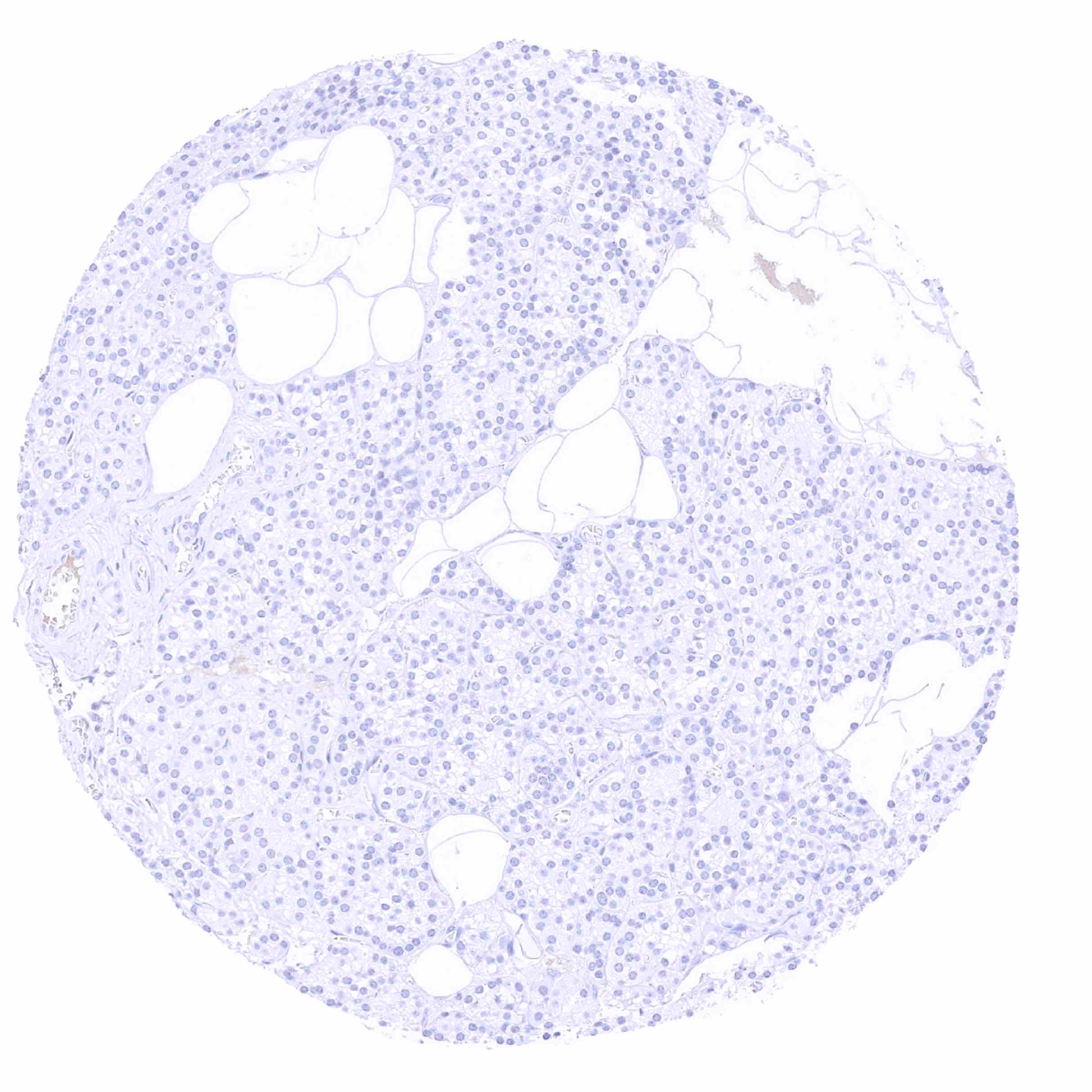
Adrenal gland
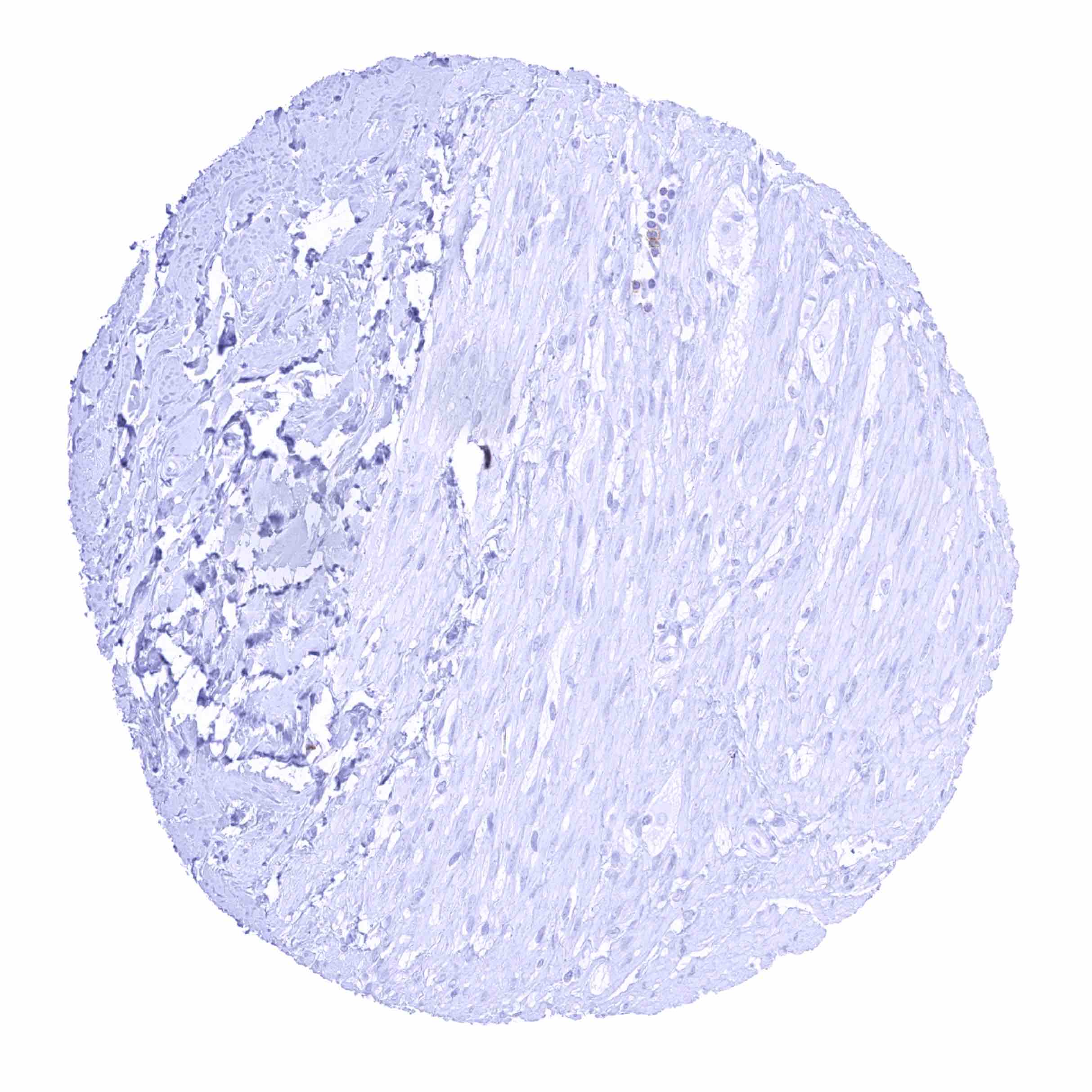
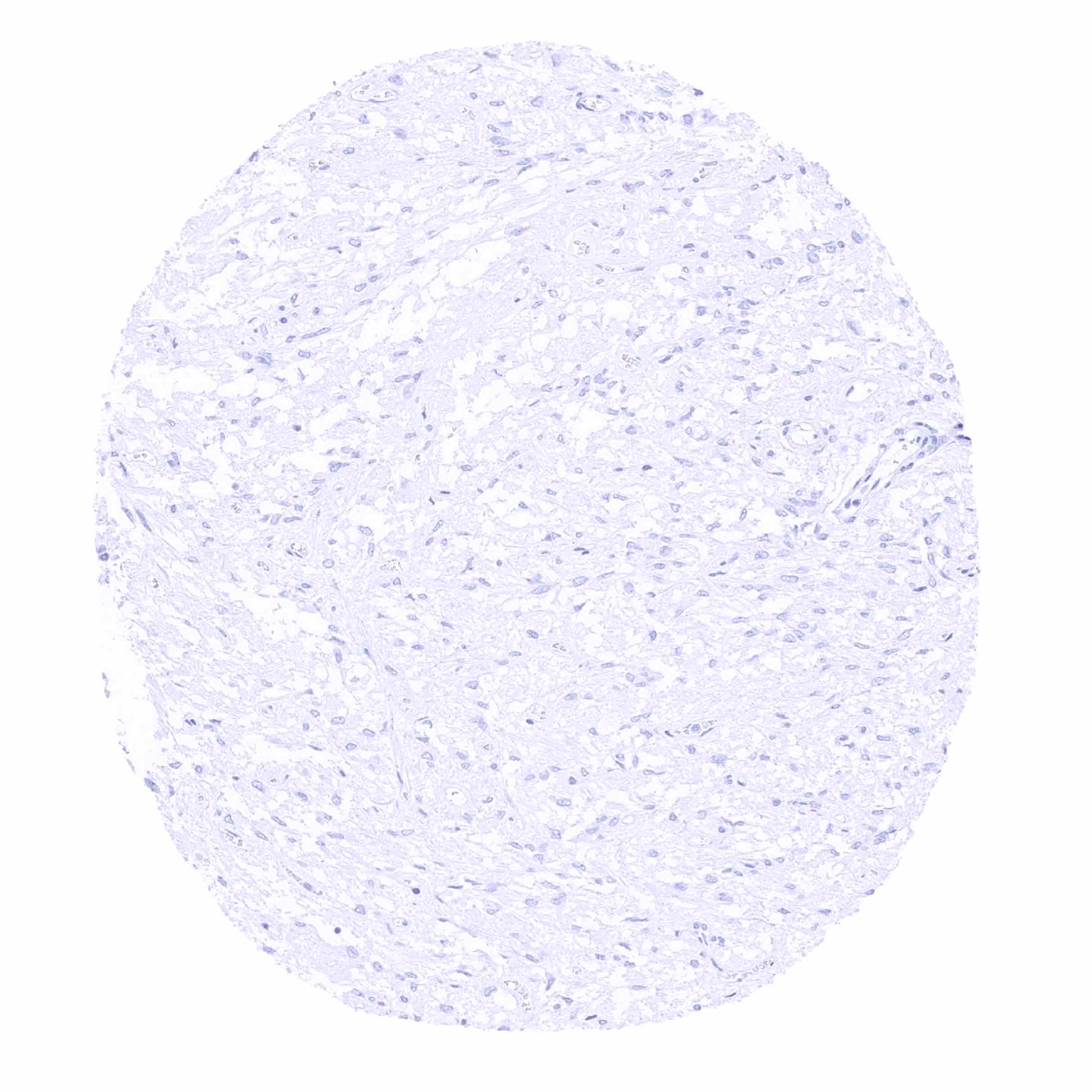
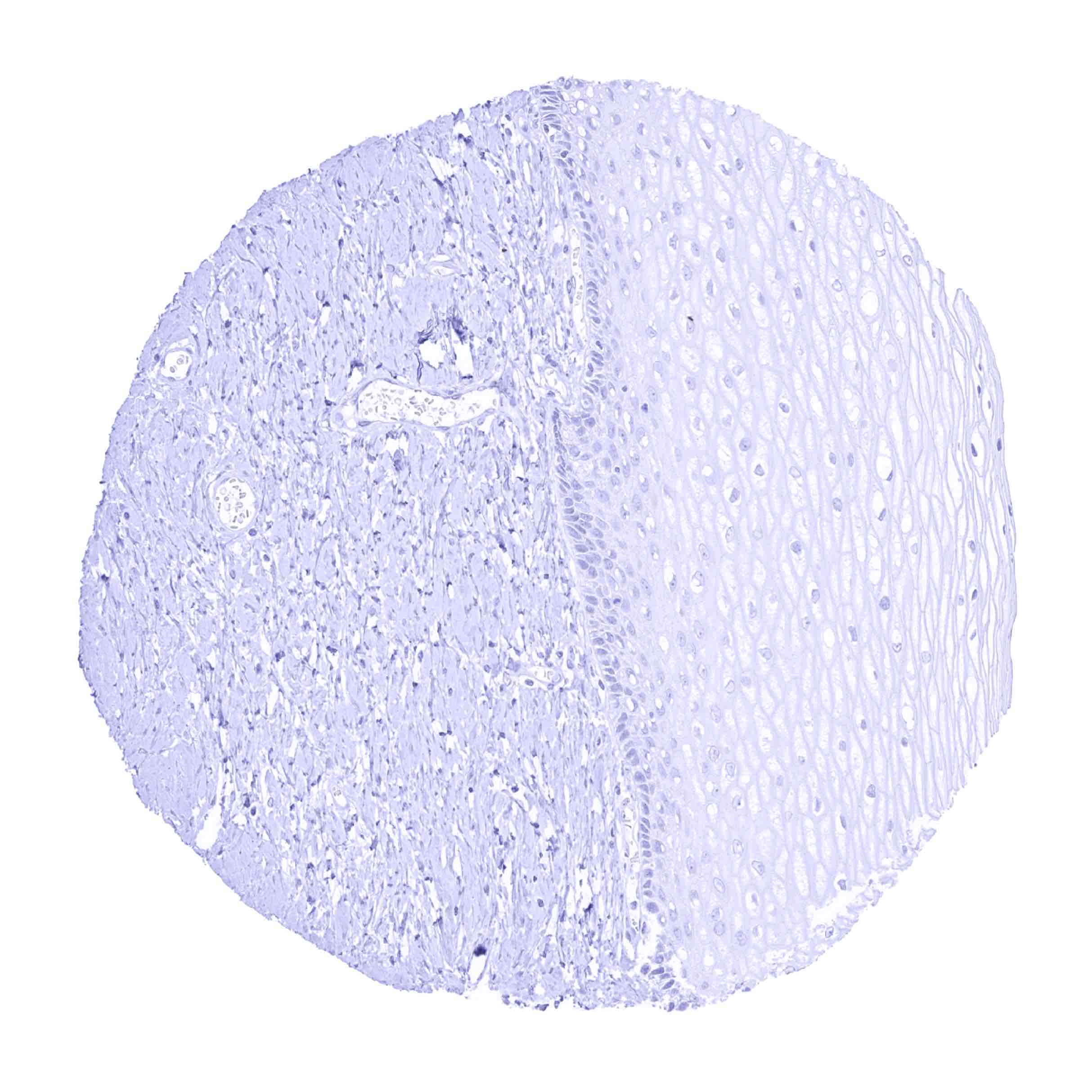
Aorta, media
Appendix, mucosa - IgD immunostaining of mantle cells surrounding a germinal centre of the appendical mucosa

Appendix, mucosa - IgD staining is largely limited to mantle cells surrounding a germinal centre of the appendical mucosa
Appendix, mucosa
Appendix, muscular wall
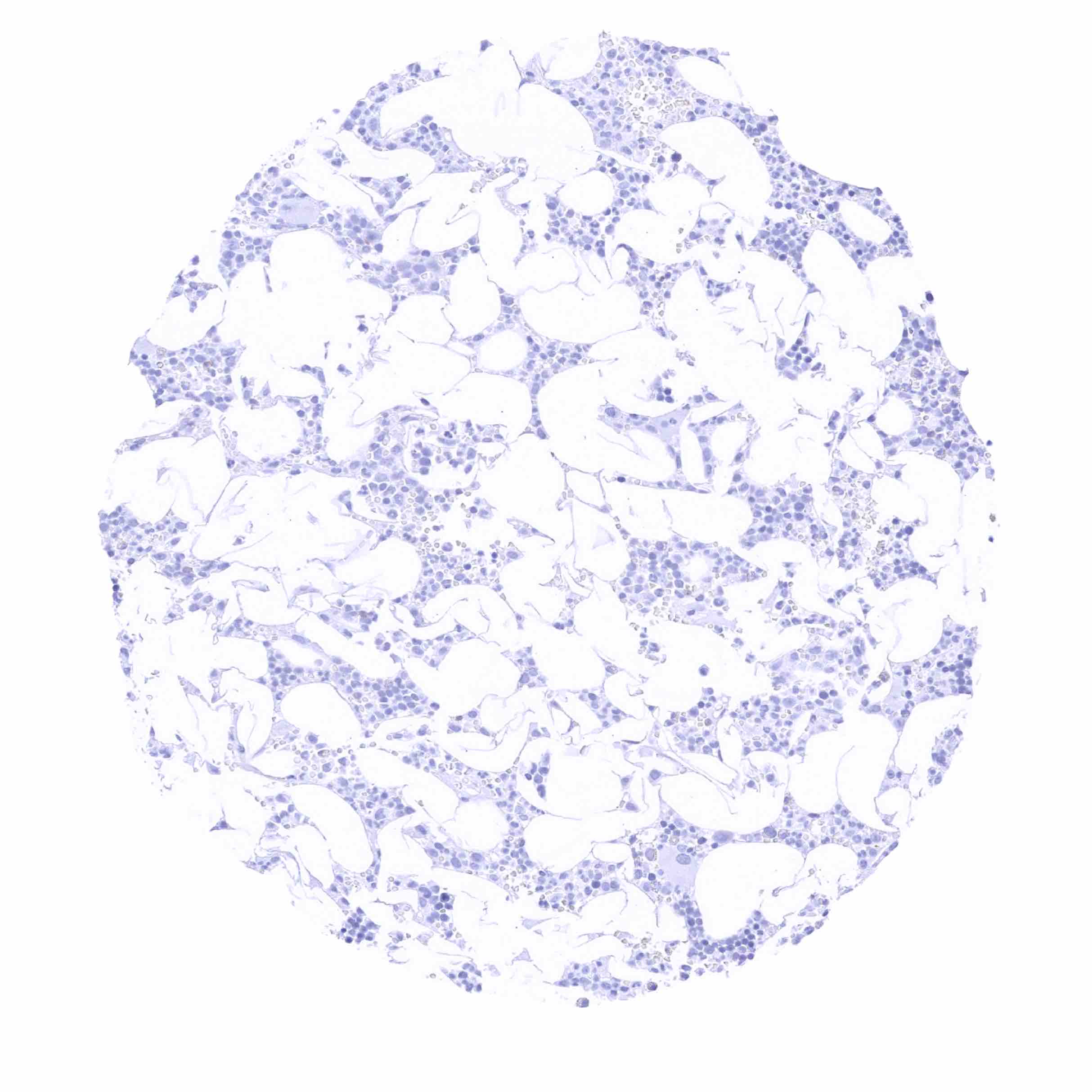
Bone marrow
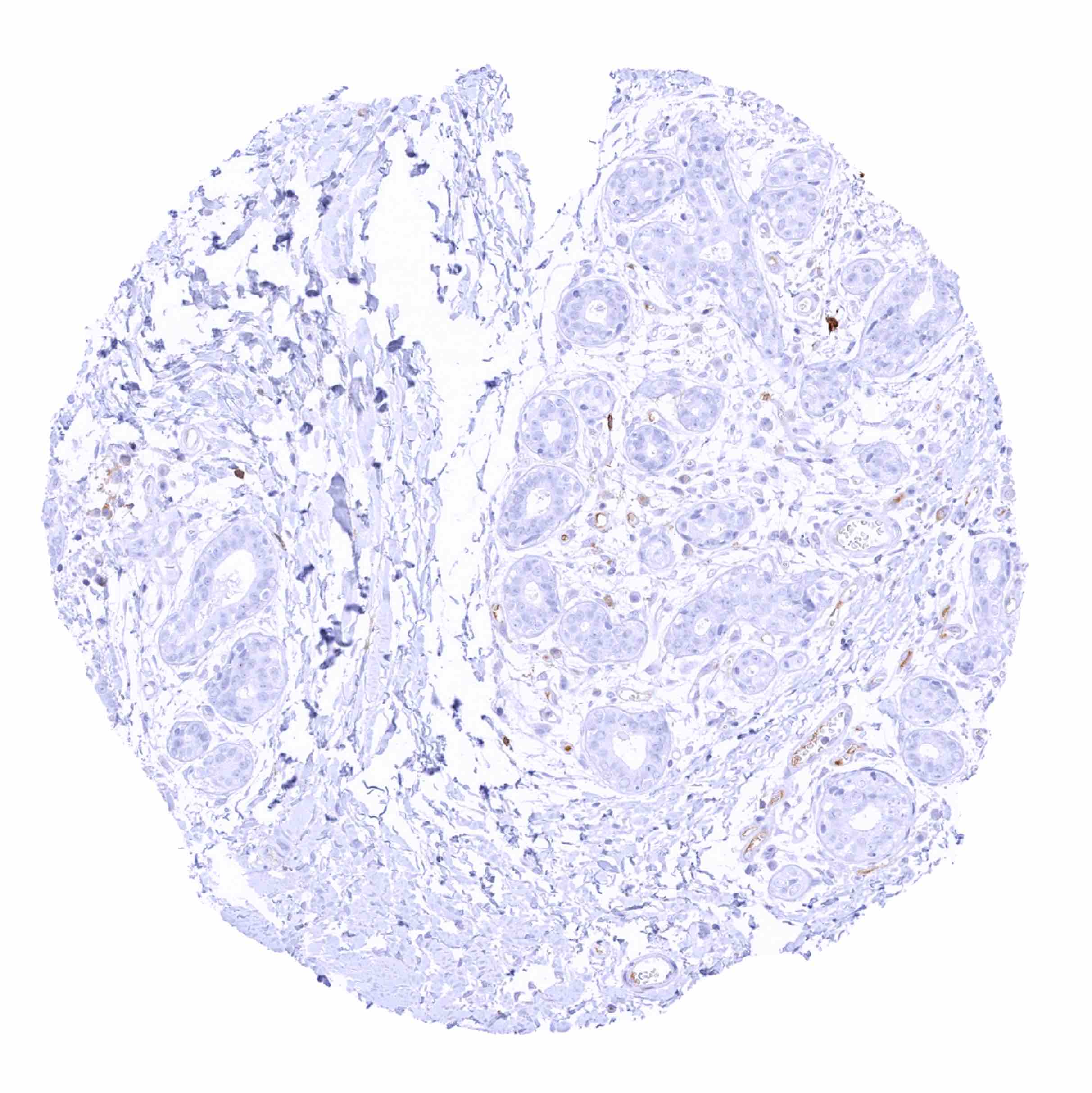
Breast
Bronchus, mucosa - Few plasma cells show strong IgD staining
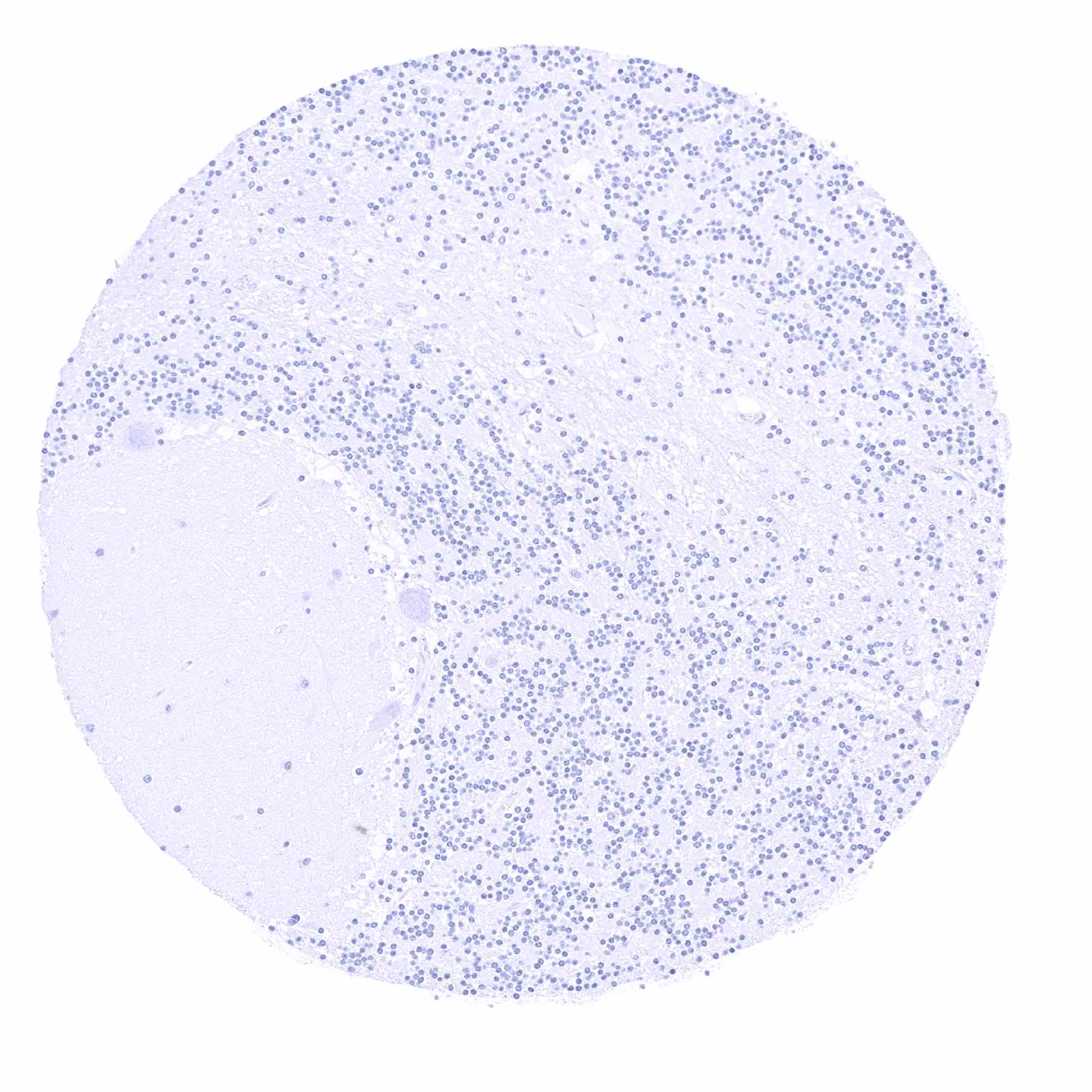
Cerebellum, cortex (Stratum moleculare)

Cerebellum, grey (Stratum neuronorum)
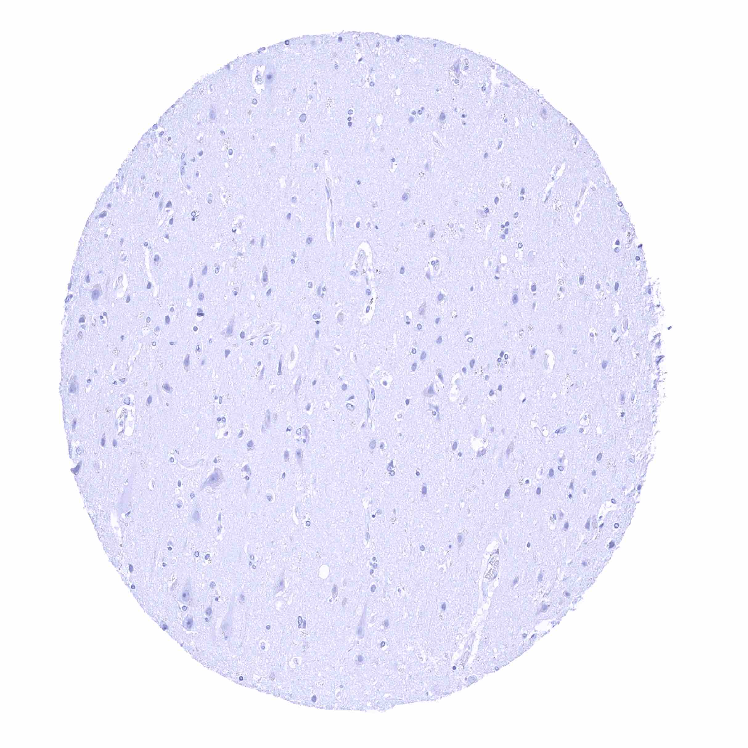
Cerebrum, grey matter
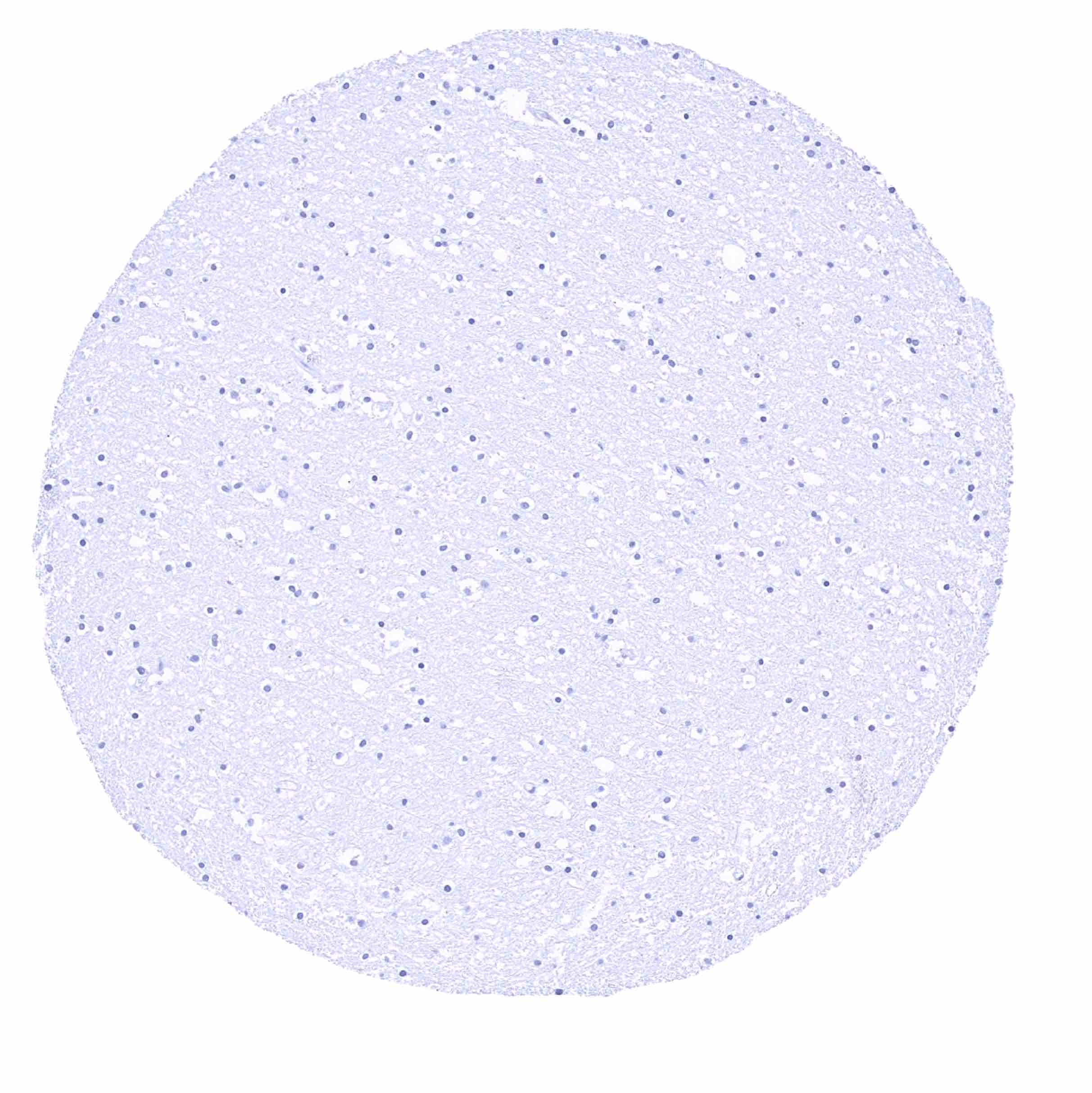
Cerebrum, white matter
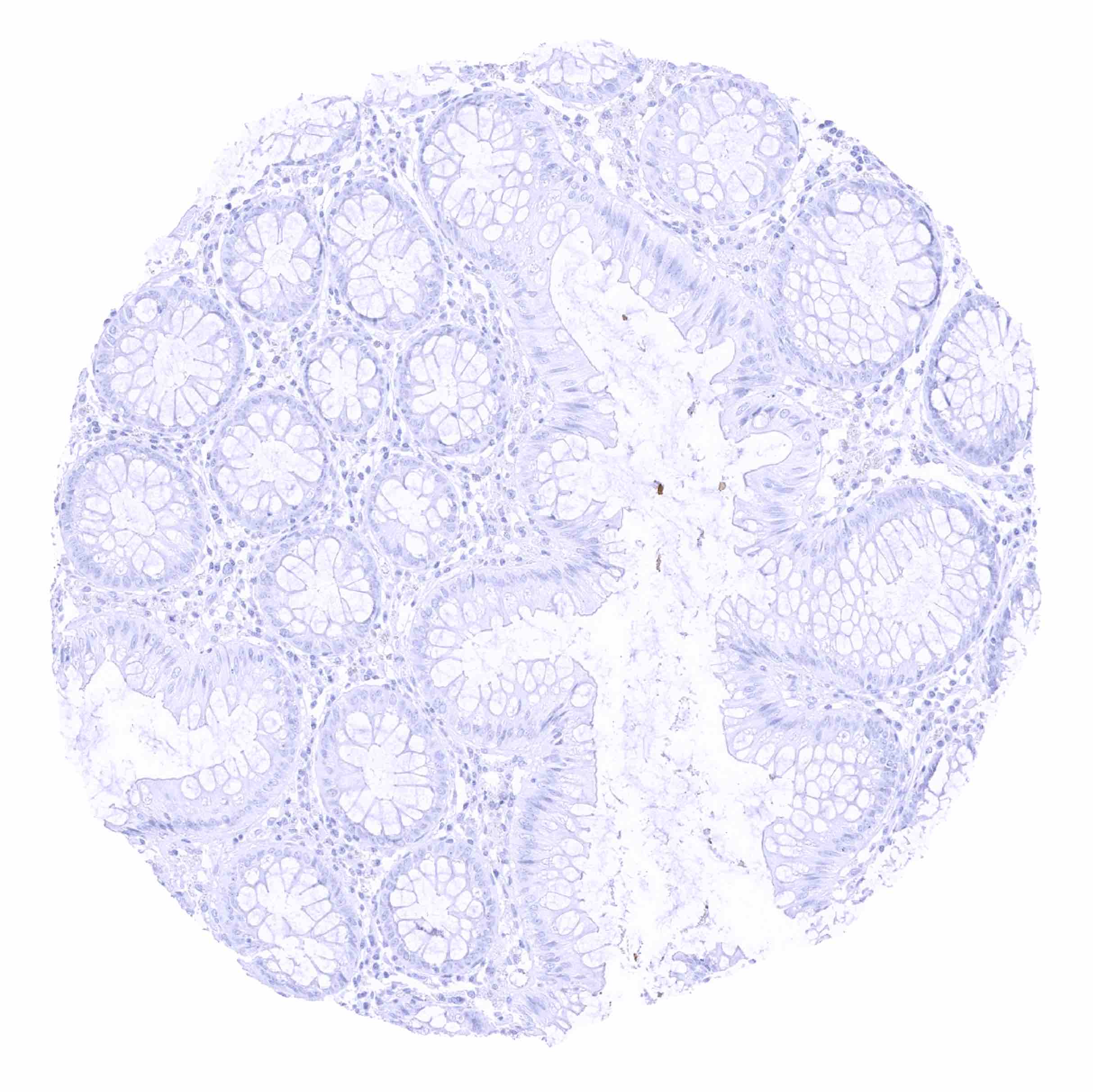
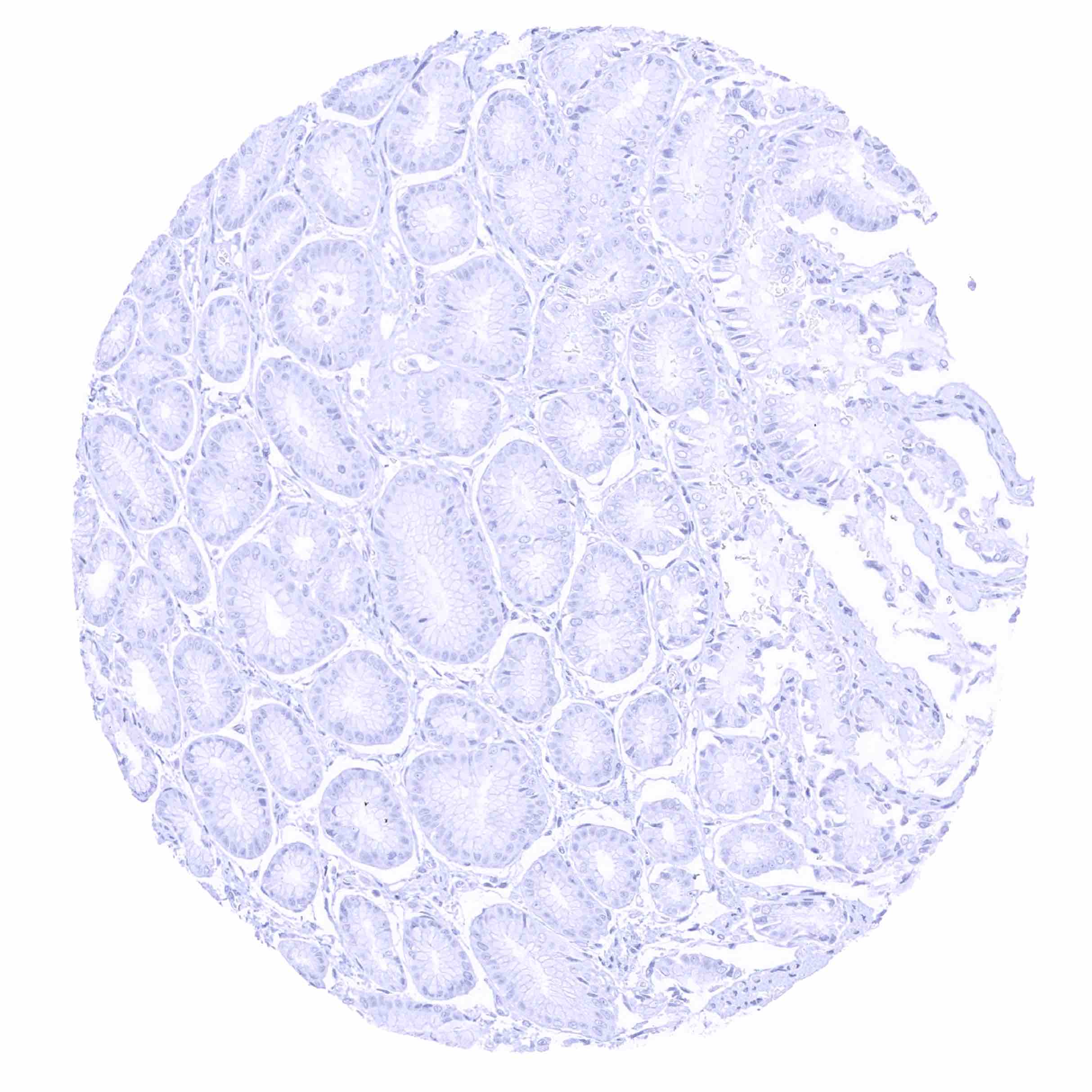
Colon descendens, mucosa

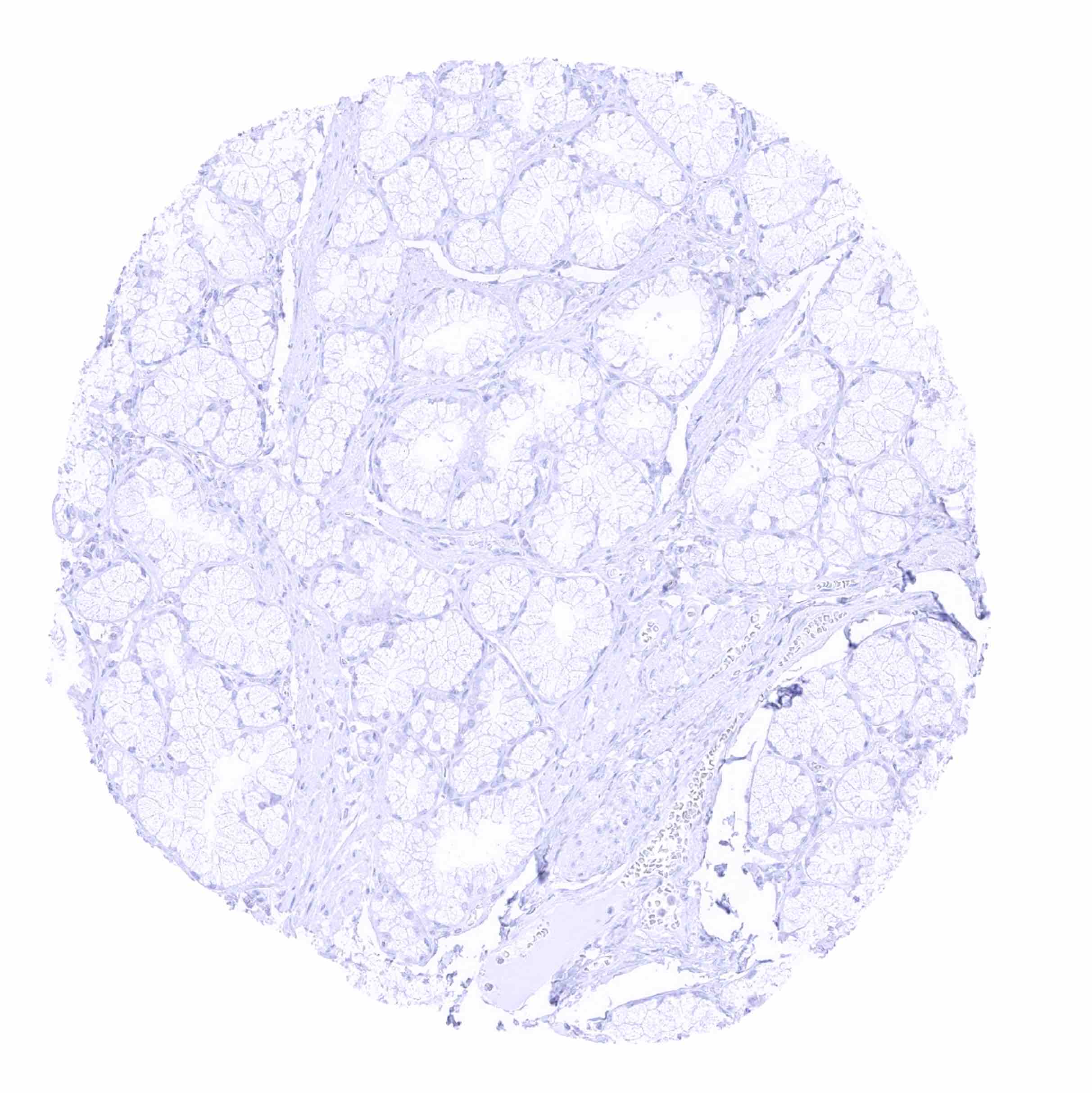
Colon descendens, muscular wall
Duodenum, Brunner gland
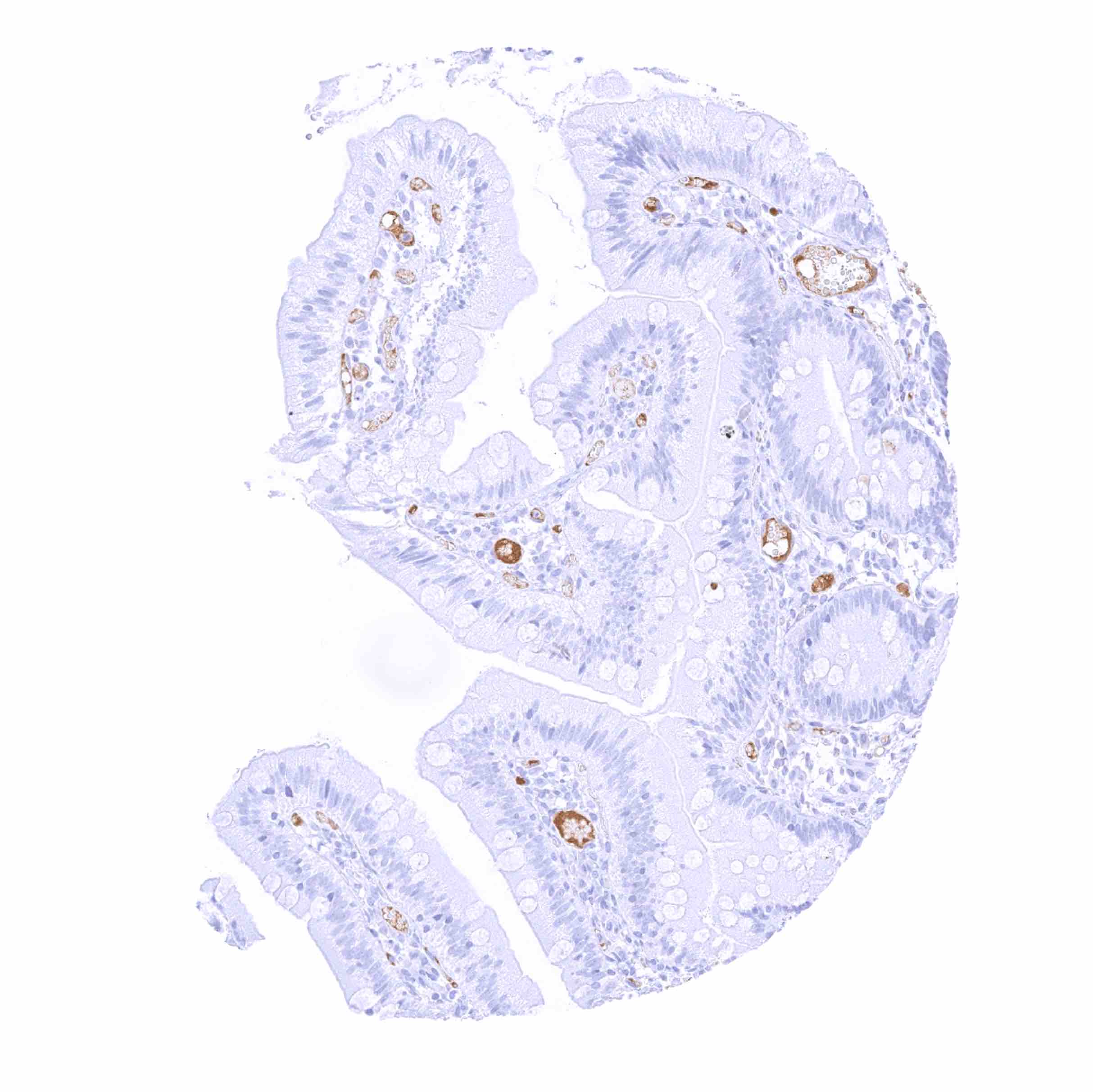
Duodenum, mucosa - IgD staining of few inflammatory cells and of intravascular material
Epididymis
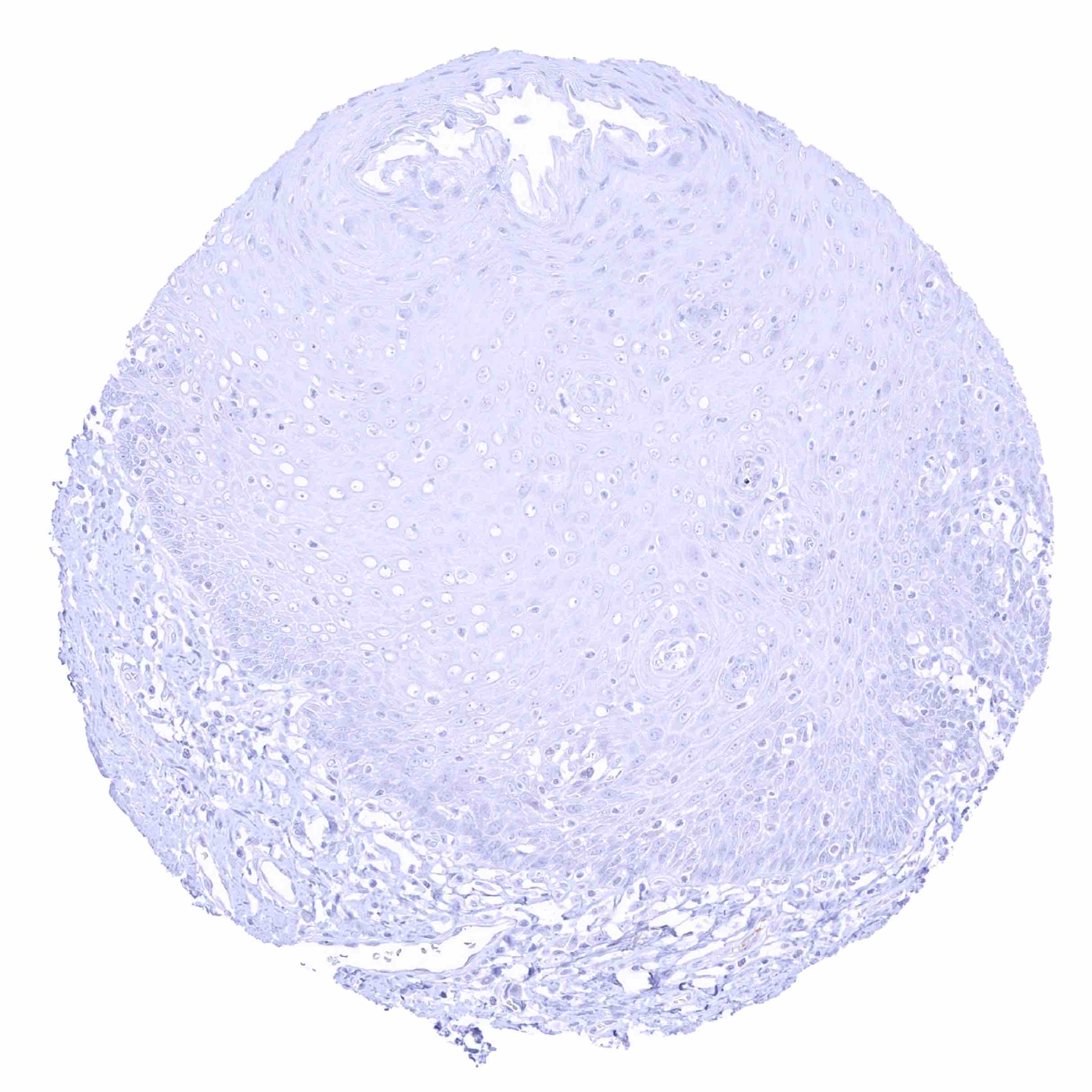
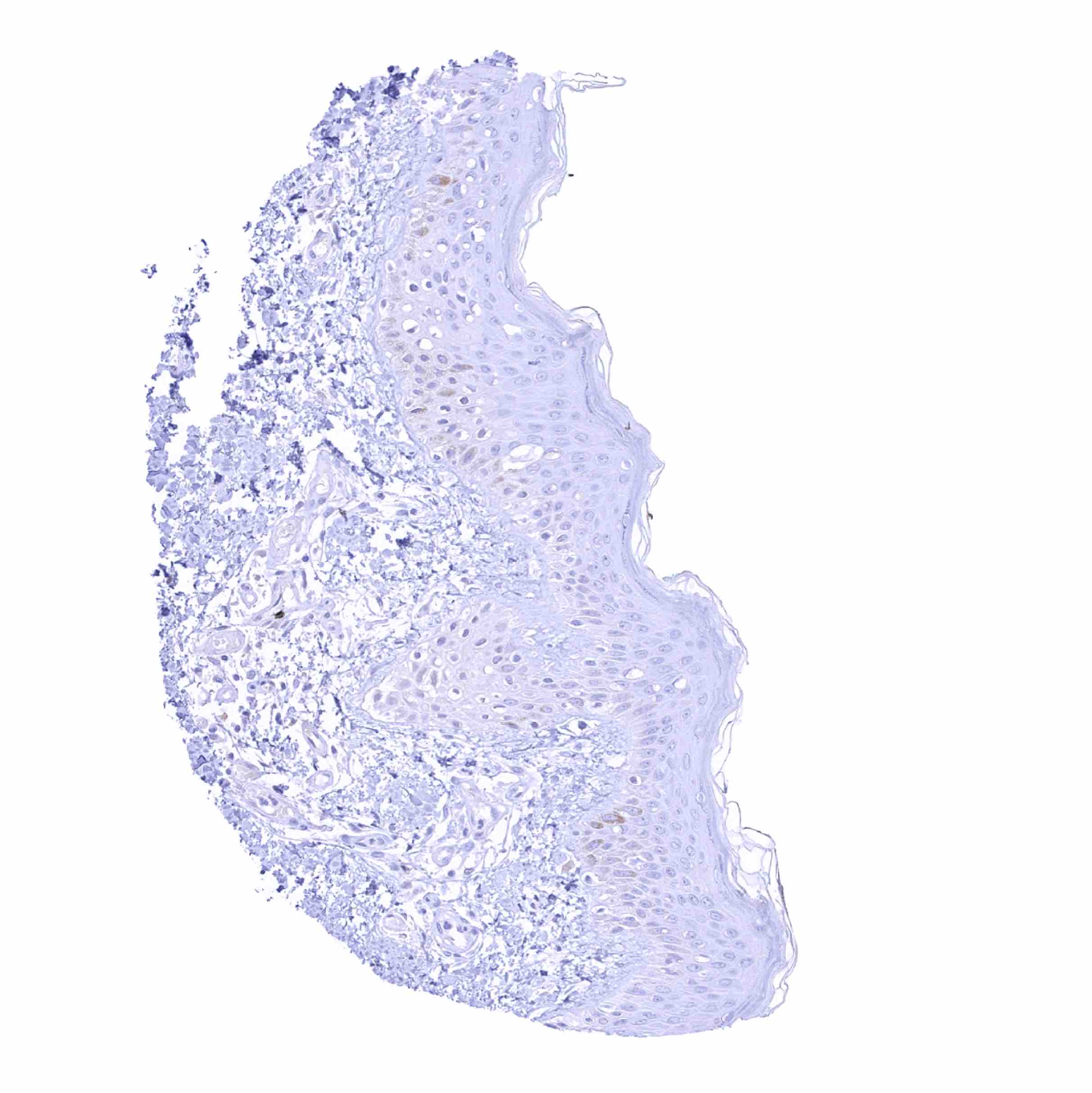
Esophagus, squamous epithelium
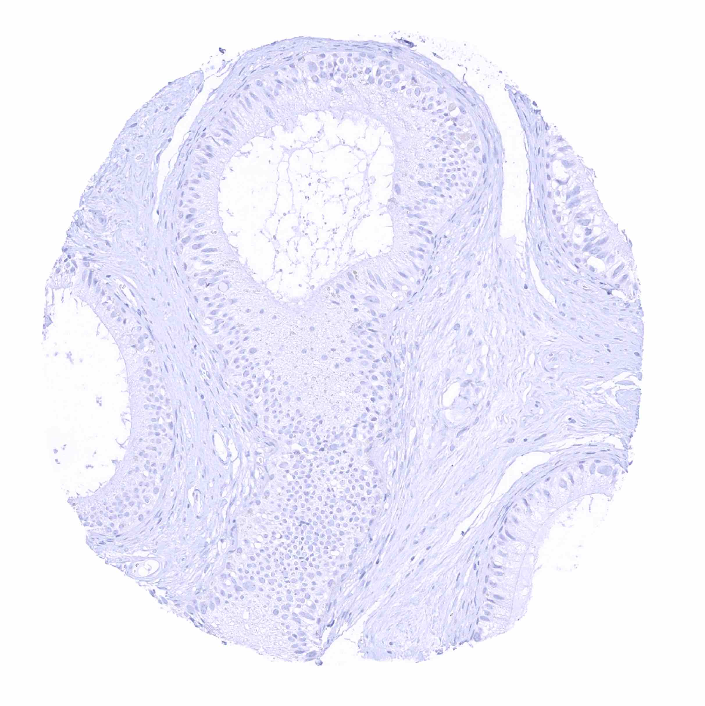
Fallopian tube, mucosa - IgD staining is largely lacking in this sample
Fallopian tube, mucosa - Substantial IgD staining of intravascular liquids. Weaker staining is also seen in adjacent stroma
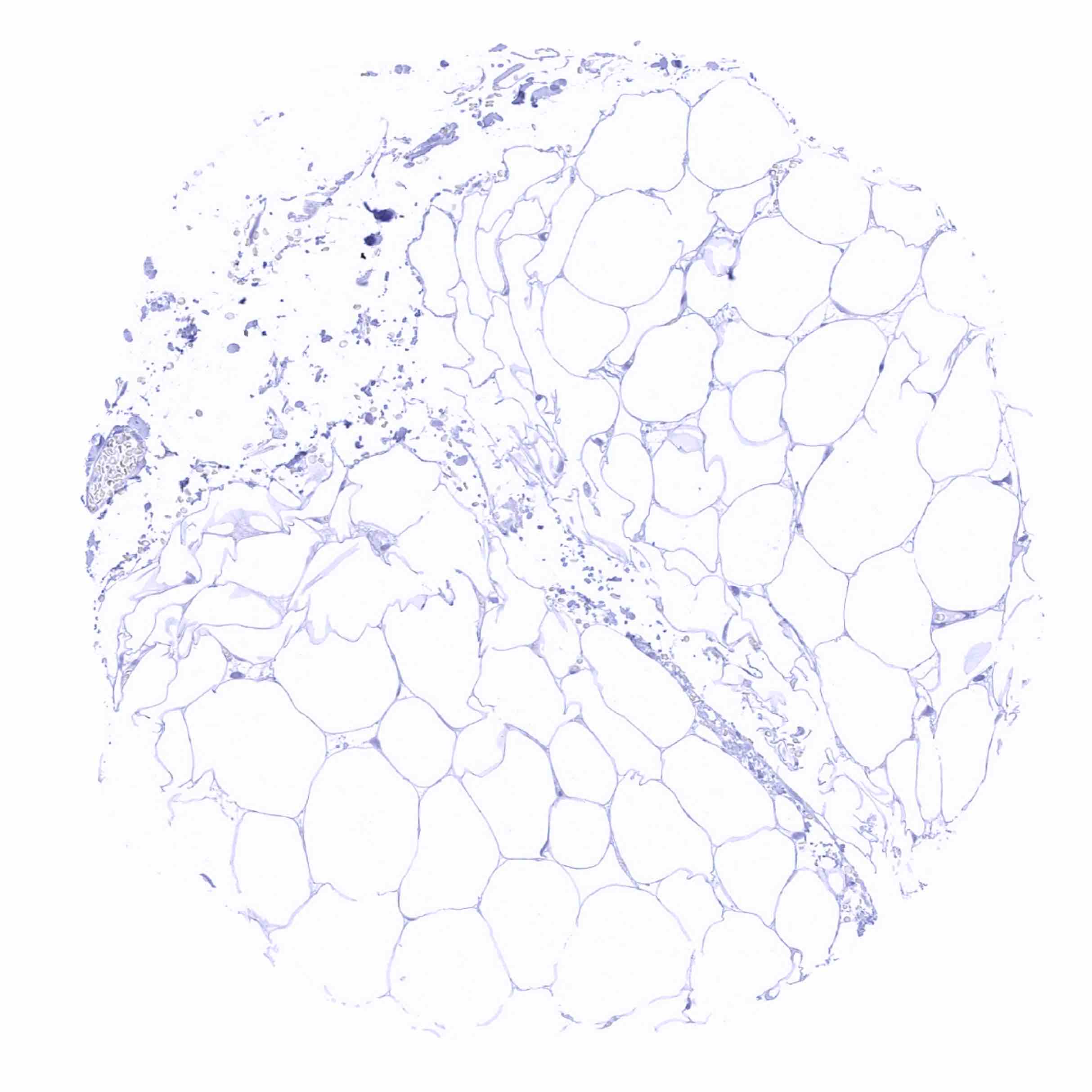
Fat
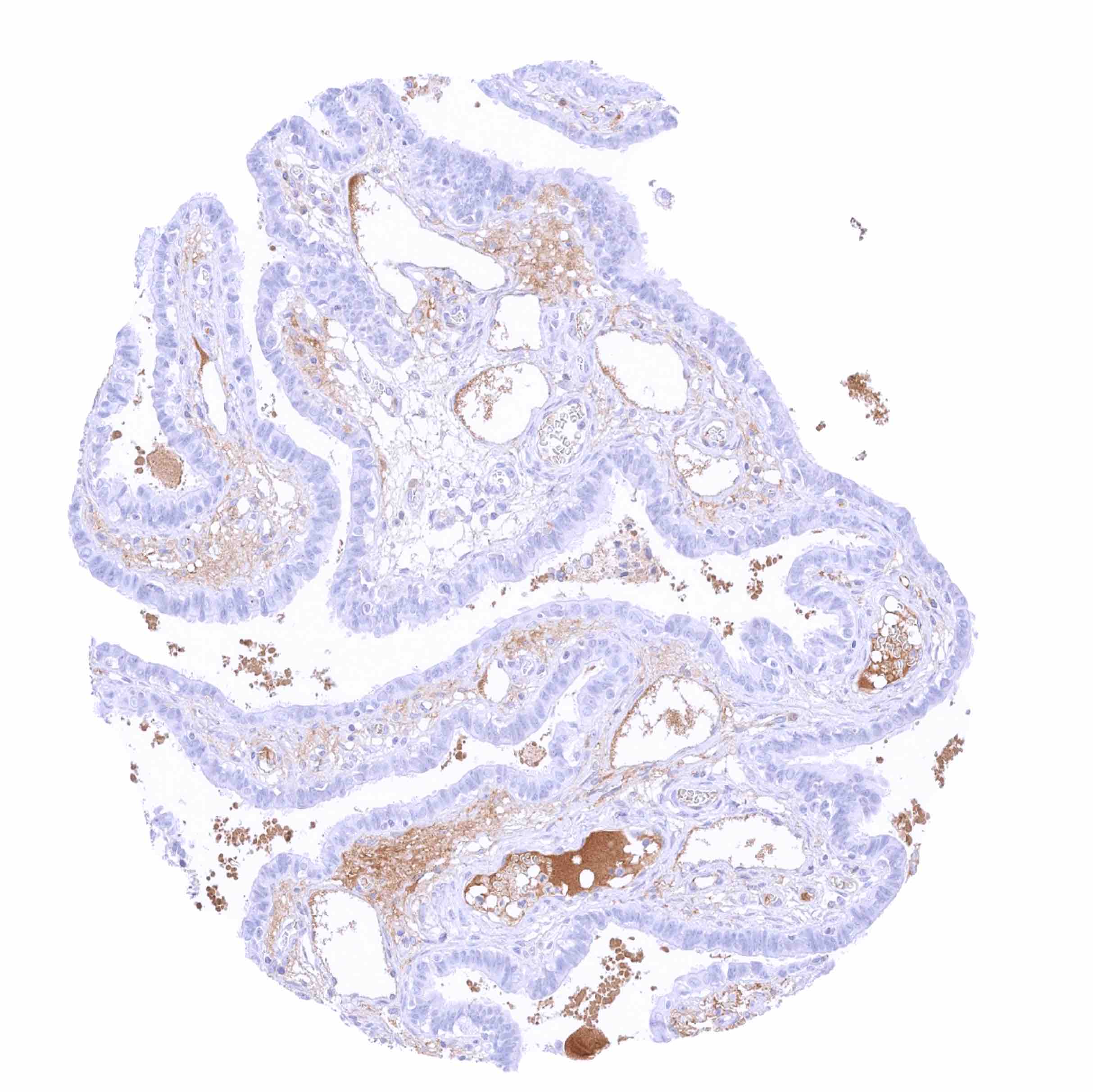
Gallbladder, epithelium
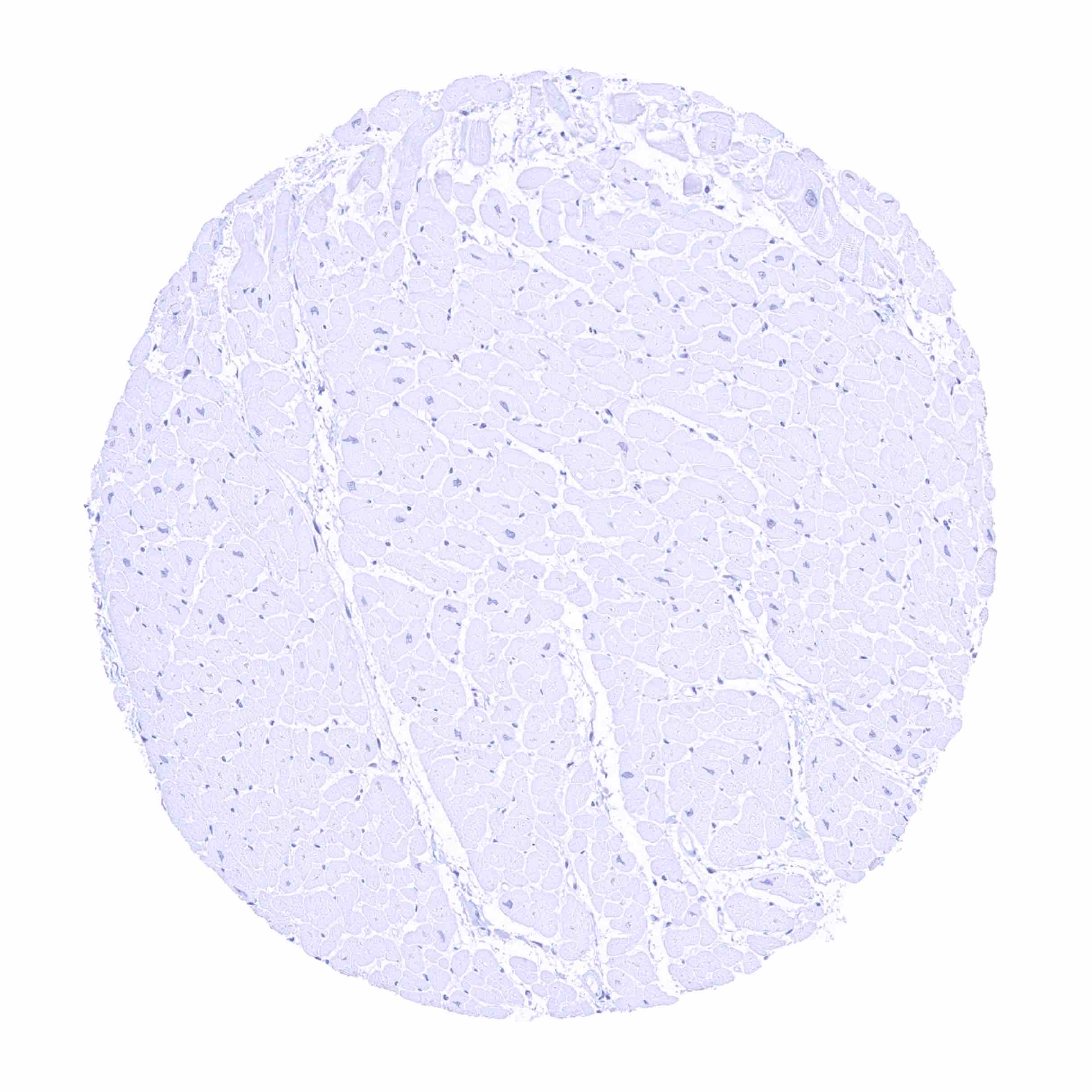
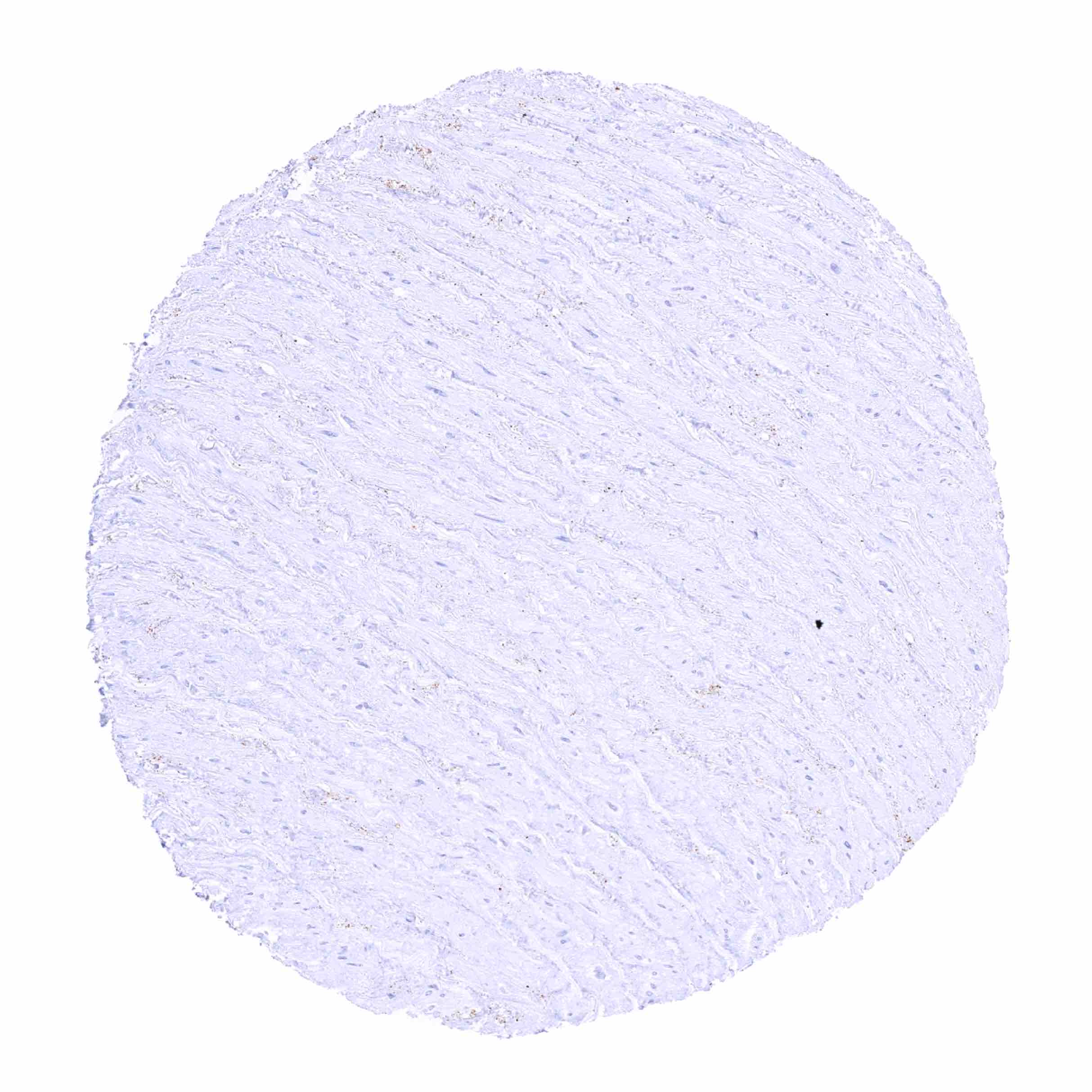
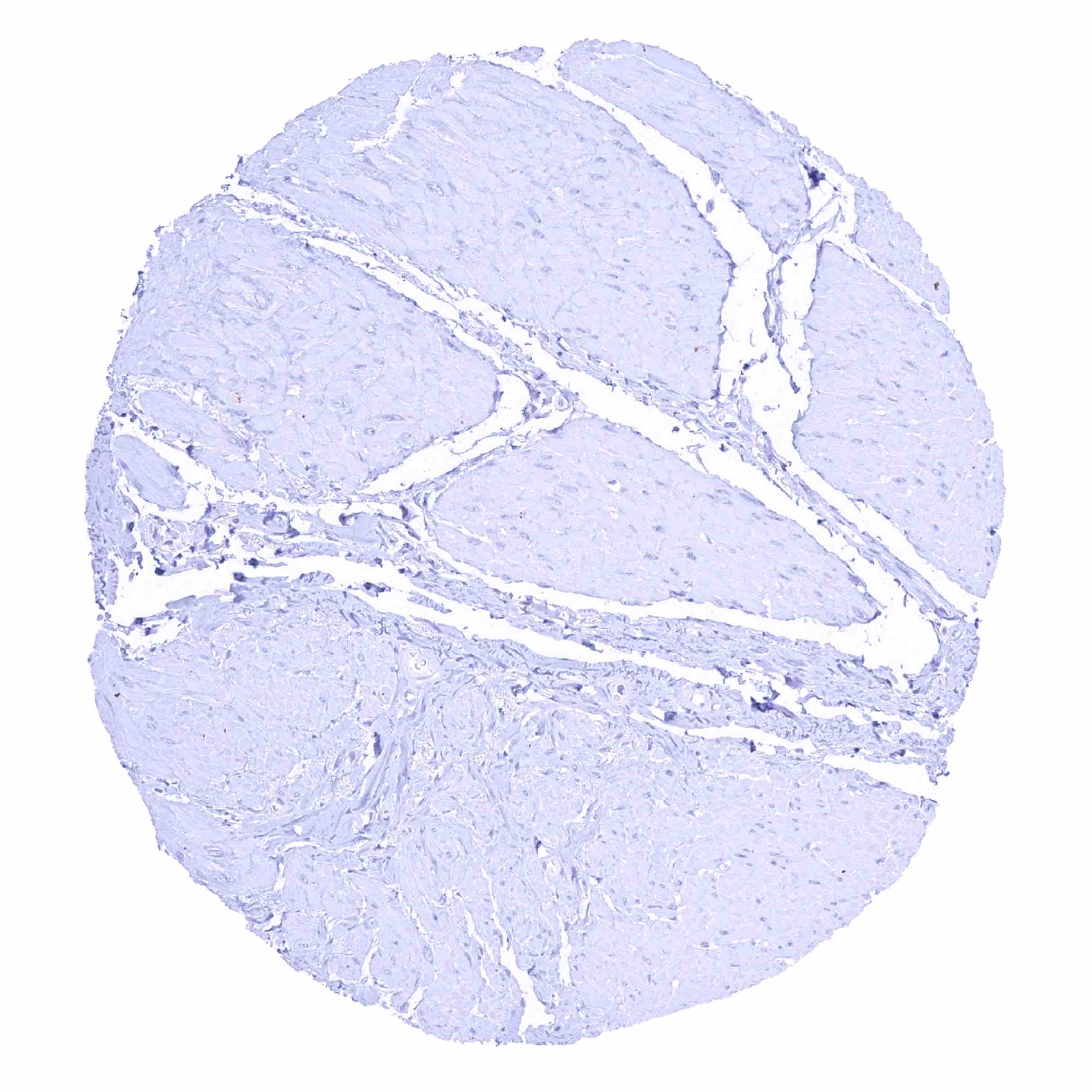
Heart muscle
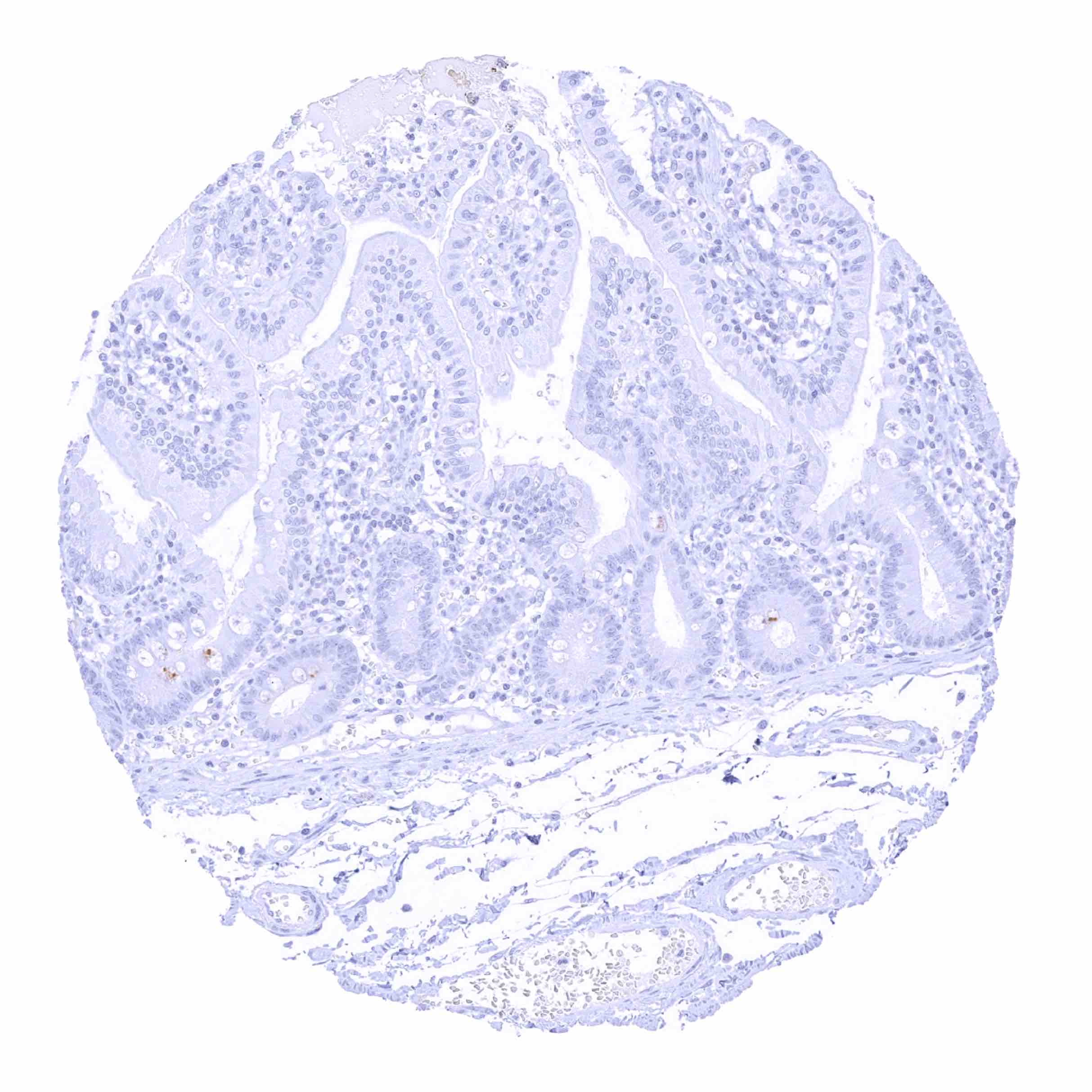
Ileum, mucosa
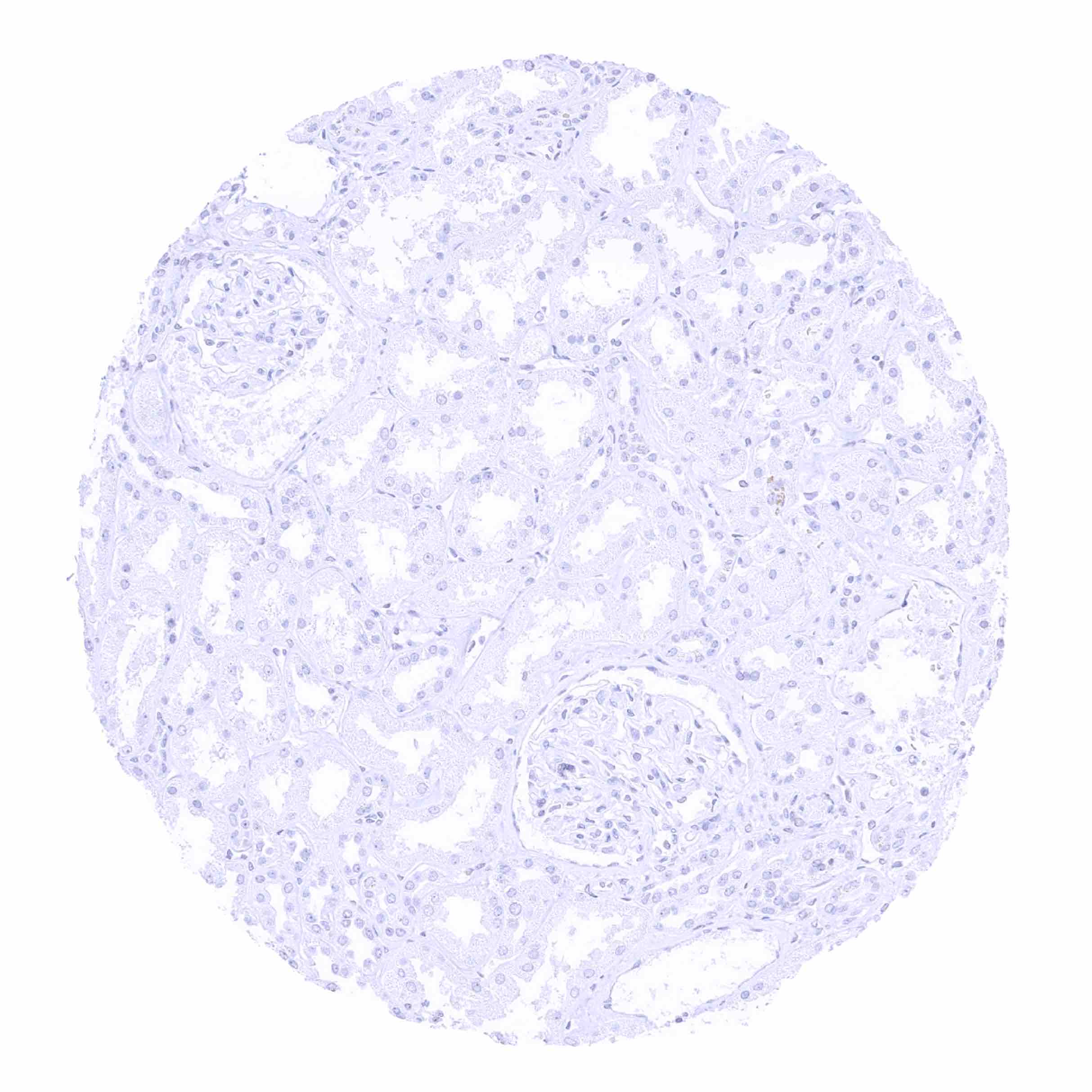
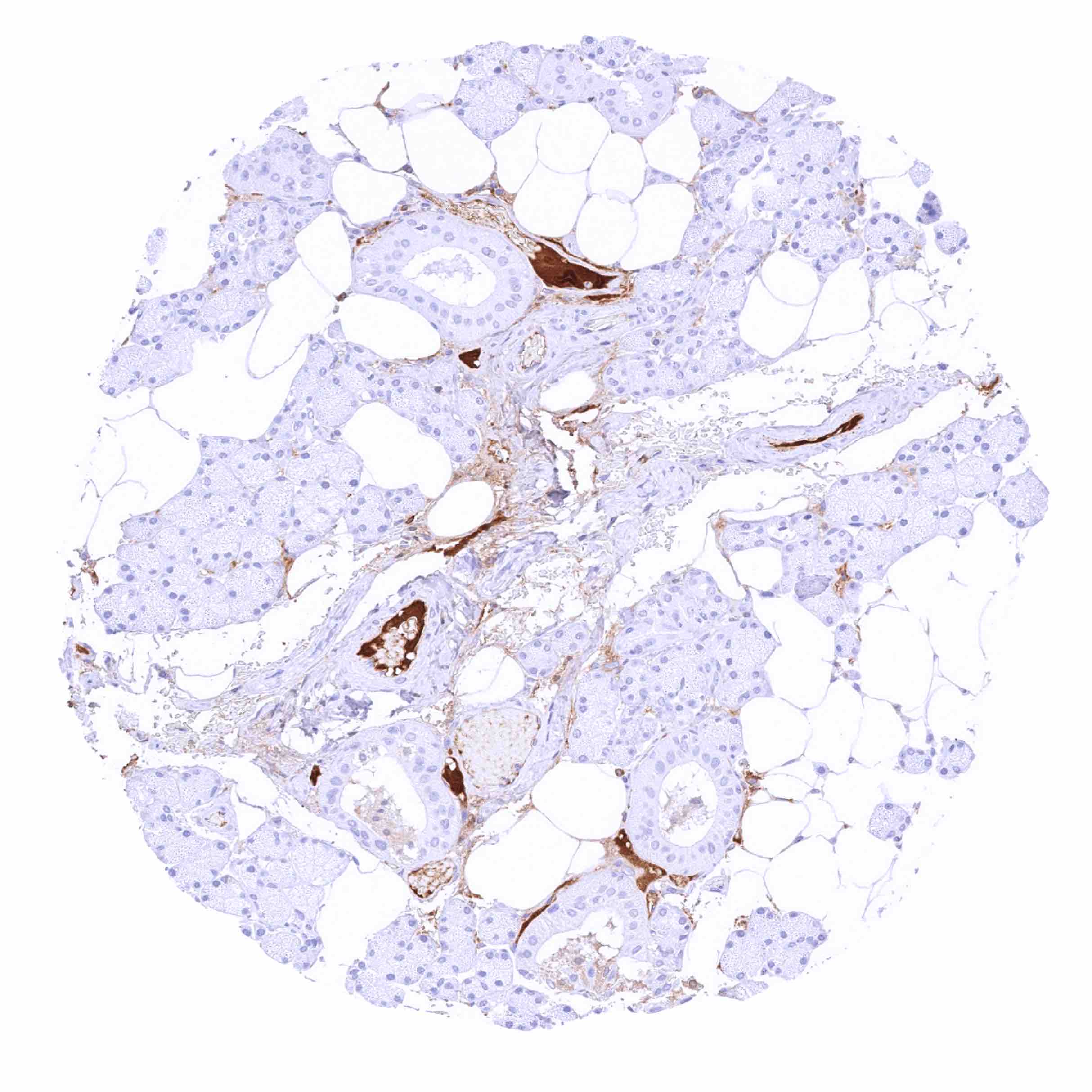
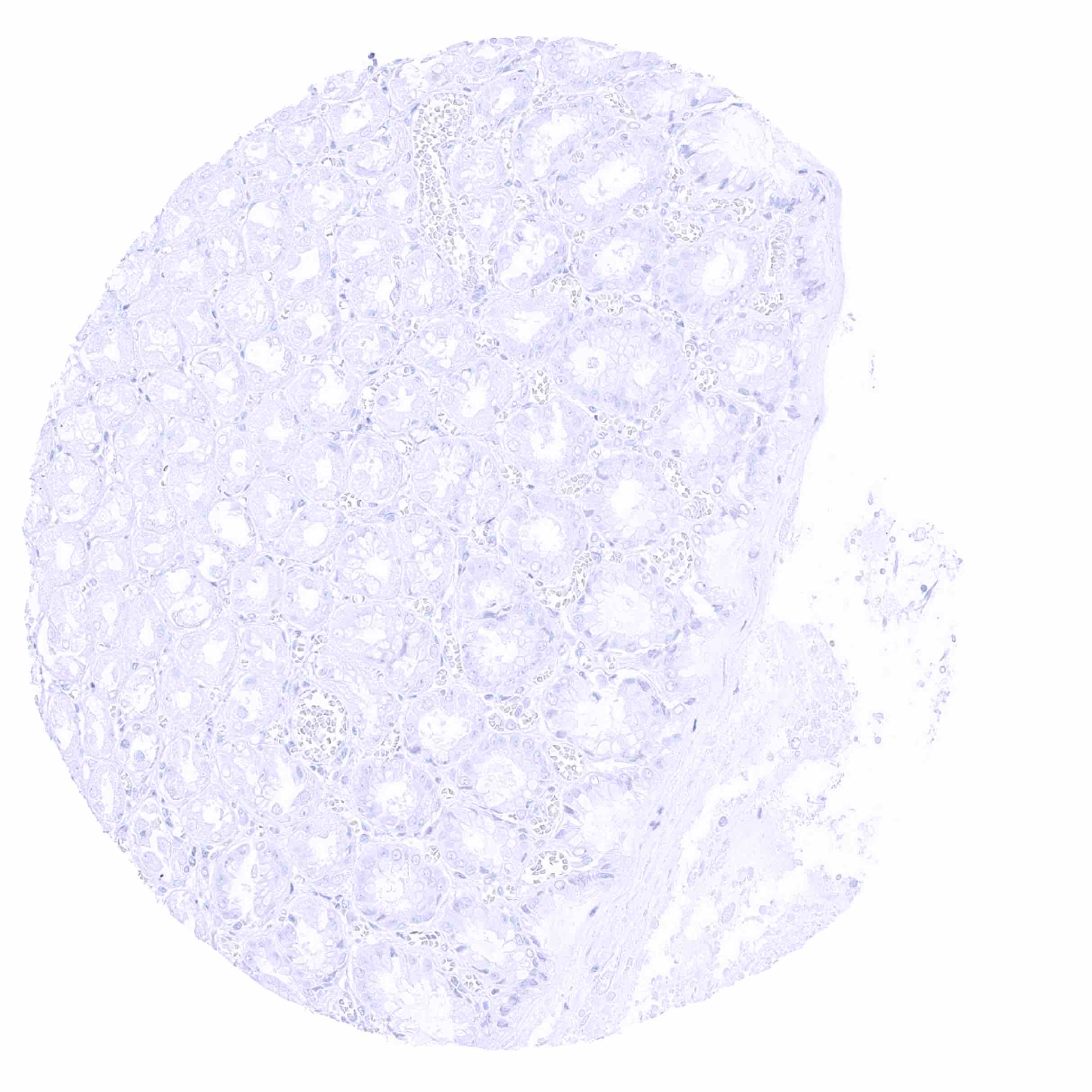
Kidney, cortex
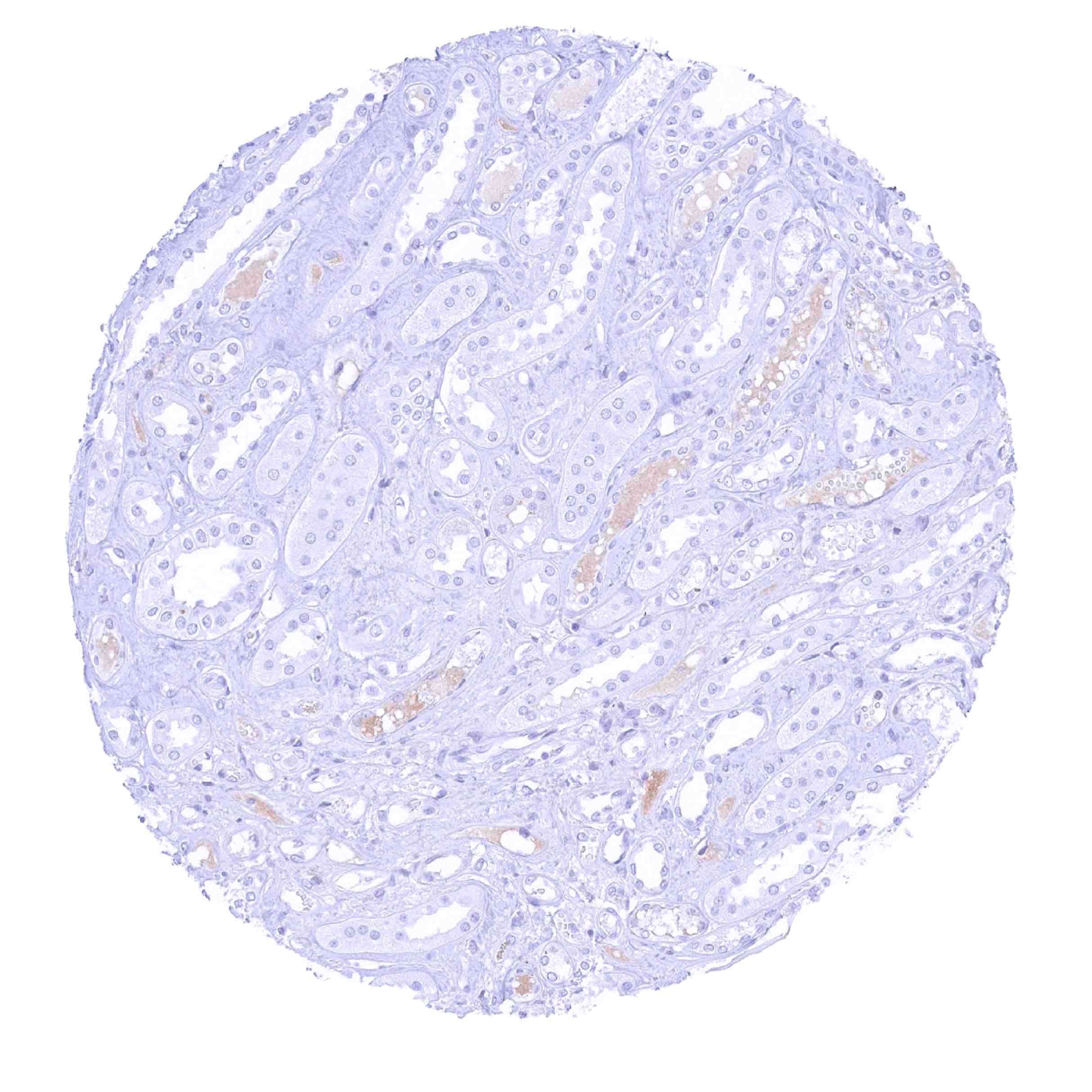
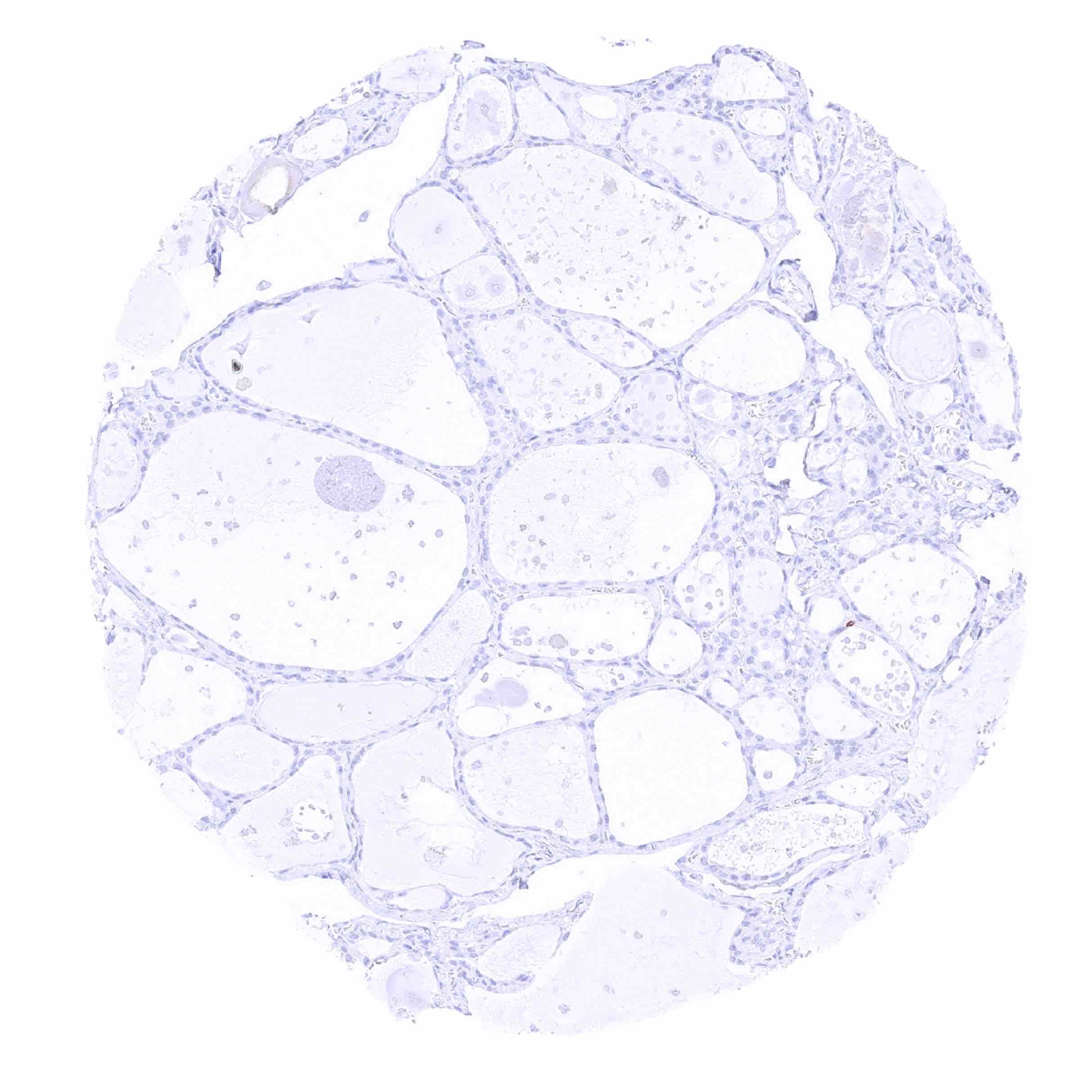
Kidney, medulla - Weak IgD staining of intratubular liquid
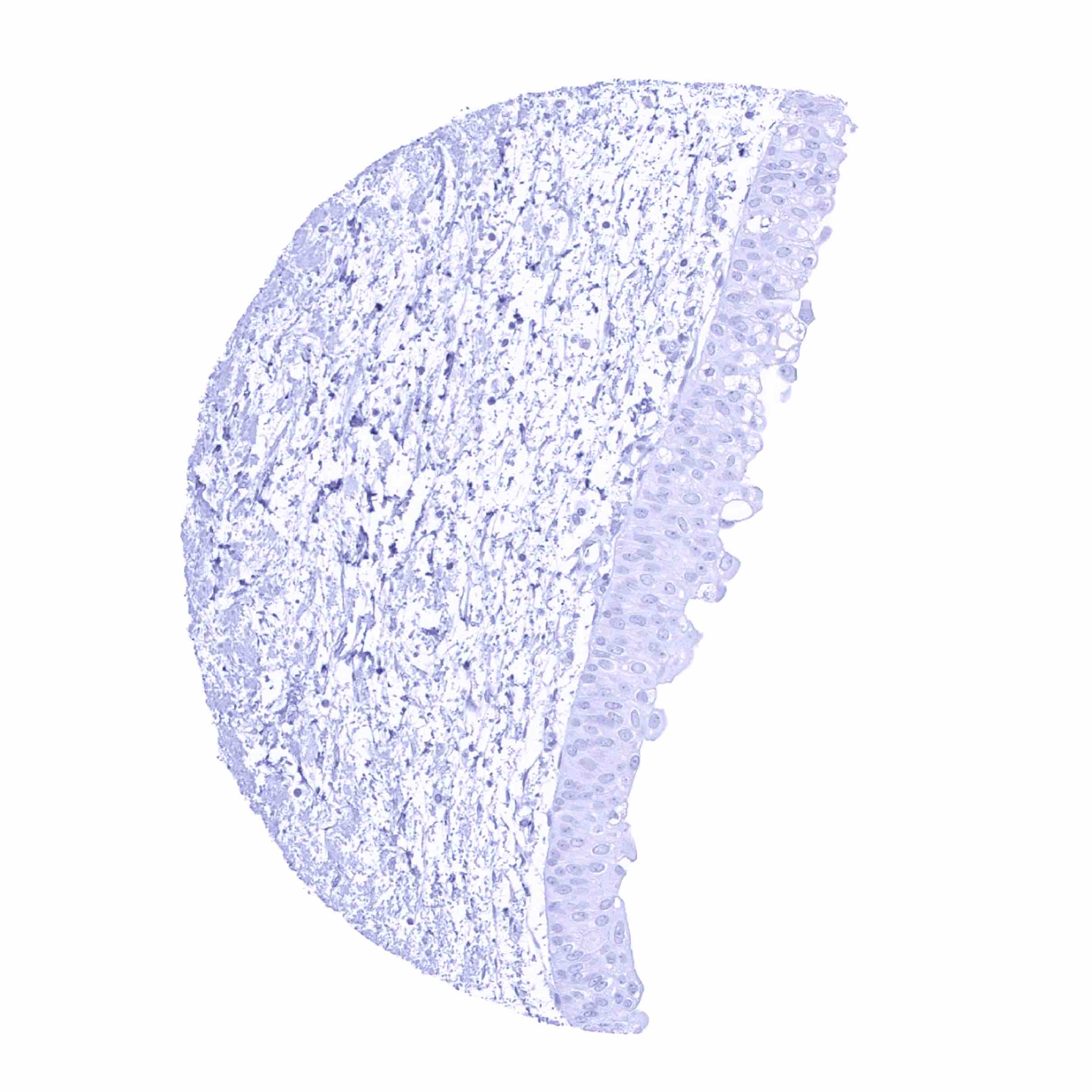
Kidney, pelvis, urothelium

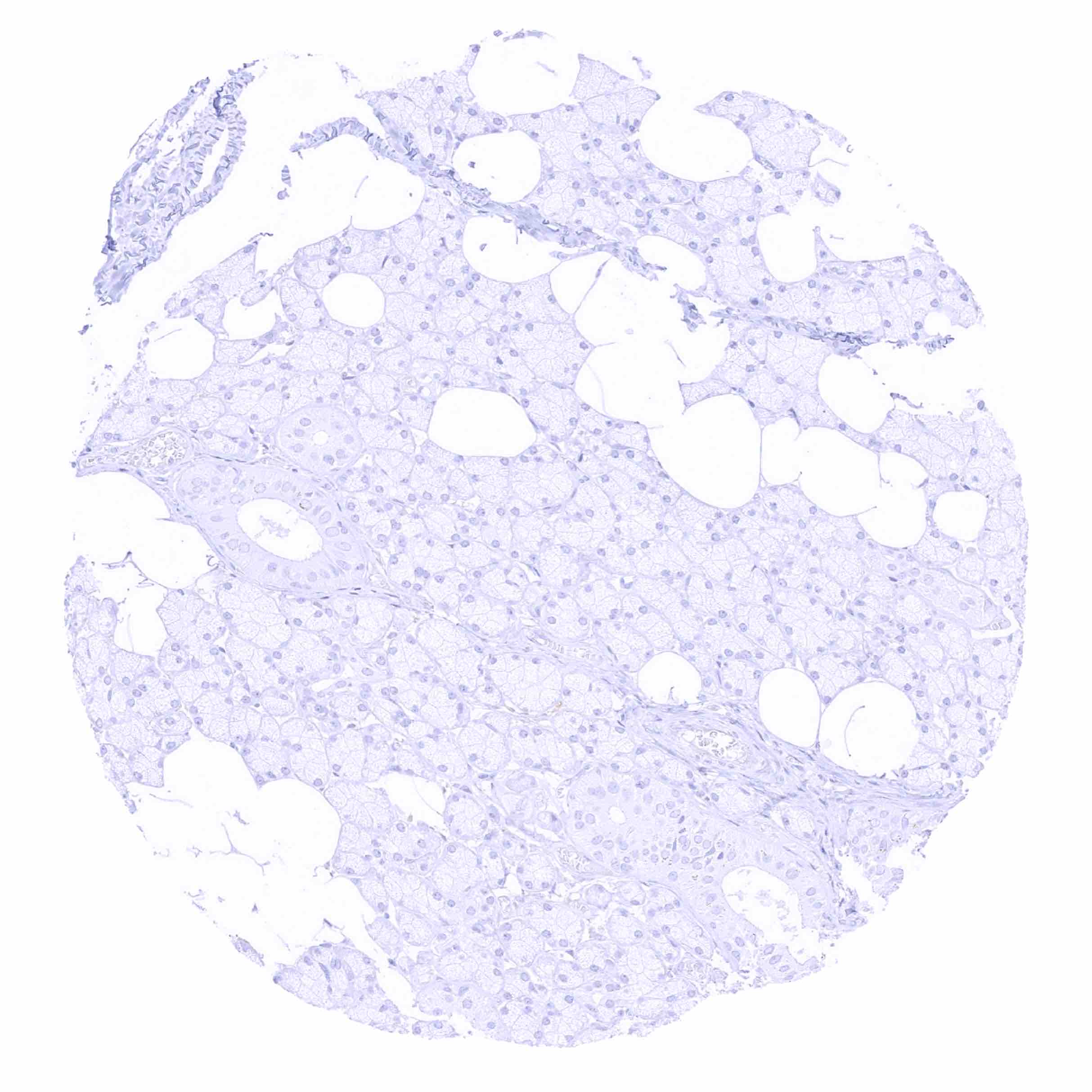
Liver
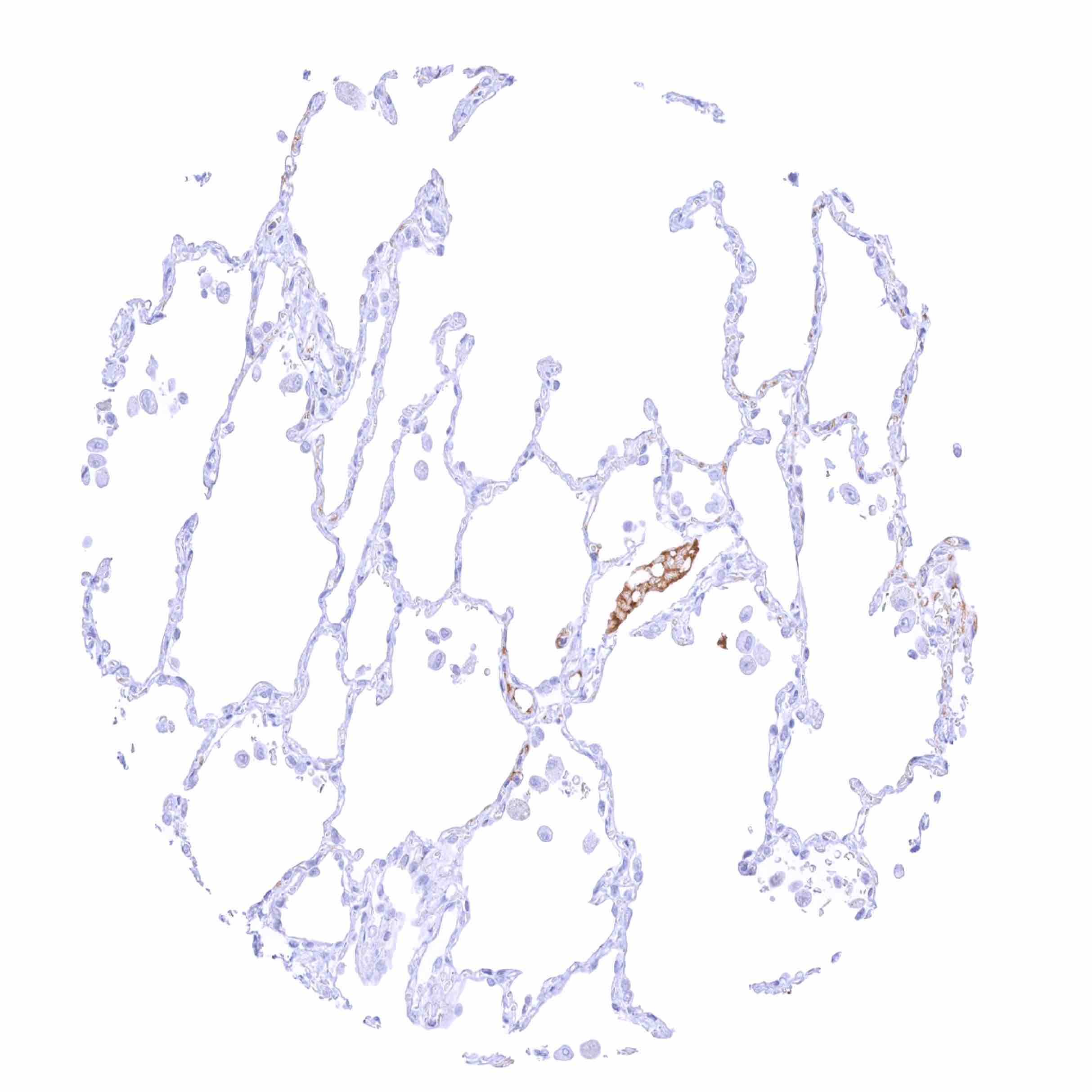
Lung - Moderate to strong IgD staining of intravascular liquid
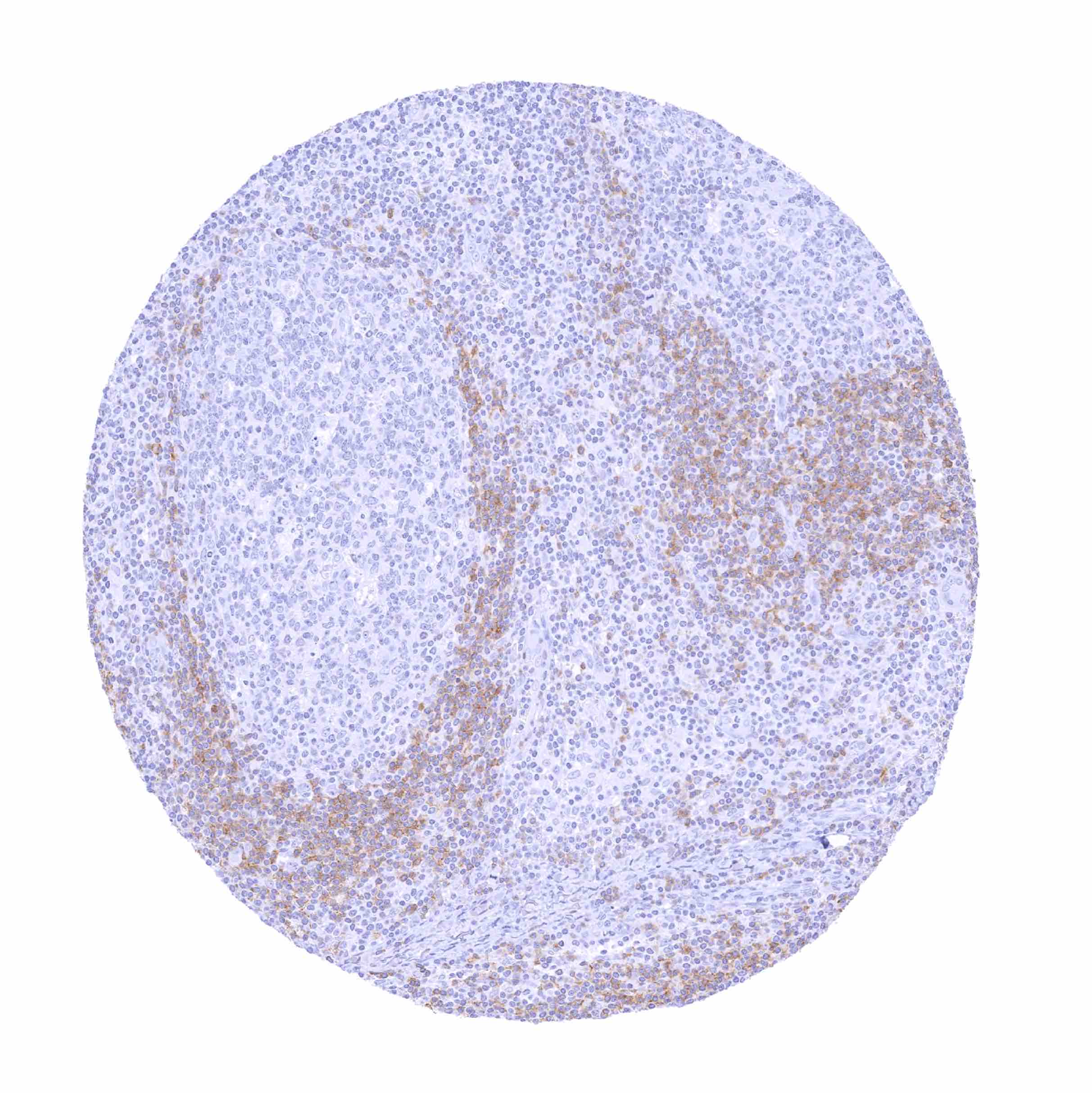
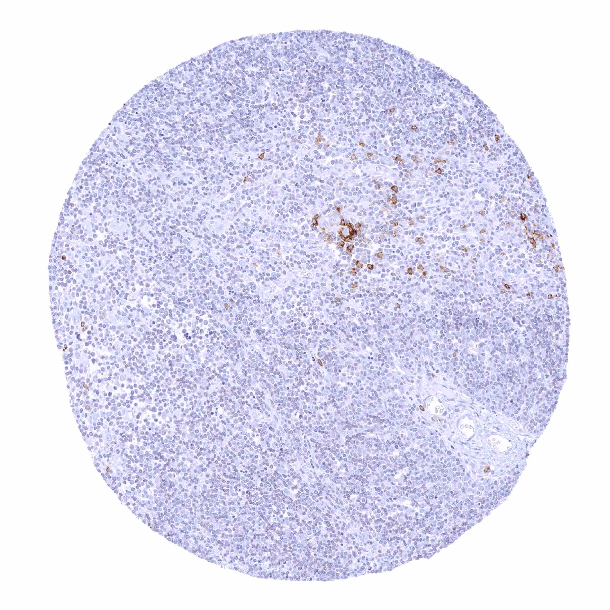
Lymph node - Significant IgD immunostaining in B-lymphocytes of the mantle zone
Lymph node - Strong IgD staining in a fraction of B-lymphocytes, especially in the mantle zone

Oral cavity
Ovary, stroma

Pancreas
Parathyroid gland
Parotid gland - Strong IgD staining of intravascular plasma
Parotid gland

Pituitary gland, anterior lobe
Pituitary gland, posterior lobe
Placenta (amnion and chorion)
Placenta early, decidua

Placenta, early - Substantial IgD staining of intervillous liquids. Weaker staining of adjacent syncytiotrophoblast may reflect a contamination artifact

Placenta, early

Placenta, mature
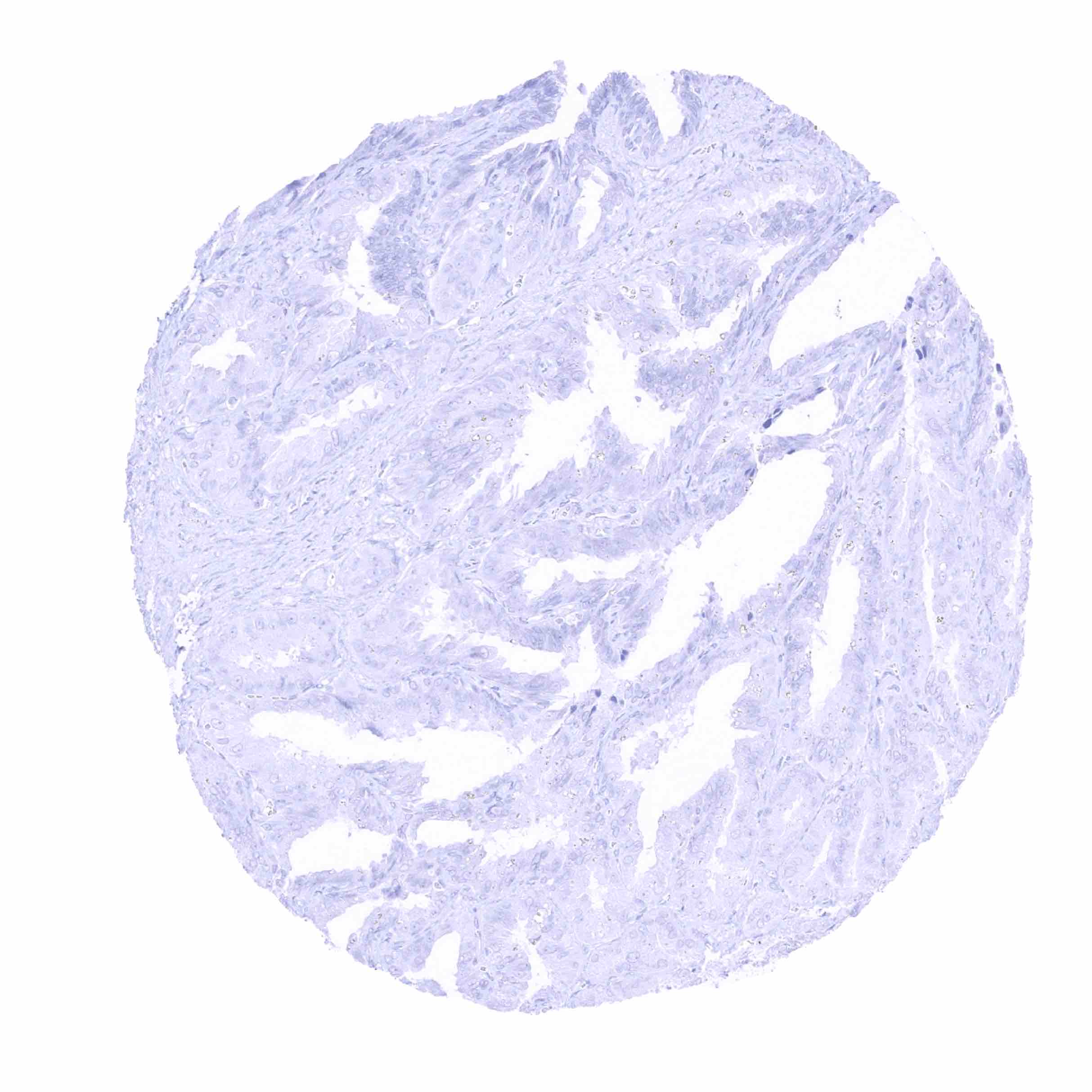
Prostate
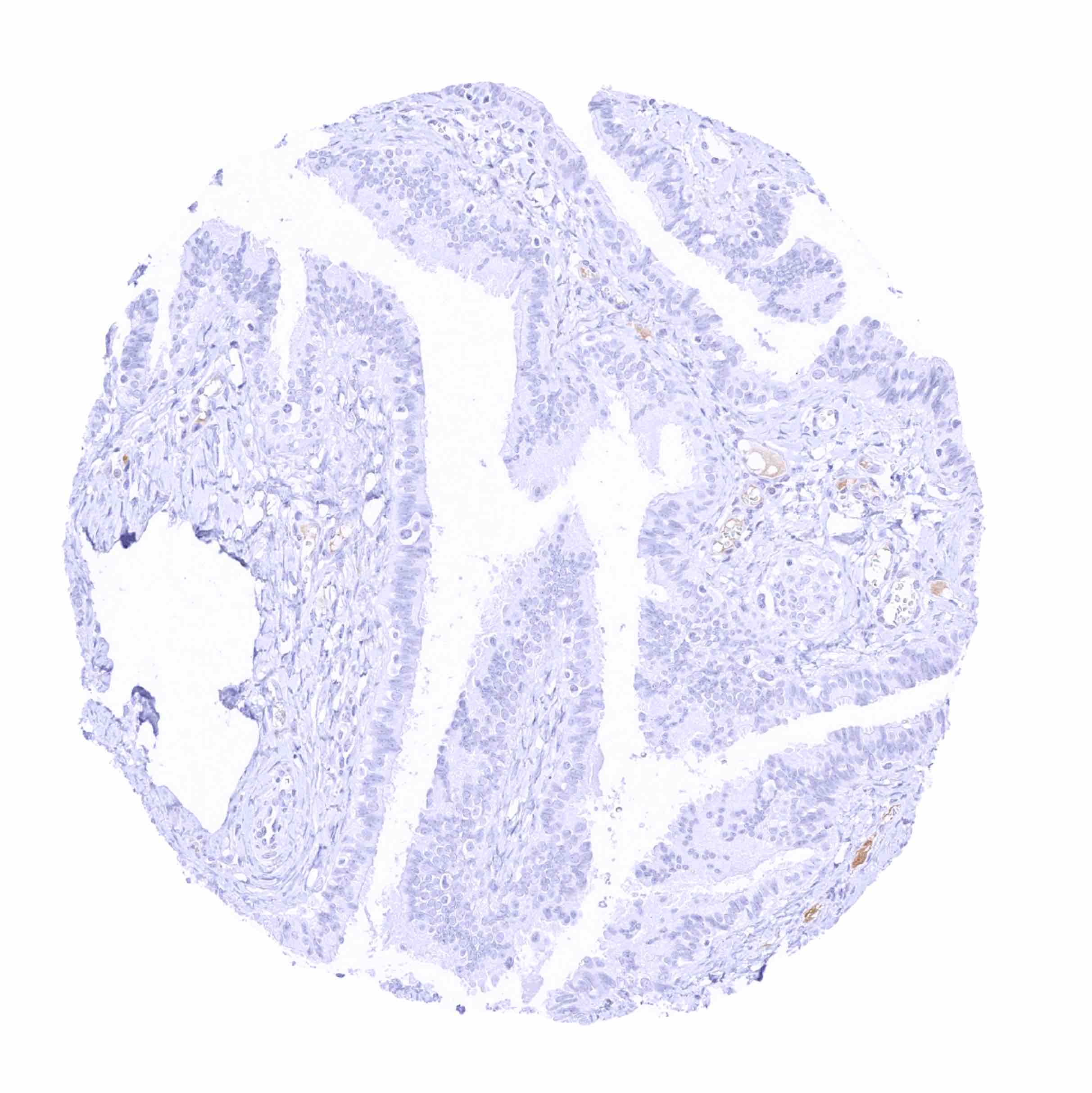
Rectum, mucosa
Seminal vesicle
Sinus paranasales
Skin

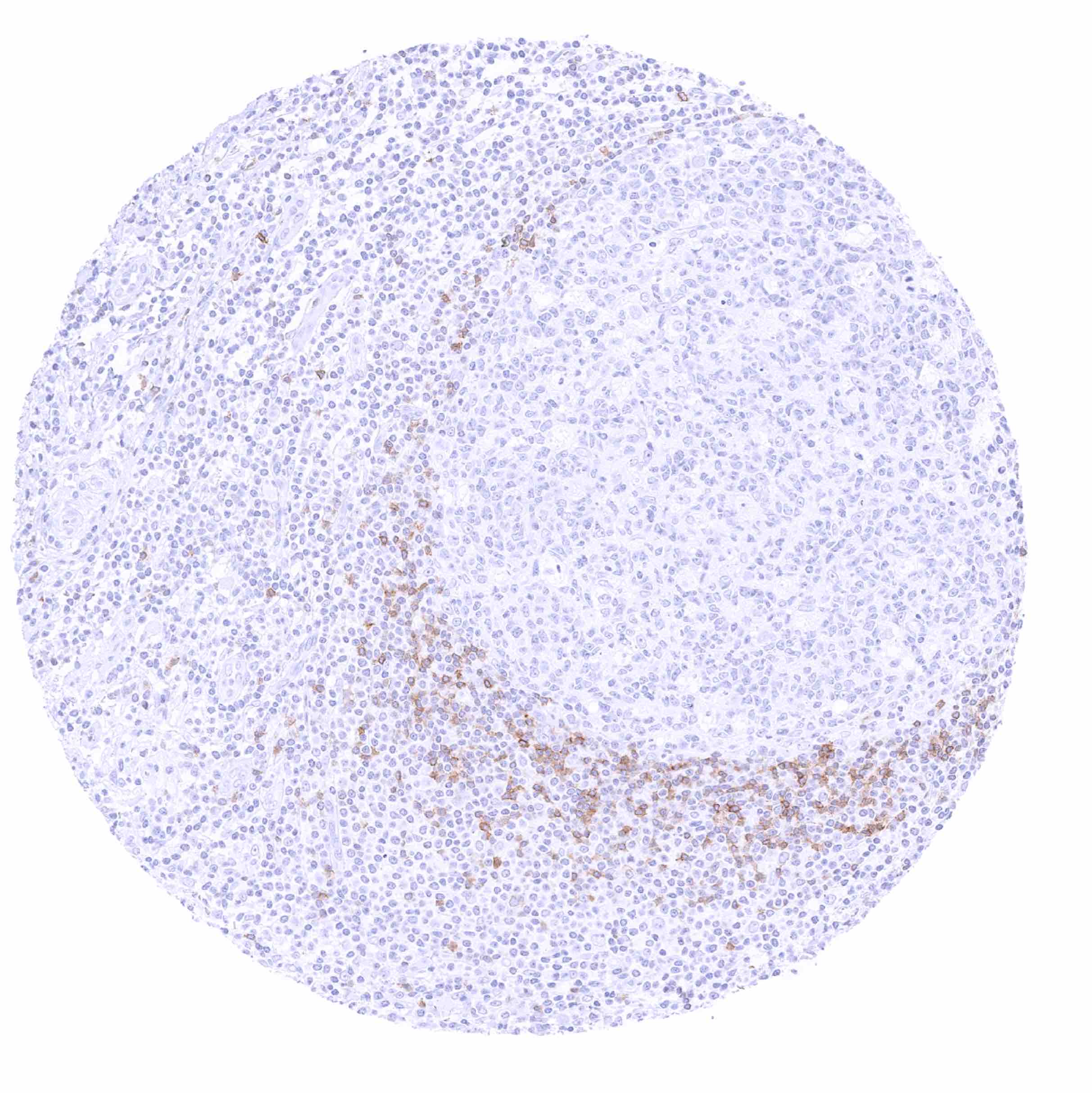
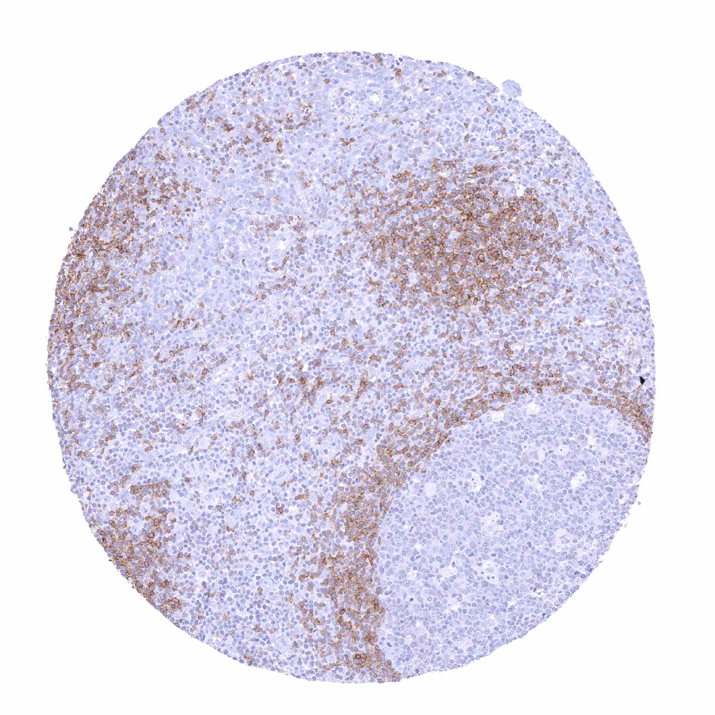
Spleen - Moderate IgD staining of a fraction of B-lymphocytes
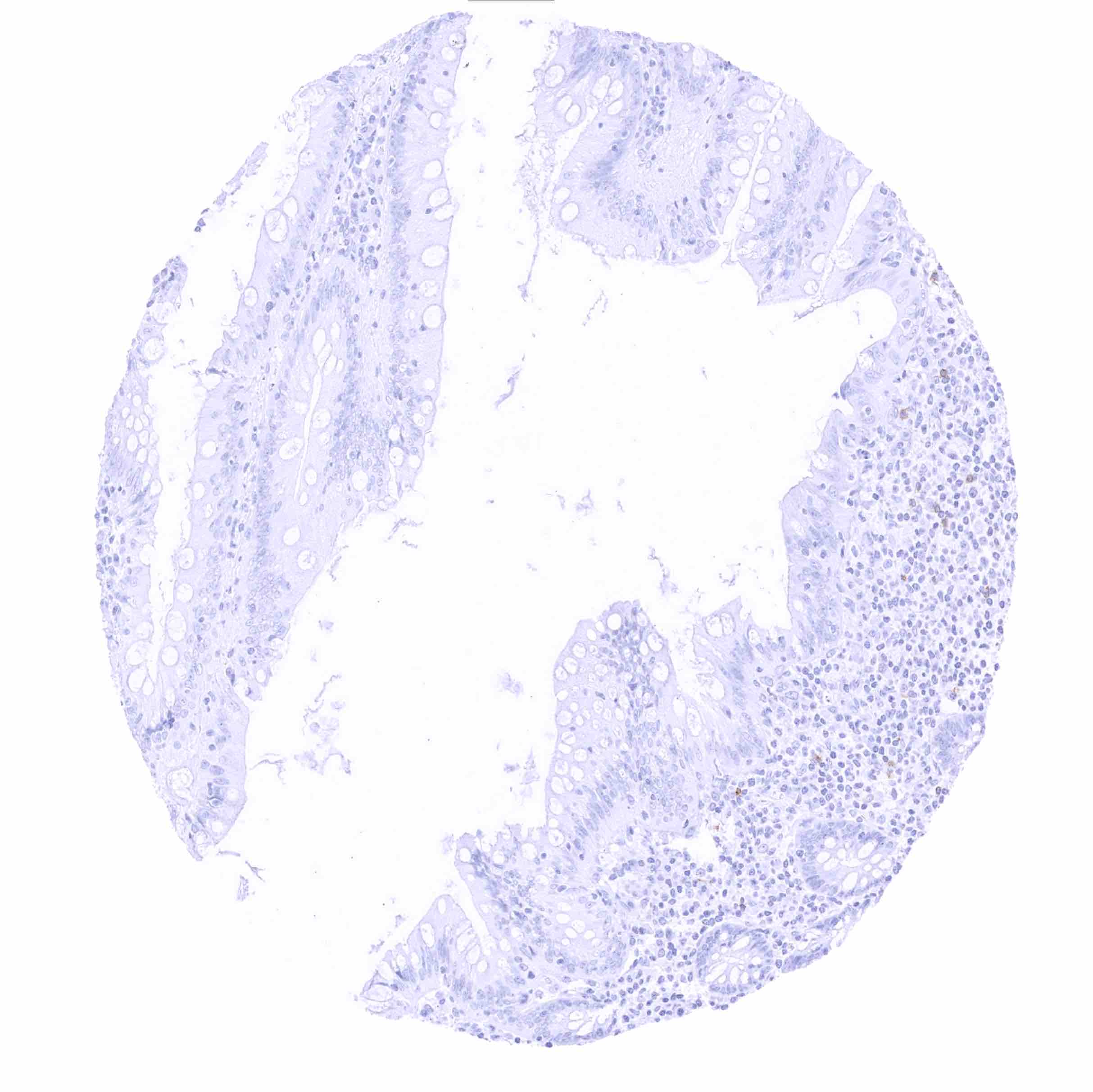
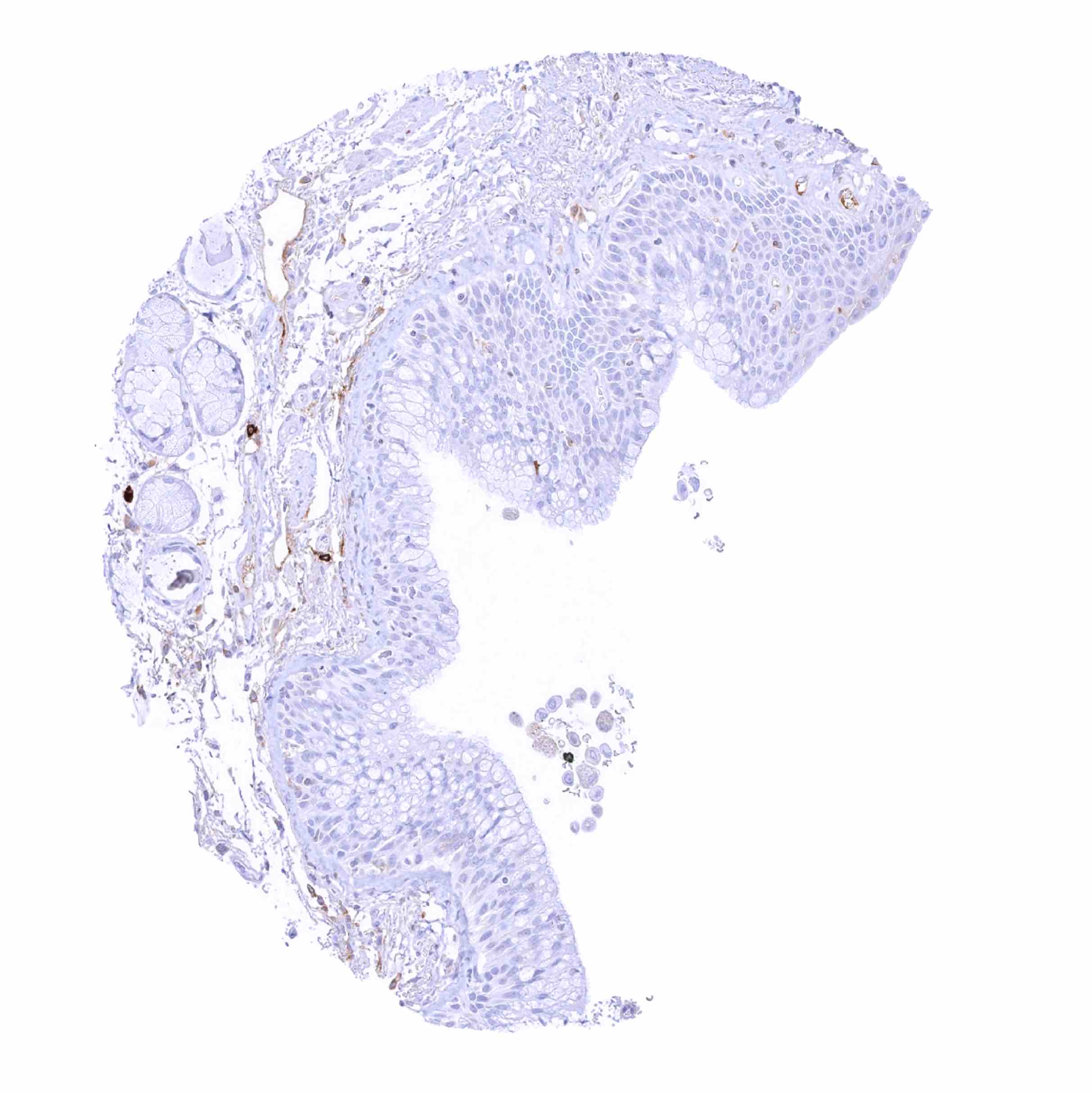
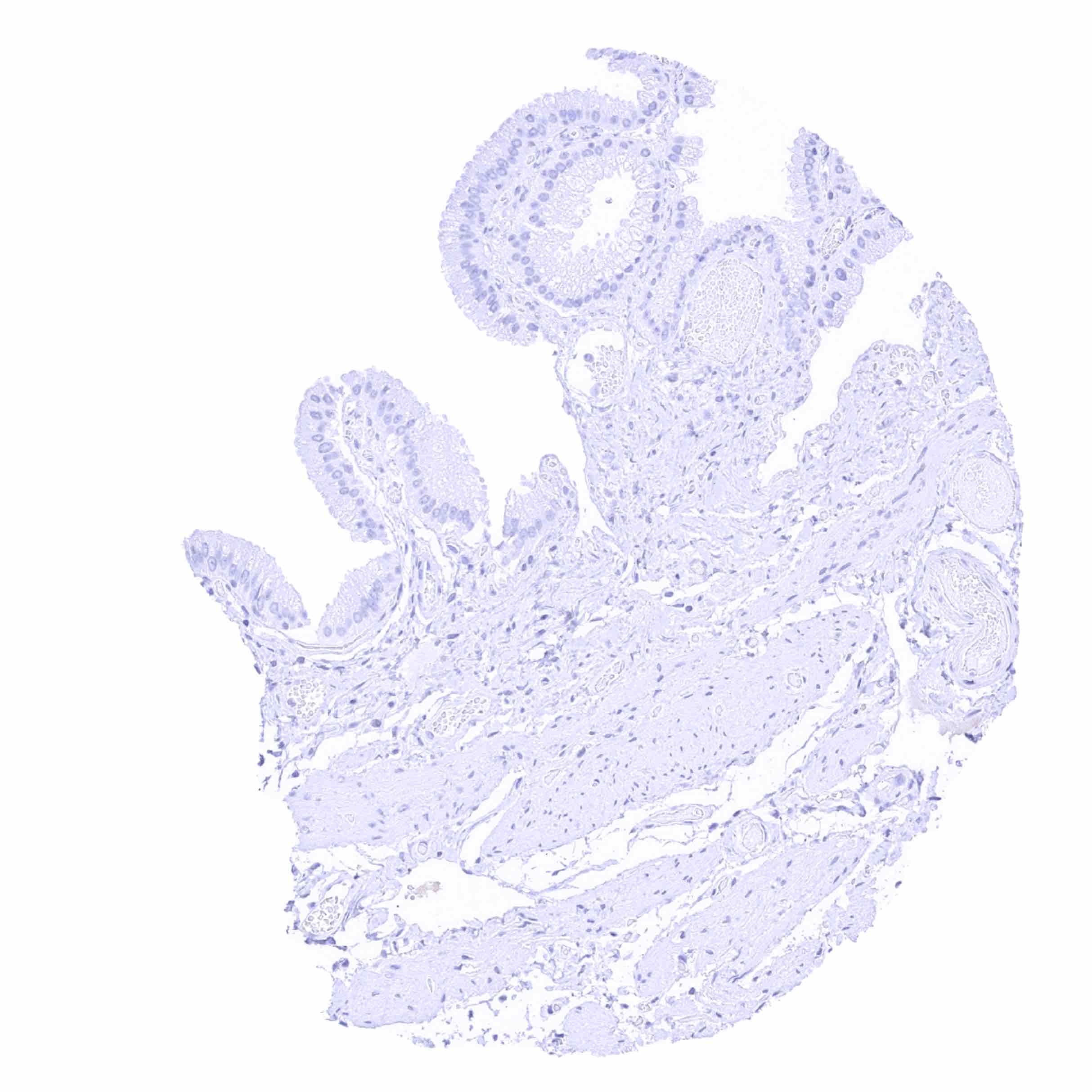
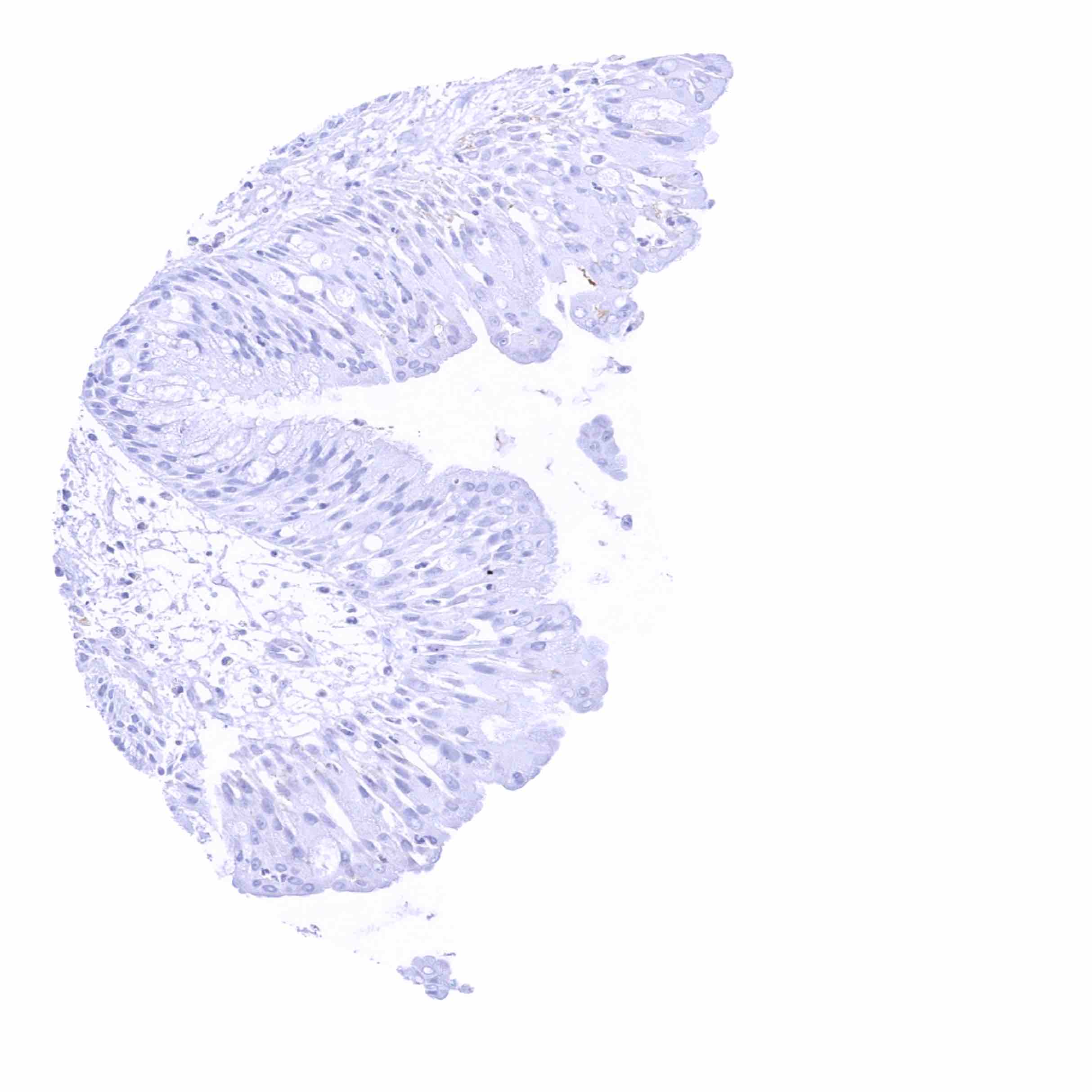
Stomach, antrum
Stomach, corpus
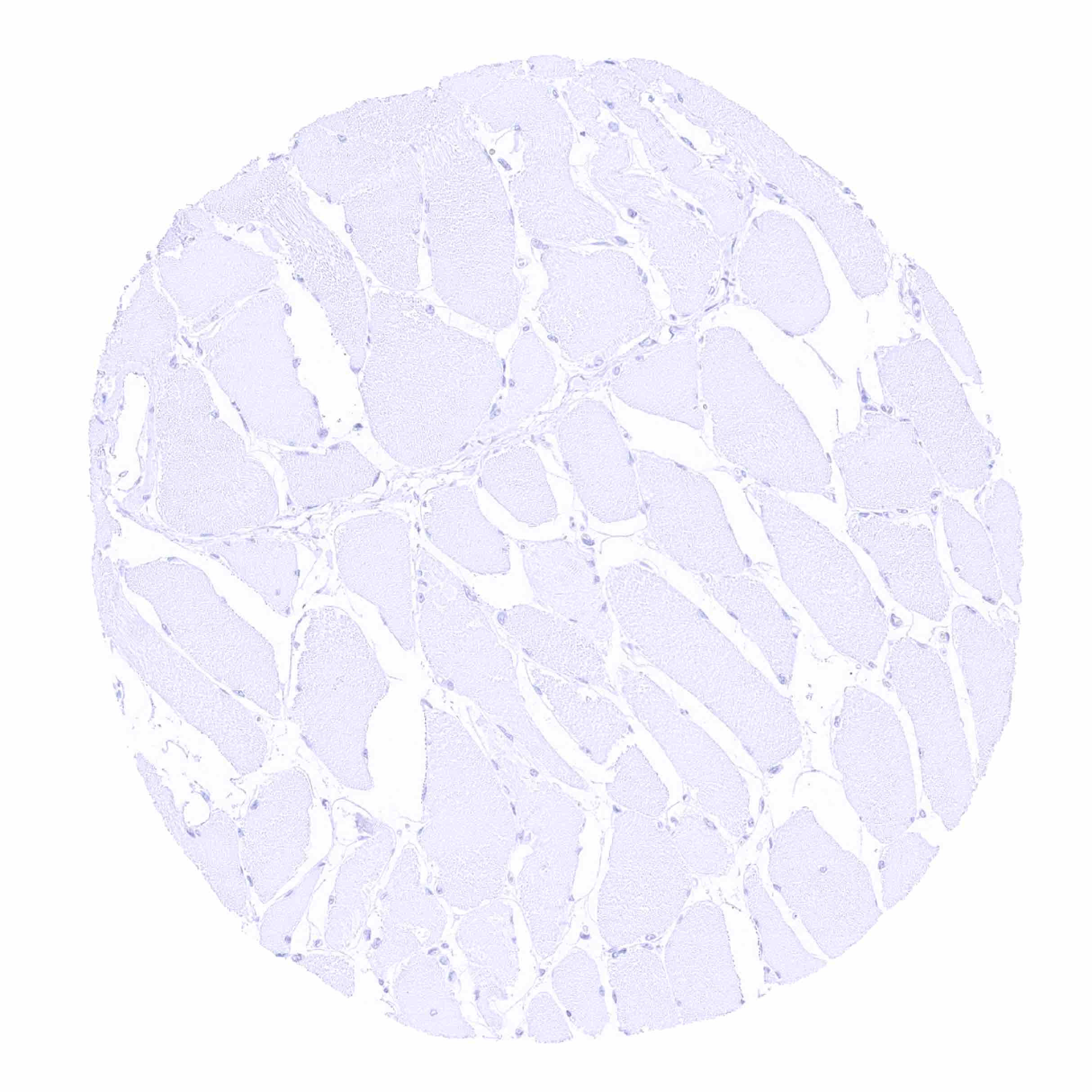
Striated muscle
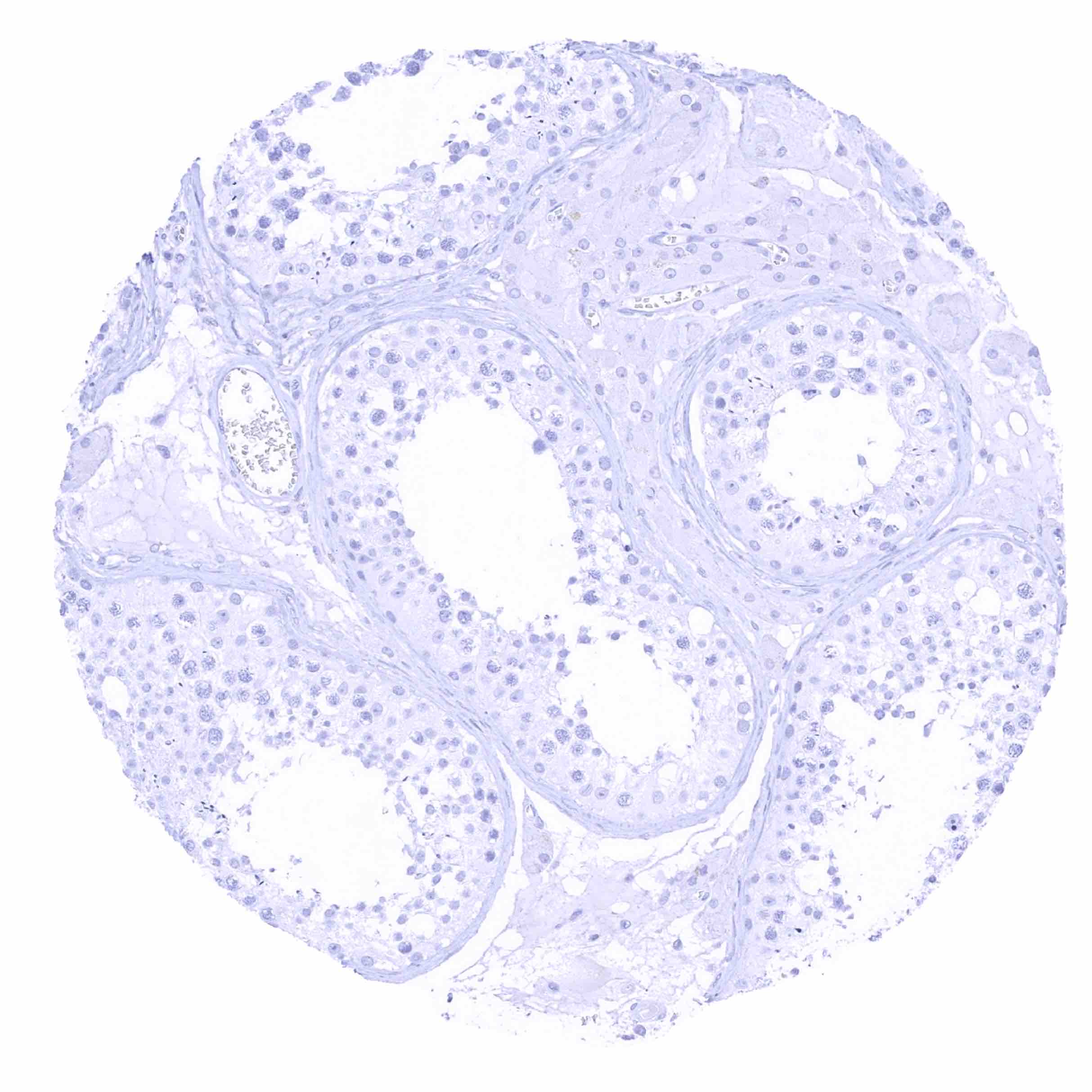
Testis - IgD staining is not seen in this sample
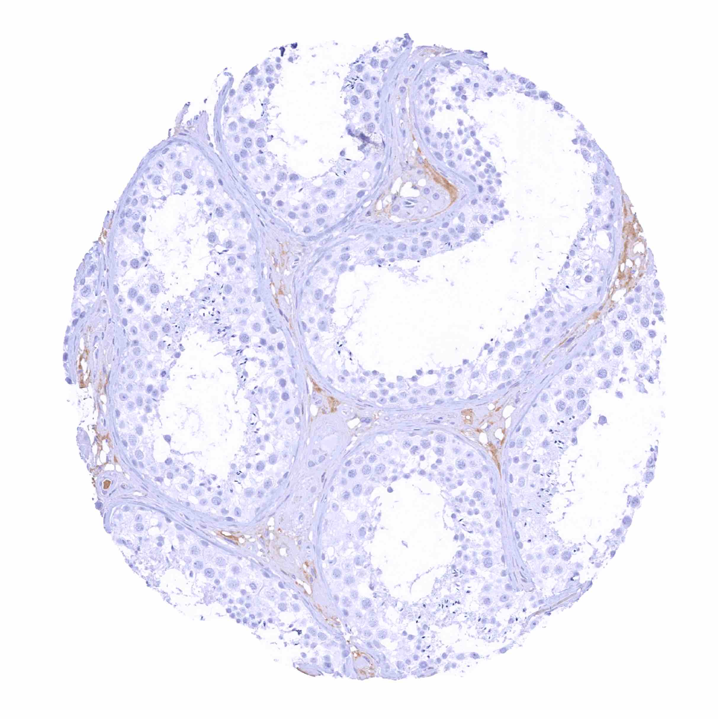
Testis - Weak IgD staining of intravascular liquid
Thymus - Only few cells show IgD staining, especially in the medulla
Thyroid gland
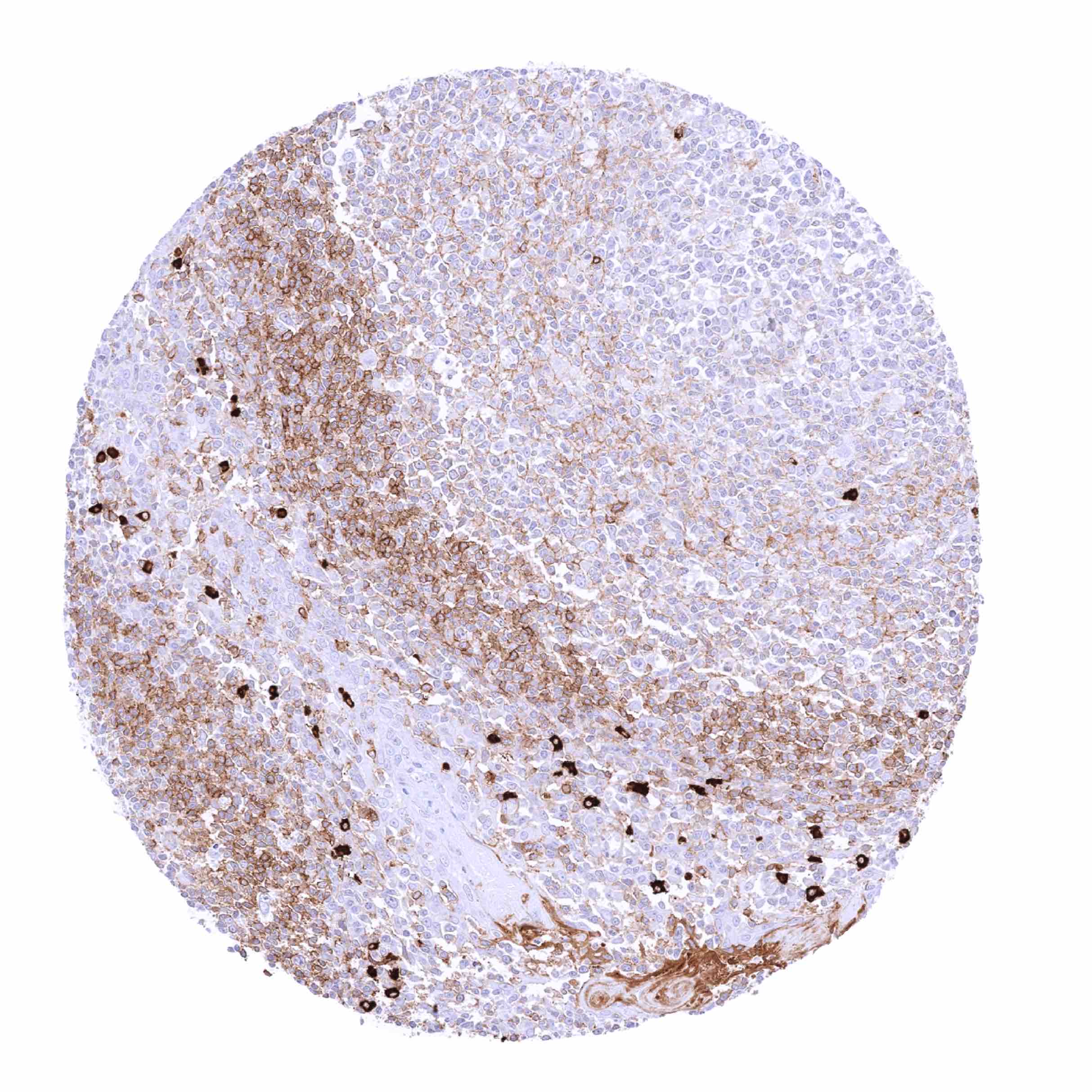
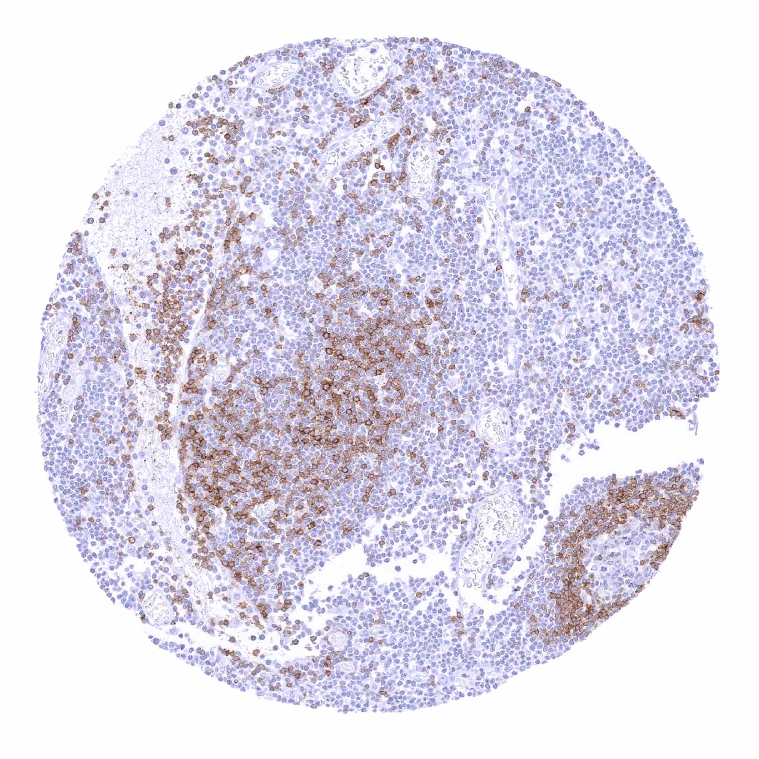
Tonsil - IgD staining of variable intensity in B-lymphocytes. Staining is particularly prominent in the mantle zone of germinal centres. Staining intensity is highest in interspersed plasma cells
Tonsil - Strong IgD staining in a fraction of B-lymphocytes, especially in the mantle zone
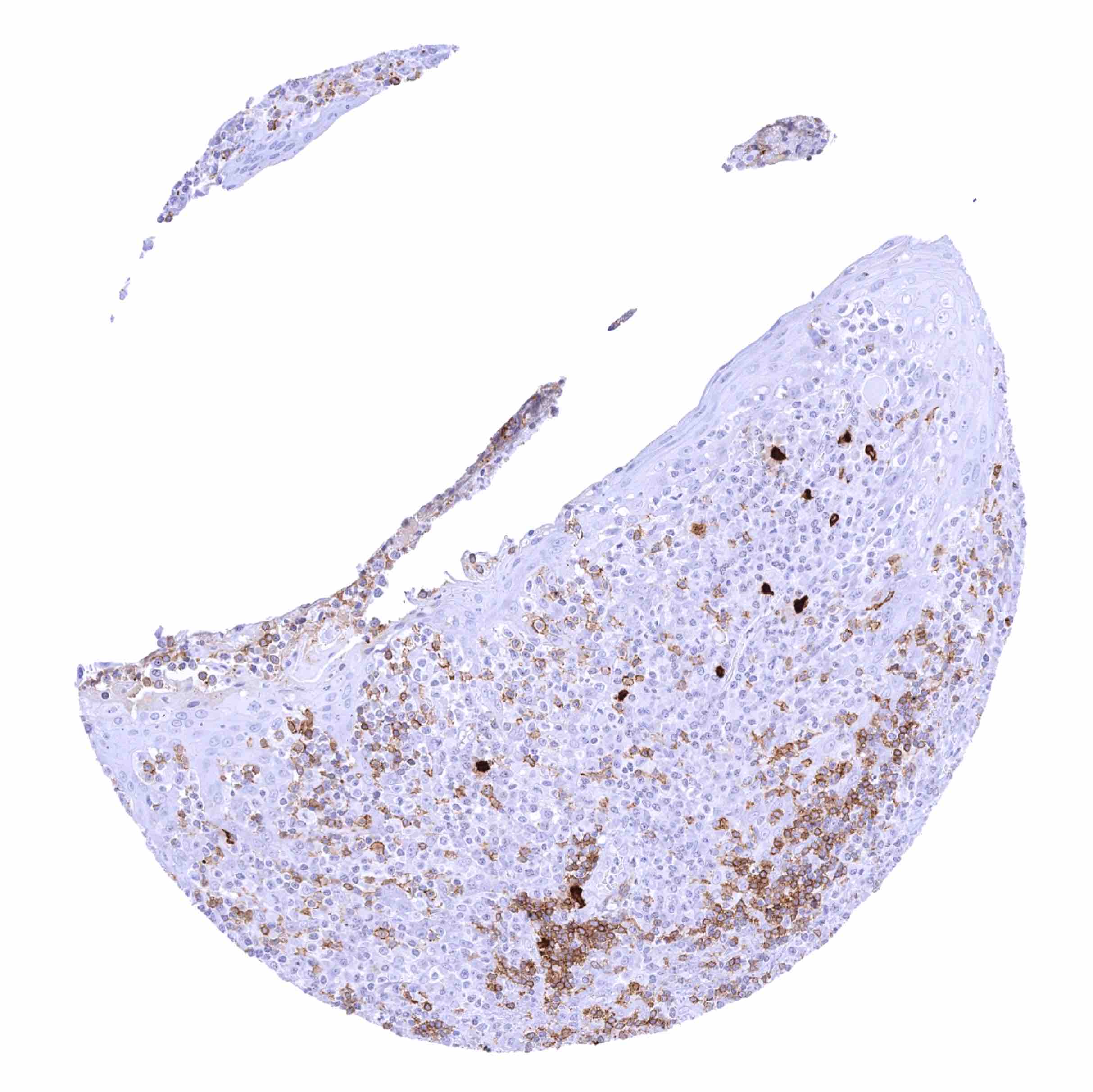
Tonsil, surface epithelium – Numerous IgD positive inflammatory cells
Urinary bladder, muscular wall
Uterus, ectocervix
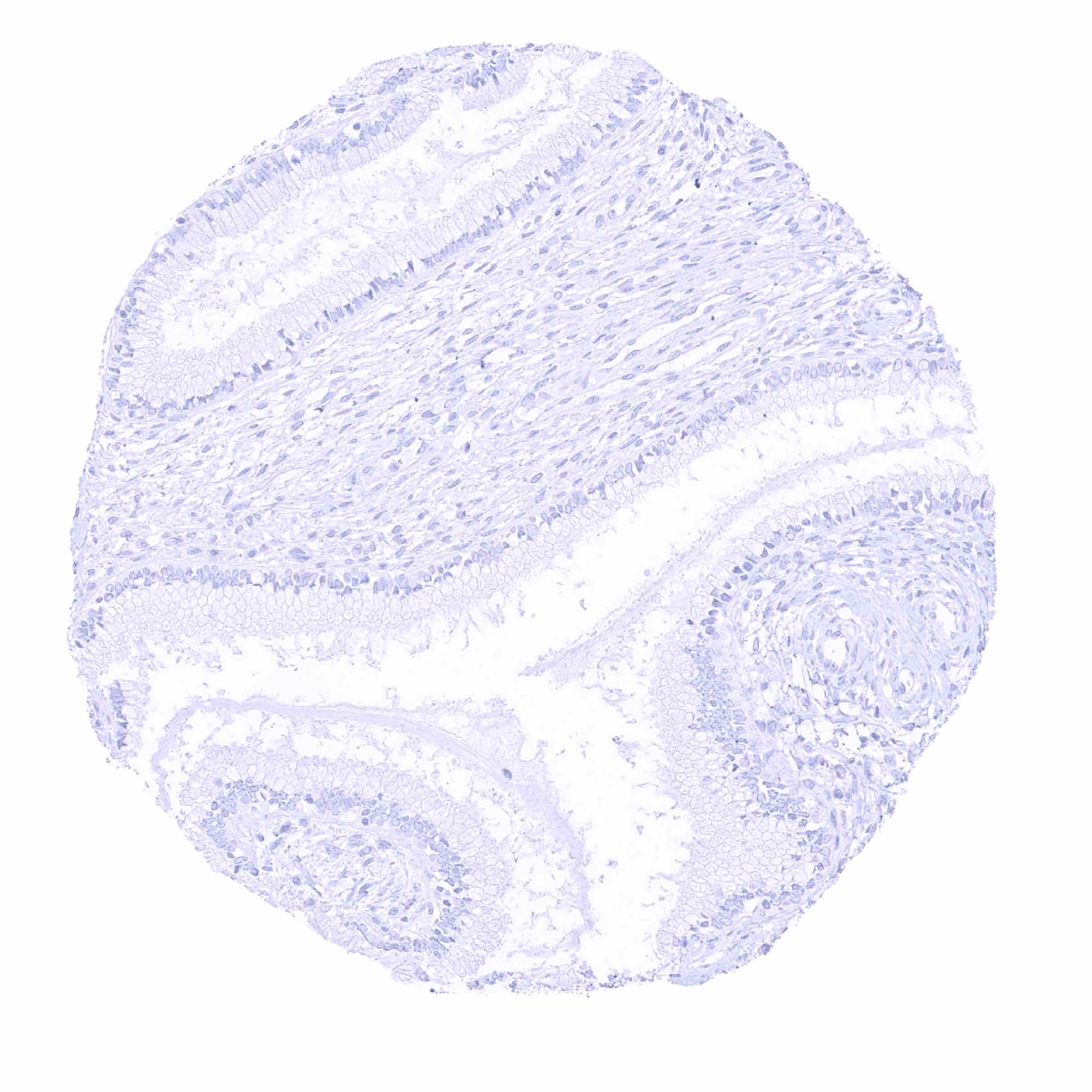
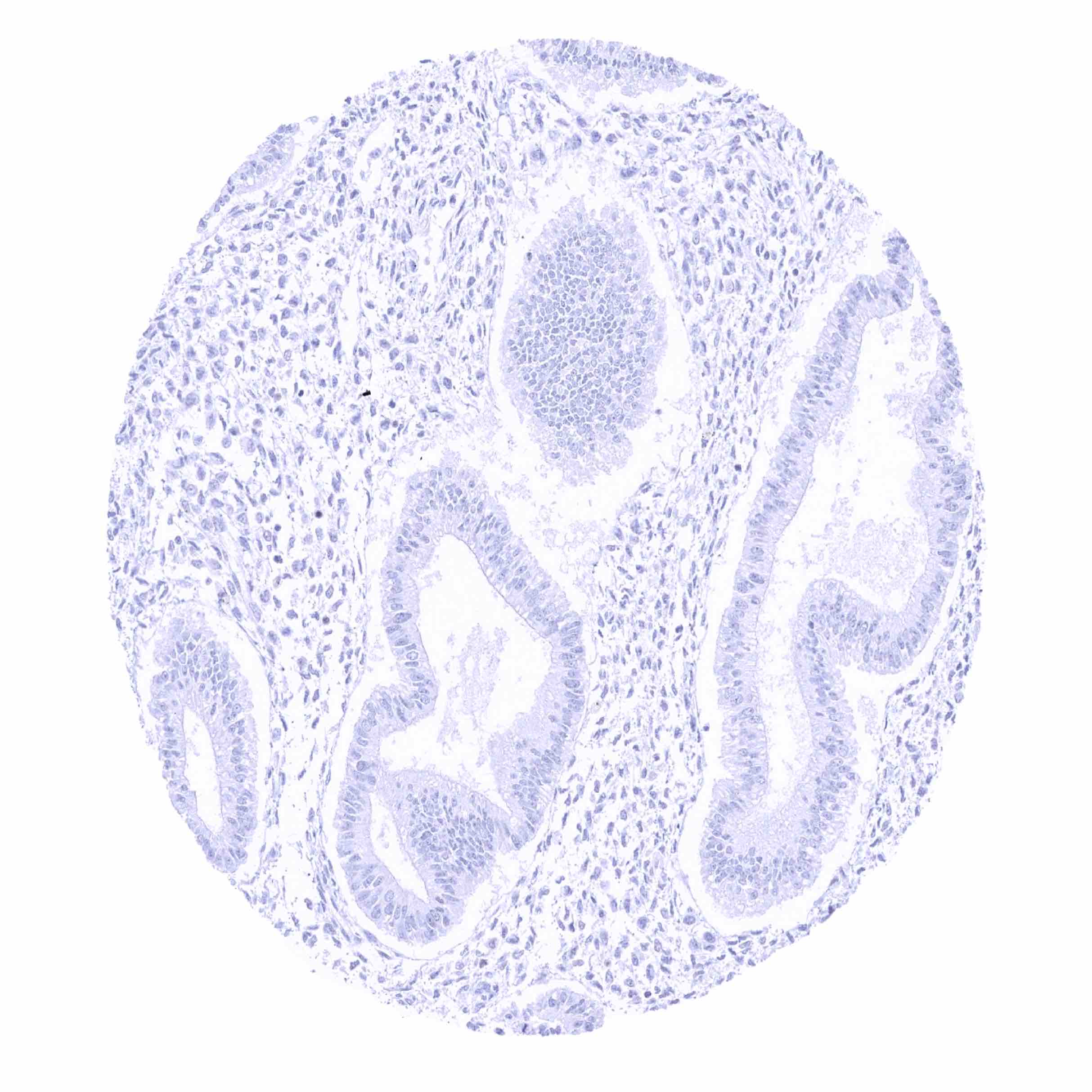
Uterus, endocervix
Uterus, endometrium (proliferation)
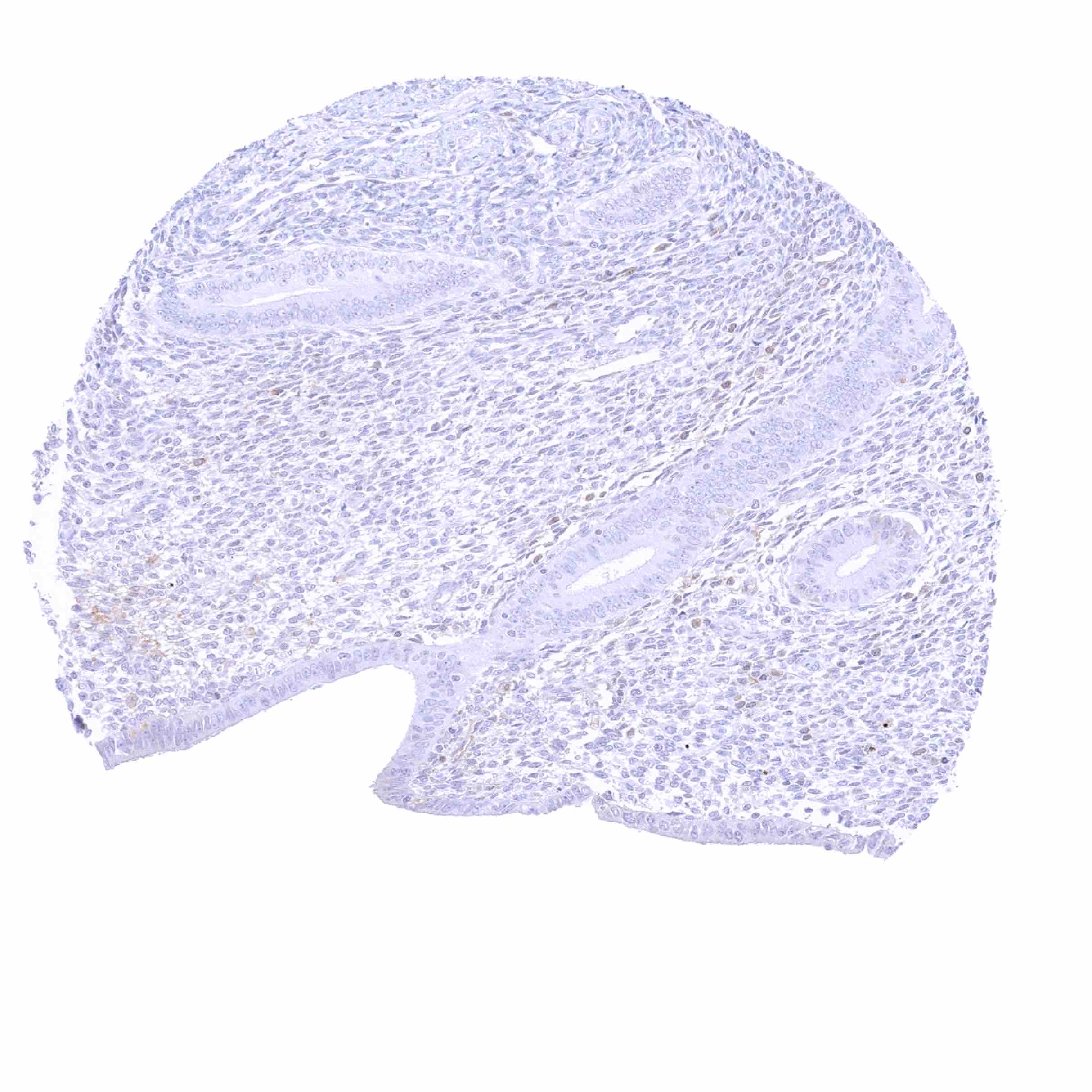
Uterus, endometrium (proliferation)
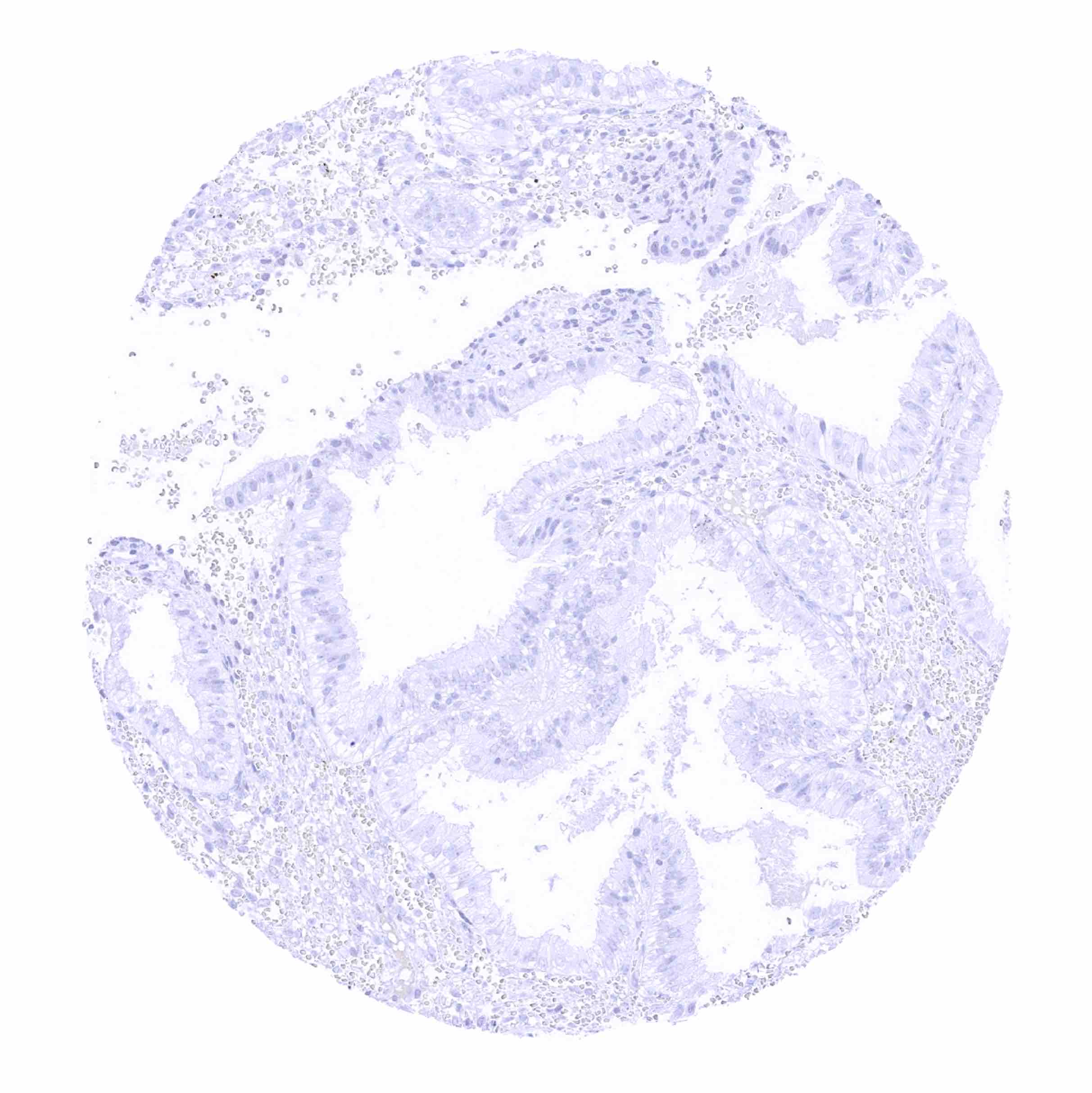
Uterus, endometrium (secretion)
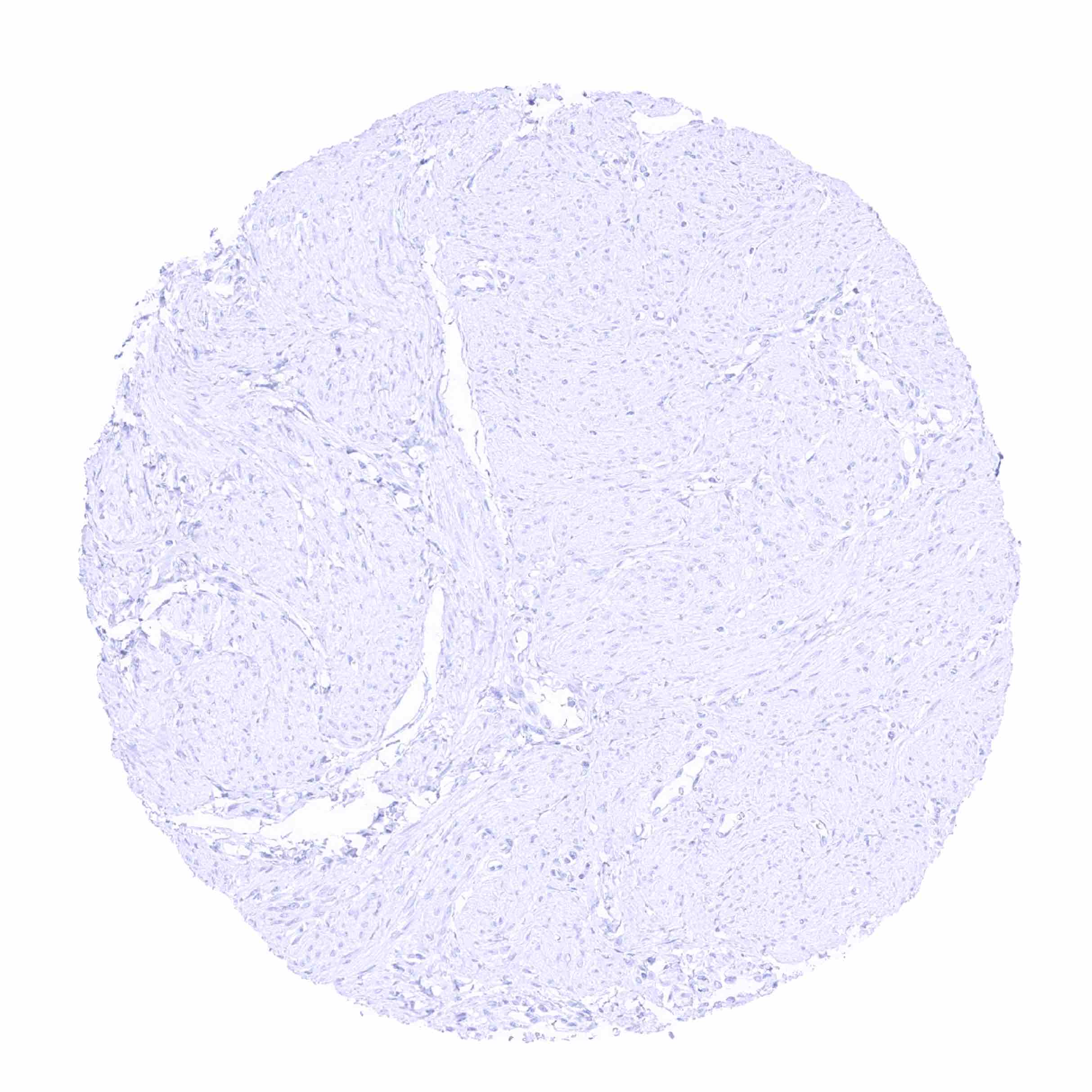
Uterus, myometrium