295,00 € – 995,00 €
Product details
Synonyms = DRB1; HLA class II histocompatibility antigen, DR-1 beta chain; HLA-DR-beta 1; HLA-DRB1; human leucocyte antigen DRB1; Leucocyte antigen DR beta 1 chain; lymphocyte antigen DRB1; major histocompatibility complex, class II, DR beta 1; MHC class II HLA-DR beta 1 chain; MHC class II HLA-DR-beta cell surface glycoprotein
Antibody type = Recombinant Rabbit monoclonal / IgG
Clone = MSVA-478R
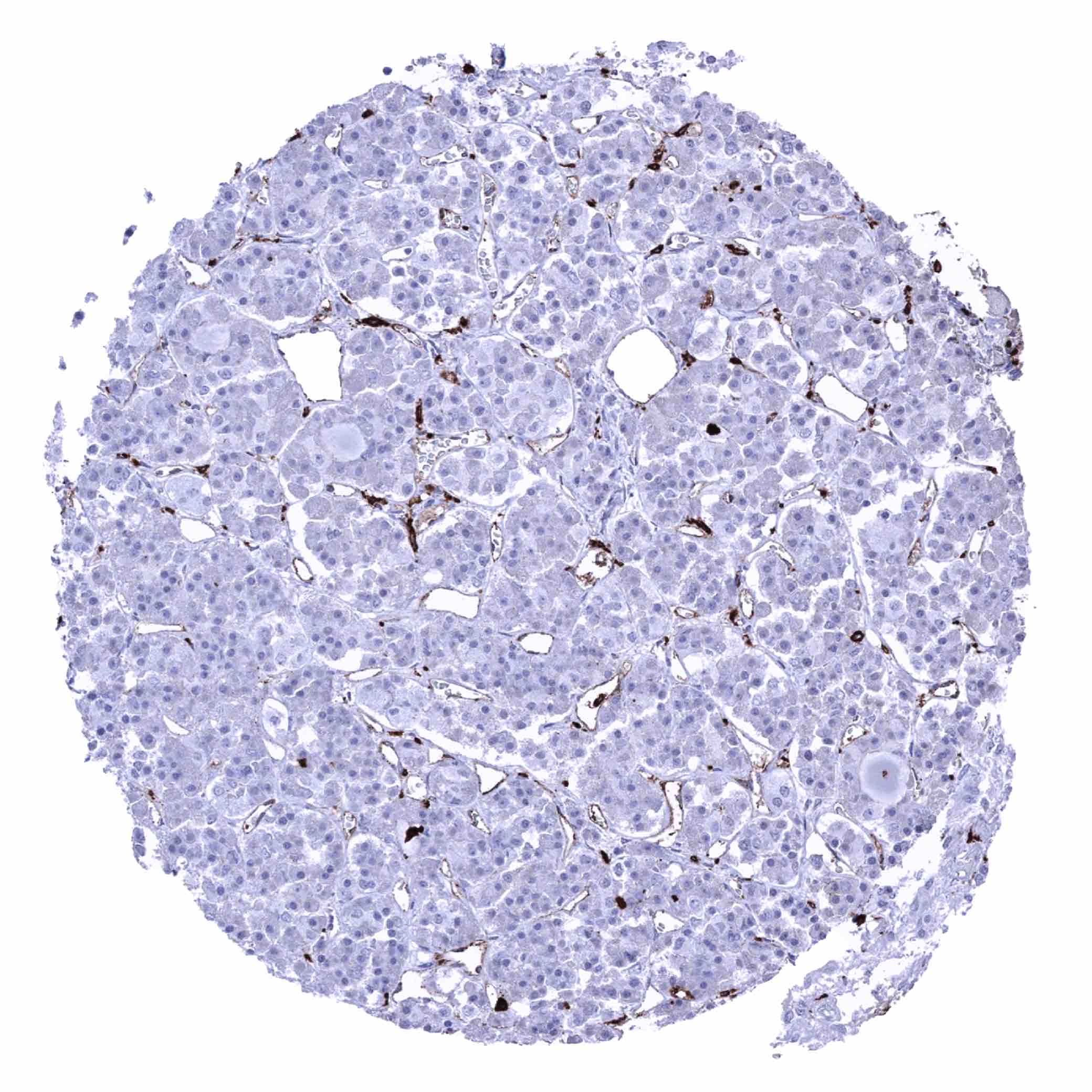
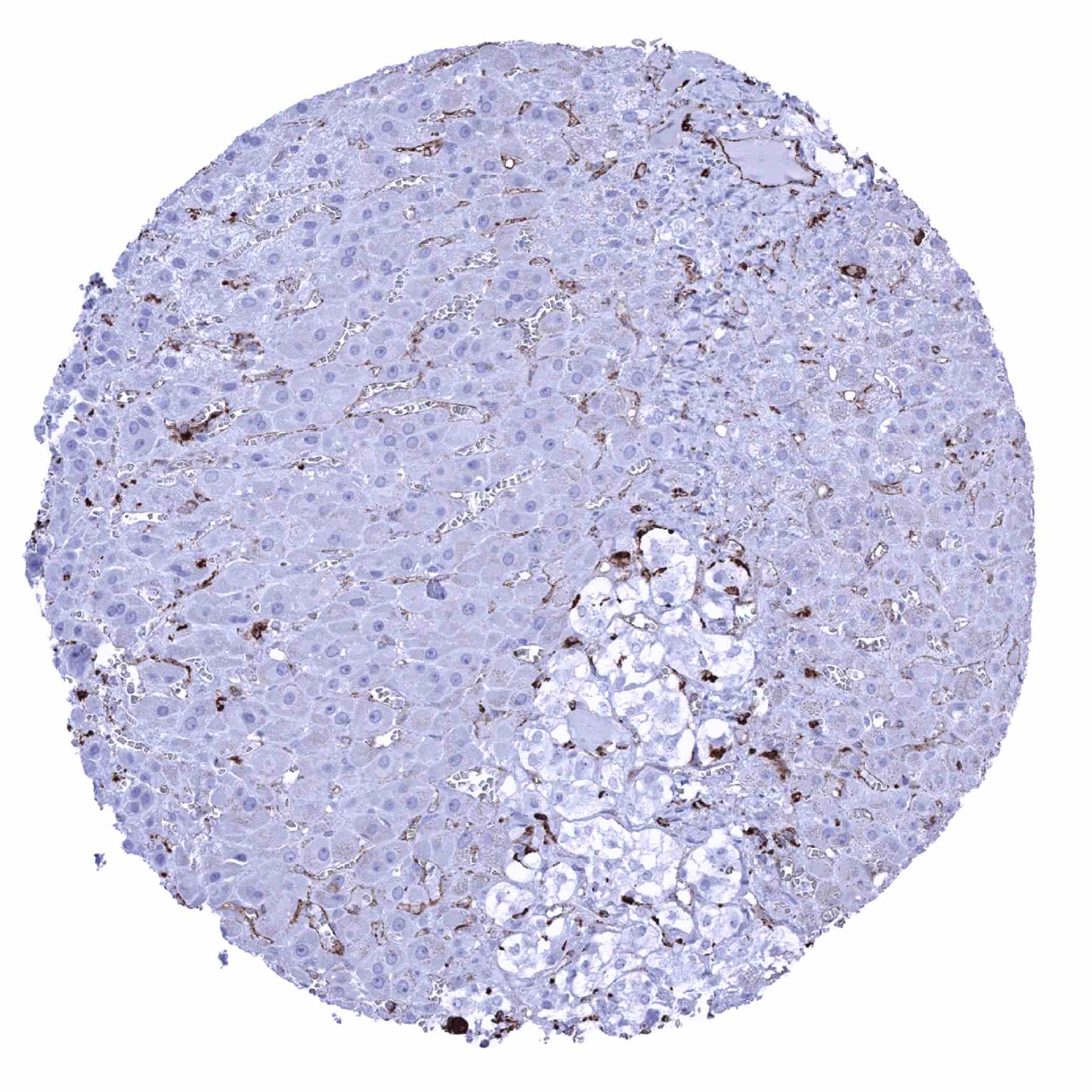
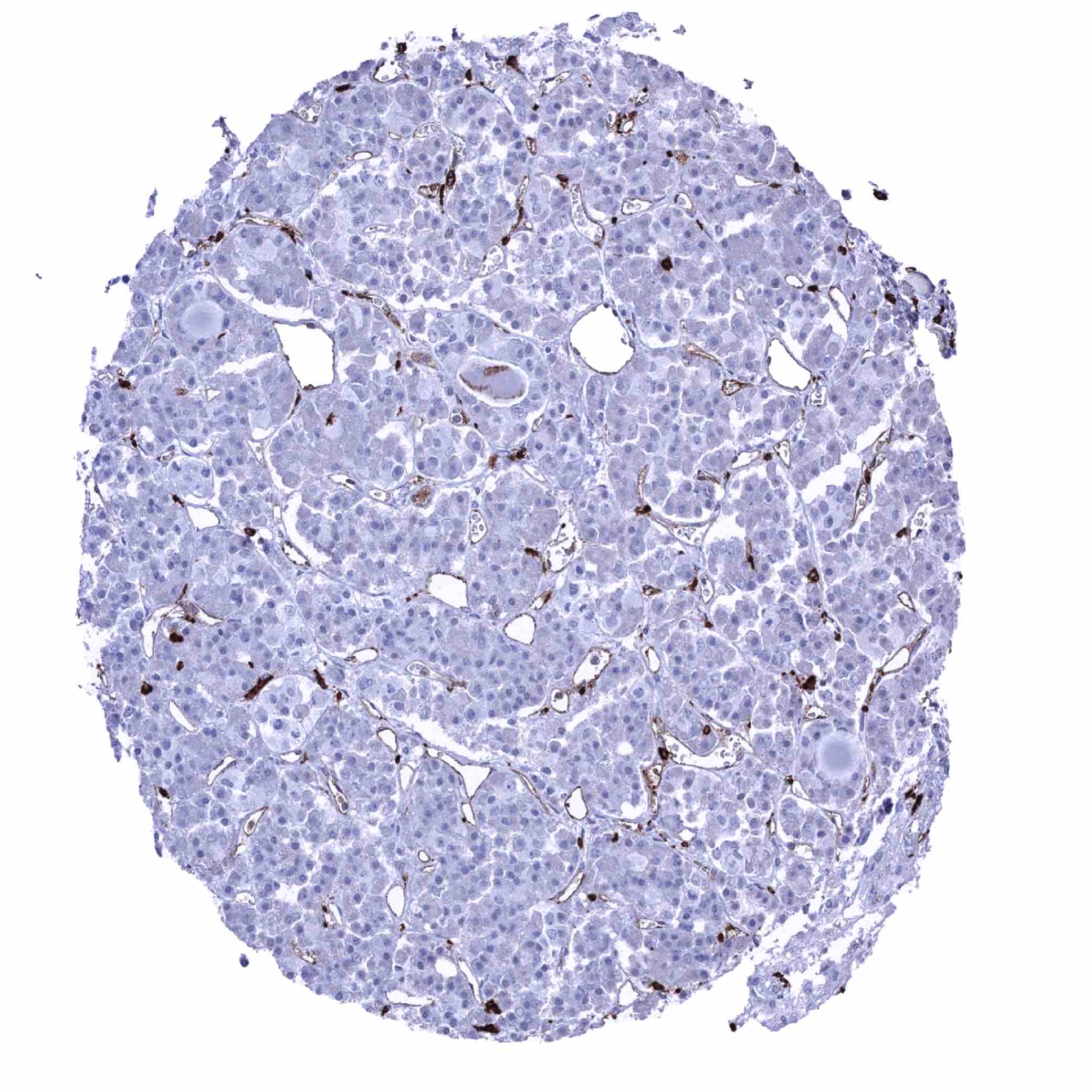
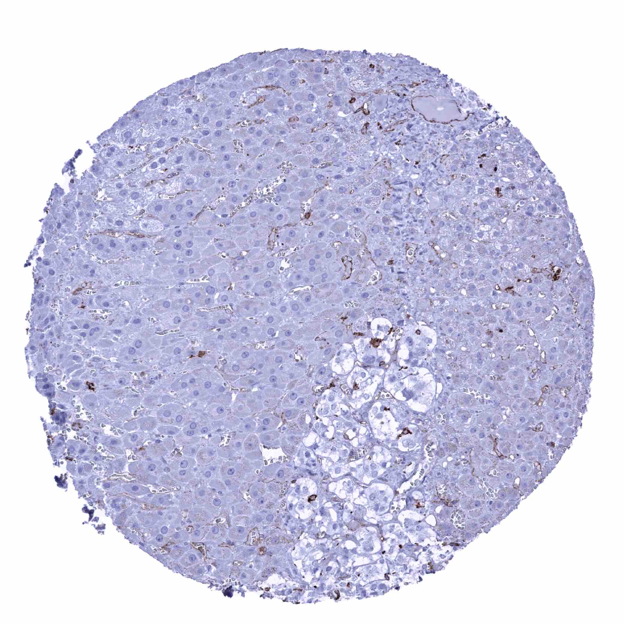
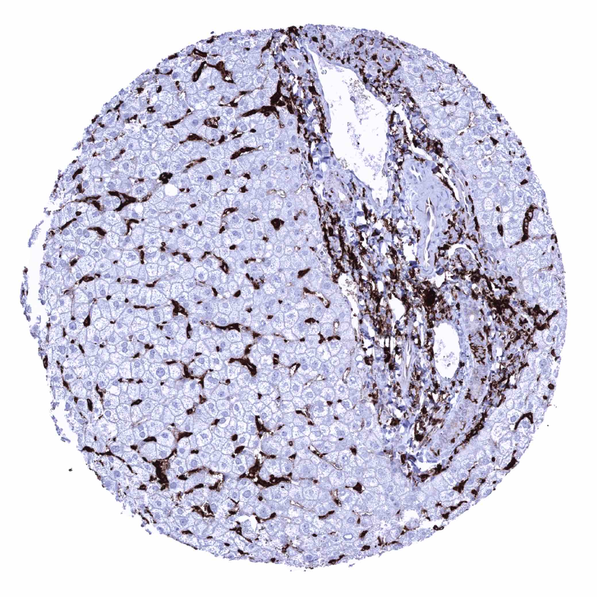
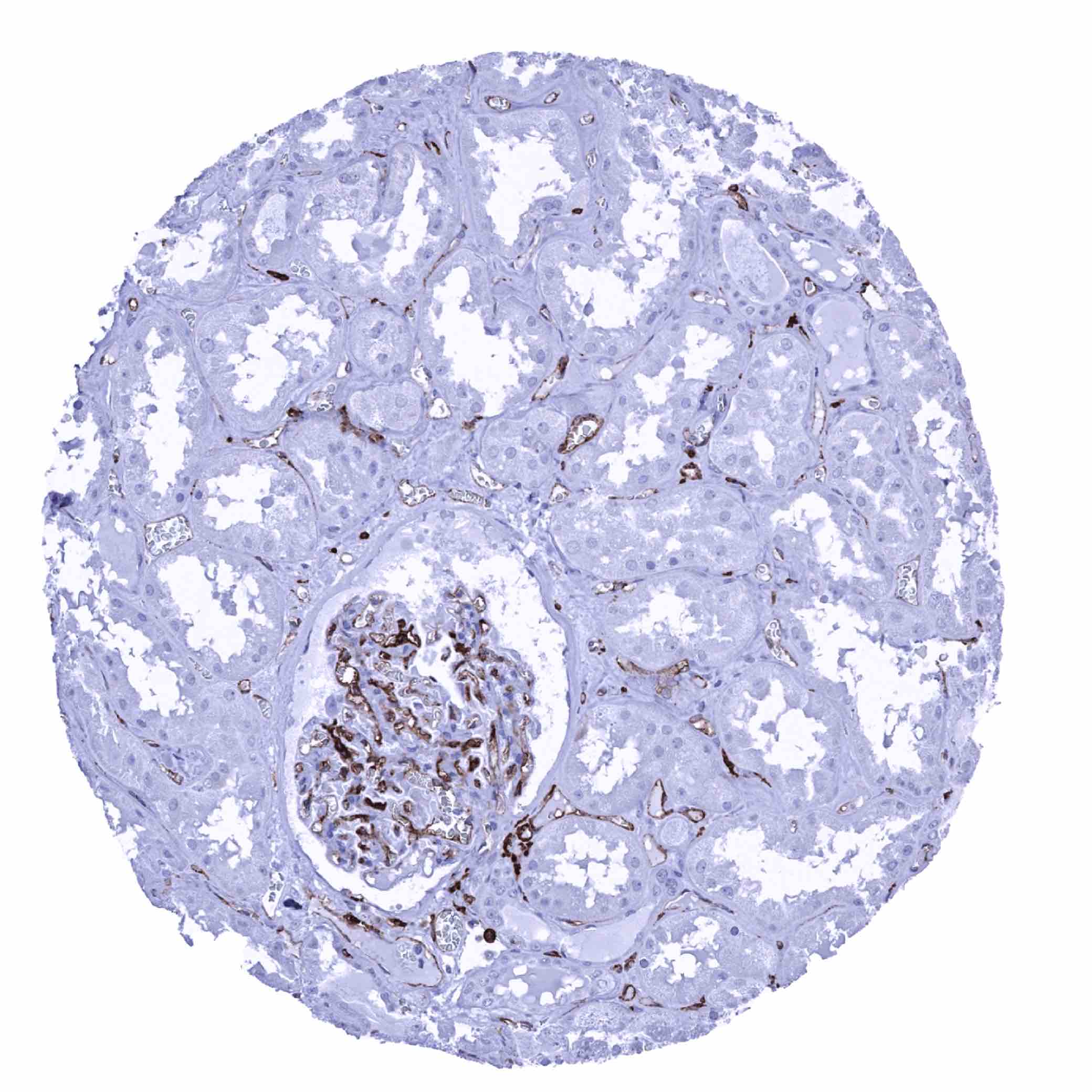
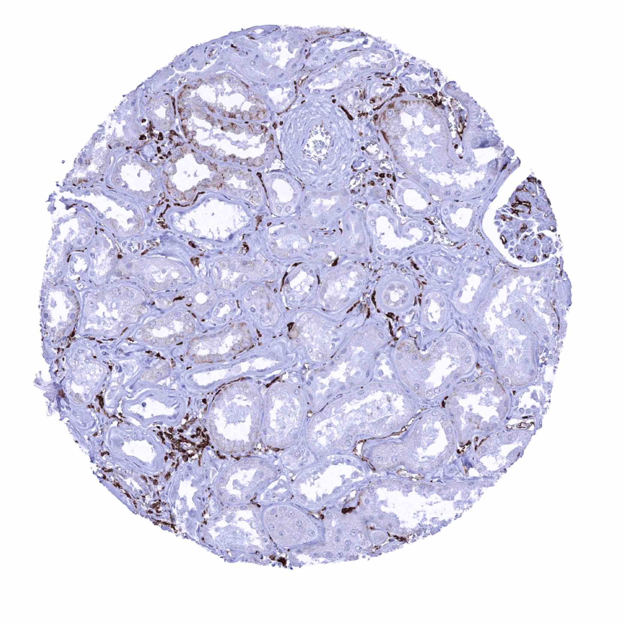
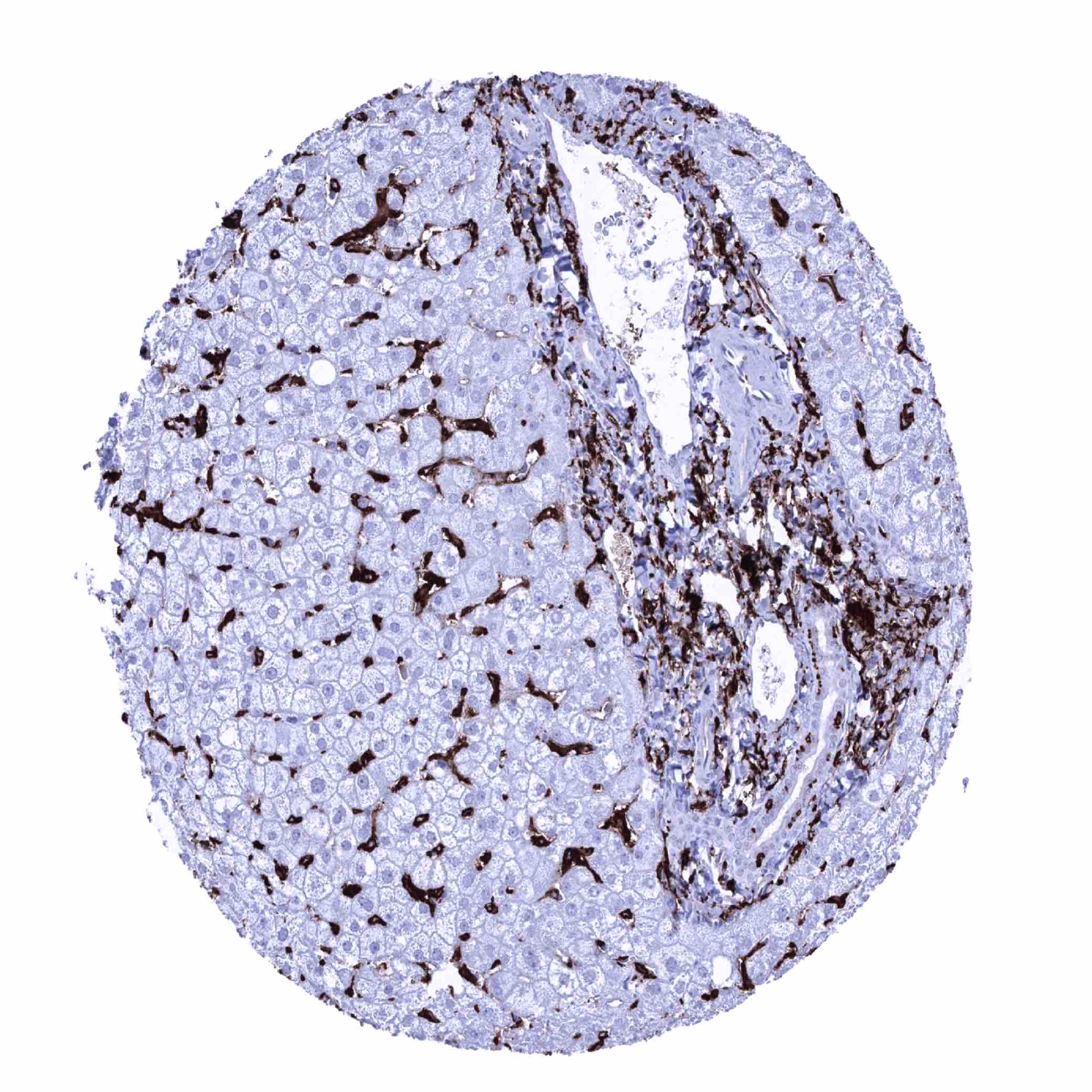
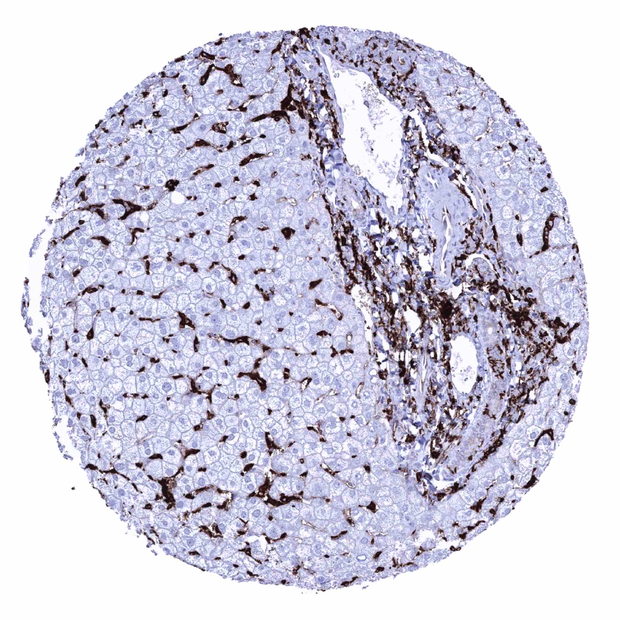
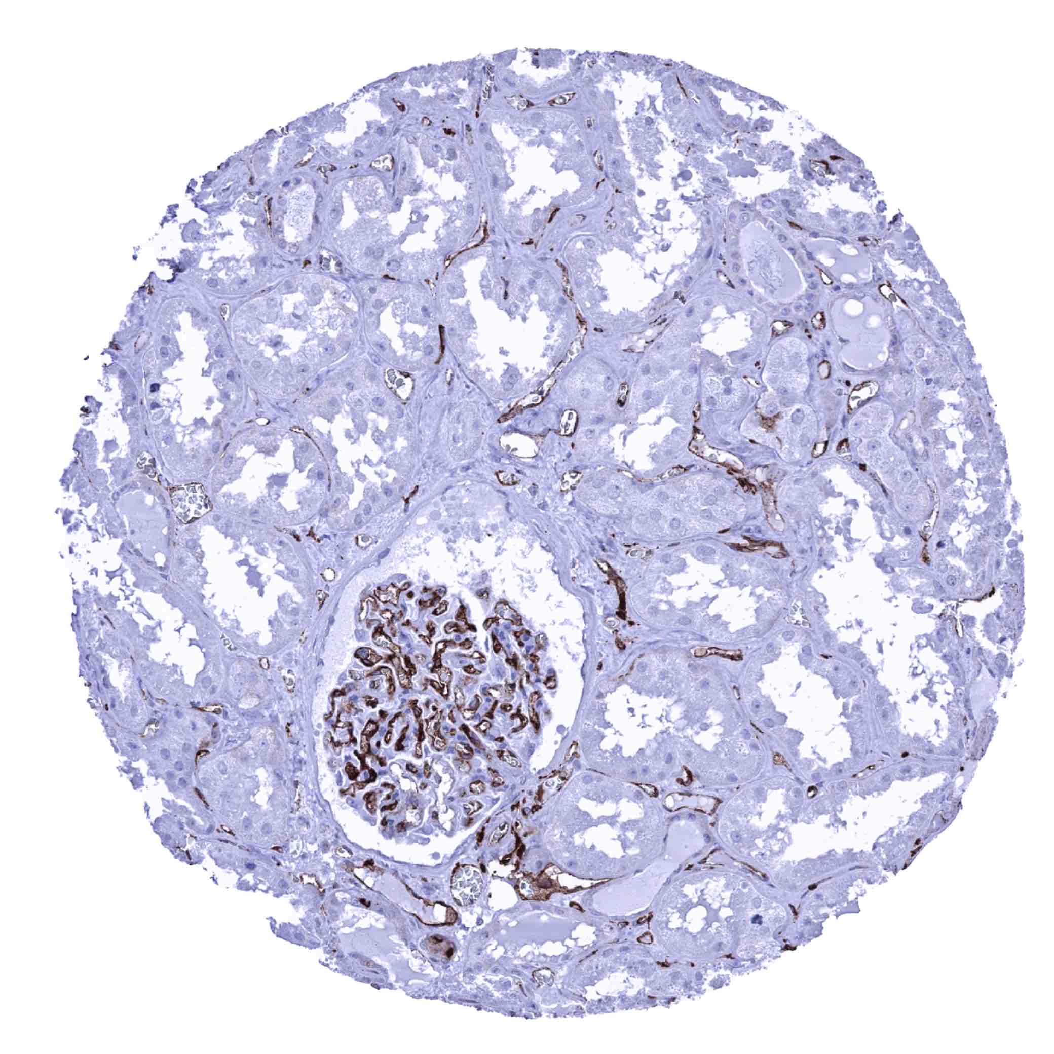
Positive control = Liver: An at least moderate staining should be seen of sinusoids and Kupffer cells. Kidney: An at least moderate staining should be seen in glomerular capillaries.
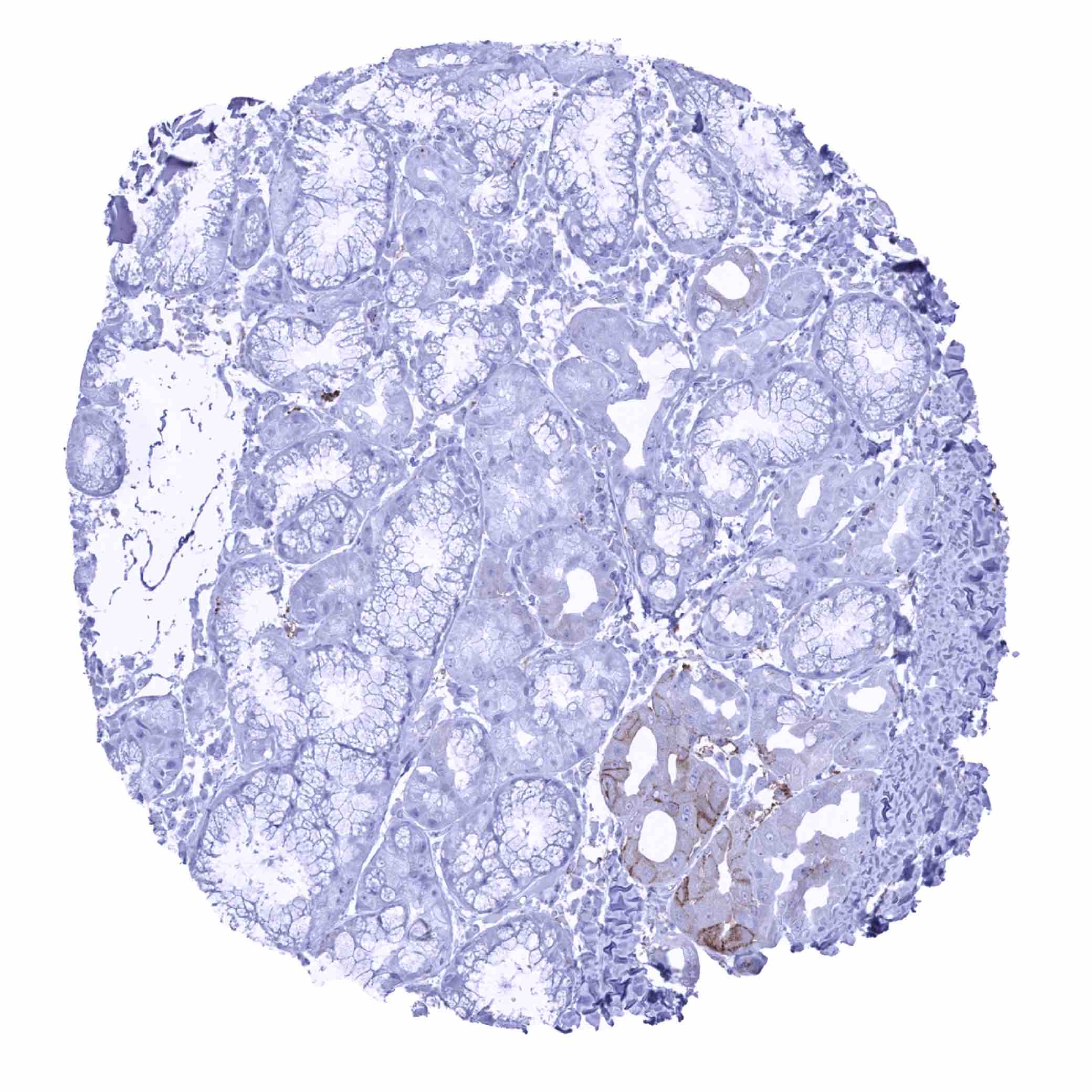
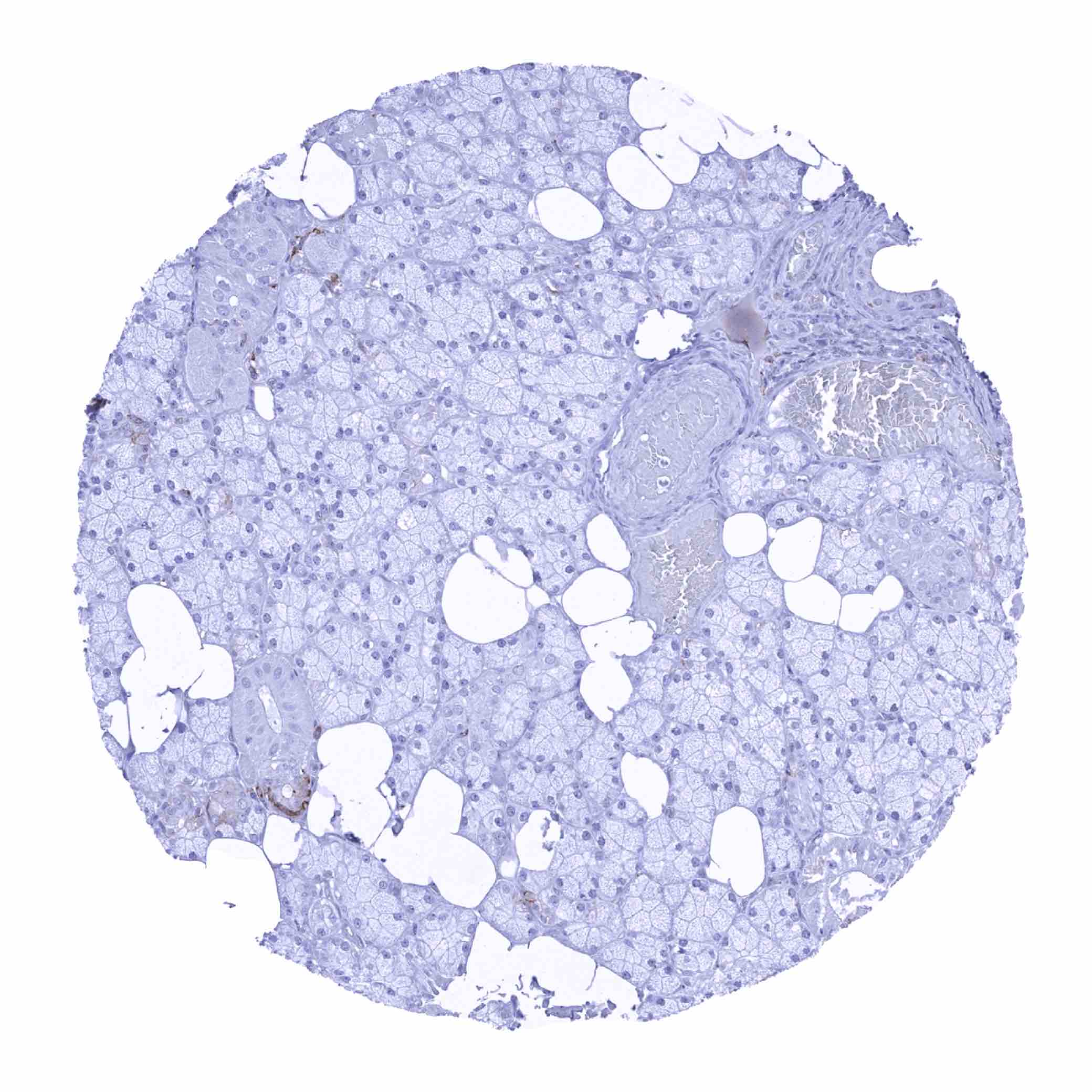
Negative control = Liver, kidney: HLA-DR staining must be absent normal in hepatocytes and in tubuli of completely normal kidney.
Cellular localization = Cell Surface
Reactivity = Human
Application = Immunohistochemistry
Dilution = 1:100 – 1:200
Intended Use = Research Use Only
Relevance of Antibody
HLA-DRB1 is a pivotal protein for immune response.
Biology Behind
HLA-DRB1 is coded by the DRB1 gene located at 6p21.31. It represents the most prevalent of 4 different beta subunits (DRB1, DRB3, DRB4, DRB5) of the HLA-DR protein complex. HLA-DRB1 is expressed in all humans and its expression level is at least 5 times is higher than what is seen for other HLA-DR β- subunits. Together with the HLA-DR α-subunit, the DR β-subunits form the HLA-DR complex which represents an αβ heterodimer protein. HLA-DR is an MHC class II cell surface receptor acting as a ligand for the T-cell receptor. The HLA-DR cell surface receptor is constitutively expressed in professional antigen-presenting cells (B lymphocytes, dendritic cells, macrophages) but can also be temporarily expressed in non-professional antigen-presenting cells. HLA-DR has a pivotal role in the immune system by presenting peptides derived from extracellular proteins to T helper cells.
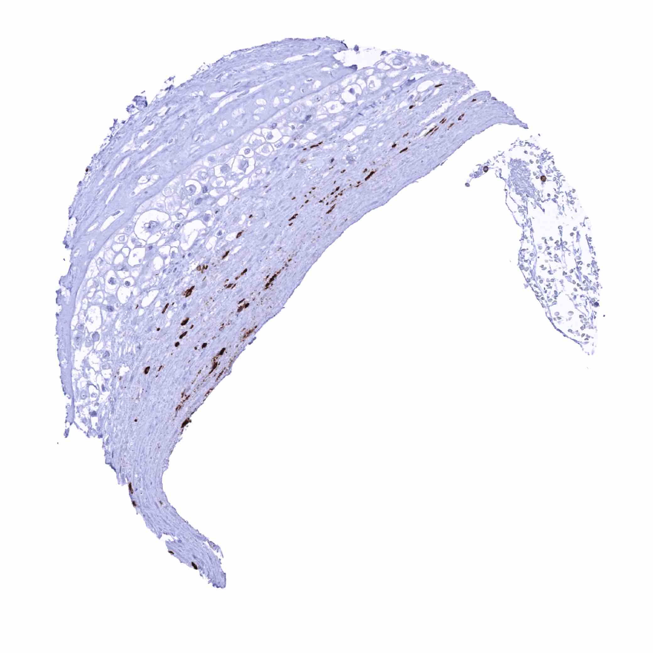
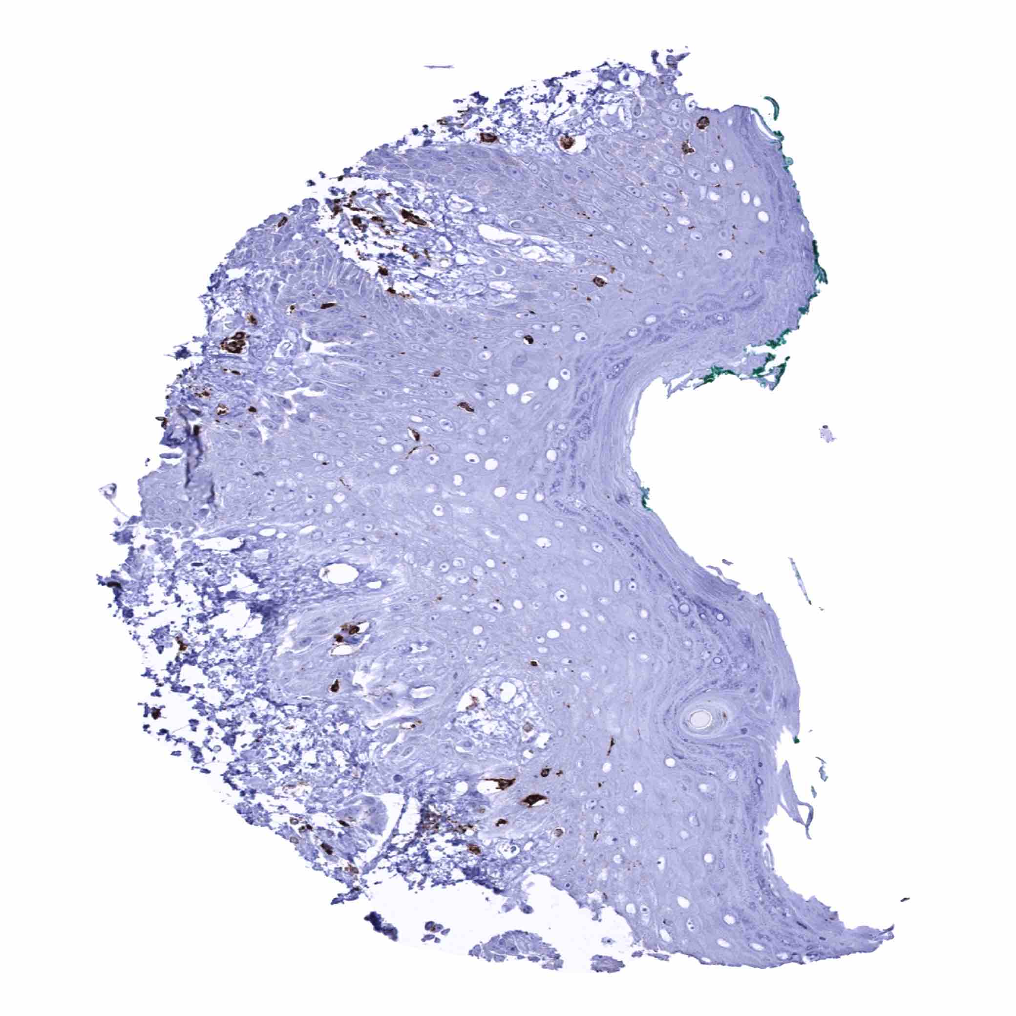
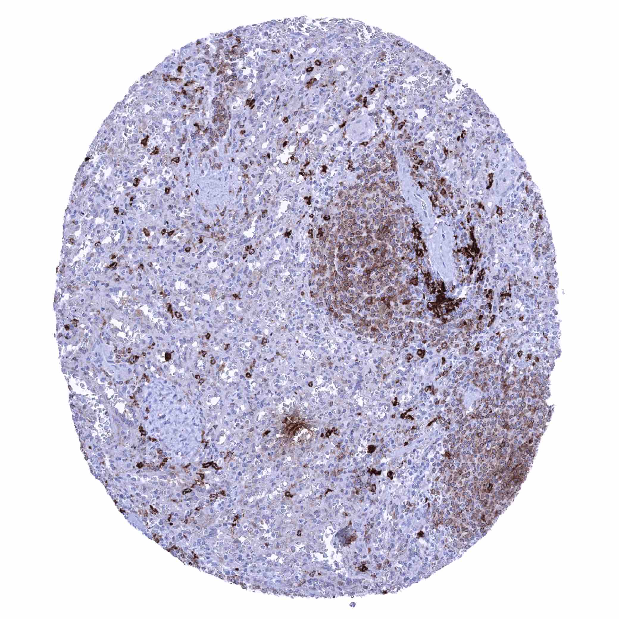
Staining Pattern in Normal Tissues
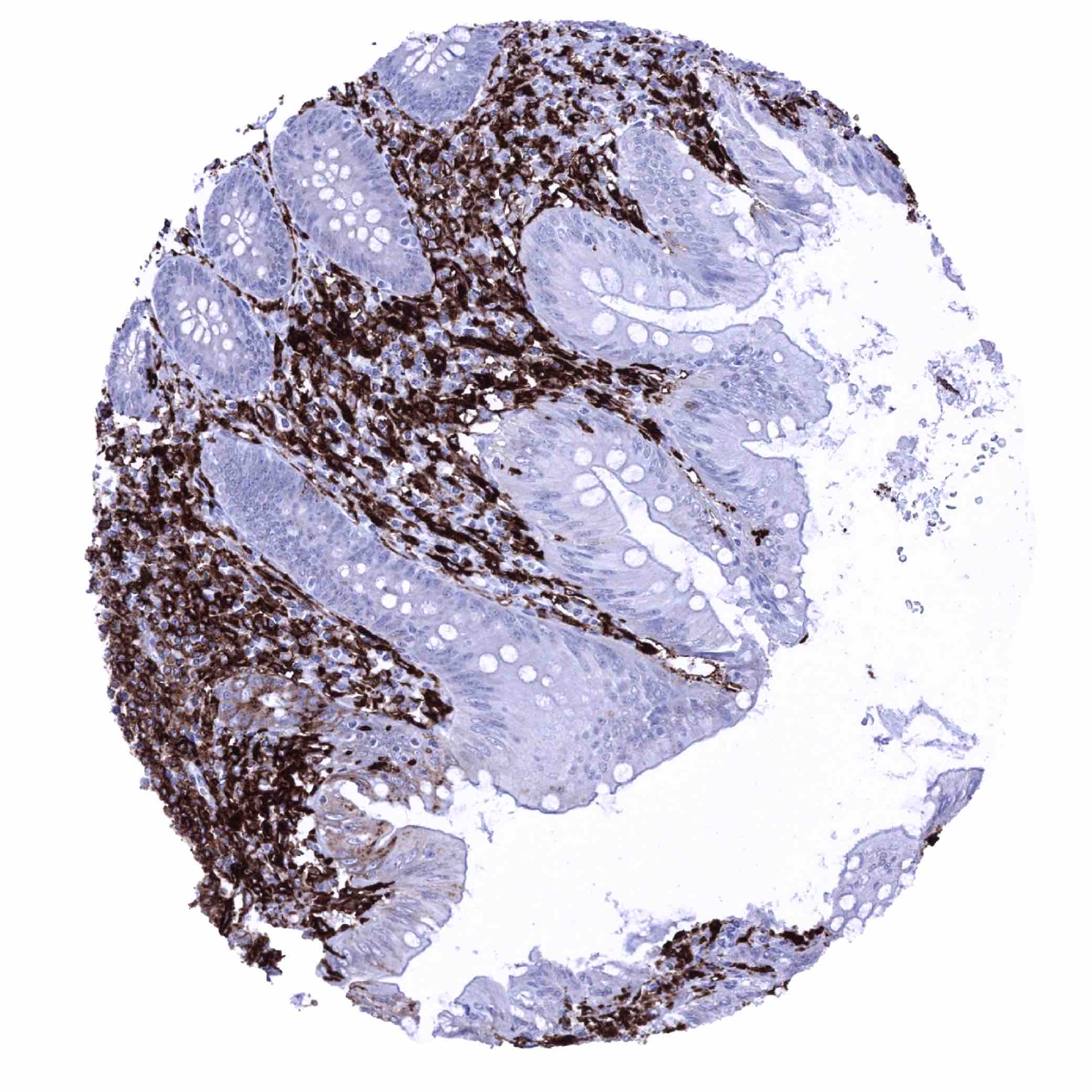
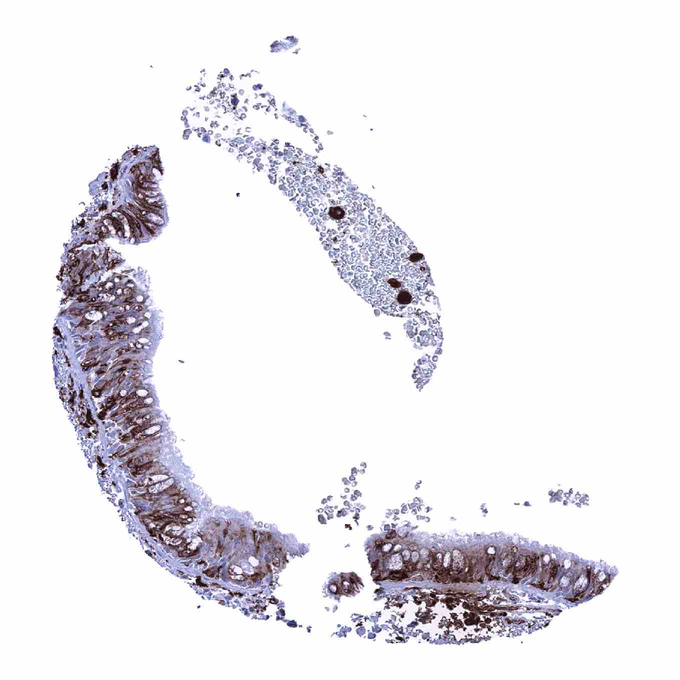
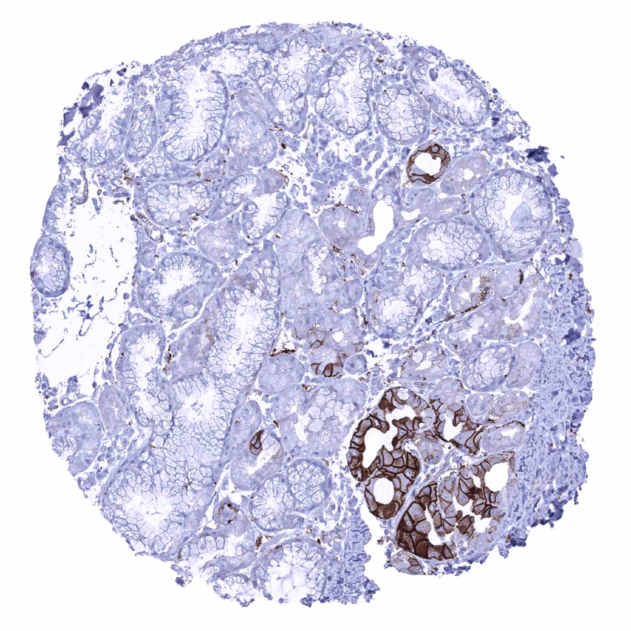
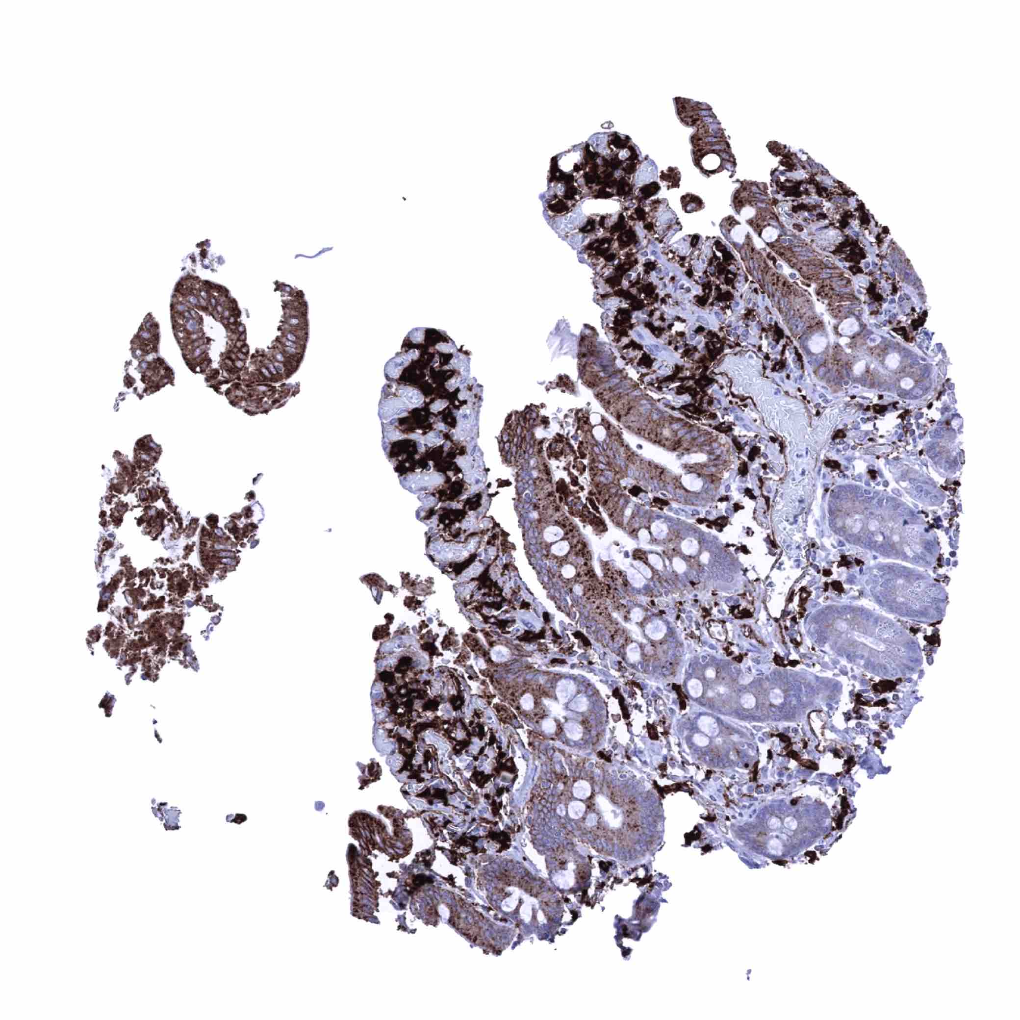
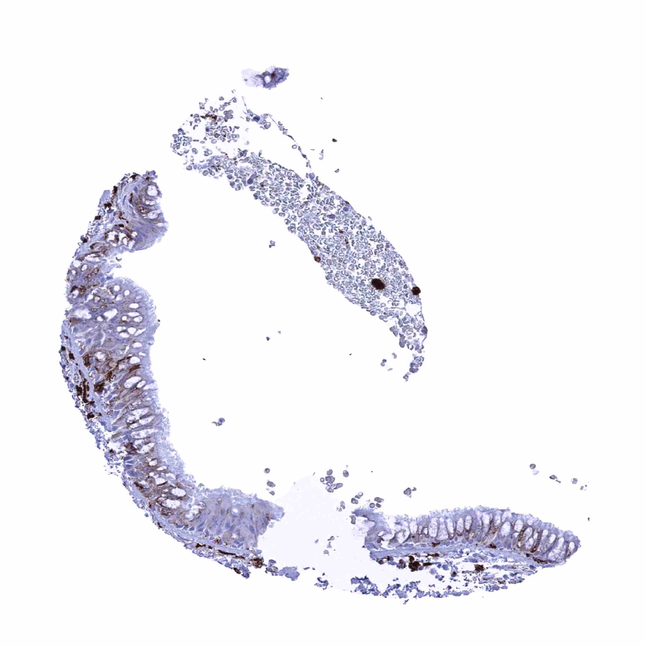
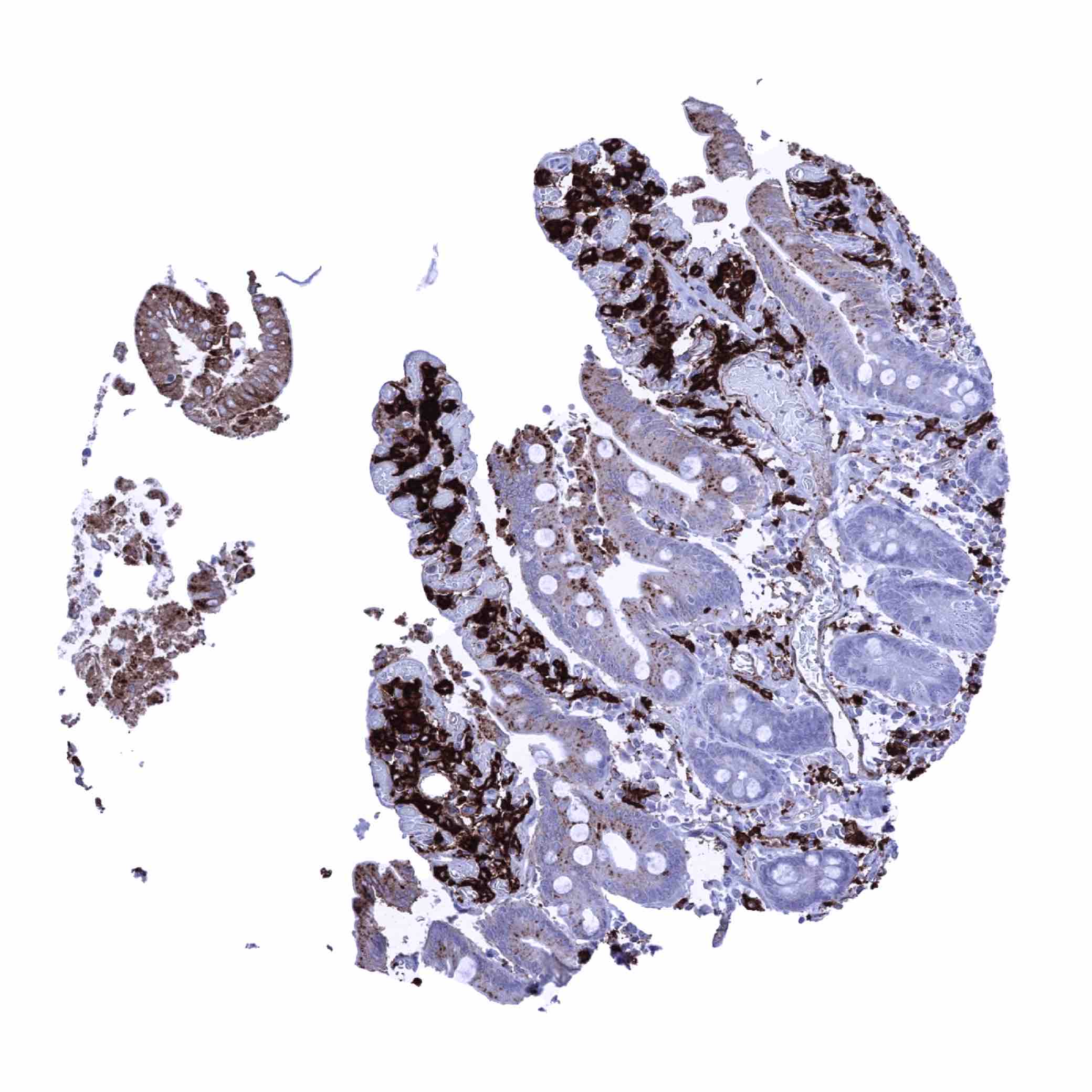
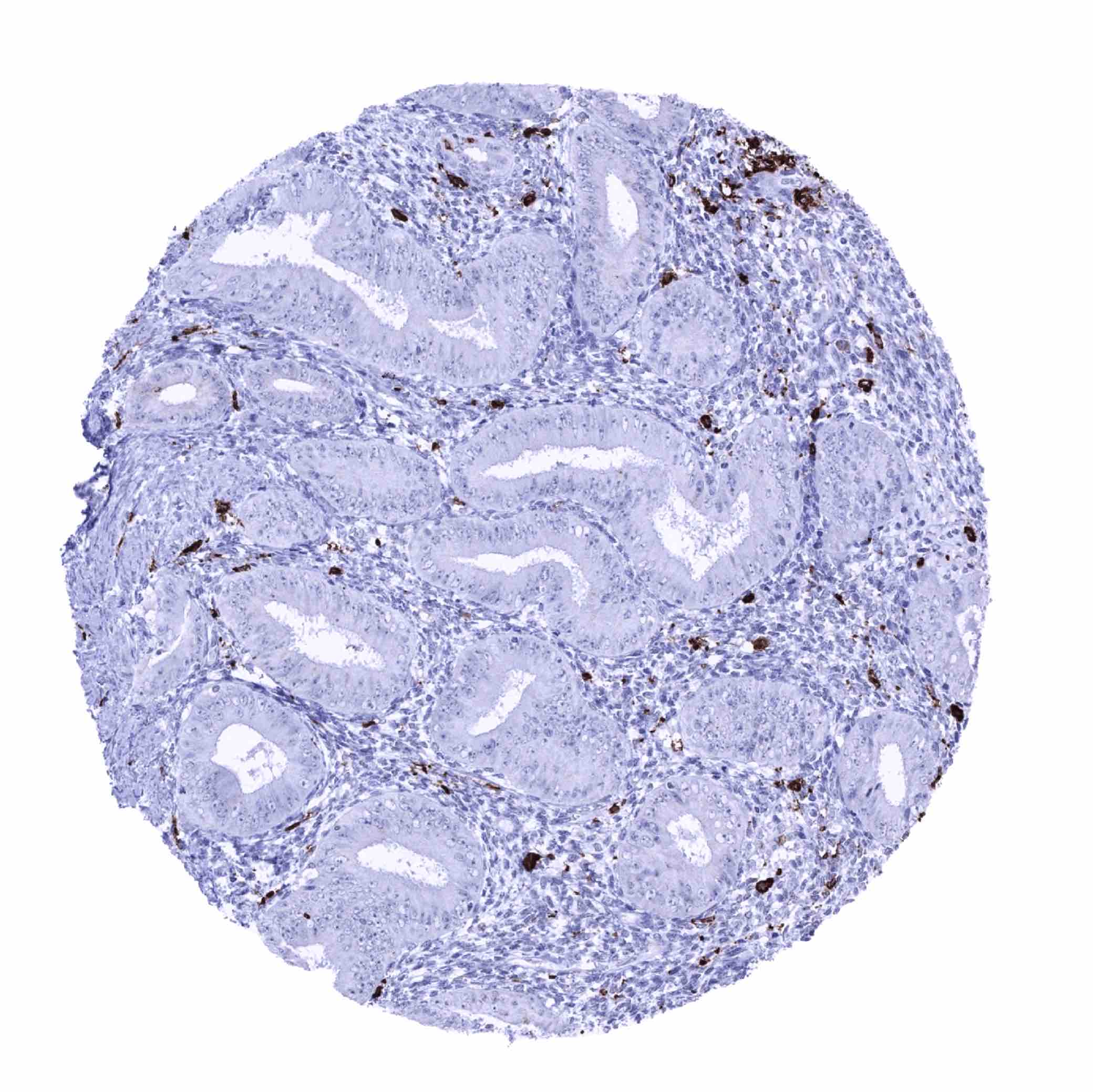
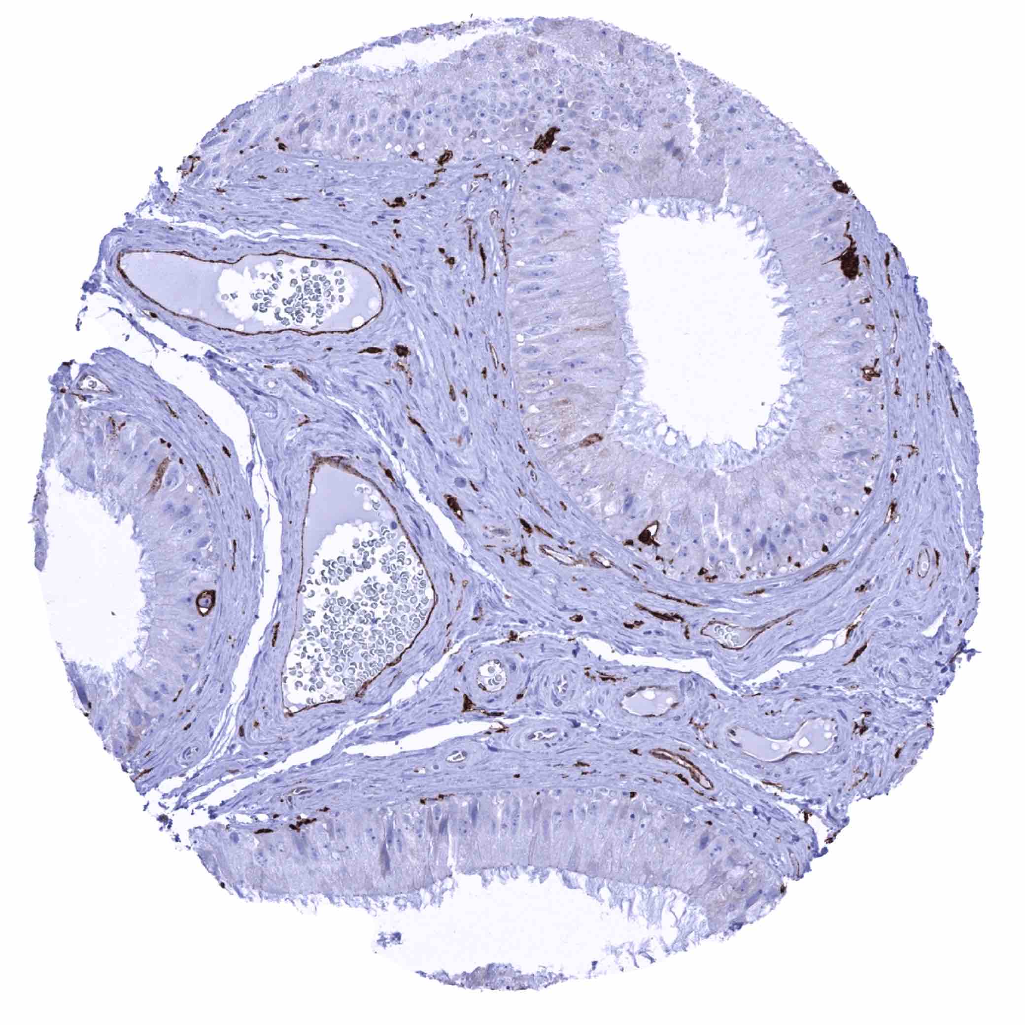
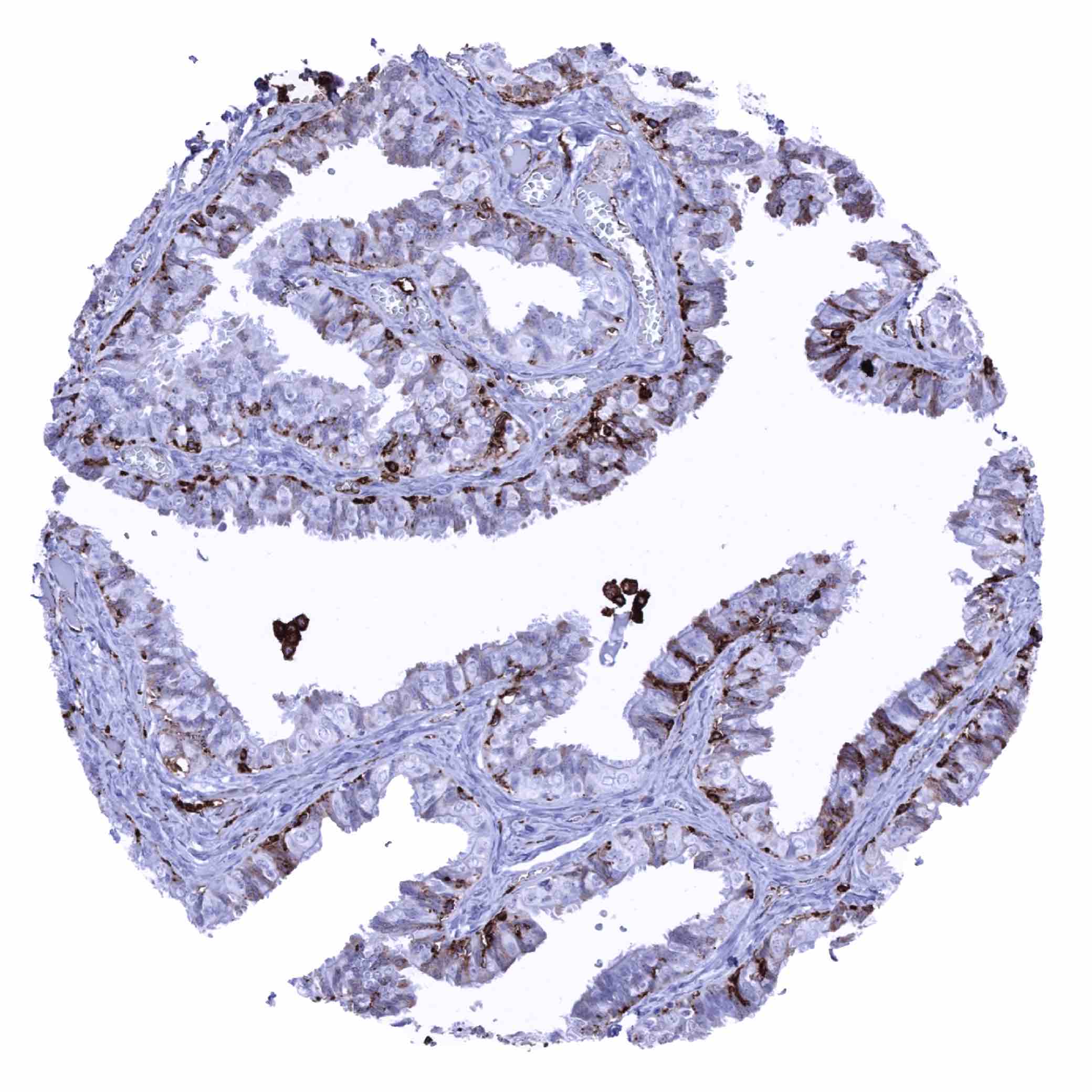
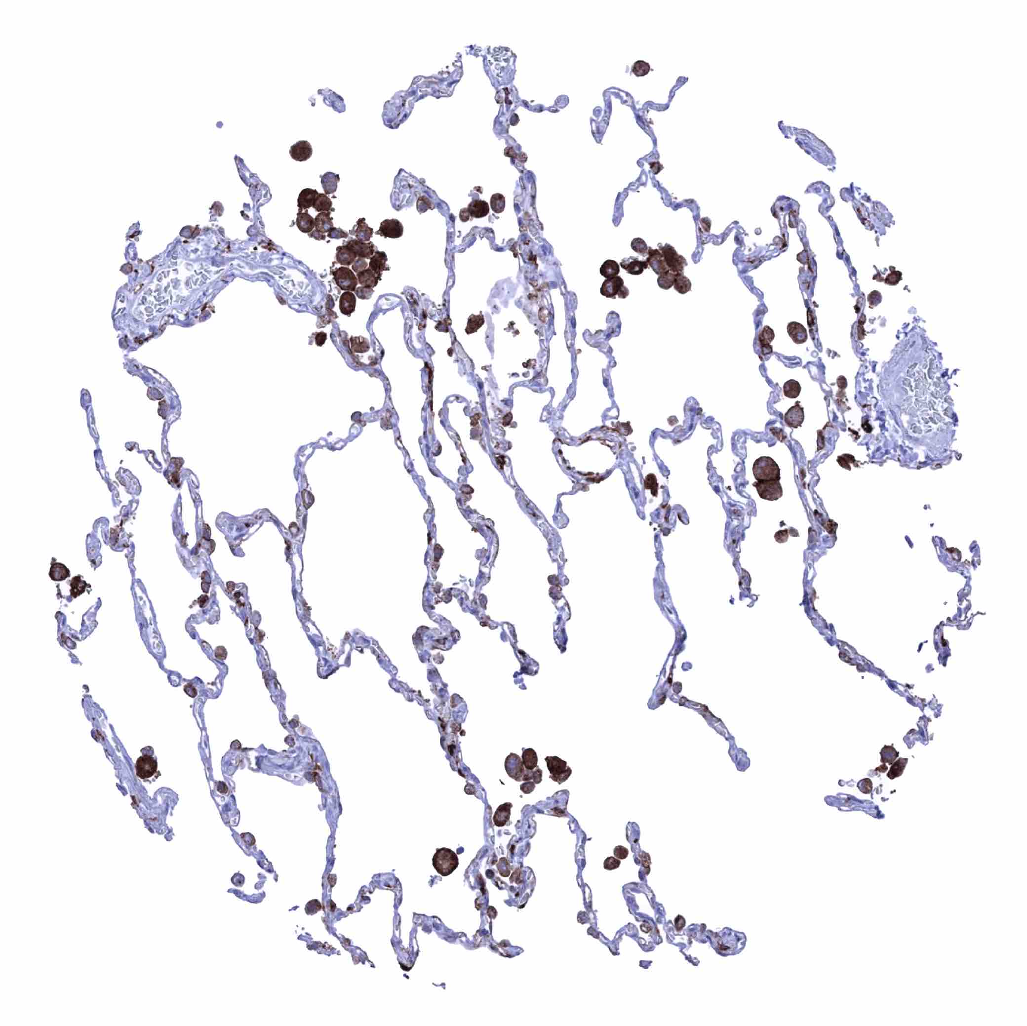
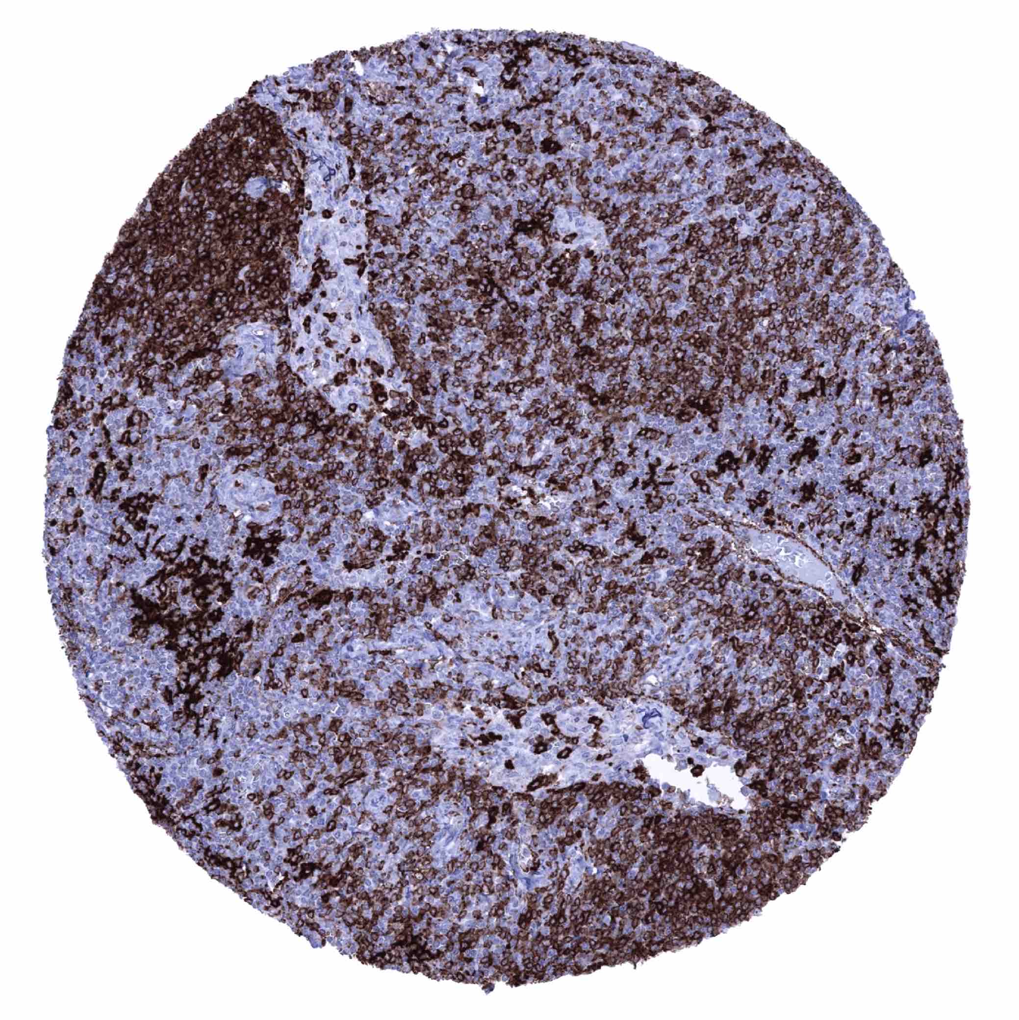
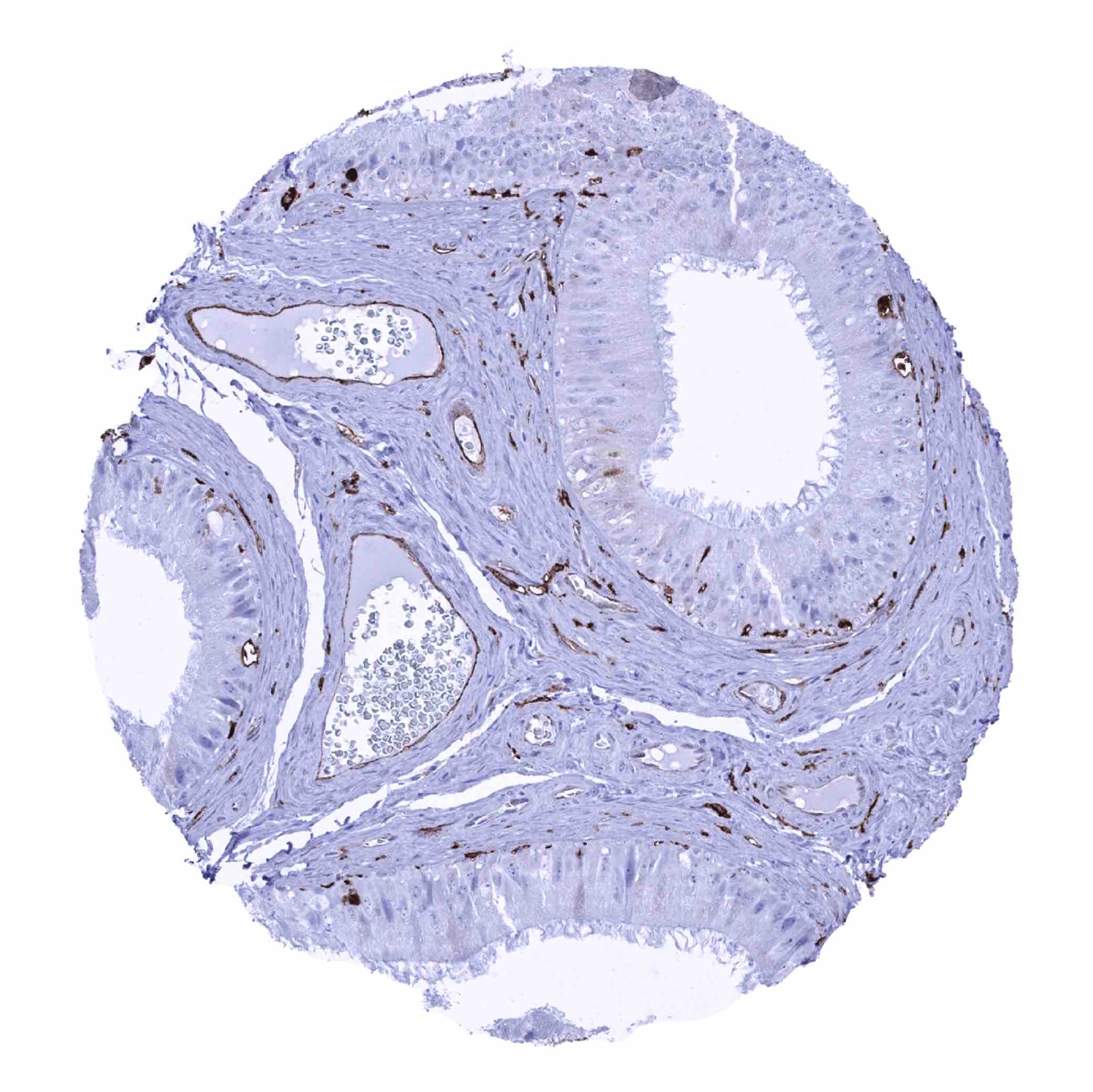
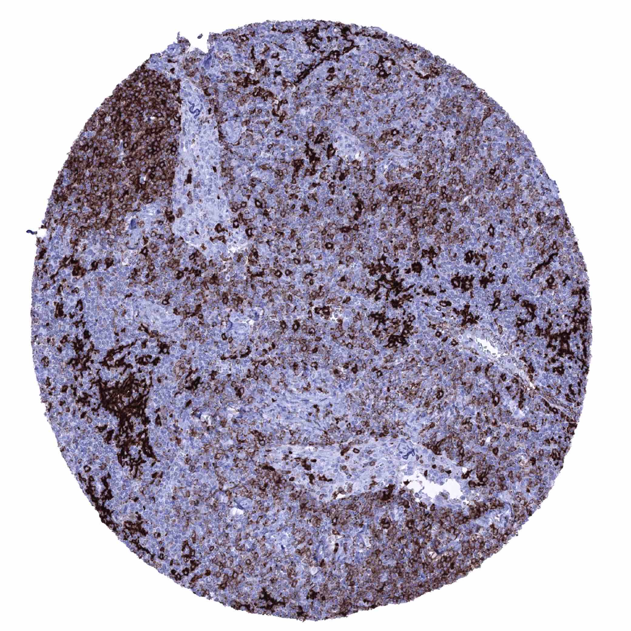
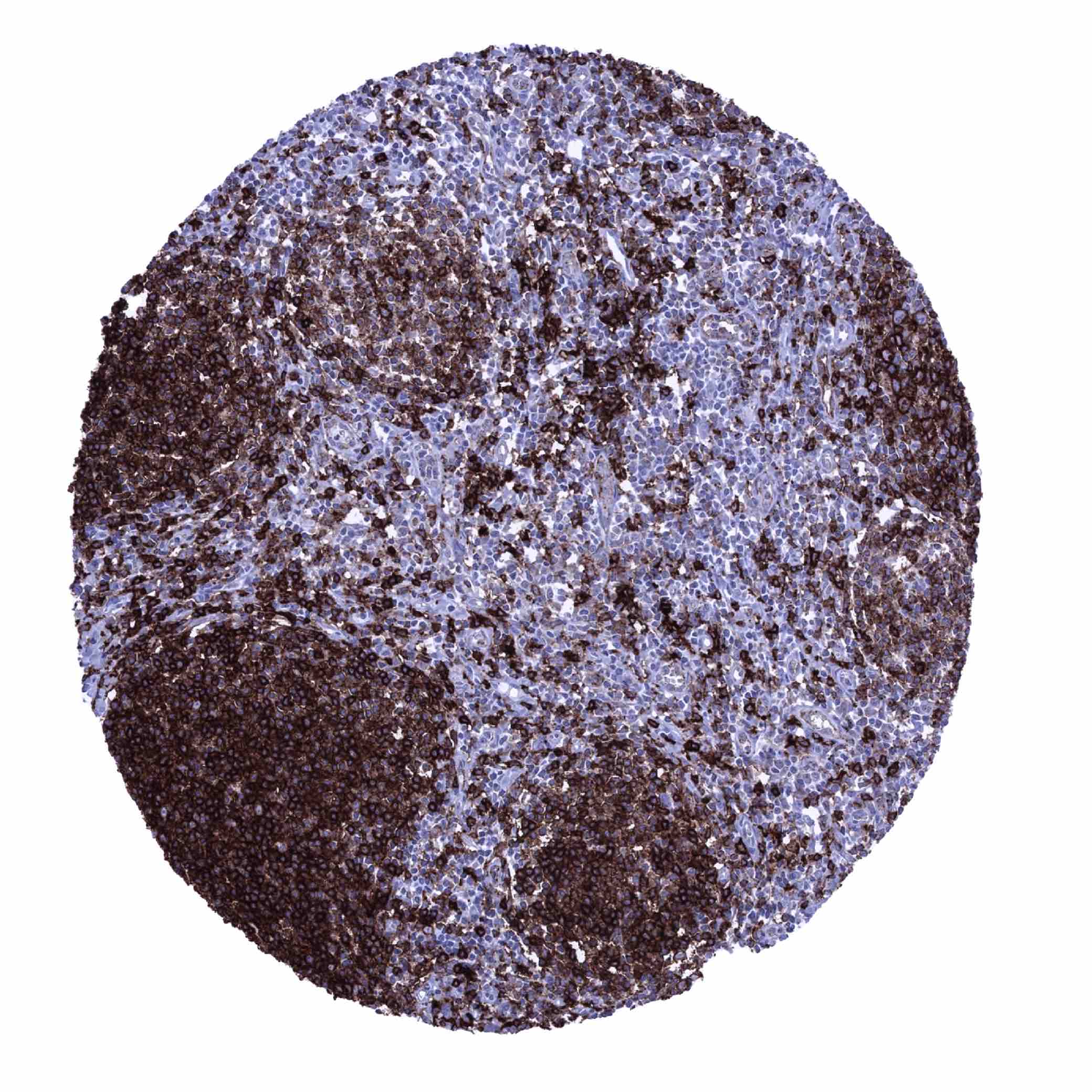
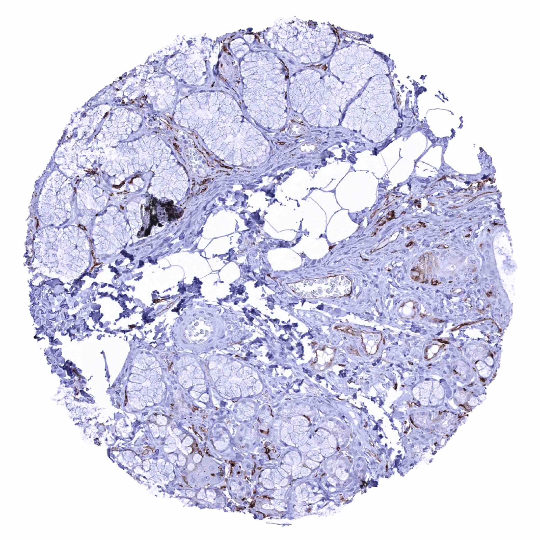
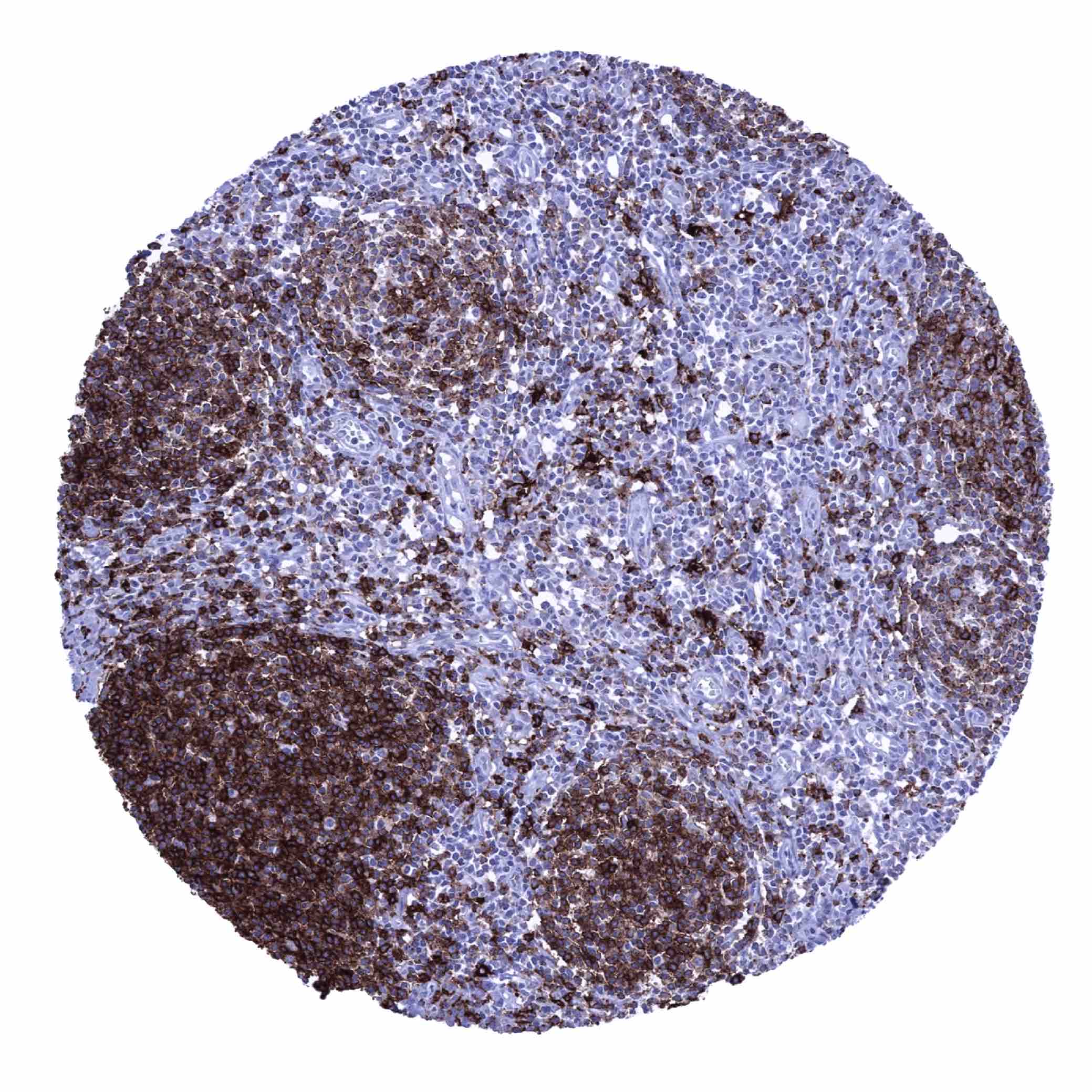
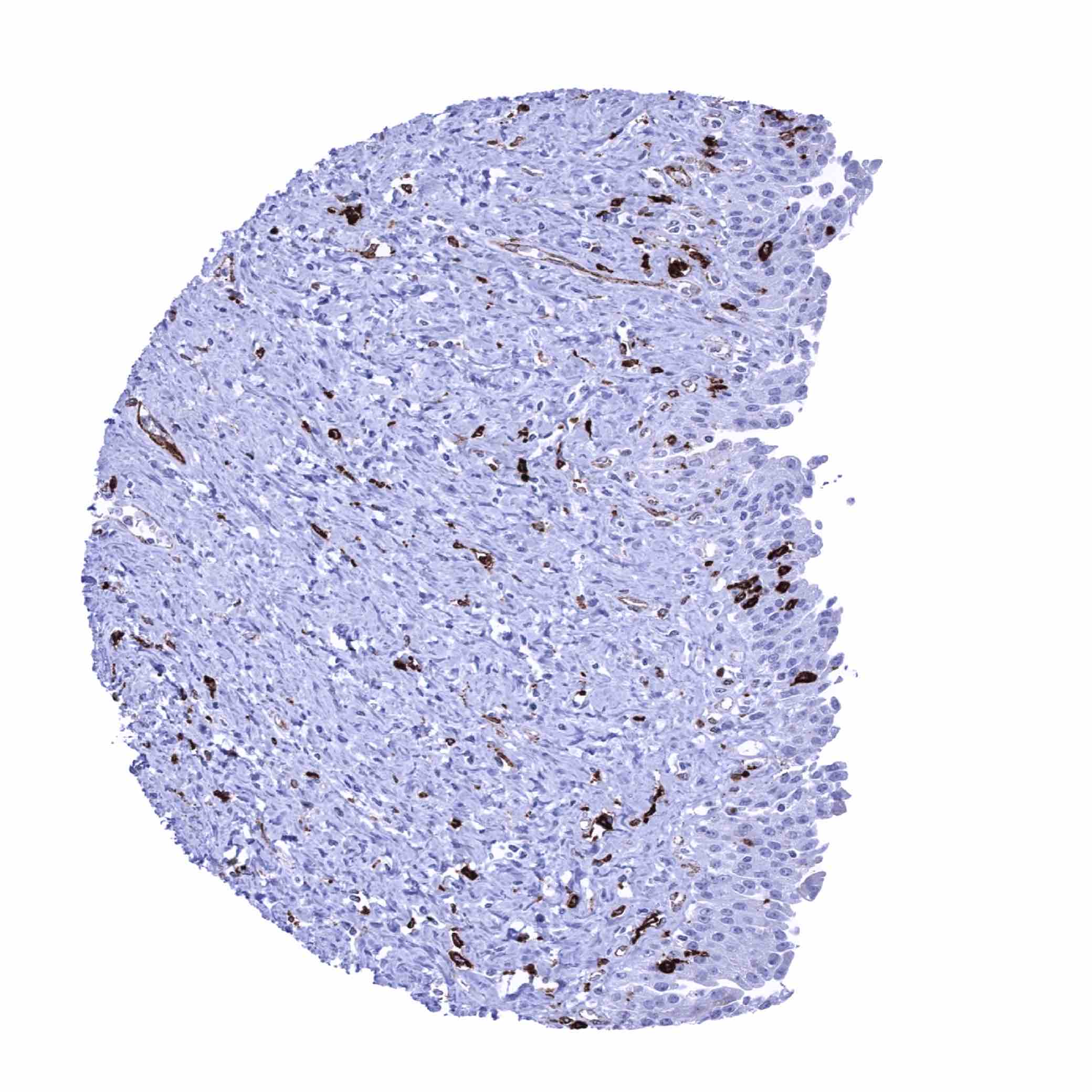
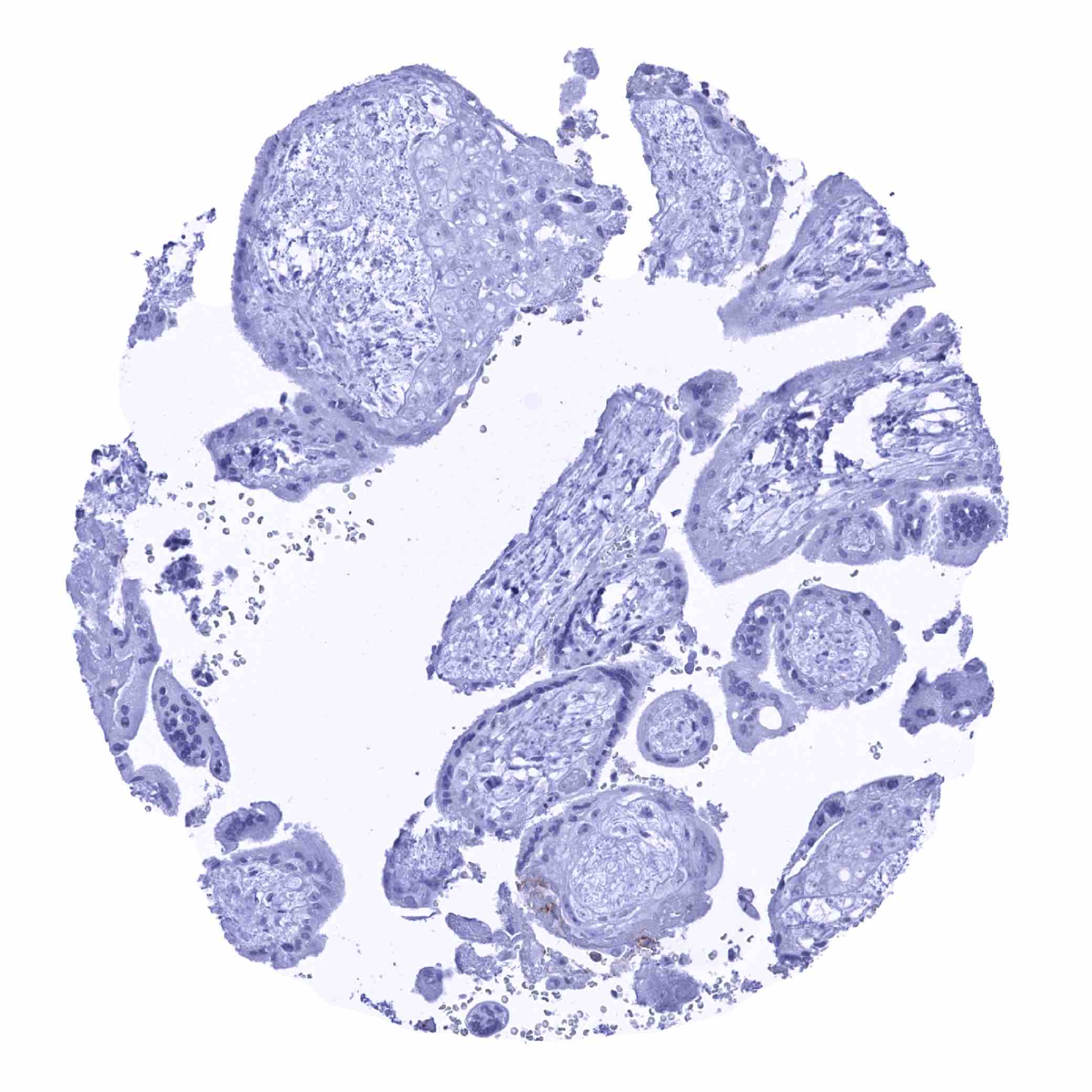
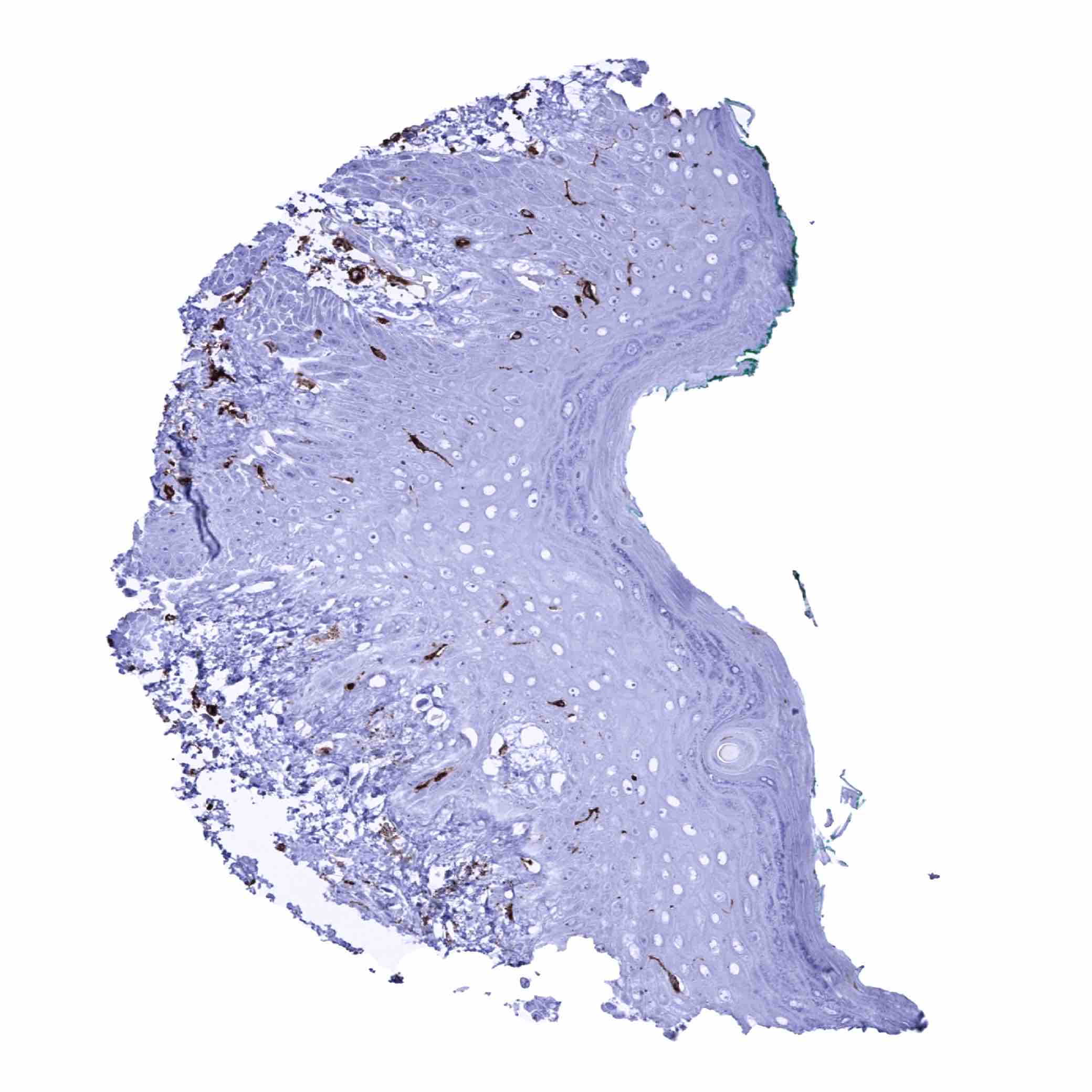
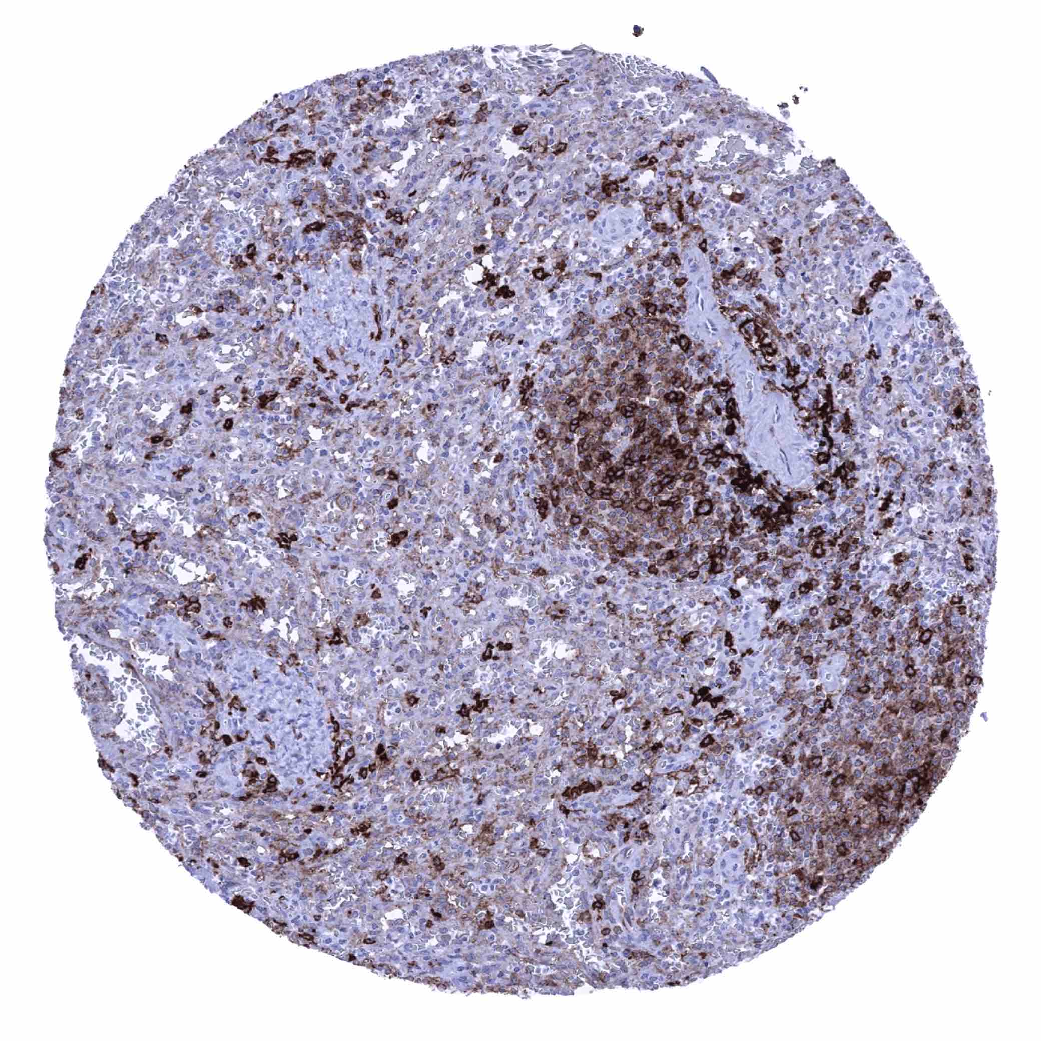
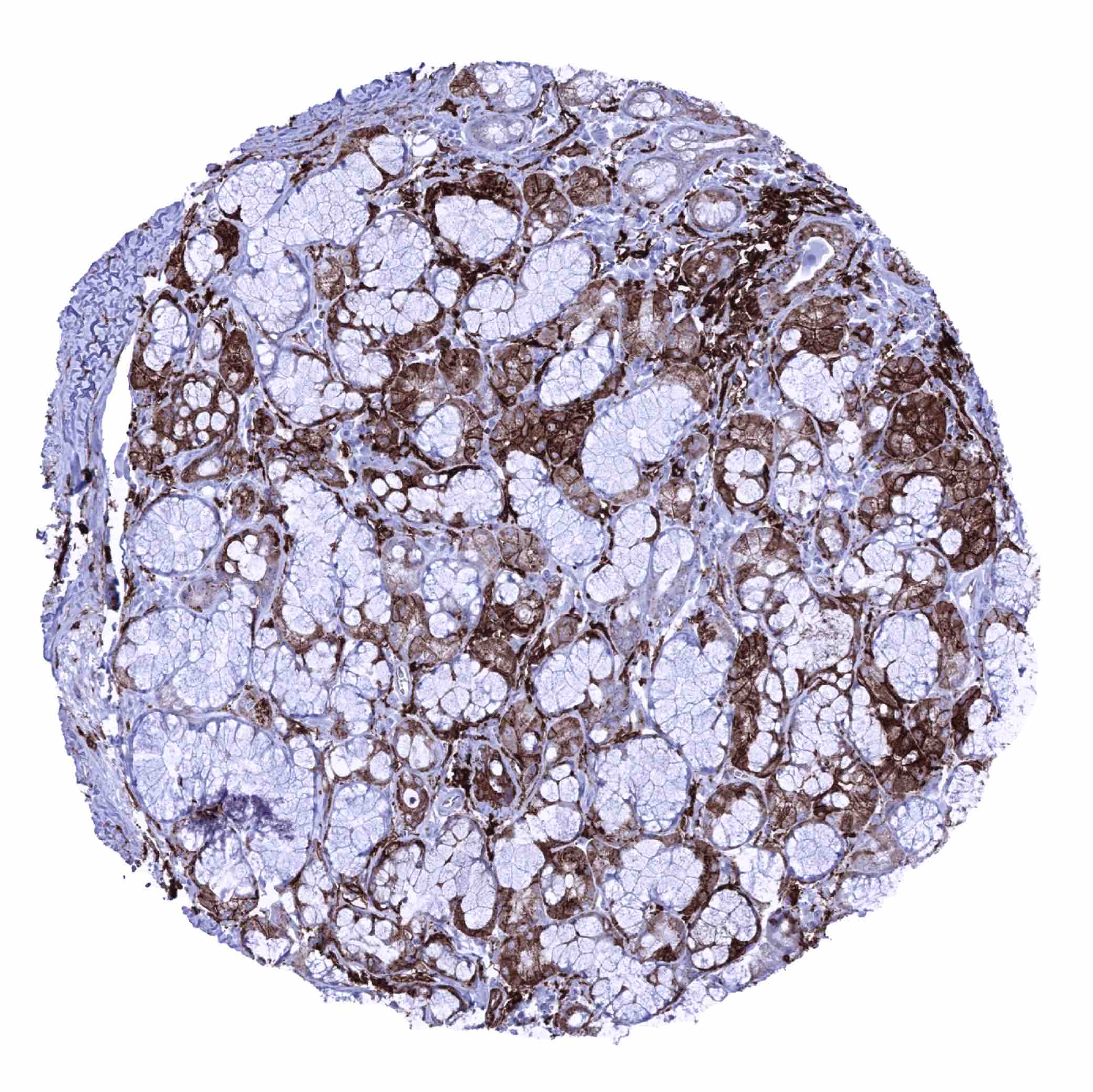
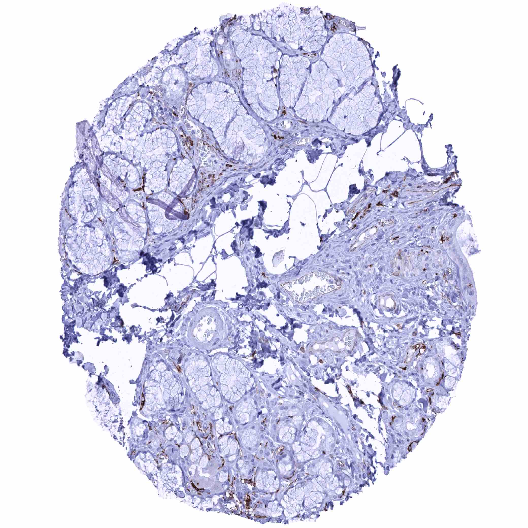
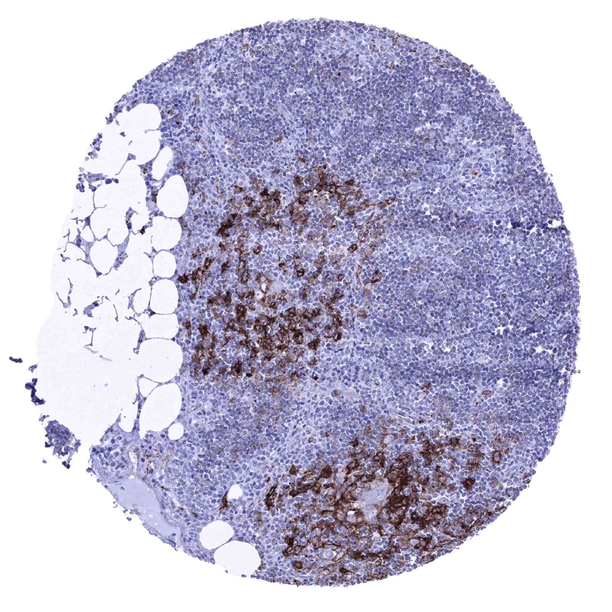
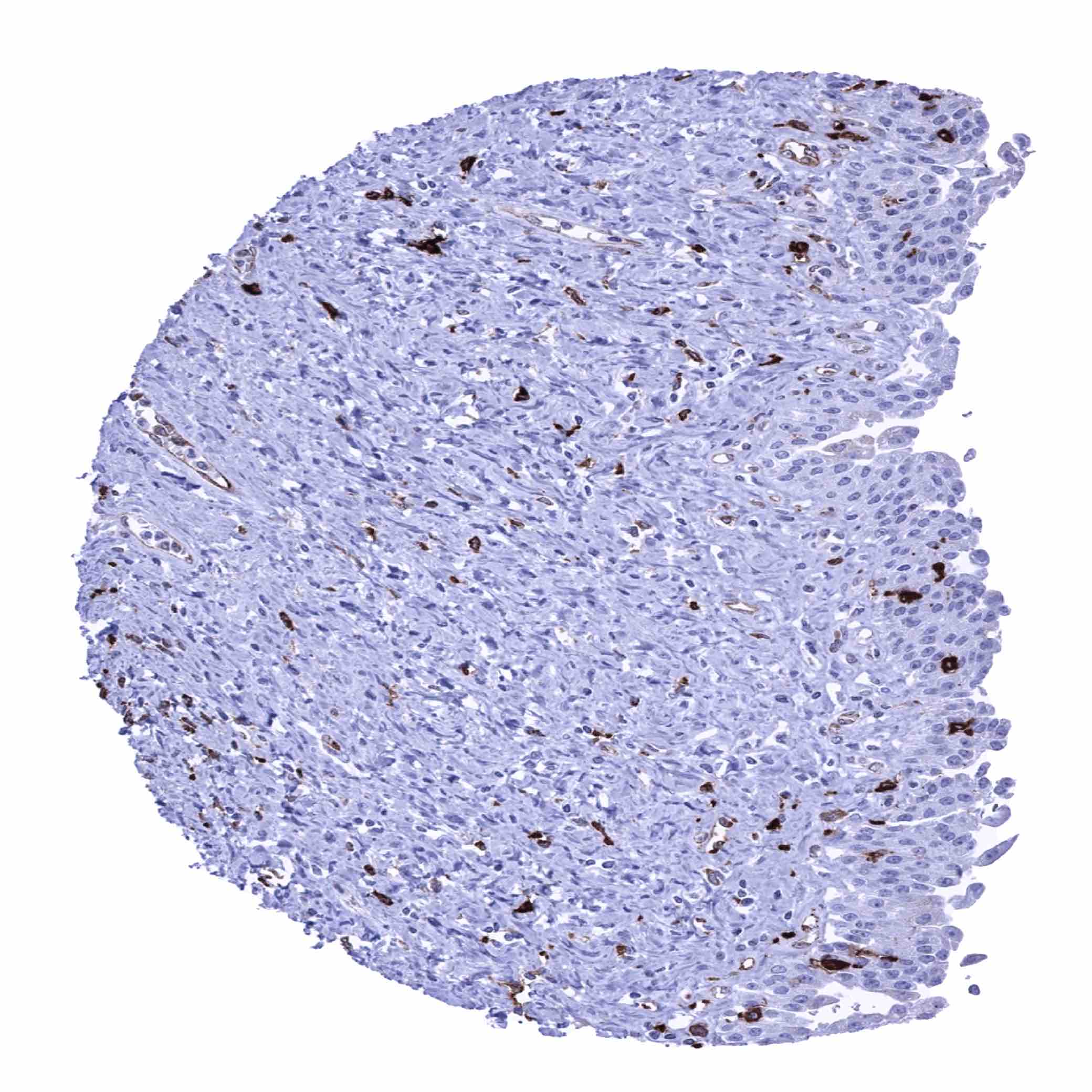
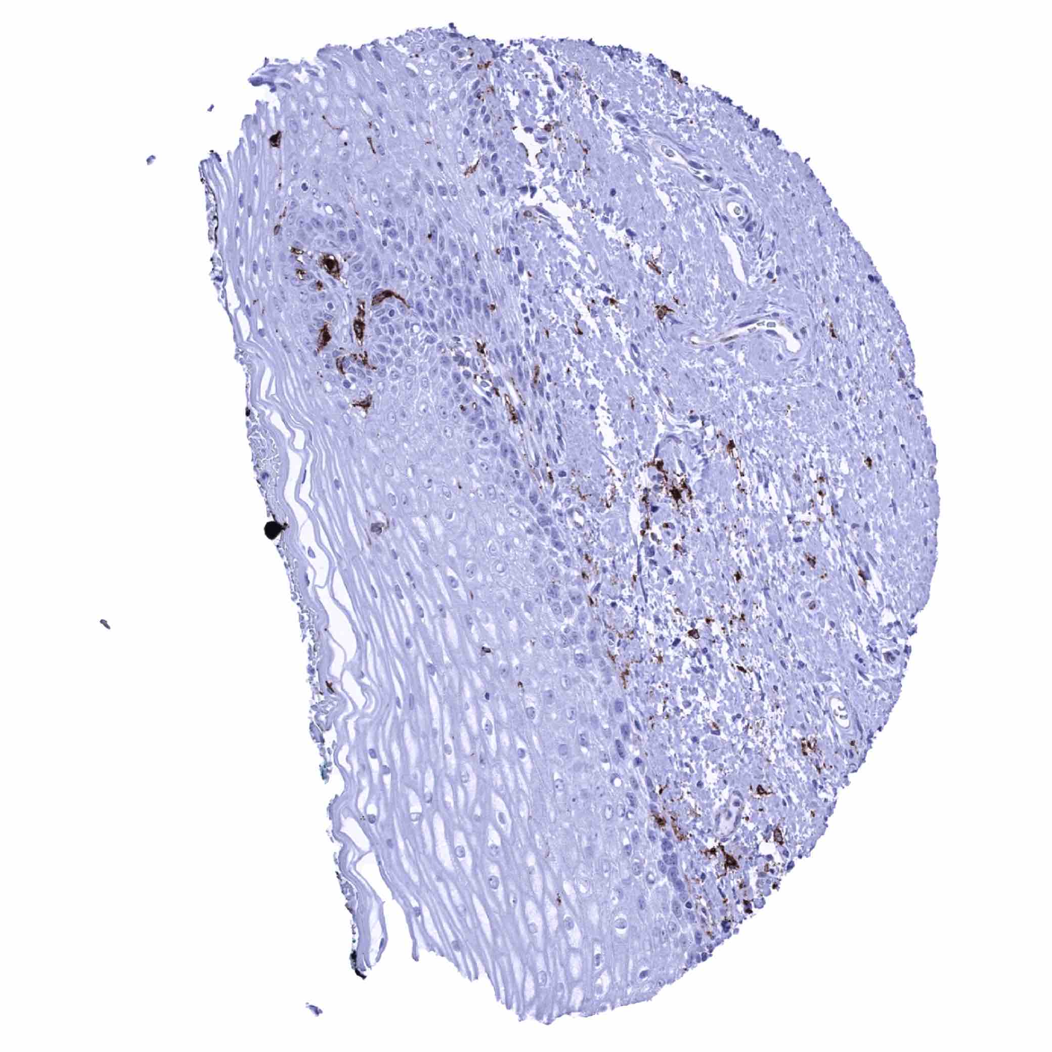
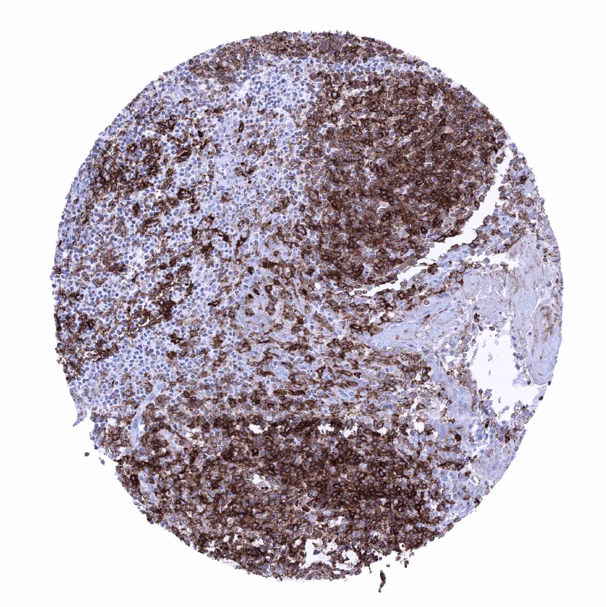
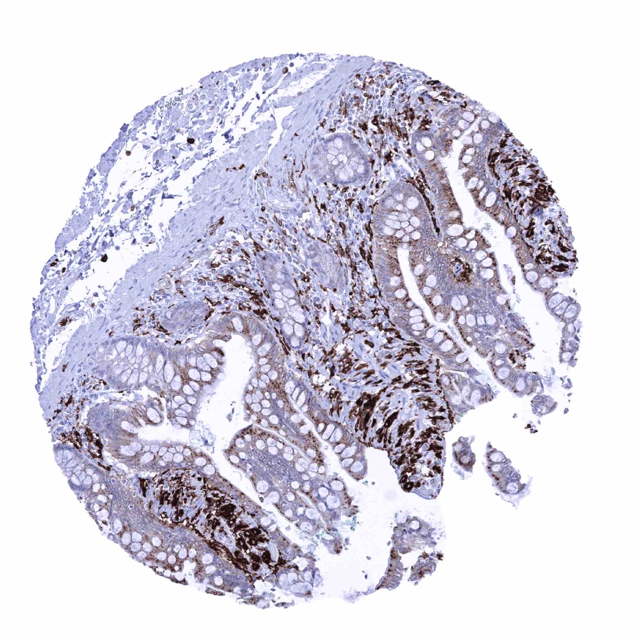
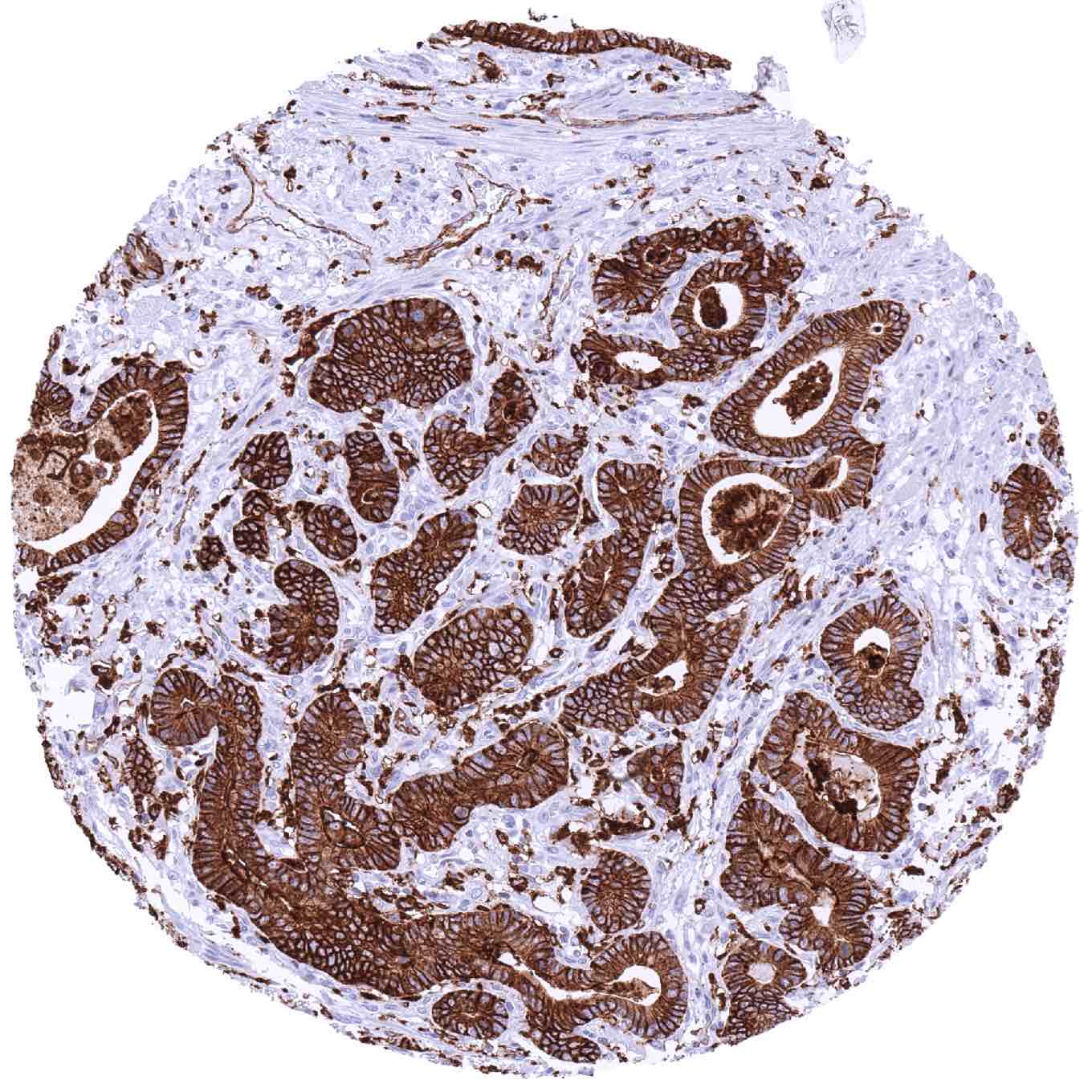
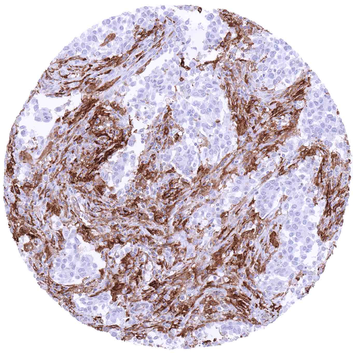
HLA-DRB1 immunostaining is regularly seen on the majority of inflammatory cells including B-lymphocytes, dendritic cells and macrophages. In these cells the staining is mostly strong but may show some variability. A moderate (to strong) HLA- DRB1 staining is also seen in endothelial cells of blood vessels. Endothelial cell staining is particularly low in the placenta. Endothelial staining also includes the liver sinus and venous sinus in the spleen. Epithelial cells do regularly show a strong, predominantly membranous staining in the small intestine (strongest staining in the duodenum) and in a subset of the surface epithelium of the fallopian tube (mosaic staining pattern). HLA-DRB1 immunostaining is also frequently seen in the surface epithelium of the stomach mucosa. Occasional cytoplasmic and membranous HLA- DRB1 staining can focally be seen in virtually all epithelial cell types, especially in case of inflammation, atrophy or regeneration.
RNA and protein expression data of HLA-DRB1 findings are also described in the Human Protein Atlas (Tissue expression HLA-DRB1)
Positive control = Liver: An at least moderate staining should be seen of sinusoids and Kupffer cells. Kidney: An at least moderate staining should be seen in glomerular capillaries.
Negative control = Liver, kidney: HLA-DR staining must be absent normal in hepatocytes and in tubuli of completely normal kidney.
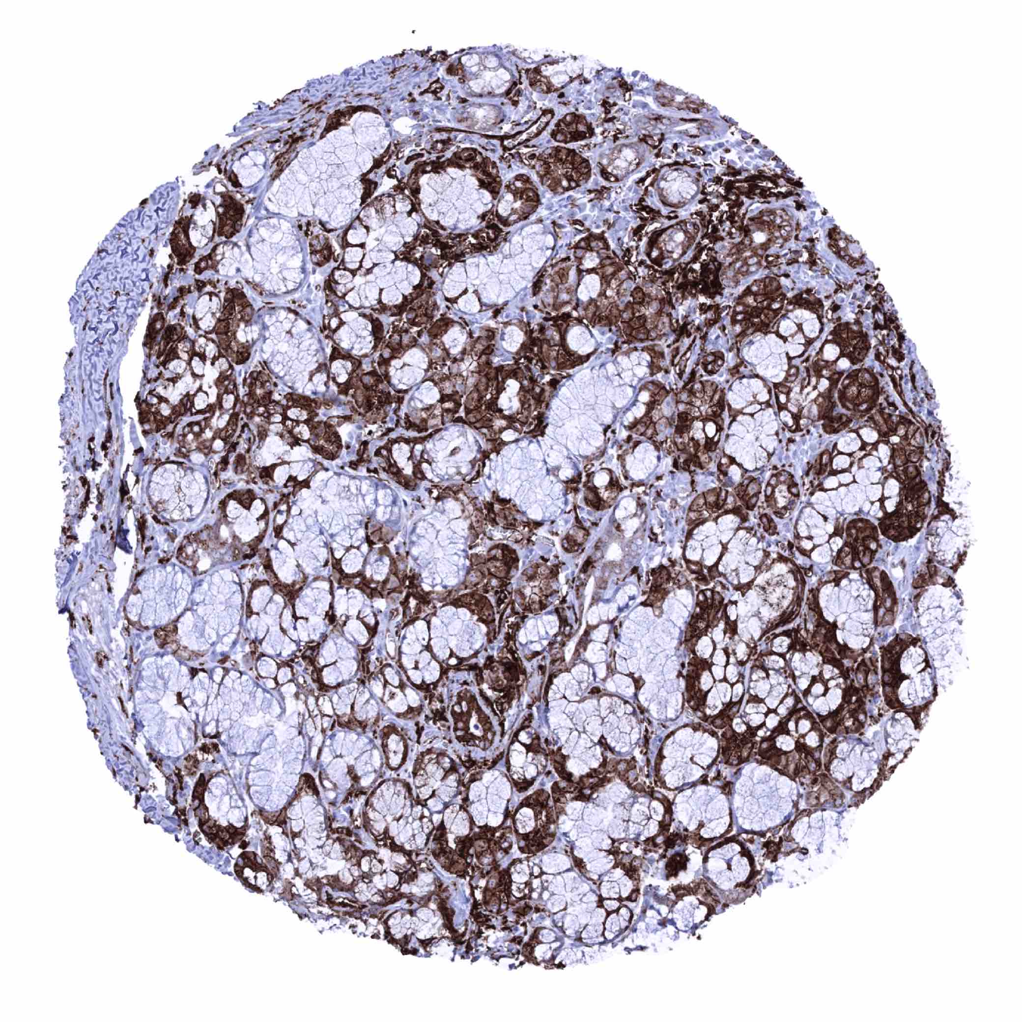
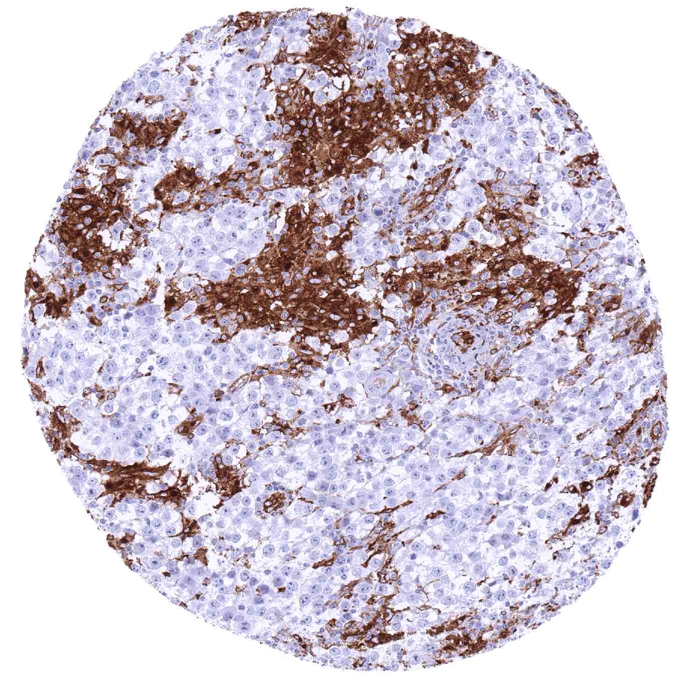
Staining Pattern in Relevant Tumor Types
HLA-DRB1 can be expressed in tumor cells of various cancer types in a fraction of cases.
The TCGA findings on HLA-DRB1 RNA expression in different tumor categories have been summarized in the Human Protein Atlas.

Compatibility of Antibodies
No data available at the moment
Protocol Recommendations
IHC users have different preferences on how the stains should look like. Some prefer high staining intensity of the target stain and even accept some background. Others favor absolute specificity and lighter target stains. Factors that invariably lead to more intense staining include higher concentration of the antibody and visualization tools, longer incubation time, higher temperature during incubation, higher temperature and longer duration of the heat induced epitope retrieval (slide pretreatment). The impact of the pH during slide pretreatment has variable effects and depends on the antibody and the target protein.
All images and data shown here and in our image galleries are obtained by the manual protocol described below. Other protocols resulting in equivalent staining are described as well.
Manual protocol
Freshly cut sections should be used (less than 10 days between cutting and staining). Heat-induced antigen retrieval for 5 minutes in an autoclave at 121°C in pH 7,8 Target Retrieval Solution buffer. Apply MSVA-478R at a dilution of 1:150 at 37°C for 60 minutes. Visualization of bound antibody by the EnVision Kit (Dako, Agilent) according to the manufacturer’s directions.
Potential Research Applications
- HLA-DRB1 expression can occur in tumors. The prognostic role of HLA-DRB1 expression and its predictive significance with respect to checkpoint inhibitor therapies needs to be investigated.
- The role of HLA-DRB1 expressing inflammatory cells and their spatial relationship to tumor cells is of interest.
Evidence for Antibody Specificity in IHC
There are two ways how the specificity of antibodies can be documented for immunohistochemistry on formalin fixed tissues. These are: 1. Comparison with a second independent method for target expression measurement across a large number of different tissue types (orthogonal strategy), and 2. Comparison with one or several independent antibodies for the same target and showing that all positive staining results are also seen with other antibodies for the same target (independent antibody strategy).
Orthogonal validation: is not well applicable for HLA-DRB1 antibodies because of the ubiquitous expression of the protein in blood vessels and inflammatory cells in virtually all organs. The particular low rate of HLA-DRB1 immunostaining in the brain and the high fraction of HLA-DRB1 positive cells in lymphatic tissues, the lung and the gastrointestinal tract is, however, consistent with data from three independent RNA screening studies, including the Human Protein Atlas (HPA) RNA-seq tissue dataset, the FANTOM5 project, and the Genotype-Tissue Expression (GTEx) project, which are all summarized in the Human Protein Atlas (Tissue expression HLA-DRB1).
Comparison of antibodies: A specific HLA-DRB1 staining of MSVA-478R in endothelial cells as well as in various epithelial and inflammatory cell types is corroborated by comparison with a commercially available independent second antibody (termed “validation antibody”). In this comparison, all stainings obtained by MSVA-478R were matched by the “validation antibody”.