295,00 € – 995,00 €
Product details
Synonyms = SMST
Antibody type = Recombinant Rabbit monoclonal / IgG
Clone = MSVA-638R
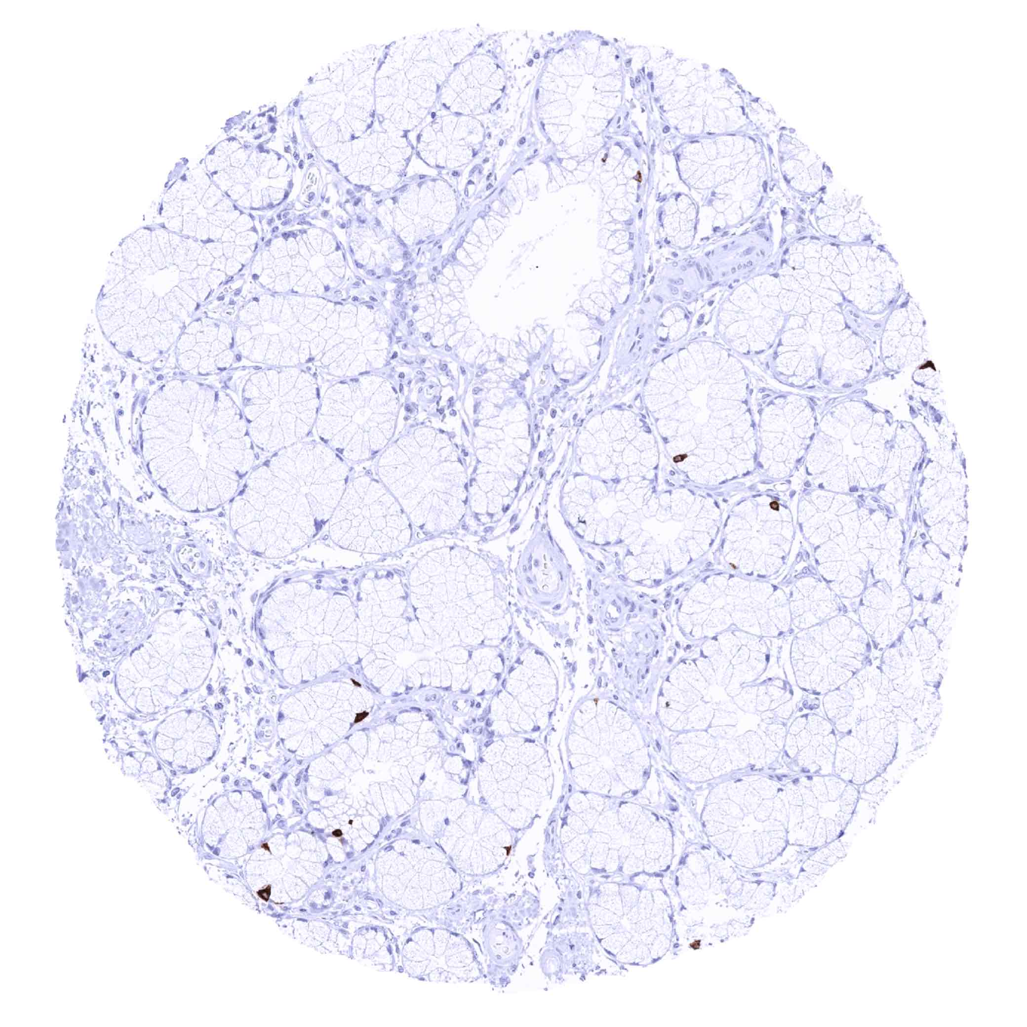
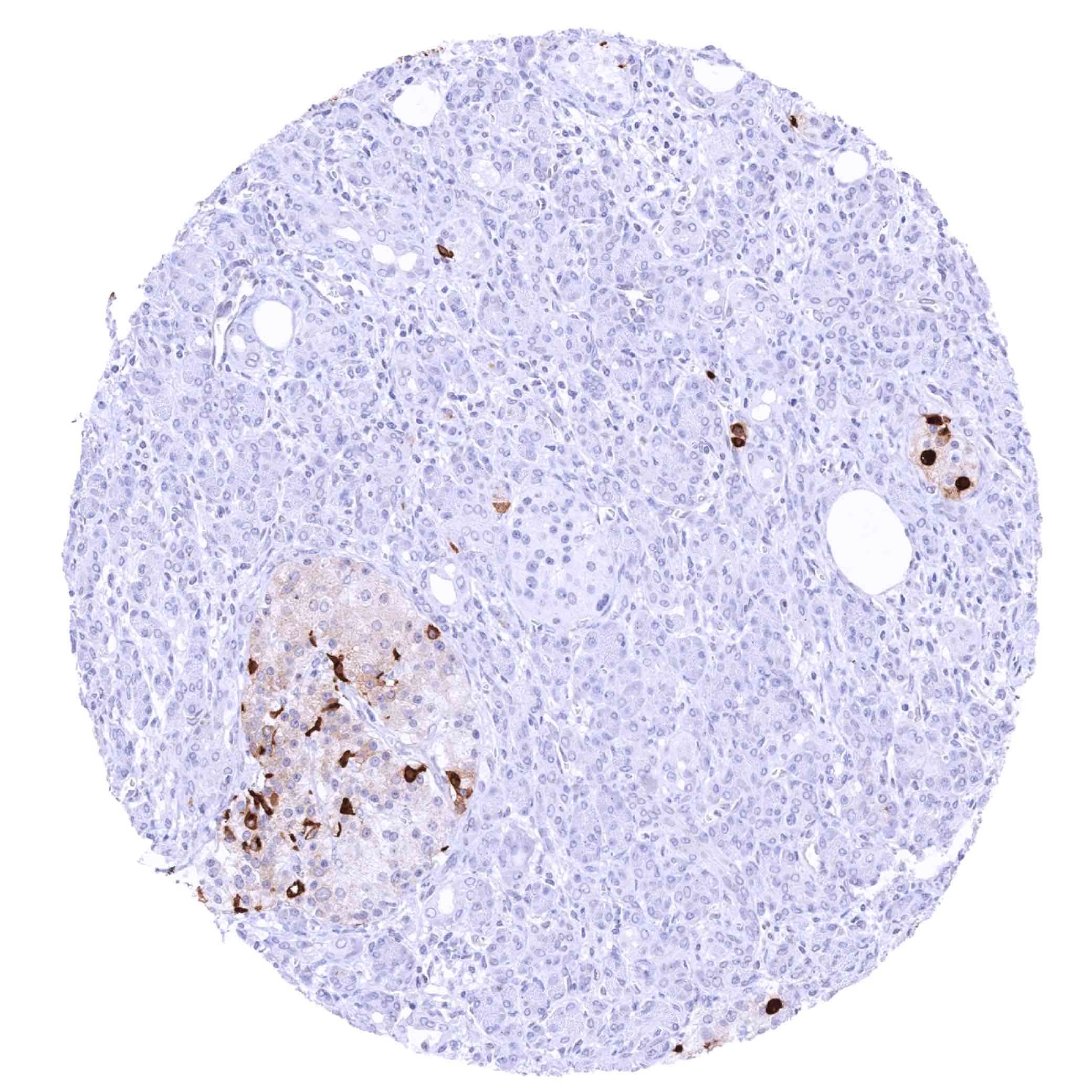
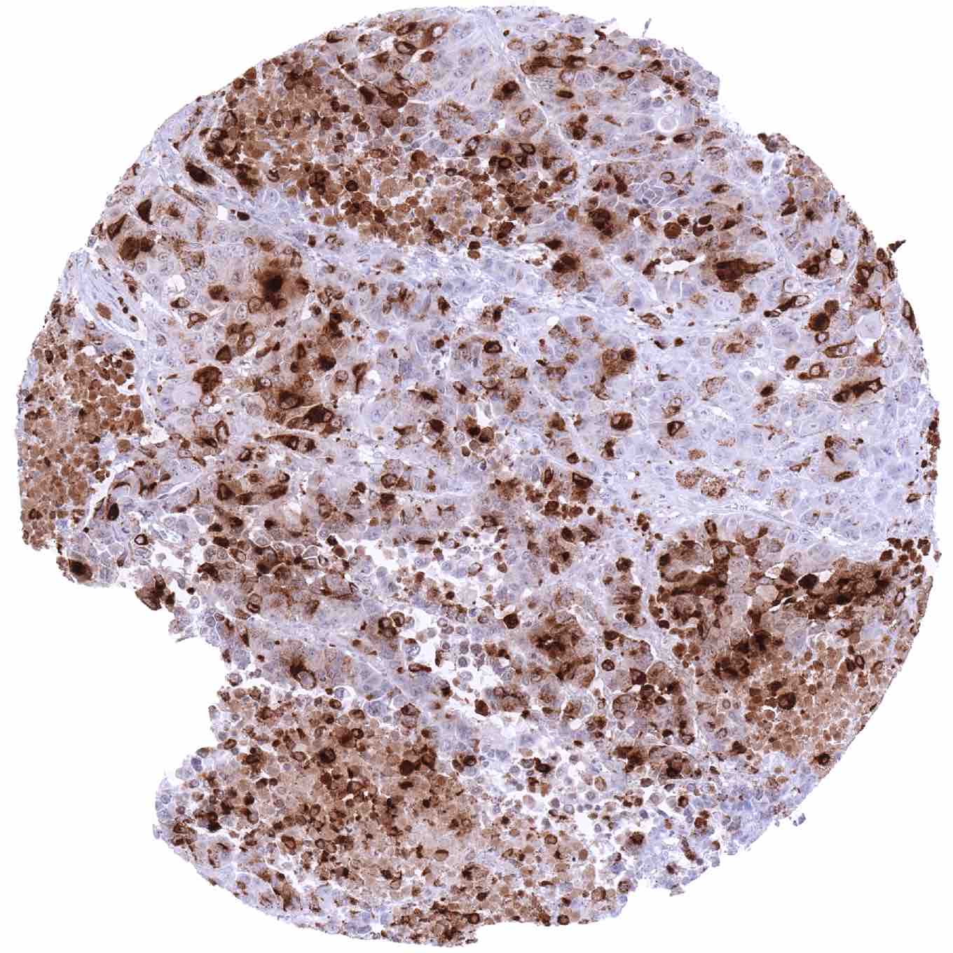
Positive control = Pancreas: A subset of pancreatic islet cells should show strong somatostatin positivity.
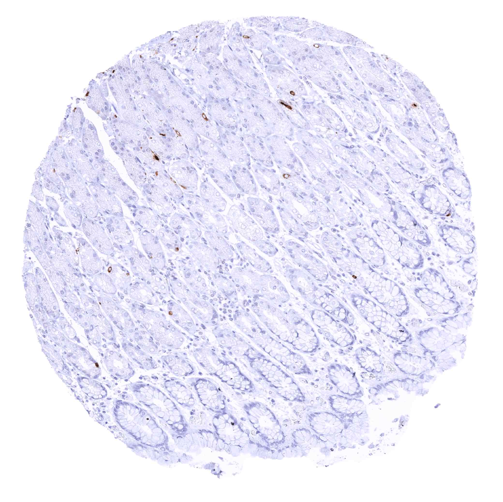
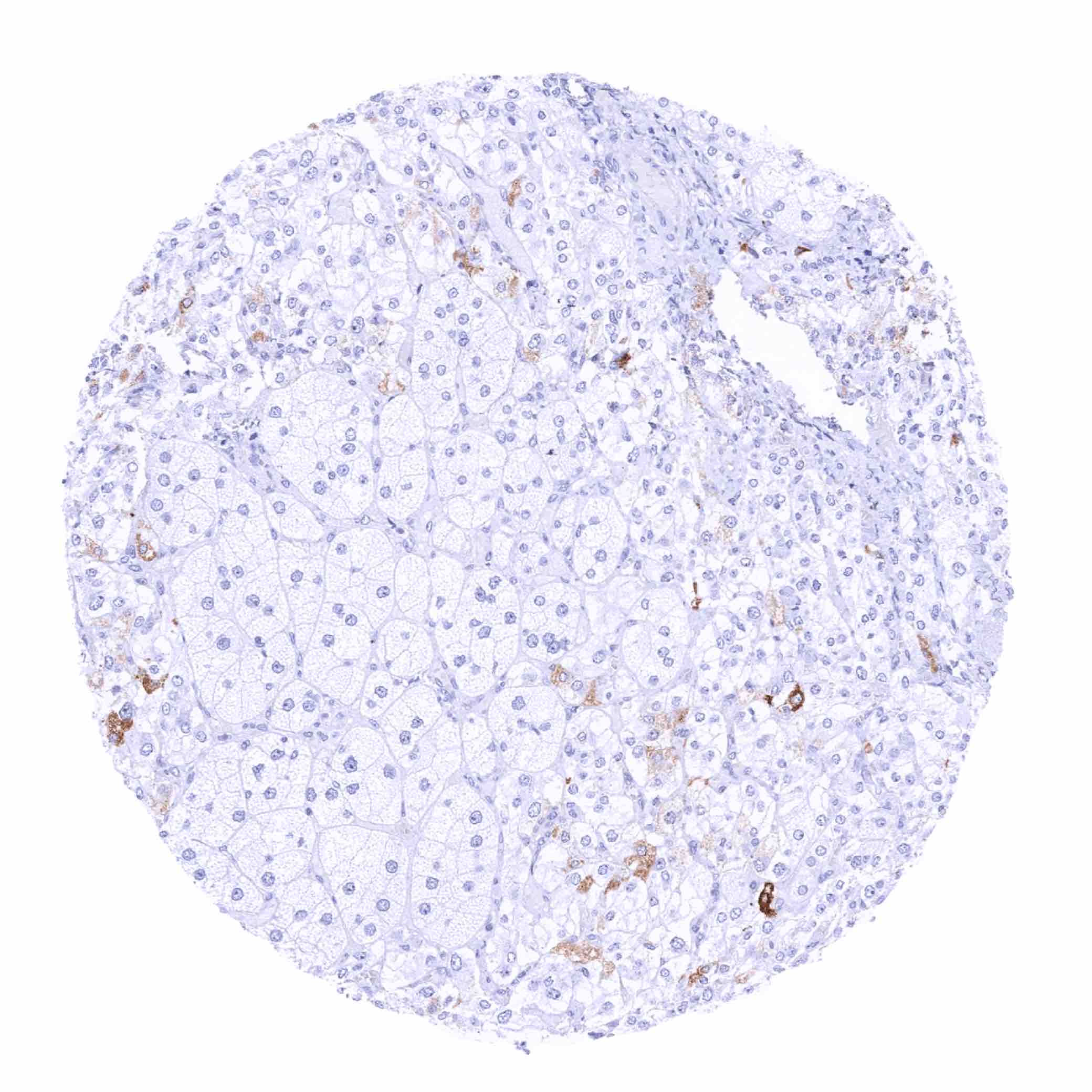
Negative control = Kidney: All cells must not show somatostatin immunostaining.
Cellular localization = Secreted
Reactivity = Human
Application = Immunohistochemistry
Dilution = 1:100 – 1:200
Intended Use = Research Use Only
Relevance of Antibody
Somatostatin is a peptide hormone with paracrine and humoral activity.
Biology Behind
Somatostatin is a peptide hormone coded by the SST gene at chromosome 3q27.3. Somatostatin has two active forms consisting of 14 and 28 amino acids produced by alternative cleavage of a single proprotein. These peptides exert humoral and paracrine functions through interaction with several somatostatin receptors. Somatostatin is secreted by delta cells in the digestive system, namely the pyloric antrum, the duodenum and the pancreatic islets. In the stomach, somatostatin antagonizes the stimulatory effect of histamine to reduce acid secretion by paracrine effects on acid-producing parietal cells. In the brain, somatostatin is produced by neuroendocrine neurons of the ventromedial nucleus of the hypothalamus. Somatostatin is then carried to the anterior pituitary gland, where it inhibits the secretion of growth hormone. Somatostatin is also produced by populations of neuroendocrine neurons in other regions of the brain. Somatostatin is an inhibitory hormone that in the anterior pituitary inhibits the release of growth hormone (GH), thyroid-stimulating hormone (TSH), and prolactin (PRL). In the gastrointestinal system, somatostatin suppresses the release of insulin and glucagon.
Staining Pattern in Normal Tissues
Images describing the somatostatin staining pattern in normal tissues obtained by the antibody MSVA-638R are shown in our “Normal Tissue Gallery”.
| Brain | Cerebrum | Few neuroendocrine neurons may show somatostatin positivity in specific regions of the brain. |
| Cerebellum | Few neuroendocrine neurons may show somatostatin positivity in specific regions of the brain. | |
| Endocrine Tissues | Thyroid | Negative. |
| Parathyroid | Negative. | |
| Adrenal gland | A subset of cells shows somatostatin positivity of variable intensity. | |
| Pituitary gland | Negative. | |
| Respiratory system | Respiratory epithelium | Negative. |
| Lung | Negative. | |
| Gastrointestinal Tract | Salivary glands | Negative. |
| Esophagus | Negative. | |
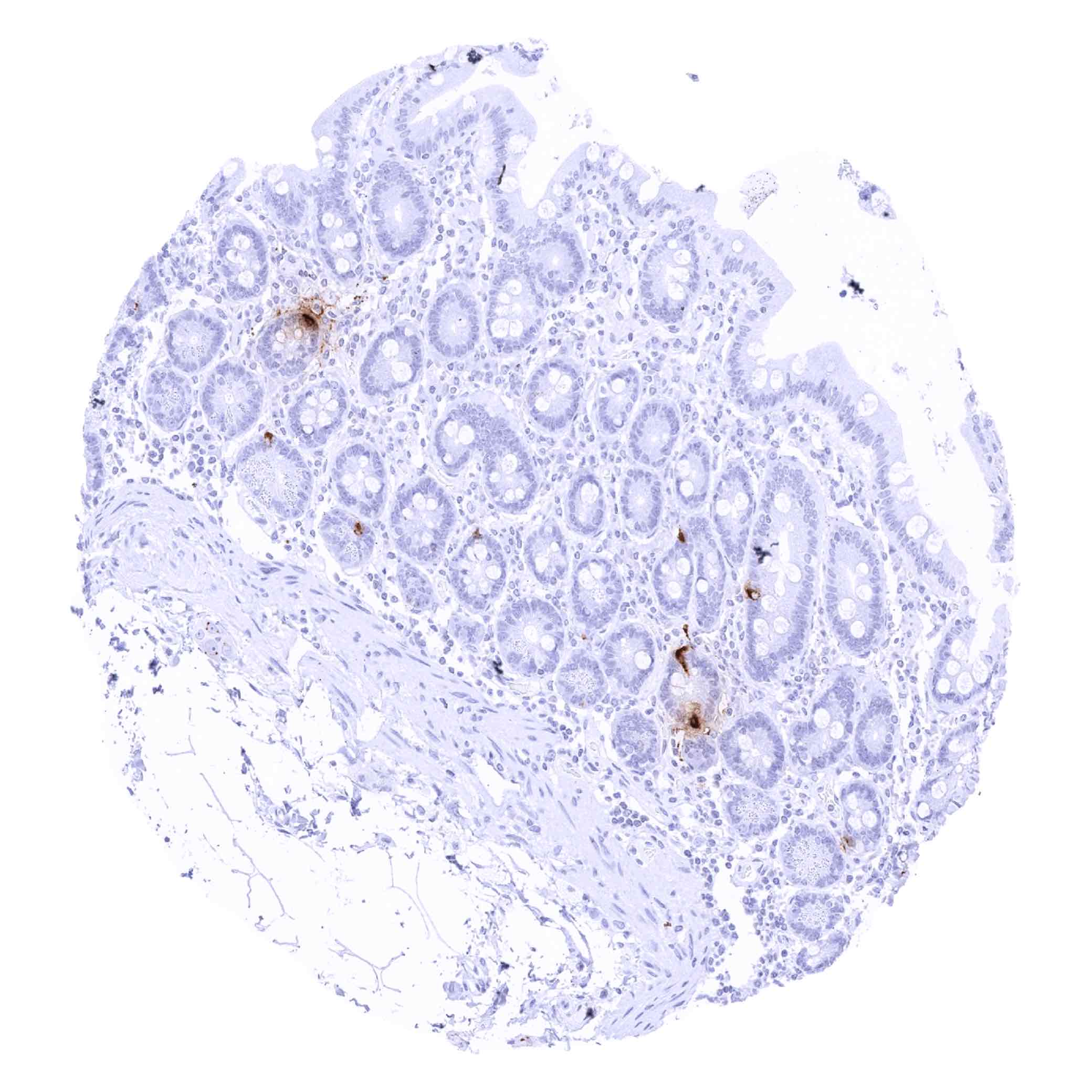
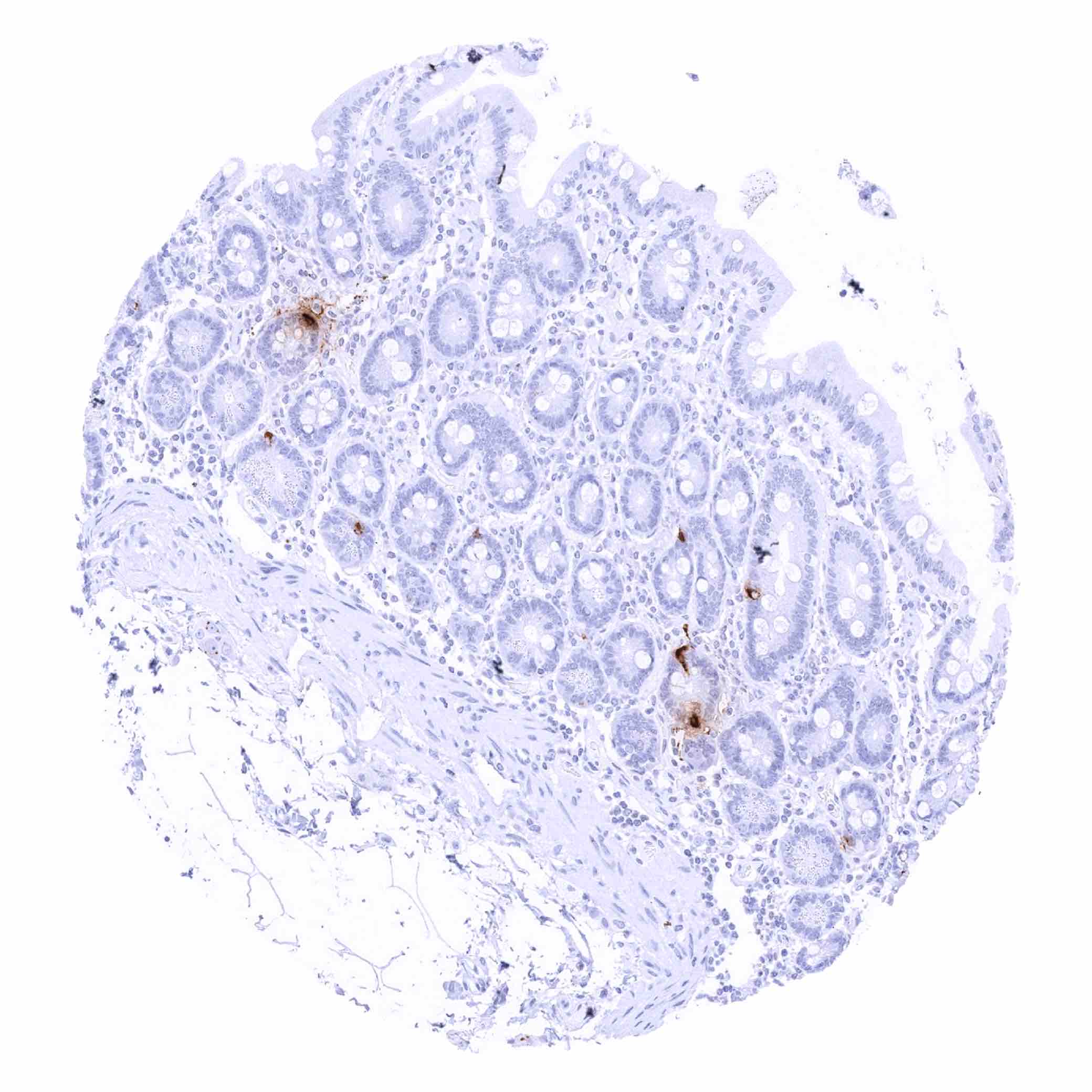
| Stomach | Distinct somatostatin staining of scattered neuroendocrine (delta) cells. | |
| Duodenum | Distinct somatostatin staining of scattered neuroendocrine (delta) cells. | |
| Small intestine | Distinct somatostatin staining of scattered neuroendocrine (delta) cells. | |
| Appendix | Distinct somatostatin staining of scattered neuroendocrine (delta) cells. | |
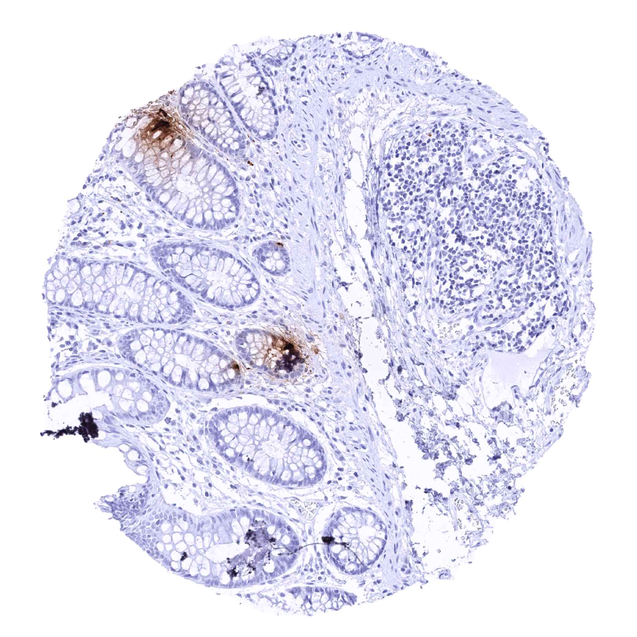
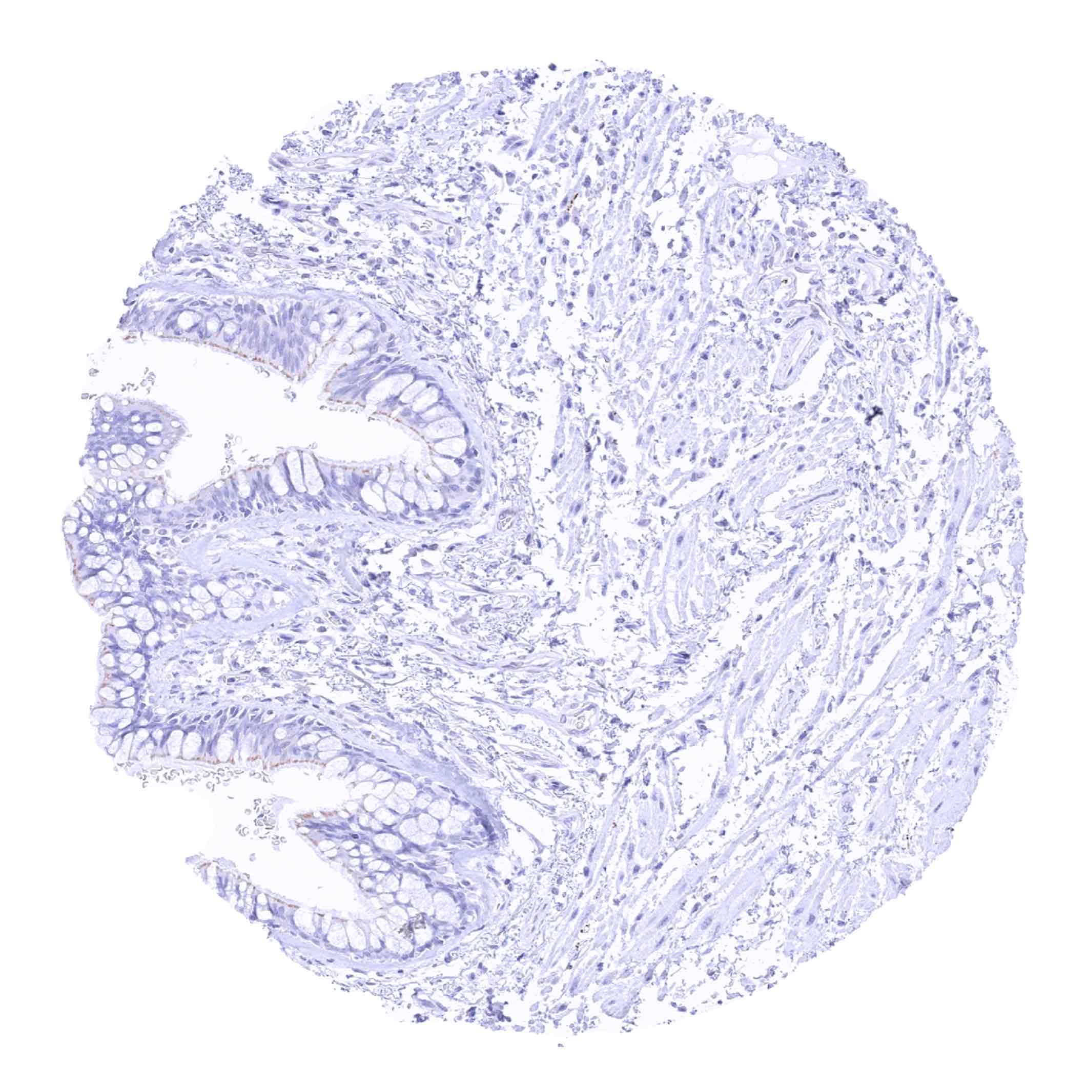
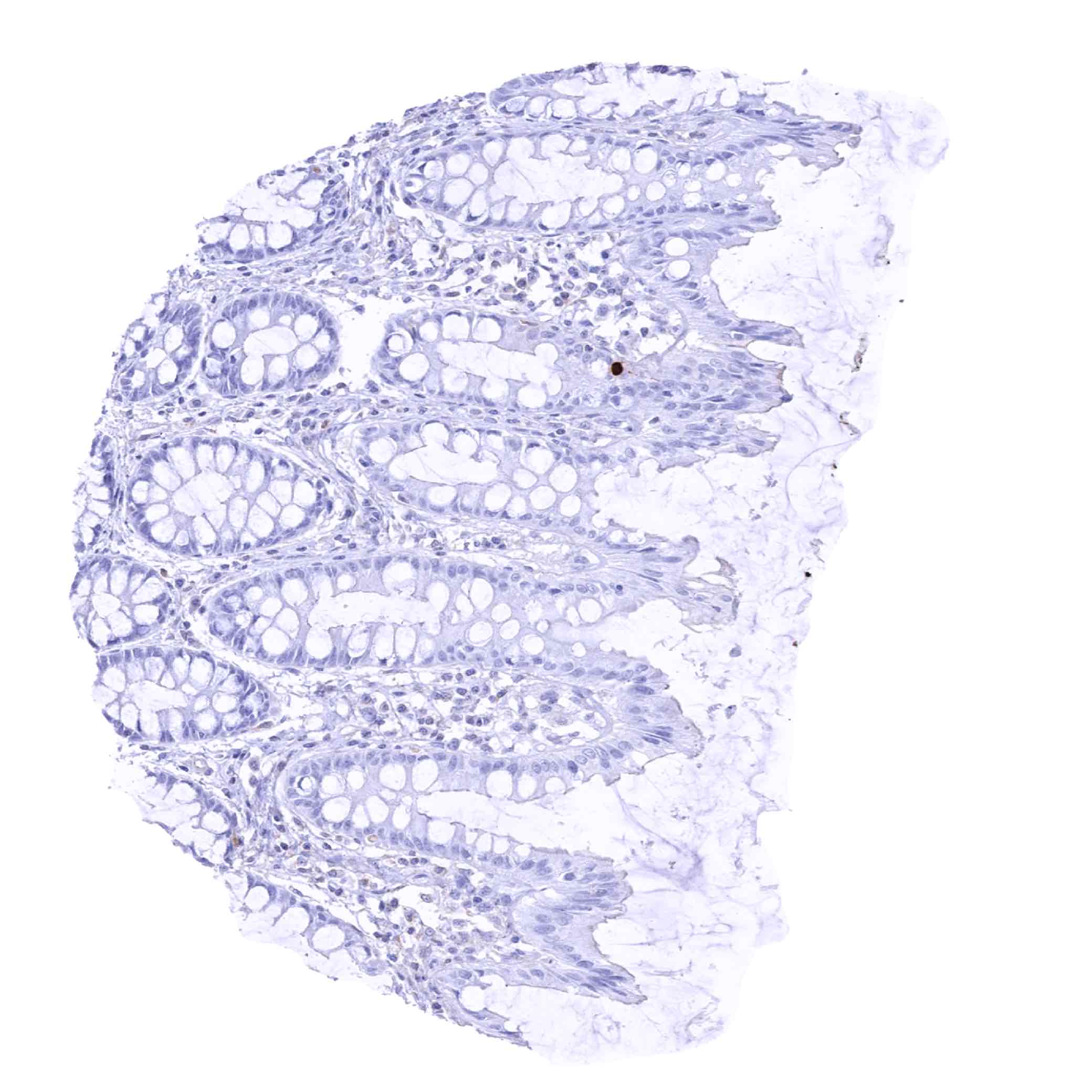
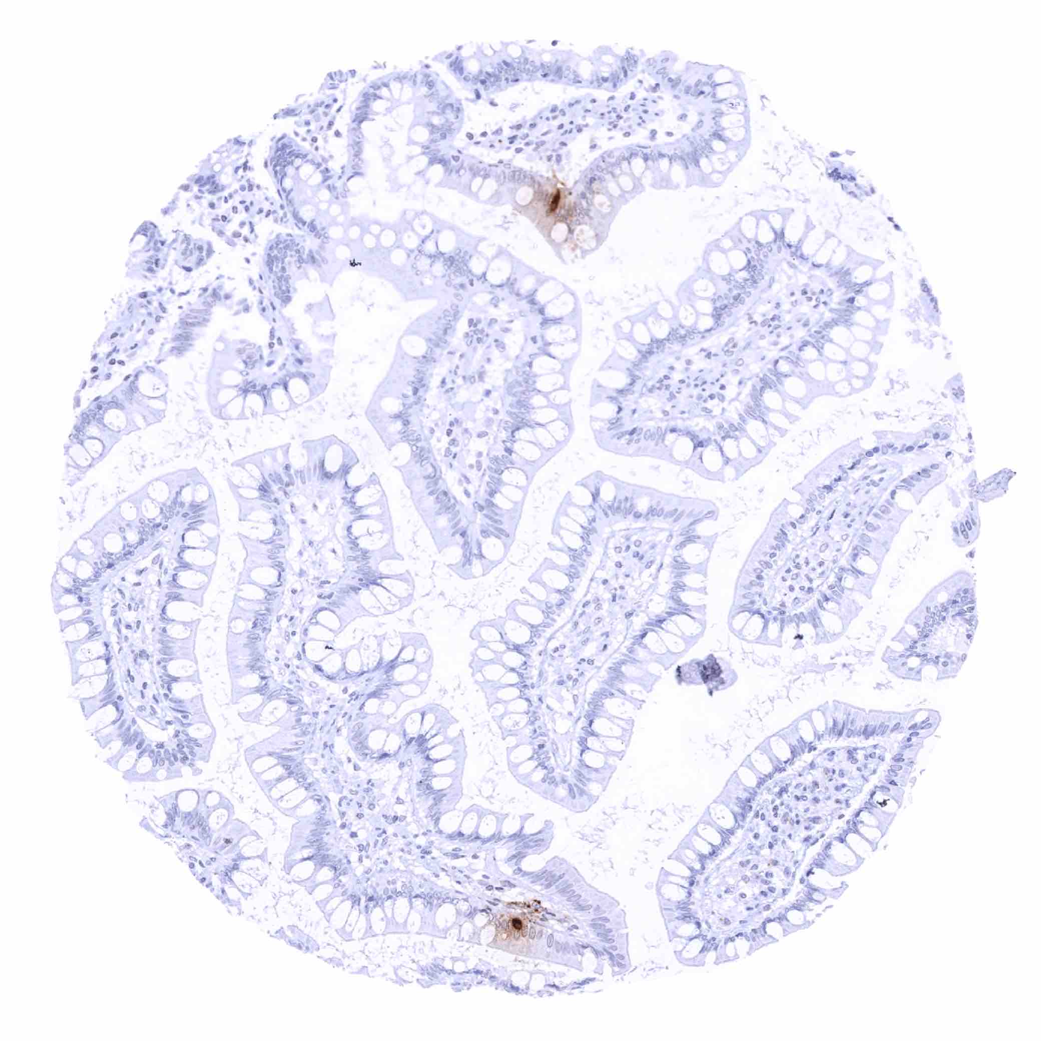
| Colon | Distinct somatostatin staining of scattered neuroendocrine (delta) cells. | |
| Rectum | Distinct somatostatin staining of scattered neuroendocrine (delta) cells. | |
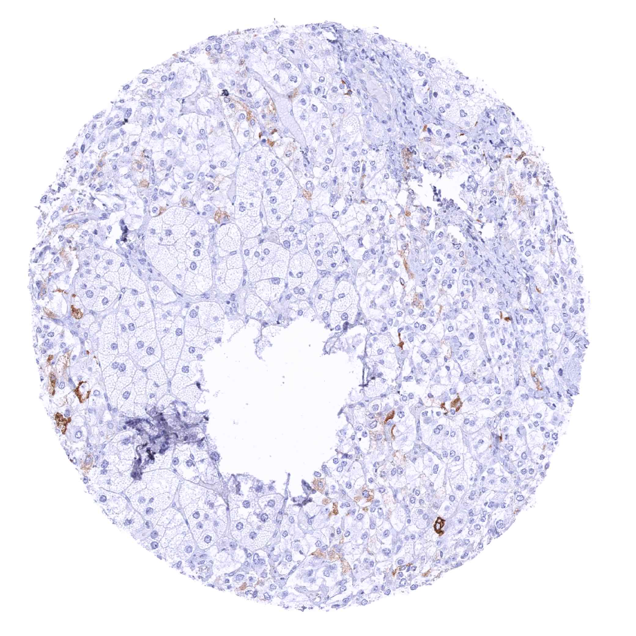
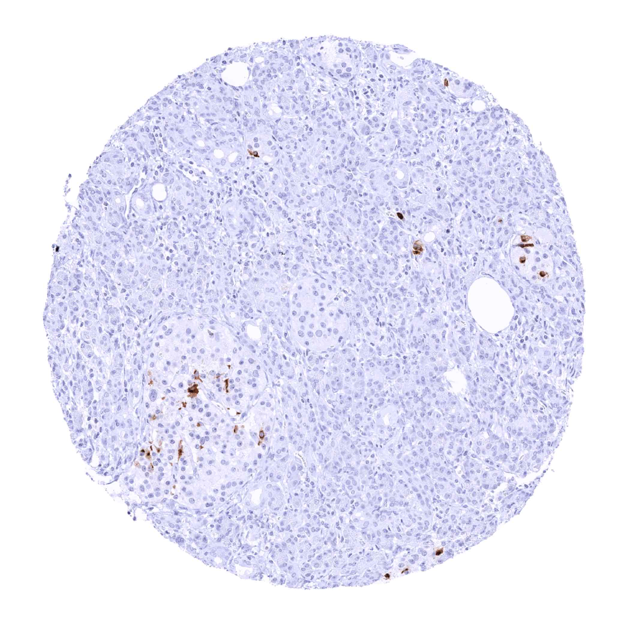
| Liver | Negative. | |
| Gallbladder | Negative. | |
| Pancreas | Strong somatostatin positivity of a subset of pancreatic islet cells. | |
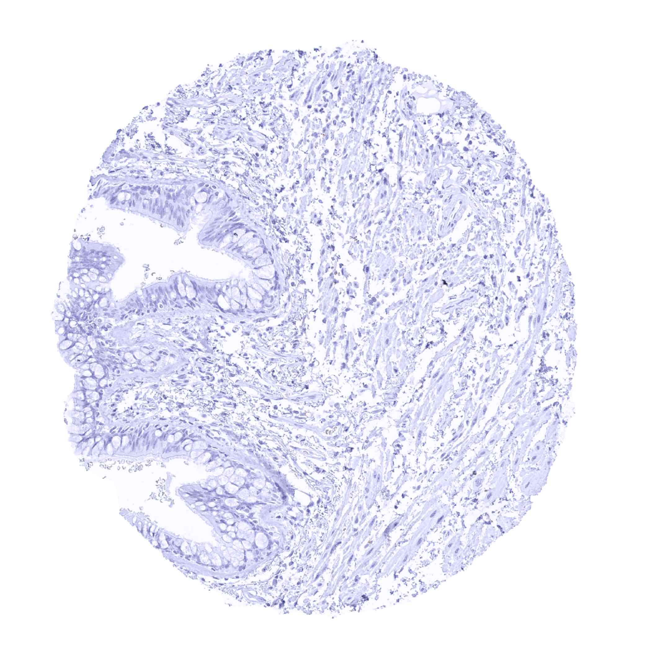
| Genitourinary | Kidney | Negative. |
| Urothelium | Negative. | |
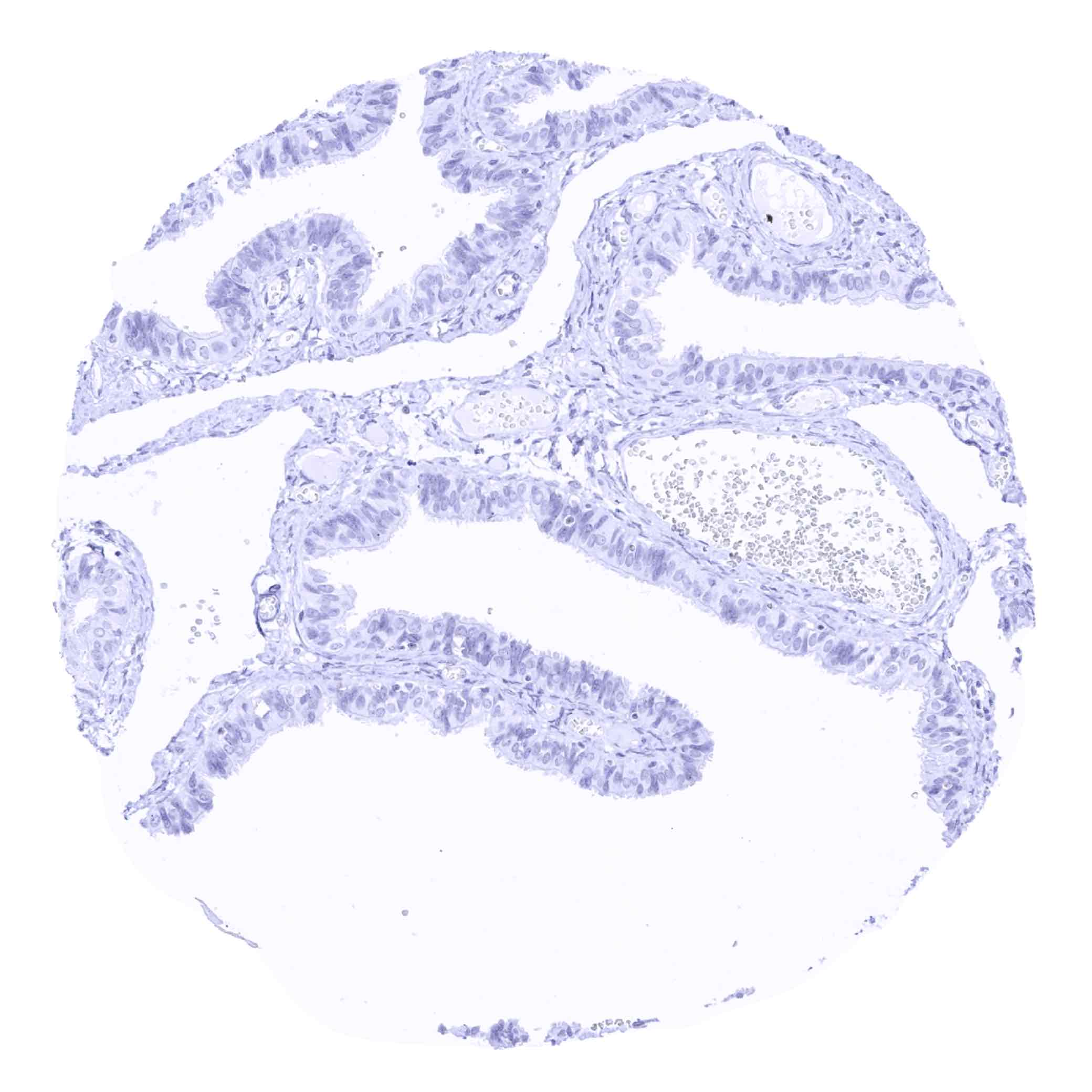
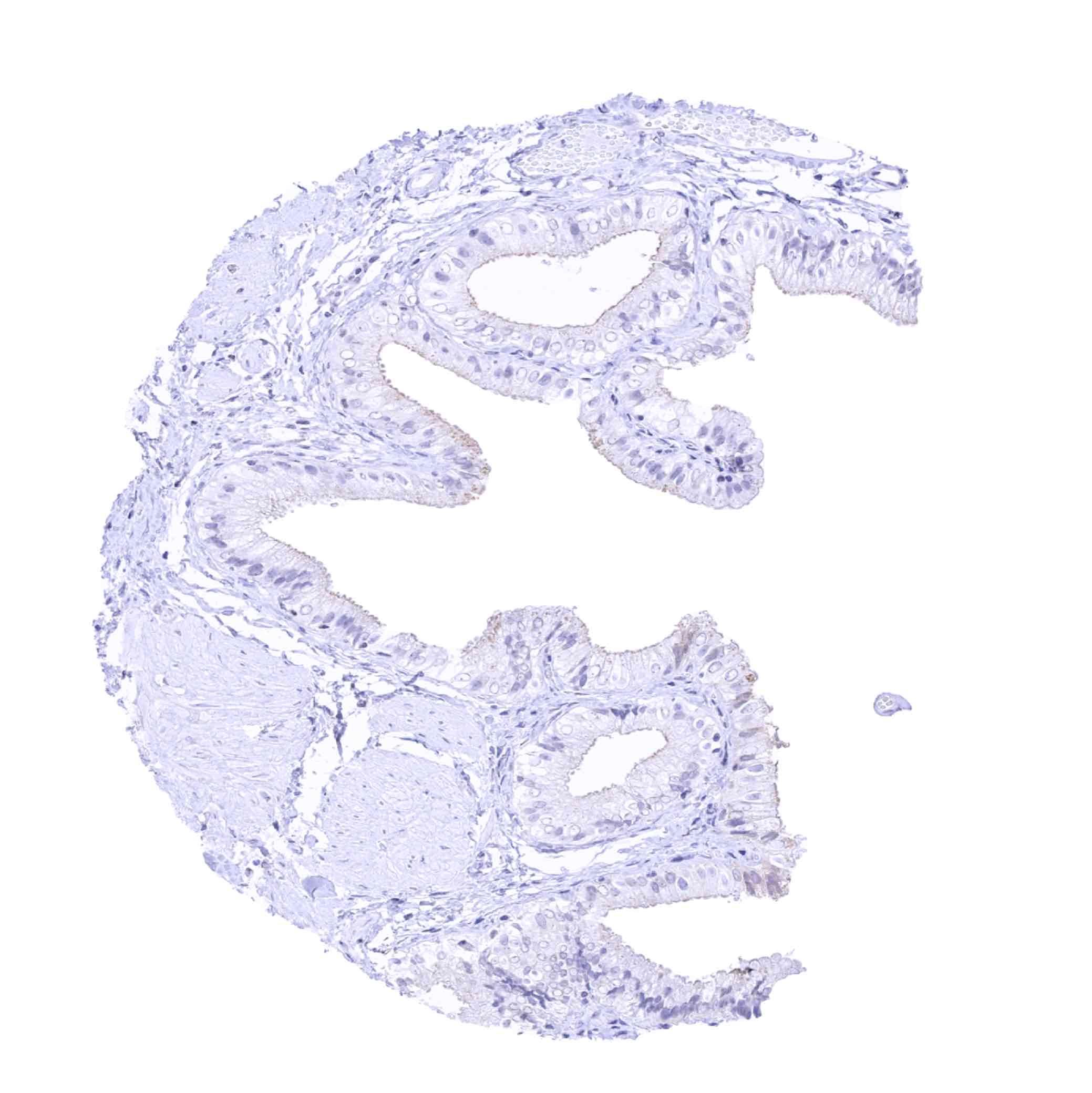
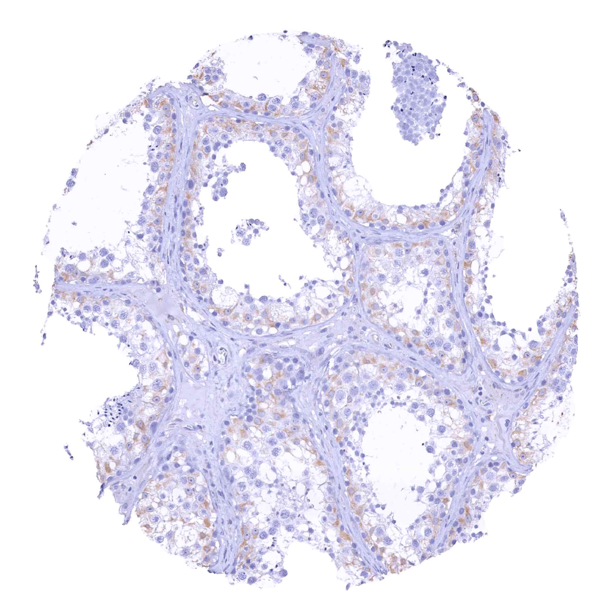
| Male genital | Prostate | Negative. |
| Seminal vesicles | Negative. | |
| Testis | Negative. | |
| Epididymis | Negative. | |
| Female genital | Breast | Negative. |
| Uterus, myometrium | Negative. | |
| Uterus, ectocervix | Negative. | |
| Uterus endocervix | Negative. | |
| Uterus, endometrium | Negative. | |
| Fallopian Tube | Negative. | |
| Ovary | Negative. | |
| Placenta early | Negative. | |
| Placenta mature | Negative. | |
| Amnion | Negative. | |
| Chorion | Negative. | |
| Skin | Epidermis | Negative. |
| Sebaceous glands | Negative. | |
| Muscle/connective tissue | Heart muscle | Negative. |
| Skeletal muscle | Negative. | |
| Smooth muscle | Negative. | |
| Vessel walls | Negative. | |
| Fat | Negative. | |
| Stroma | Negative. | |
| Endothelium | Negative. | |
| Bone marrow/lymphoid tissue | Bone marrow | Negative. |
| Lymph node | Negative. | |
| Spleen | Negative. | |
| Thymus | Negative. | |
| Tonsil | Negative. | |
| Remarks | Due to very high levels of somatostatin expression in some of the positive cells, contamination artifacts of adjacent cells may occur. |
RNA and protein expression data of Somatostatin findings are also described in the Human Protein Atlas (Tissue expression Somatostatin)
Positive control = Pancreas: A subset of pancreatic islet cells should show strong somatostatin positivity.
Negative control = Kidney: All cells must not show somatostatin immunostaining.

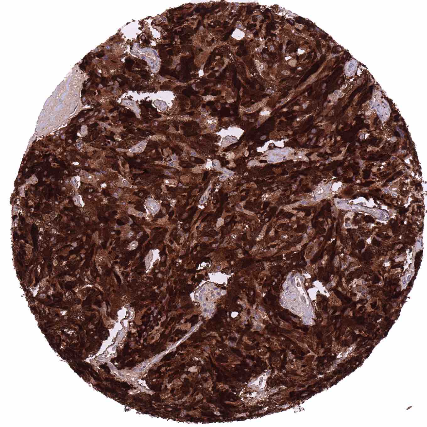
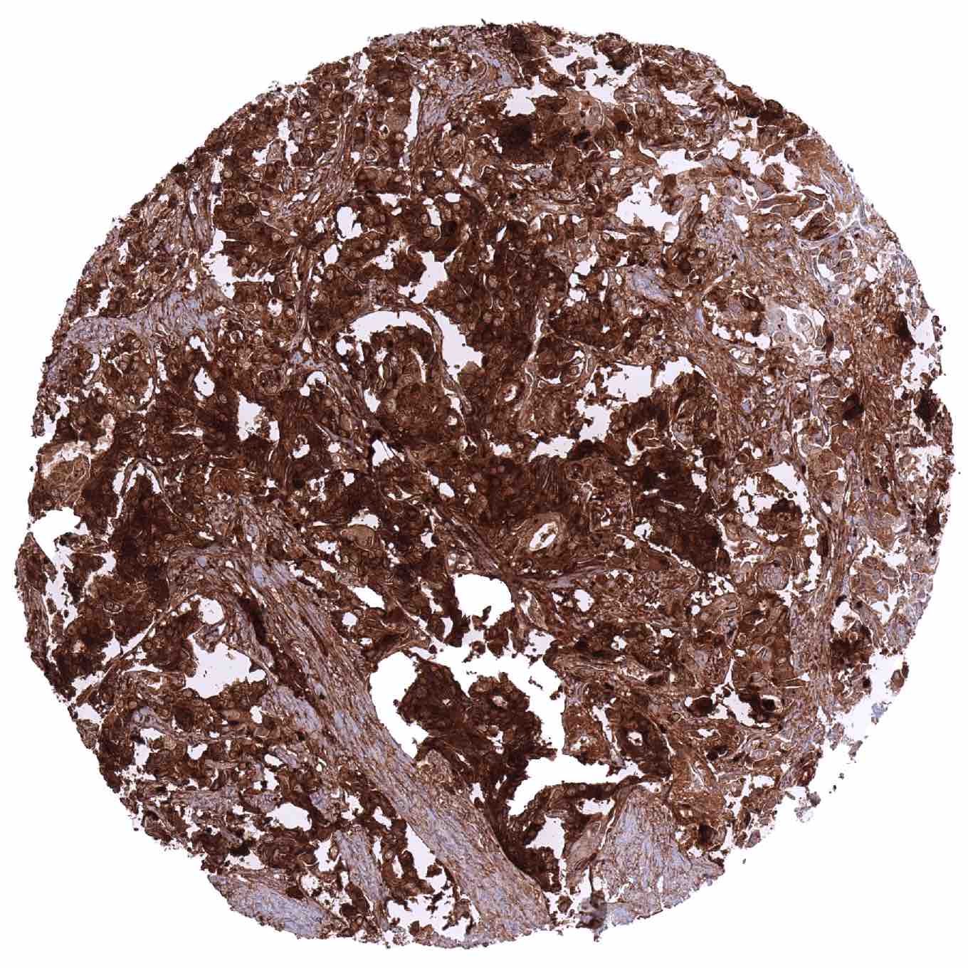
Staining Pattern in Relevant Tumor Types
A positive somatostatin immunostaining can be seen in a fraction of neuroendocrine tumors (in particular if these are derived from the pancreas), a fraction of pheochromocytomas and rarely also in other tumors.
The TCGA findings on Somatostatin RNA expression in different tumor categories have been summarized in the Human Protein Atlas.
Compatibility of Antibodies
No data available at the moment
Protocol Recommendations
IHC users have different preferences on how the stains should look like. Some prefer high staining intensity of the target stain and even accept some background. Others favor absolute specificity and lighter target stains. Factors that invariably lead to more intense staining include higher concentration of the antibody and visualization tools, longer incubation time, higher temperature during incubation, higher temperature and longer duration of the heat induced epitope retrieval (slide pretreatment). The impact of the pH during slide pretreatment has variable effects and depends on the antibody and the target protein.
All images and data shown here and in our image galleries are obtained by the manual protocol described below. Other protocols resulting in equivalent staining are described as well.
Manual protocol
Freshly cut sections should be used (less than 10 days between cutting and staining). Heat-induced antigen retrieval for 5 minutes in an autoclave at 121°C in pH 7,8 Target Retrieval Solution buffer. Apply MSVA-638R at a dilution of 1:150 at 37°C for 60 minutes. Visualization of bound antibody by the EnVision Kit (Dako, Agilent) according to the manufacturer’s directions.
Potential Research Applications
- The clinical significance of somatostatin expression in cancer is unclear.
Evidence for Antibody Specificity in IHC
There are two ways how the specificity of antibodies can be documented for immunohistochemistry on formalin fixed tissues. These are: 1. Comparison with a second independent method for target expression measurement across a large number of different tissue types (orthogonal strategy), and 2. Comparison with one or several independent antibodies for the same target and showing that all positive staining results are also seen with other antibodies for the same target (independent antibody strategy).
Orthogonal validation: For the antibody MSVA-638R specificity is suggested by the complete concordance of the immunostaining data with data from three independent RNA screening studies, including the Human Protein Atlas (HPA) RNA-seq tissue dataset, the FANTOM5 project, and the Genotype-Tissue Expression (GTEx) project, which are all summarized in the Human Protein Atlas (Tissue expression Somatostatin). Somatostatin RNA expression had only been described in organs (adrenal gland, gastrointestinal tract, pancreas, brain) where immunostaining was observed by MSVA-638R.
Comparison of antibodies: Specific somatostatin staining by MSVA-638R was also suggested by a confirmation of all MSVA-638R stainings by an independent – commercially available – second antibody termed “validation antibody”. Independence of the two antibodies MSVA-638R and “validation antibody” is documented by additional cytoplasmic staining in testicular cells and apical membranous staining of gallbladder, fallopian tube and respiratory epithelium seen by “validation antibody” but not by MSVA-638R. These staining are considered cross-reactivities of the “validation antibody”.