
295,00 € – 995,00 €
Product details
Synonyms = DR alpha chain; DRB1; DRB4; HLA class II histocompatibility antigen DR alpha chain; HLA DR1B; HLA DR3B; HLA DRA; HLA-DRA; HLADR4B; HLADRA1; HLADRB; Major histocompatibility complex class II DR alpha; Major histocompatibility complex class II DR beta 1/3/4/5
Antibody type = Recombinant Rabbit monoclonal / IgG
Clone = MSVA-470R
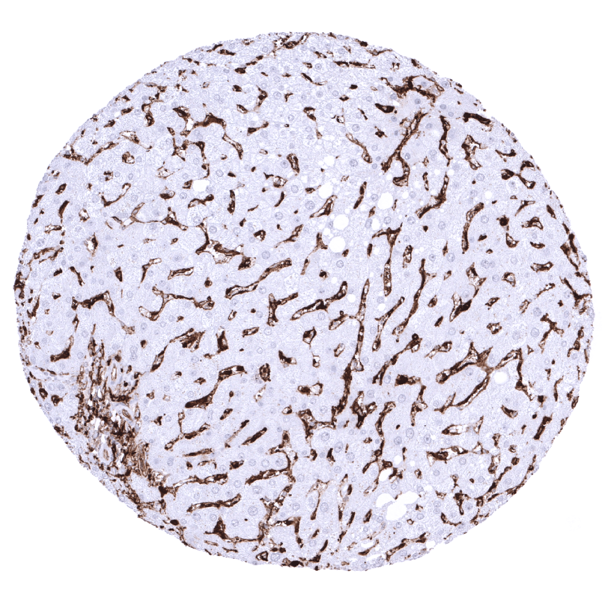
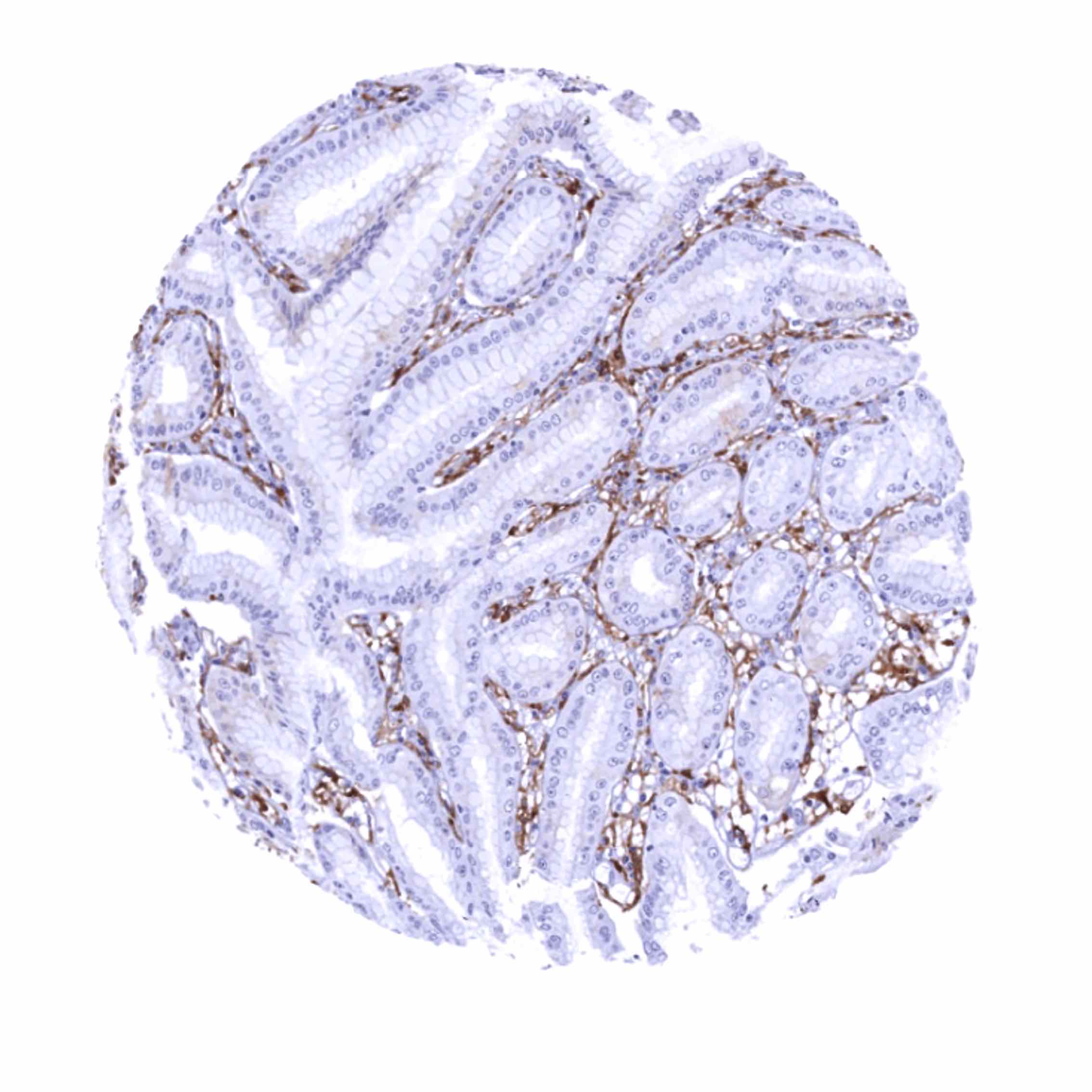
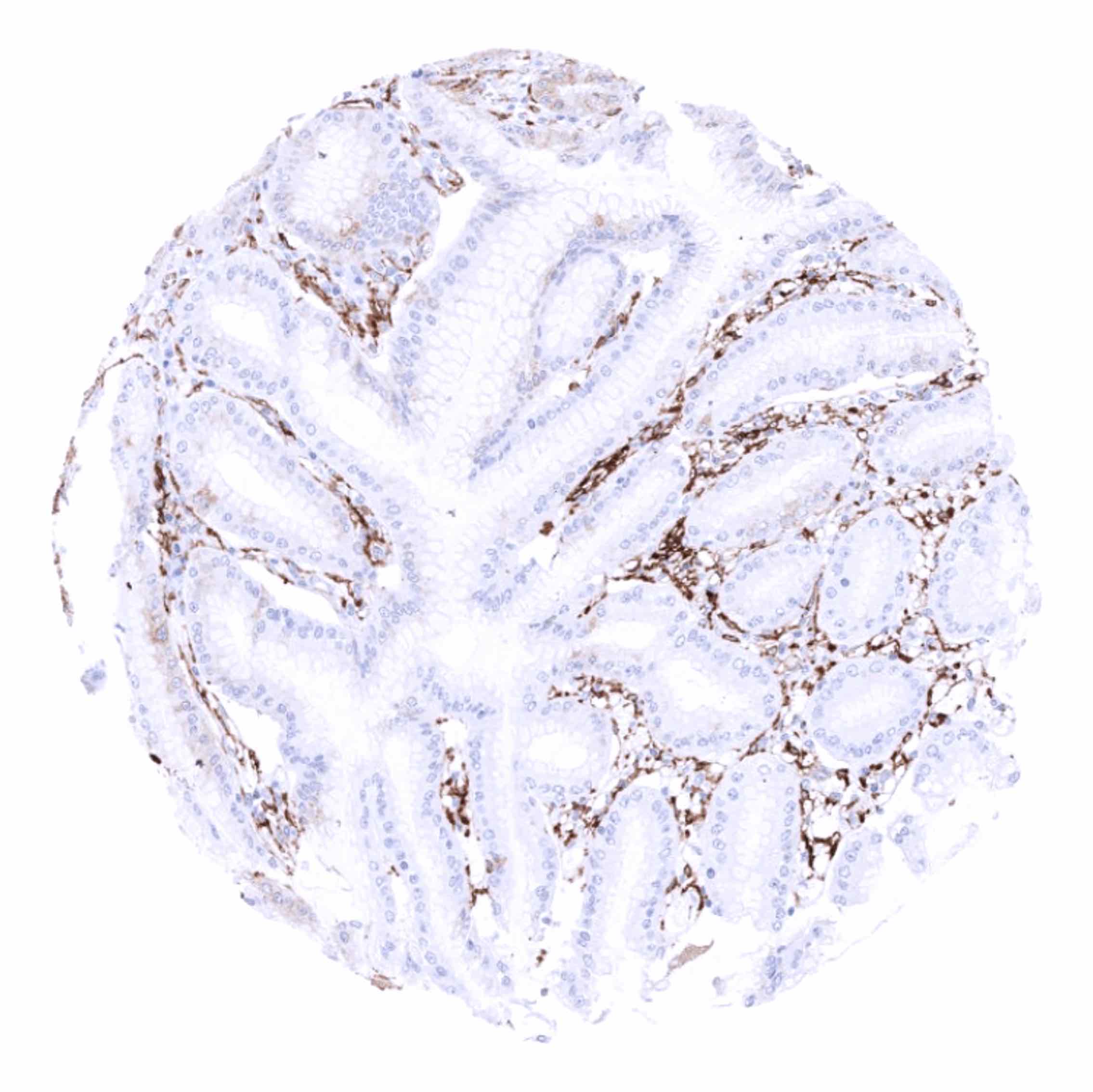
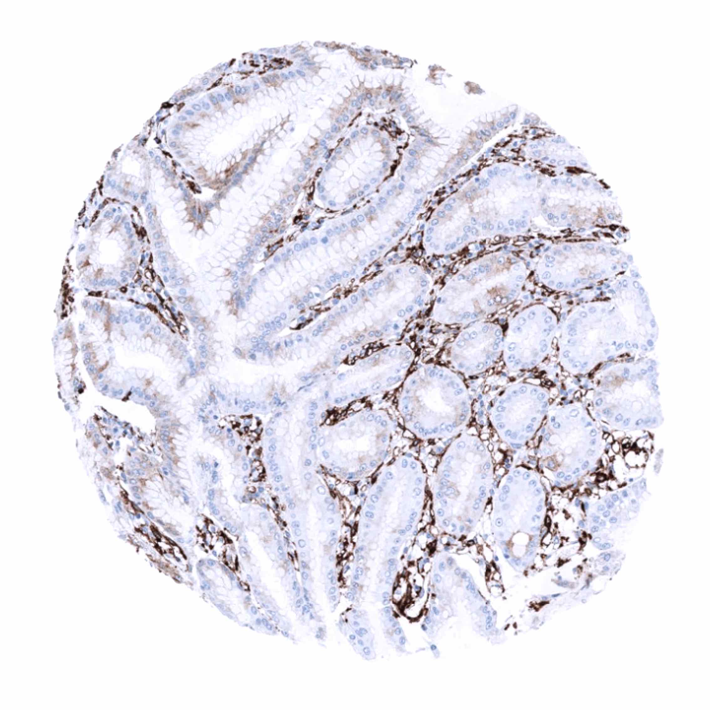
Positive control = Liver: An at least moderate staining should be seen of sinusoids and Kupffer cells. Kidney: An at least moderate staining should be seen in small capillaries of glomeruli.
Negative control = Absence of HLA-DRa staining is expected in hepatocytes of a completely normal liver and in tubuli of completely normal kidney.
Cellular localization = Cell Surface
Reactivity = Human
Application = Immunohistochemistry
Dilution = 1:100 – 1:200
Intended Use = Research Use Only
Relevance of Antibody
HLA-DRa is a pivotal protein for immune response.
Biology Behind
HLA-DR is an MHC class II cell surface receptor acting as a ligand for the T-cell receptor. HLA-DR is an αβ heterodimer protein. The DR α-chain is encoded by the HLA-DRA gene. The DR β-chain is encoded by 4 different gene loci (DRB1, DRB3, DRB4, DRB5), which are all located close together with HLA-DRα gene locus at 6p21.31. Of these 4 DR β-chain gene loci, no more than 3 are functional in a single individual, and no more than two can be functional on a single chromosome. The main function of HLA-DRa is to present “foreign appearing” peptide antigens to the immune system in order to induce T-(helper)-cell responses that eventually result in the production of antibodies against the same peptide antigen. There is a high level of allelic diversity at HLA-DRB1, especially in positions that affect its peptide binding groove. HLA-DRB1 variability thus affects the spectrum of peptides that can be recognized as “foreign” and the efficiency of their presentation to antigen presenting cells. In organ transplantation, good matching of these antigens between host and donor is critical for graft survival. HLA-DRa alleles also impact the risk for autoimmune disease, disease susceptibility and disease resistance. HLA-DRa antigens show restricted tissue distribution. HLA-DRa is expressed in antigen-presenting cells (macrophages, B-cells, and dendritic cells) but can also be found in epithelial cells in case of inflammation or neoplastic transformation.
Staining Pattern in Normal Tissues
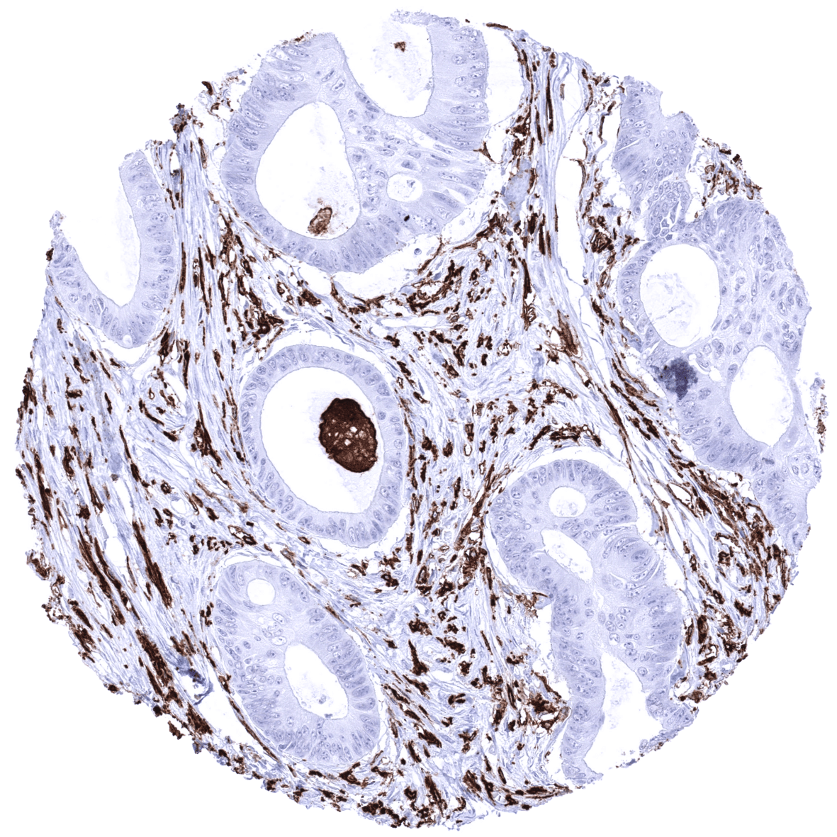
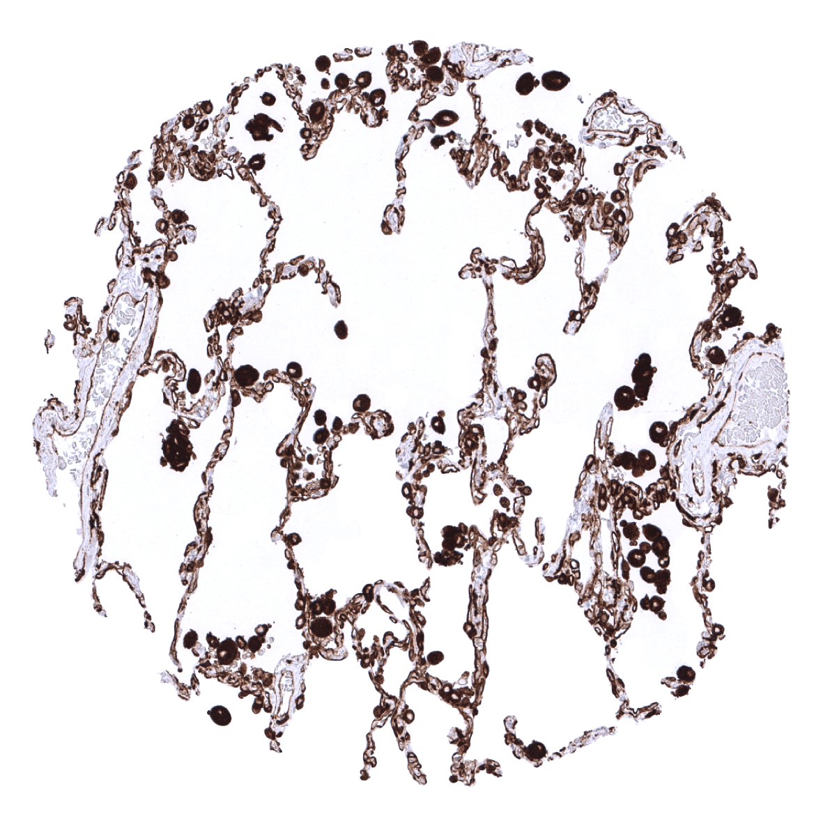
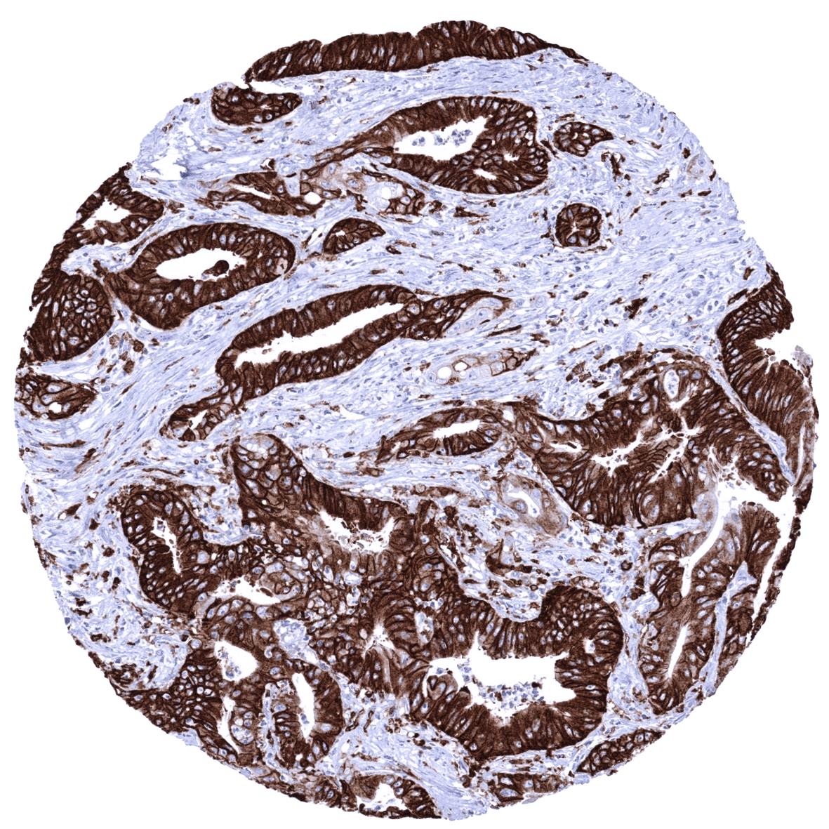
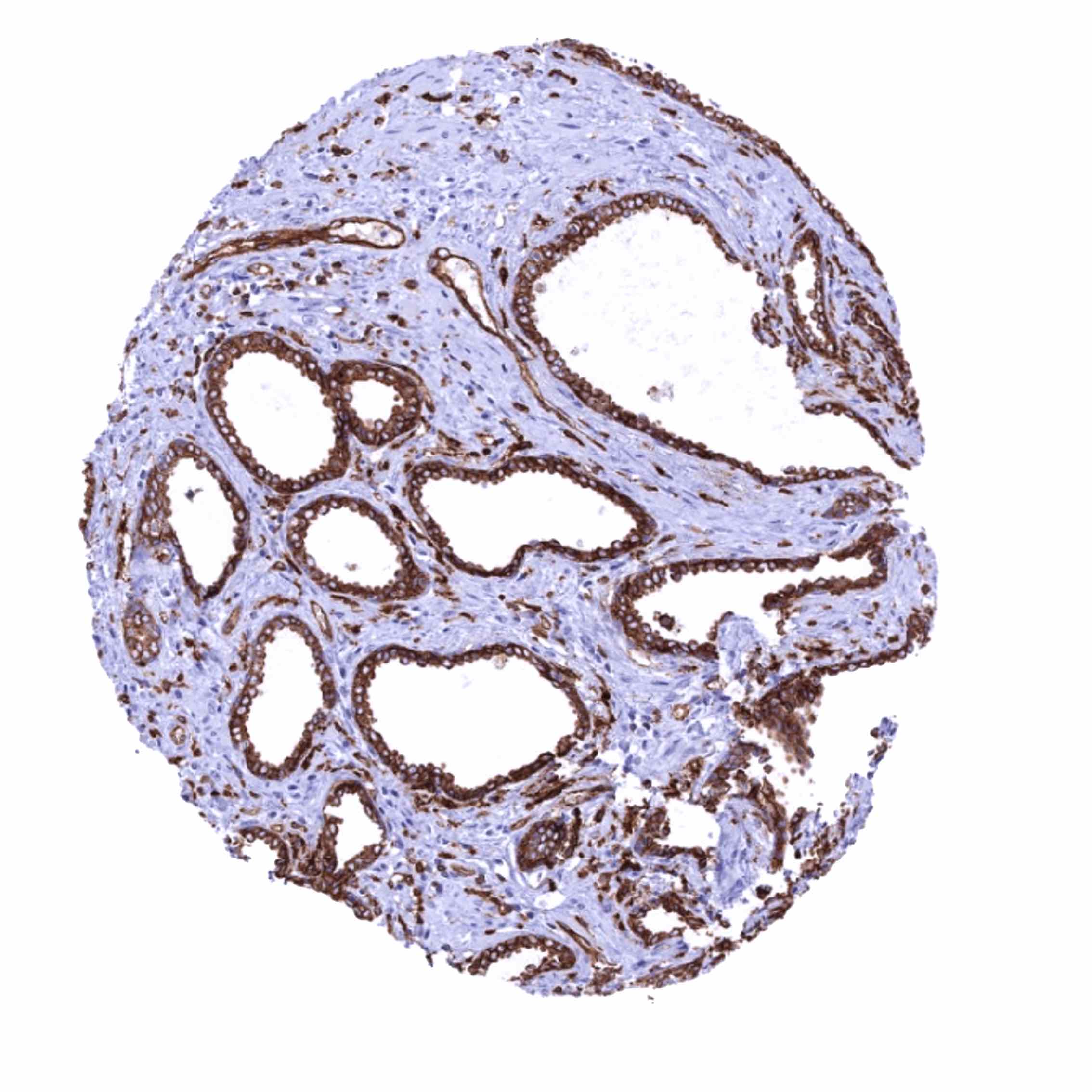
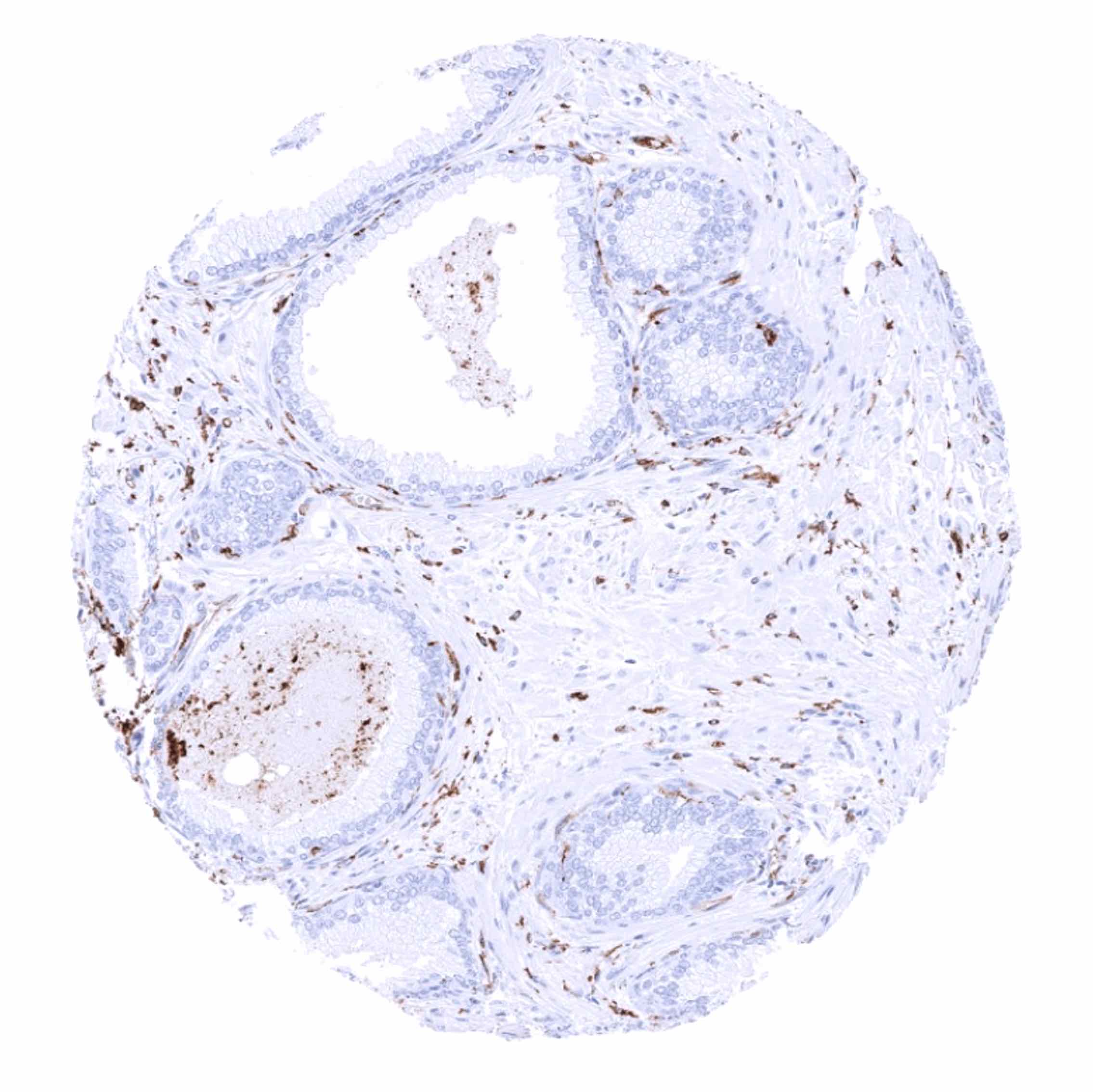
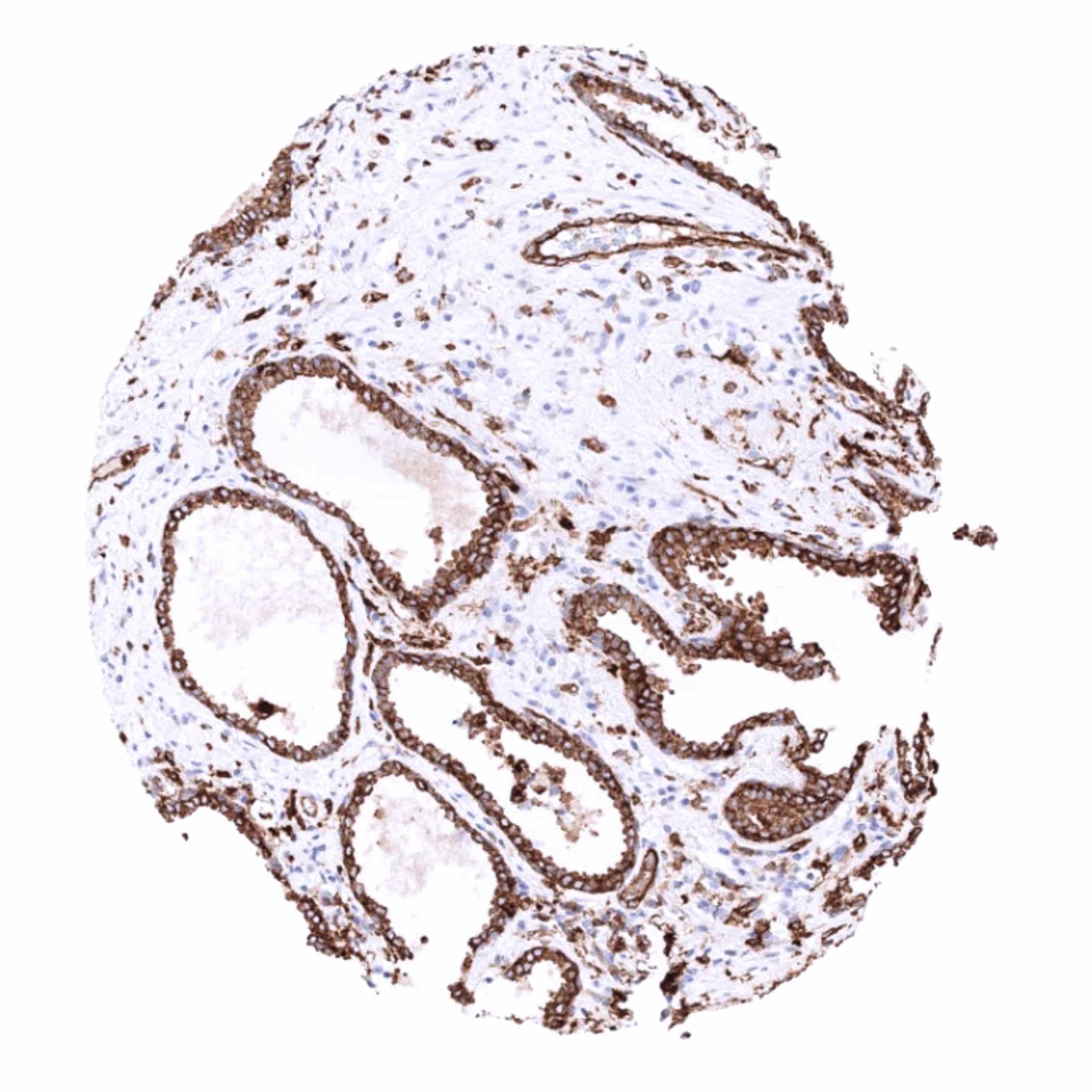
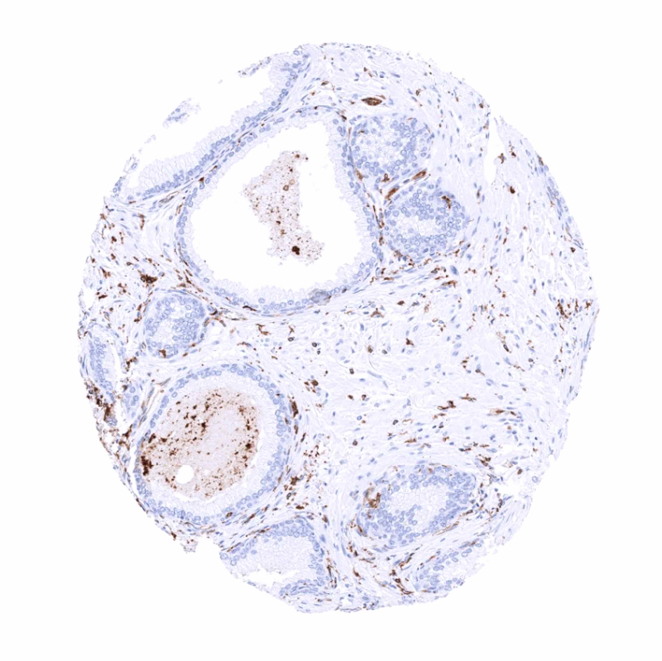
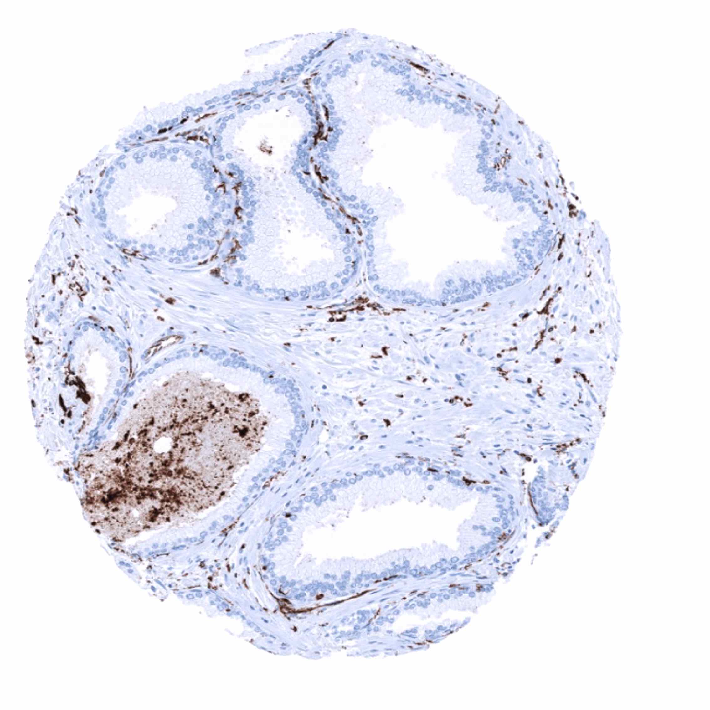
HLA-DRa immunostaining is regularly seen on the majority of inflammatory cells including dendritic cells and macrophages. In these cells the staining is mostly strong but may show some variability. A moderate (to strong) HLA-DRa staining is also seen in endothelial cells of blood vessels except in the placenta. This includes the liver sinus and venous sinus in the spleen. Epithelial cells do regularly show a strong, predominantly membranous staining in the small intestine (strongest staining in the duodenum) and in a subset of the surface epithelium of the fallopian tube (mosaic staining pattern). HLA-DRa immunostaining is also frequently seen in the surface epithelium of the stomach mucosa. Occasional cytoplasmic and membranous HLA-DRa staining can focally be seen in virtually all epithelial cell types, especially in case of inflammation, atrophy or regeneration.
These findings are largely comparable to the RNA and protein data described in the Human Protein Atlas (Tissue expression HLA-DRa) with the exception of corpus luteum which are not described there. For corpus luteum this is due to the fact that this tissue type is not analyzed in the protein atlas.
Positive control: Liver: An at least moderate staining should be seen of sinusoids and Kupffer cells. Kidney: An at least moderate staining should be seen in small capillaries of glomeruli.
Negative control: Absence of HLA-DRa staining is expected in hepatocytes of a completely normal liver and in tubuli of completely normal kidney.


Staining Pattern in Relevant Tumor Types
HLA-DRa can be expressed in tumor cells of various cancer types in a fraction of cases. HLA-DRa expression has been described to be related to favorable patient prognosis in colorectal cancer, gastric cancer, and squamous cell carcinoma of the larynx, but was associated with poor prognosis in glioma and esophageal squamous cell carcinoma in at least one study each. HLA-DRa expression of tumor infiltrating inflammatory cells can be seen in all tumors to a variable extent.
The TCGA findings on HLA-DRa RNA expression in different tumor categories have been summarized in the Human Protein Atlas.


Compatibility of Antibodies
No data available at the moment
Protocol Recommendations
IHC users have different preferences on how the stains should look like. Some prefer high staining intensity of the target stain and even accept some background. Others favor absolute specificity and lighter target stains. Factors that invariably lead to more intense staining include higher concentration of the antibody and visualization tools, longer incubation time, higher temperature during incubation, higher temperature and longer duration of the heat induced epitope retrieval (slide pretreatment). The impact of the pH during slide pretreatment has variable effects and depends on the antibody and the target protein.
All images and data shown here and in our image galleries are obtained by the manual protocol described below. Other protocols resulting in equivalent staining are described as well.
Manual protocol
Freshly cut sections should be used (less than 10 days between cutting and staining). Heat-induced antigen retrieval for 5 minutes in an autoclave at 121°C in pH 9,0 Target Retrieval Solution buffer. Apply MSVA-470R at a dilution of 1:150 at 37°C for 60 minutes. Visualization of bound antibody by the EnVision Kit (Dako, Agilent) according to the manufacturer’s directions.

Agilent / Dako – Autostainer Link 48
Pretreatment in PT-Link for 30 minutes at 95°C (pH high); FLEX peroxidase blocking for 5 minutes (room temperature), MSVA-470R 1:150 for 20 minutes (room temperature), FLEX+ mouse/rabbit (LINKER) for 15 minutes (room temperature), horseradish peroxidase (HRP) for 20 minutes (room temperature), FLEX DAB+Sub-Chromo for 10 minutes (room temperature), FLEX hematoxylin for 5 minutes (room temperature).

These images reflect stainings by the protocol described above. It is of note that a comparable staining result can also be obtained by different protocols. In general, a longer pretreatment, a longer incubation time of the primary antibody, a higher antibody concentration, and a longer incubation time of FLEX+LINKER result in stronger staining, potentially at the cost of more background staining. Modifications of the protocol with a strengthening effect on staining intensity in combination with changes of other parameters that result in lower staining intensity can result in a comparable result as shown above.
Leica – BOND RX
Dewax at 72°C for 30 seconds; Pretreatment in Bond Epitope Retrieval Solution (ER2 – EDTA pH9) for 20 minutes at 100°C; Peroxidase blocking for 5 minutes (room temperature), MSVA-470R 1:125 for 15 minutes (room temperature), Post primary (rabbit anti mouse) for 8 minutes (room temperature), Polymer (goat anti rabbit) for 8 minutes (room temperature), mixed DAB refine for 10 minutes (room temperature), hematoxylin for 5 minutes (room temperature).

These images reflect stainings by the protocol described above. It is of note that a comparable staining result can also be obtained by different protocols. In general, a longer pretreatment, a longer incubation time of the primary antibody, a higher antibody concentration, a higher temperature during incubation, and a longer incubation time of Post primary and or the Polymer result in stronger staining, potentially at the cost of more background staining. Modifications of the protocol with a strengthening effect on staining intensity in combination with changes of other parameters that result in lower staining intensity can result in a comparable result as shown above.
Roche – Ventana Discovery ULTRA
Pretreatment for 64 minutes at 100°C (pH 8,4); CM peroxidase blocking for 12 minutes (room temperature), MSVA-470R 1:50 for 20 minutes at 36°C, secondary antibody (anti-rabbit HQ) for 12 minutes at 36°C, anti-HQ HRP for 12 minutes at room temperature, DAB at room temperature, hematoxylin II at room temperature for 8 minutes, bluing reagent at room temperature for 4 minutes.

These images depict staining results obtained by the protocol described above. It is of note, that the Ventana machines generally require higher antibody concentrations than other commonly used autostainers because the antibodies are automatically diluted during the procedure. Various other protocols can result in an identical result as shown above. A longer pretreatment, a longer incubation time of the primary antibody, a higher antibody concentration, a higher temperature during incubation, and a longer incubation time of secondary antibody and or the anti-HQ HRP result in stronger staining, potentially at the cost of more background staining.
Potential Research Applications
- HLA-DR expression can occur in a fraction of tumors where it appears to be linked to favorable prognosis. The prognostic role of HLA-DR expression and its predictive significance with respect to checkpoint inhibitor therapies needs to be investigated.
- The role of HLA-DR expressing cells and their spatial relationship to tumor cells is of interest.
- HLA-DR immunostaining can be used to distinguish macrophage subtypes. HLA-DR expression may be lacking in a fraction of M2 macrophages. Monocytes with diminished or absent HLA-DR expression have emerged as important mediators of tumor-induced immunosuppression.
Evidence for Antibody Specificity in IHC
There are two ways how the specificity of antibodies can be documented for immunohistochemistry on formalin fixed tissues. These are: 1. comparison with a second independent method for target expression measurement across a large number of different tissue types (orthogonal strategy), and 2. Comparison with one or several independent antibodies for the same target and showing that all positive staining results are also seen with other antibodies for the same target (independent antibody strategy).
Orthogonal validation: For the antibody MSVA-470R specificity is suggested by the strong concordance of the immunostaining with RNA expression data derived from the Human Protein Atlas (HPA) RNA-seq tissue dataset, the FANTOM5 project, and the Genotype-Tissue Expression (GTEx) project which are all summarized in the Human Protein Atlas (Tissue expression HLA-DRa). Immunostaining by using MSVA-470R was almost exclusively detected in organs with documented RNA expression.
Comparison of antibodies: True expression of HLA-DRa in various epithelial cell types is further suggested by similar findings obtained by the use of the antibody CAB002798.
