

445,00 € – 2.995,00 €
Product details
Synonyms = CD366; HAVR2; Hepatitis A virus cellular receptor 2 (HAVCR2); Kidney injury molecule 3 (KIM3); T-cell immunoglobulin and mucin domain-containing protein 3; T-cell immunoglobulin mucin receptor 3; T-cell membrane protein 3; TIM3; TIMD3
Antibody type = Recombinant Rabbit monoclonal / IgG
Clone = MSVA-366R
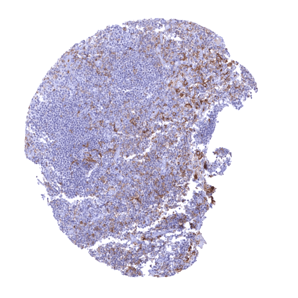
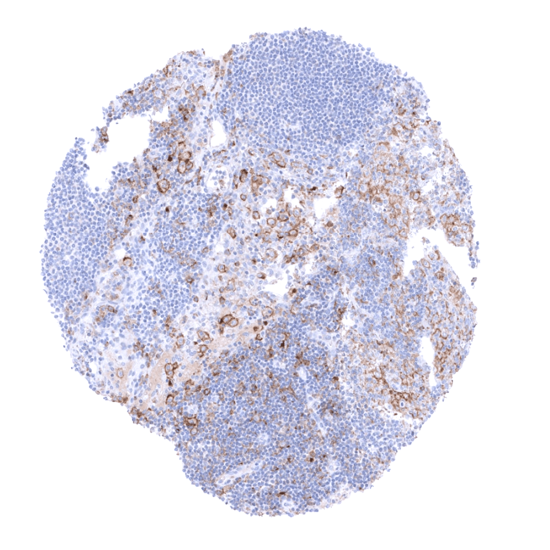
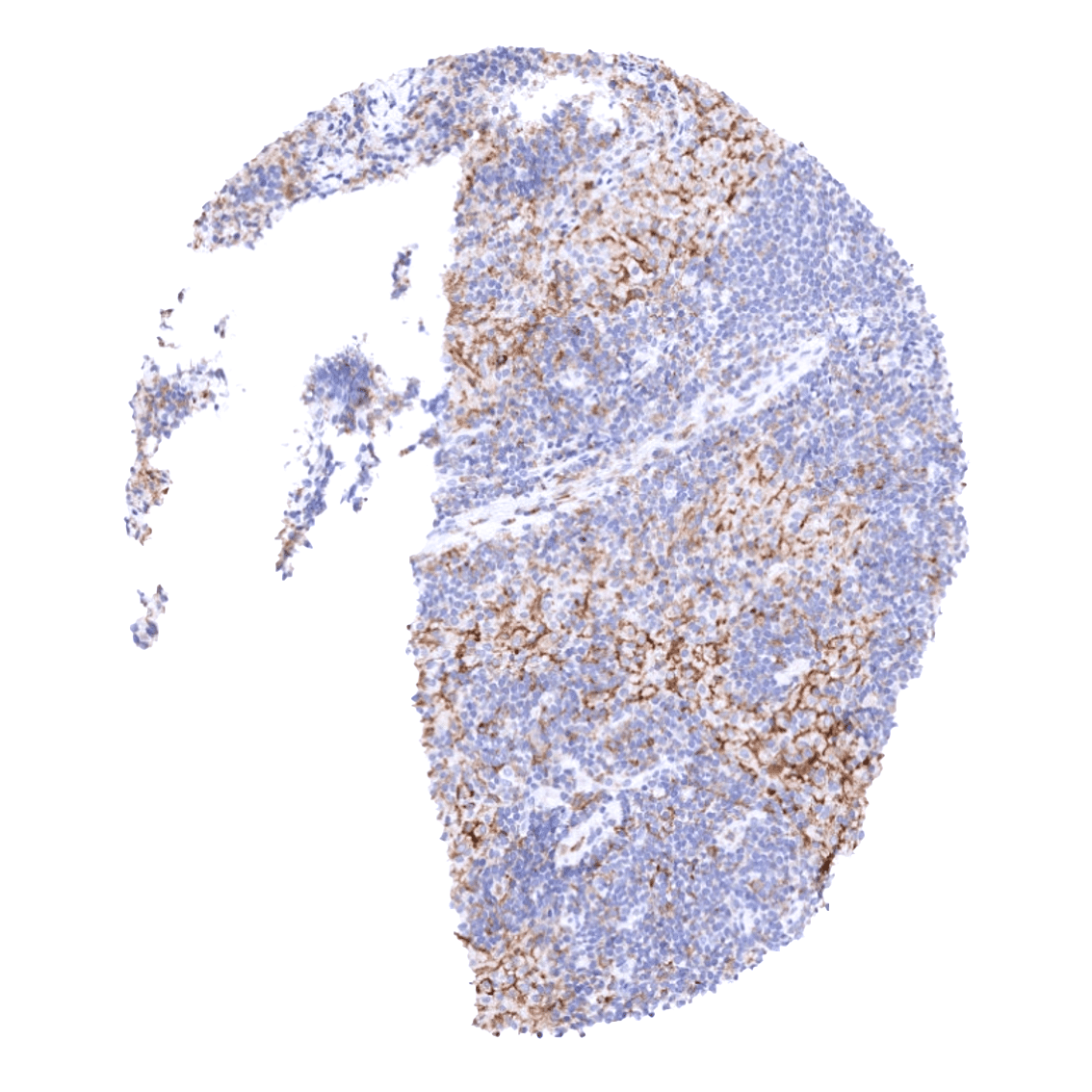
Positive control = Tonsil: A moderate to strong TIM-3 immunostaining should be seen in a significant fraction of T-cells, while only few TIM-3 positive cells are seen in the germinal centre and the mantle zone.
Negative control = Tonsil: Epithelial cells should be negative.
Cellular localization = Cell Surface
Reactivity = Human
Application = Immunohistochemistry
Dilution = 1:100 – 1:200
Intended Use = Research Use Only
Relevance of Antibody
TIM-3 is an immune checkpoint that constitutes a “hot topic” in immune-oncology research.
Biology Behind
TIM-3 (T-cell immunoglobulin and mucin-domain containing-3) also termed Hepatitis A virus cellular receptor 2 (HAVCR2) is a 33 kDa cell surface receptor protein coded by the TIM-3 gene on 5q33.3. TIM-3 belongs to TIM family cell surface receptor proteins. These proteins share a similar structure with an extracellular region containing a variable immunoglobulin domain (IgV) and a glycosylated mucin domain of variable length. TIM-3 is an immune checkpoint. It mediates CD8+ T-cell exhaustion together with other inhibitory receptors such as PD-1 and LAG3. TIM-3 is primarily activated by galectin-9. The interaction with galectin-9 leads to stimulation of an influx of calcium to the intracellular space and induction of apoptosis. Other known TIM-3 ligands include phosphatidylserine (PtdSer), High Mobility Group Protein 1 (HMGB1) and Carcinoembryonic Antigen Related Cell Adhesion Molecule 1 (CEACAM1). TIM-3 is viewed as a promising drug target. Multiple phase 1/2 clinical trials with anti-TIM3 monoclonal antibodies are ongoing in various cancer types.
Staining Pattern in Normal Tissues
TIM-3 staining pattern in Normal Tissues with antibody MSVA-366R (images are shown in our “Normal Tissue Gallery”)
| Brain | Cerebrum | Negative. |
| Cerebellum | Negative. | |
| Endocrine Tissues | Thyroid | Negative. |
| Parathyroid | Negative. | |
| Adrenal gland | TIM-3 positivity of some tissue associated macrophages. | |
| Pituitary gland | Negative. | |
| Respiratory system | Respiratory epithelium | TIM-3 positivity of some tissue associated macrophages and/or lymphocytes. |
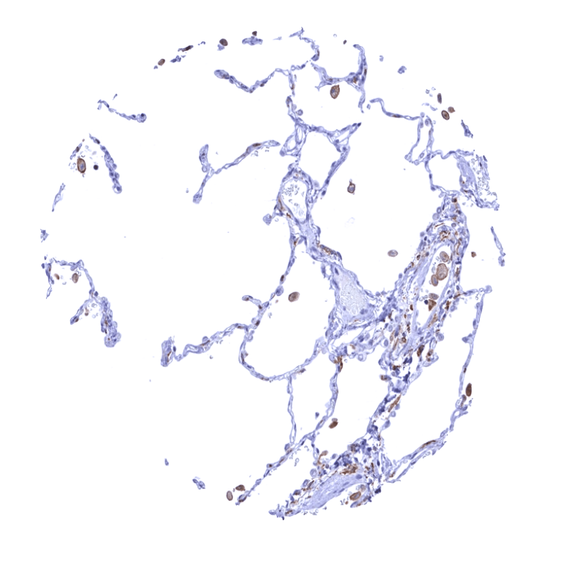
| Lung | Strong TIM-3 positivity of alveolar macrophages. | |
| Gastrointestinal Tract | Salivary glands | Epithelial cells are TIM-3 negative. |
| Esophagus | Epithelial cells are TIM-3 negative. | |
| Stomach | Epithelial cells are TIM-3 negative. | |
| Duodenum | Moderate TIM-3 staining of apical membranes of surface epithelial may reflect a (tolerable) cross-reactivity. | |
| Small intestine | Moderate TIM-3 staining of apical membranes of surface epithelial may reflect a (tolerable) cross-reactivity. | |
| Appendix | TIM-3 positivity of a fraction of tissue associated macrophages. | |
| Colon | TIM-3 positivity of a fraction of tissue associated macrophages. | |
| Rectum | TIM-3 positivity of a fraction of tissue associated macrophages.. | |
| Liver | Moderate membranous TIM-3 staining of Kupffer cells. | |
| Gallbladder | Epithelial cells are TIM-3 negative. | |
| Pancreas | Epithelial cells are TIM-3 negative. | |
| Genitourinary | Kidney | Weak to moderate membranous TIM-3 staining of apical membranes of a fraction of tubuli. |
| Urothelium | Epithelial cells are TIM-3 negative. | |
| Male genital | Prostate | Epithelial cells are TIM-3 negative. |
| Seminal vesicles | Epithelial cells are TIM-3 negative. | |
| Testis | TIM-3 positivity may be seen in some associated inflammatory cells. | |
| Epididymis | Strong membranous TIM-3 staining of apical membranes of tall columnar cells of the epididymis. | |
| Female genital | Breast | Epithelial cells are TIM-3 negative. |
| Uterus, myometrium | Negative. | |
| Uterus, ectocervix | Epithelial cells are TIM-3 negative. | |
| Uterus endocervix | Epithelial cells are TIM-3 negative. | |
| Uterus, endometrium | TIM-3 positivity of a fraction of tissue associated macrophages and/or lymphocytes. | |
| Fallopian Tube | Epithelial cells are TIM-3 negative. | |
| Ovary | Negative. | |
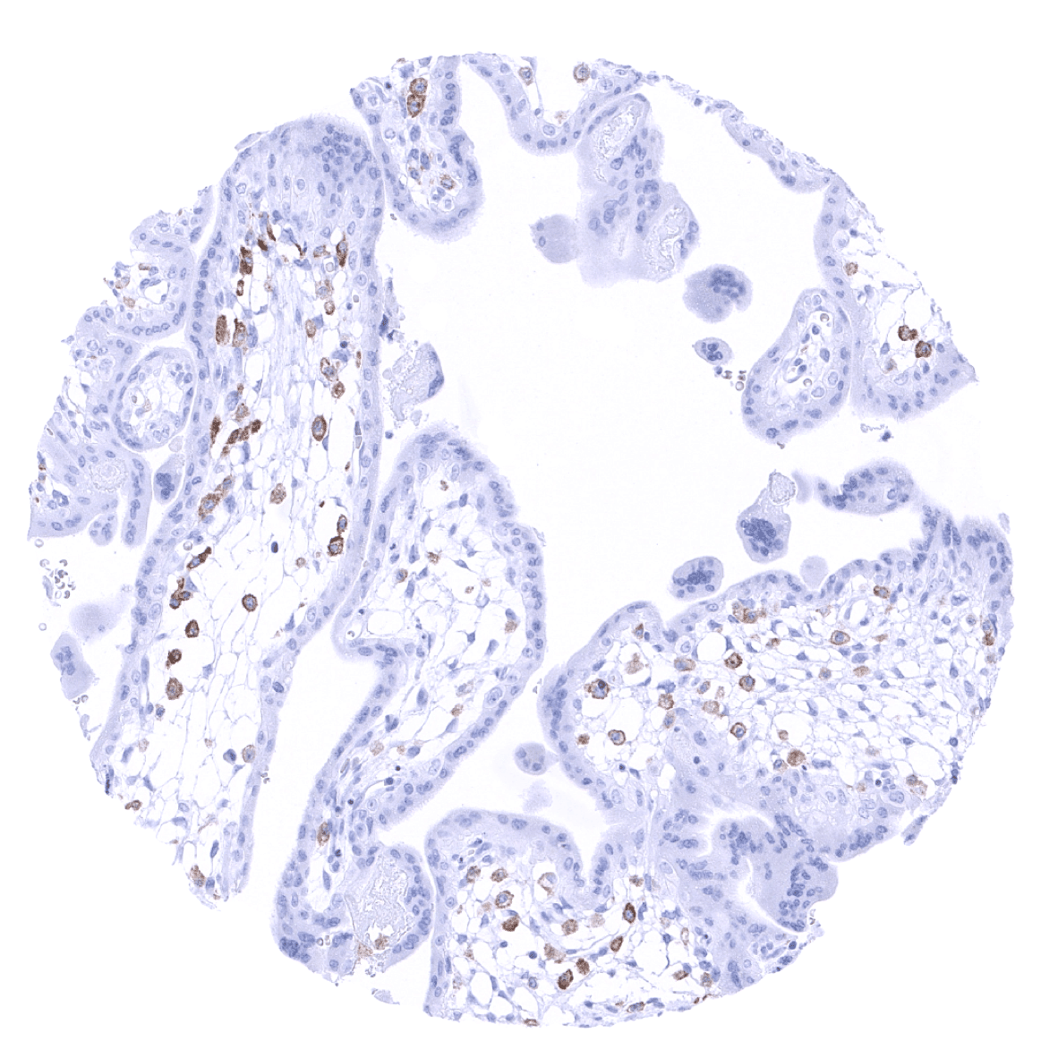
| Placenta early | Strong TIM-3 positivity in placenta macrophages. | |
| Placenta mature | Strong TIM-3 positivity in placenta macrophages. Significant TIM-3 positivity of fractions of macrophages and possibly also T-lymphocytes around decidua cells. | |
| Amnion | Epithelial cells are TIM-3 negative. | |
| Chorion | Epithelial cells are TIM-3 negative. | |
| Skin | Epidermis | Epithelial cells are TIM-3 negative. |
| Sebaceous glands | Epithelial cells are TIM-3 negative. | |
| Muscle/connective tissue | Heart muscle | Negative. |
| Skeletal muscle | Negative. | |
| Smooth muscle | Negative. | |
| Vessel walls | Negative. | |
| Fat | Negative. | |
| Stroma | Variable TIM-3 positivity in a fraction of T-lymphocytes and of macrophages/dendritic cells. | |
| Endothelium | Mostly negative. | |
| Bone marrow/ lymphoid tissue | Bone marrow | TIM-3 positivity in a fraction of T-lymphocytes and of macrophages/dendritic cells. |
| Lymph node | Significant TIM-3 positivity of fractions of tissue associated macrophages and T-lymphocytes. | |
| Spleen | Weak to moderate TIM-3 staining of endothelial cells of venous sinusoids. | |
| Thymus | Significant TIM-3 positivity in macrophages but cortical T-lymphocytes show little expression. | |
| Tonsil | Variable levels of TIM-3 staining in macrophages and lymphocytes. | |
| Remarks | TIM-3 immunostaining in a fraction of T-lymphocytes and of macrophages/dendritic cells in all organs. |
These findings are largely comparable to the RNA and protein data described in the Human Protein Atlas (Tissue expression TIM3)
Positive control = Tonsil: A moderate to strong TIM-3 immunostaining should be seen in a significant fraction of T-cells, while only few TIM-3 positive cells are seen in the germinal centre and the mantle zone.
Negative control = Tonsil: Epithelial cells should be negative.
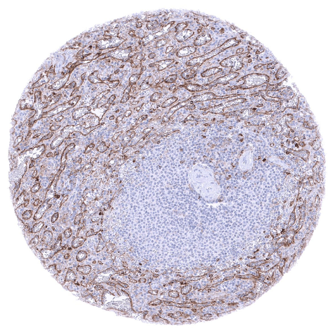
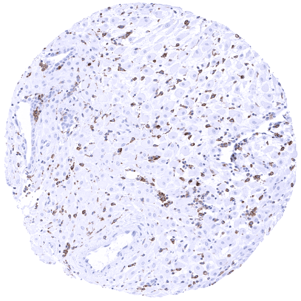
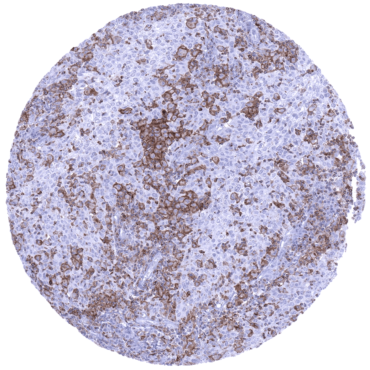
Staining Pattern in Relevant Tumor Types
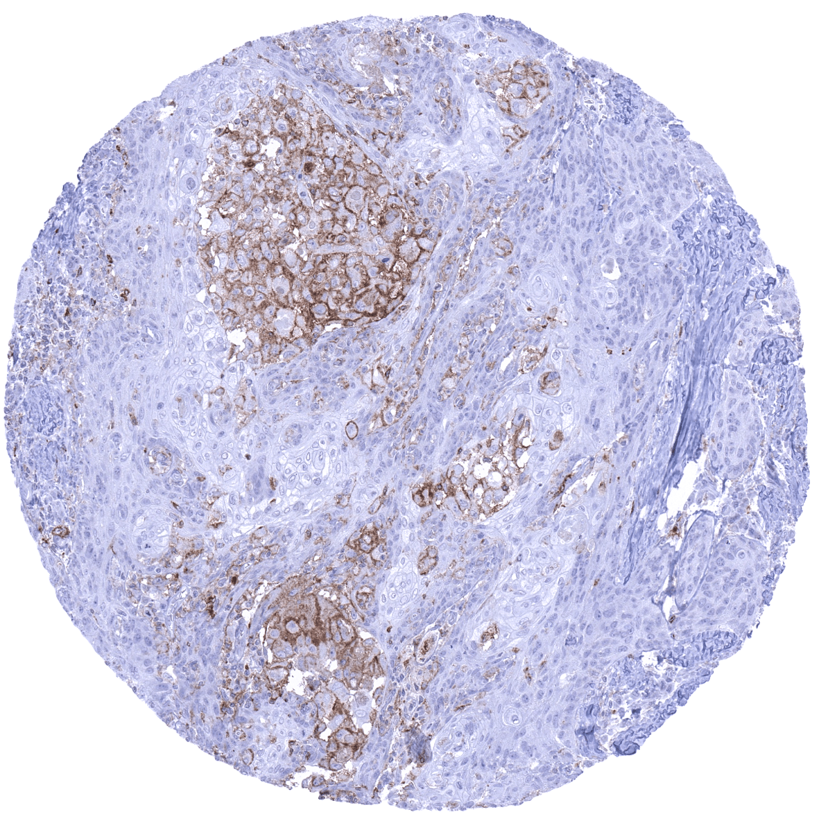
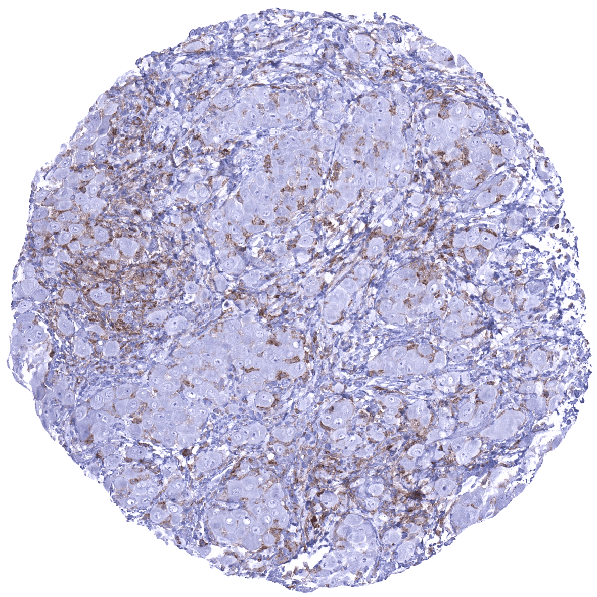
TIM-3 positive lymphocytes and macrophages occur at variable density in virtually all cancers. TIM-3 can also be expressed on cancer cells such as for example in a fraction of renal cell carcinomas.
The TCGA findings on TIM-3 RNA expression in different tumor categories have been summarized in the Human Protein Atlas.

Compatibility of Antibodies
No data available at the moment
Protocol Recommendations
IHC users have different preferences on how the stains should look like. Some prefer high staining intensity of the target stain and even accept some background. Others favor absolute specificity and lighter target stains. Factors that invariably lead to more intense staining include higher concentration of the antibody and visualization tools, longer incubation time, higher temperature during incubation, higher temperature and longer duration of the heat induced epitope retrieval (slide pretreatment). The impact of the pH during slide pretreatment has variable effects and depends on the antibody and the target protein.
All images and data shown here and in our image galleries are obtained by the manual protocol described below. Other protocols resulting in equivalent staining are described as well.
Manual protocol
Freshly cut sections should be used (less than 10 days between cutting and staining). Heat-induced antigen retrieval for 5 minutes in an autoclave at 121°C in pH 7,8 Target Retrieval Solution buffer. Apply MSVA-366R at a dilution of 1:150 at 37°C for 60 minutes. Visualization of bound antibody by the EnVision Kit (Dako, Agilent) according to the manufacturer’s directions.
Agilent / Dako – Autostainer Link 48
Pretreatment in PT-Link for 30 minutes at 95°C (pH high); FLEX peroxidase blocking for 5 minutes (room temperature), MSVA-366R 1:150 for 20 minutes (room temperature), FLEX+ mouse/rabbit (LINKER) for 15 minutes (room temperature), horseradish peroxidase (HRP) for 20 minutes (room temperature), FLEX DAB+Sub-Chromo for 10 minutes (room temperature), FLEX hematoxylin for 5 minutes (room temperature).

These images reflect stainings by the protocol described above. It is of note that a comparable staining result can also be obtained by different protocols. In general, a longer pretreatment, a longer incubation time of the primary antibody, a higher antibody concentration, and a longer incubation time of FLEX+LINKER result in stronger staining, potentially at the cost of more background staining. Modifications of the protocol with a strengthening effect on staining intensity in combination with changes of other parameters that result in lower staining intensity can result in a comparable result as shown above.
Leica – BOND RX
Dewax at 72°C for 30 seconds; Pretreatment in Bond Epitope Retrieval Solution (ER2 – EDTA pH9) for 20 minutes at 100°C; Peroxidase blocking for 5 minutes (room temperature), MSVA-366R 1:200 for 15 minutes (room temperature), Post primary (rabbit anti mouse) for 8 minutes (room temperature), Polymer (goat anti rabbit) for 8 minutes (room temperature), mixed DAB refine for 10 minutes (room temperature), hematoxylin for 5 minutes (room temperature).

These images reflect stainings by the protocol described above. It is of note that a comparable staining result can also be obtained by different protocols. In general, a longer pretreatment, a longer incubation time of the primary antibody, a higher antibody concentration, a higher temperature during incubation, and a longer incubation time of Post primary and or the Polymer result in stronger staining, potentially at the cost of more background staining. Modifications of the protocol with a strengthening effect on staining intensity in combination with changes of other parameters that result in lower staining intensity can result in a comparable result as shown above.
Roche – Ventana Discovery ULTRA
Pretreatment for 64 minutes at 100°C (pH 8,4); CM peroxidase blocking for 12 minutes (room temperature), MSVA-366R 1:100 for 20 minutes at 36°C, secondary antibody (anti-rabbit HQ) for 12 minutes at 36°C, anti-HQ HRP for 12 minutes at room temperature, DAB at room temperature, hematoxylin II at room temperature for 8 minutes, bluing reagent at room temperature for 4 minutes.


These images depict staining results obtained by the protocol described above. It is of note, that the Ventana machines generally require higher antibody concentrations than other commonly used autostainers because the antibodies are automatically diluted during the procedure. Various other protocols can result in an identical result as shown above. A longer pretreatment, a longer incubation time of the primary antibody, a higher antibody concentration, a higher temperature during incubation, and a longer incubation time of secondary antibody and or the anti-HQ HRP result in stronger staining, potentially at the cost of more background staining.
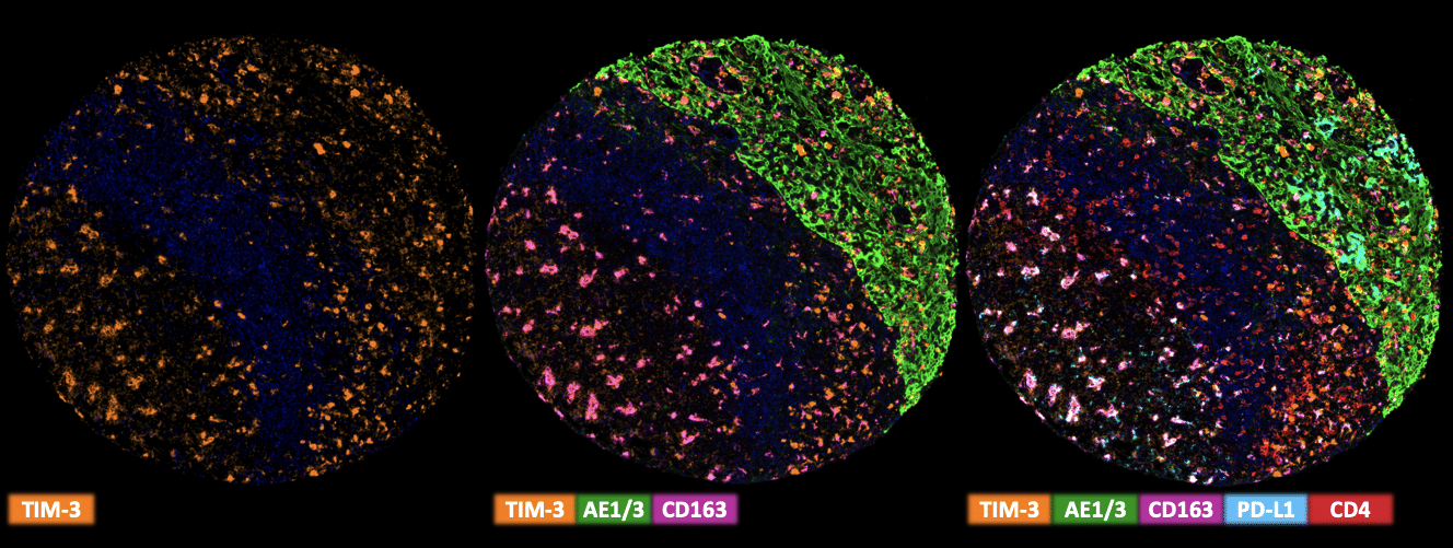
Multiplex Fluorescence Immunohistochemistry
Freshly cut sections should be used (less than 10 days between cutting and staining). Slides were initially boiled in an autoclave (30 min at 100–120 °C in pH9 buffer) for antigen retrieval. The antibody panel consists of TIM3 (MSVA-366R – 1:150), Pan, Cytokeratin (MSVA-000R – 1:150), PD-L1 (MSVA-711R – 1:150), CD4 (MSVA-004R – 1:150) & CD163 (MSVA-163M – 1:150). The OPAL dye kit (Cat# NEL811001KT, AKOYA Biosciences, Menlo Park, California, United States) according to the manufacturer’s directions was used to detect the primary antibodies.
Potential Research Applications
- TIM-3 is an important immune checkpoint, the function of which deserves further evaluation.
- As TIM-3 can be expressed in epithelial cells (kidney, epididymis), the expression of TIM-3 in tumor cells of cancerous tissues needs to be investigated.
- The clinical significance of the density of TIM-3 positive lymphocytes and macrophages needs to be investigated.
Evidence for Antibody Specificity in IHC
There are two ways, how the specificity of antibodies can be documented for immunohistochemistry on formalin fixed tissues. These are: 1. comparison with a second independent method for target expression measurement across a large number of different tissue types (orthogonal strategy), and 2. Comparison with one or several independent antibodies for the same target and showing that all positive staining results are also seen with other antibodies for the same target (independent antibody strategy).
As a standard validation process for MSVA antibodies, RNA data summarized in the protein atlas are used for comparison. However, this has limited validity for proteins that are expressed in significant subsets of lymphocytes and macrophages because these cells occur in virtually all organs. The validation of MSVA-366R is thus based on a comparison with other antibodies.
The MSVA-366R findings of a significant TIM-3 protein expression in:
- Kupffer cells of the liver
- epididymis
- spleen vessels
- and the kidney were confirmed by positive staining with the antibody CAB026003 shown in the human protein atlas.
As a staining of surface epithelium of small intestine was not seen by using CAB026003 we consider this a potential cross-reactivity. As this staining is strictly limited to the small intestine, we consider it tolerable.
Moreover, no staining was seen in tissues notorious for non-specific IHC background such as colonic mucosa, and epidermis.
