
195,00 € – 695,00 €
Product details
Synonyms = CMD1I; CSM1; CSM2; DES; Intermediate filament protein
Antibody type = Recombinant Mouse monoclonal / IgG
Clone = MSVA-651M
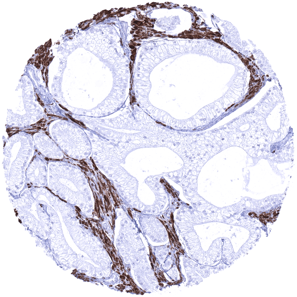
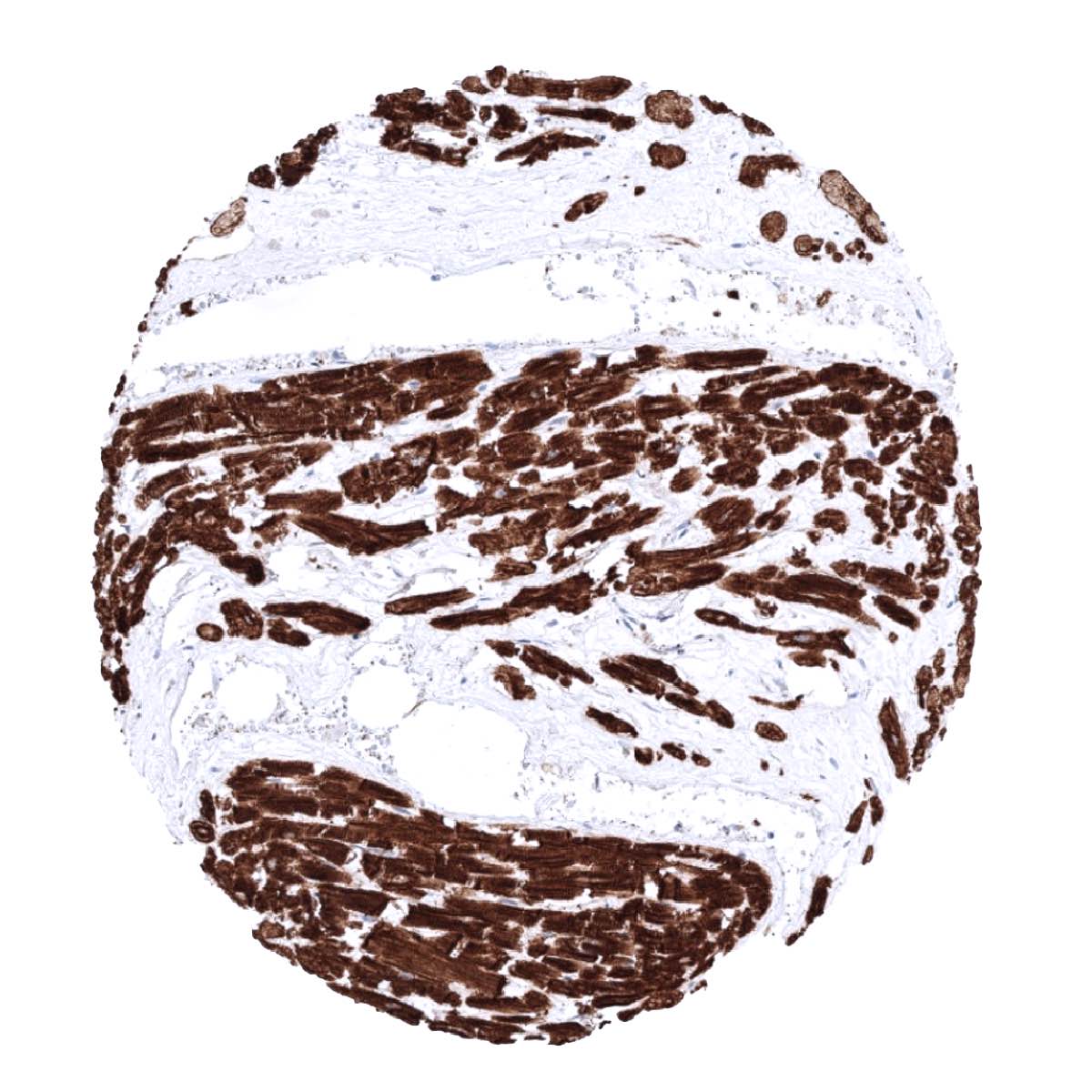
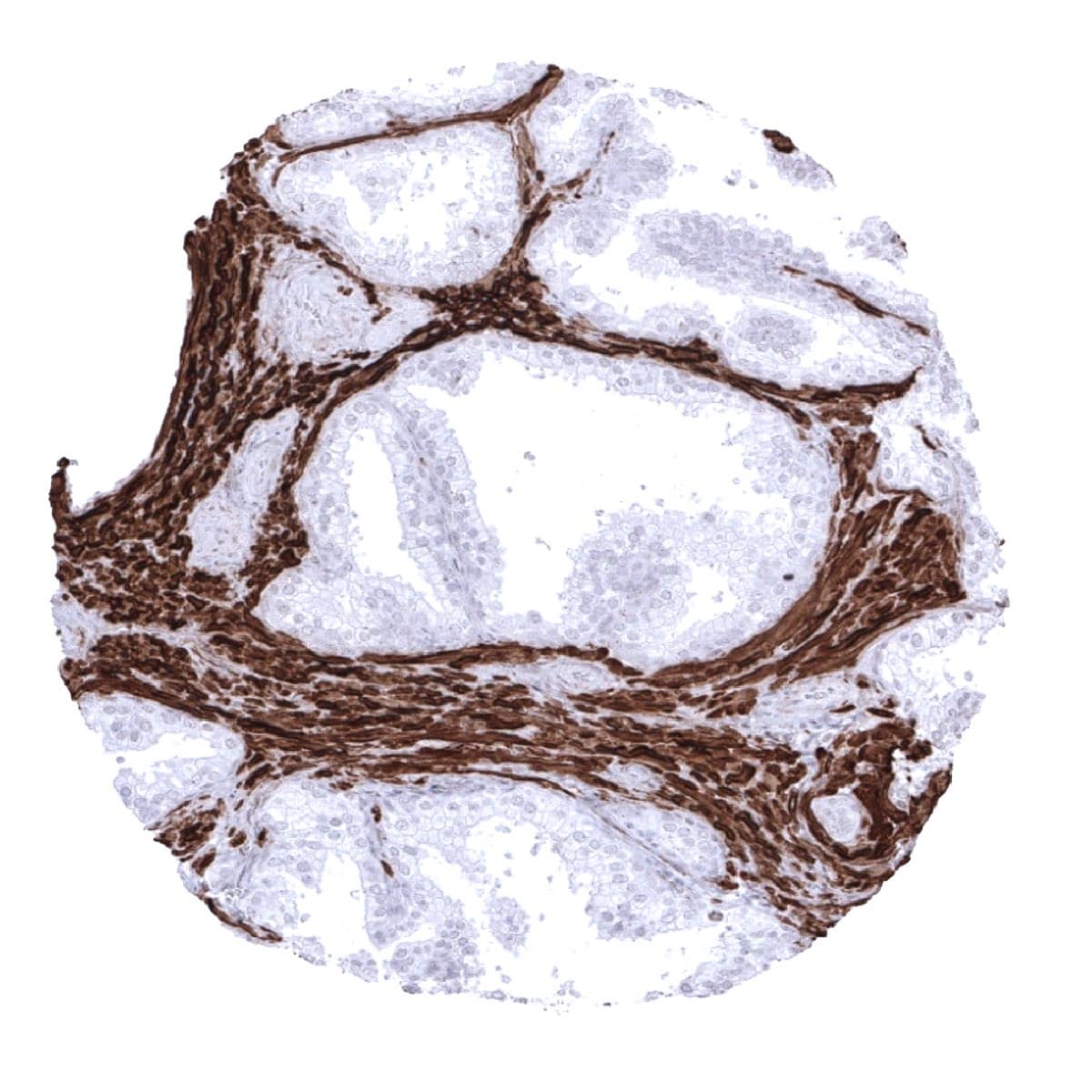
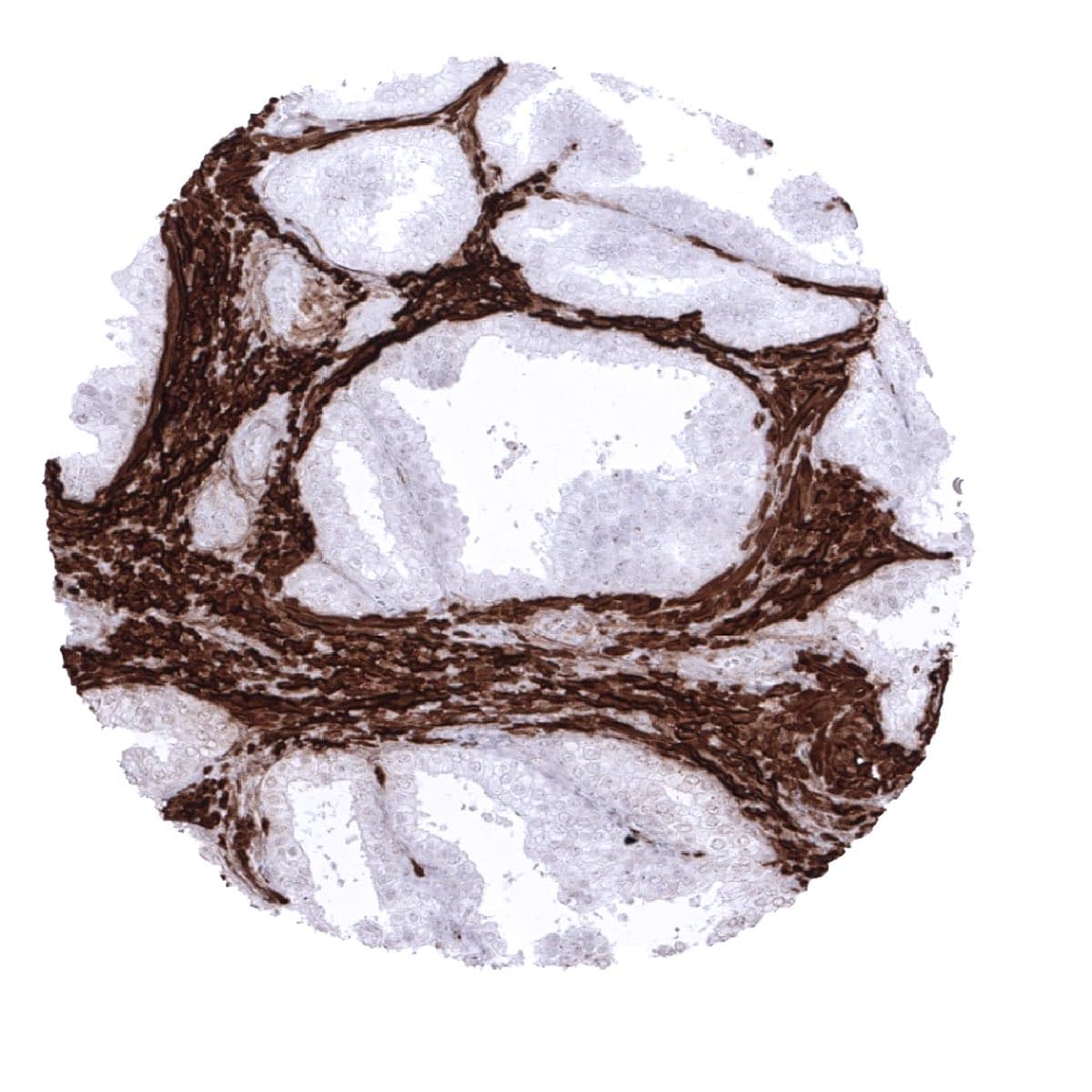
Positive control = Appendix: Smooth muscle cells in the lamina muscularis mucosae and muscularis propria must show a strong cytoplasmic staining while staining should be at least moderate in most smooth muscle cells of blood vessels and in dispersed myofibroblasts.
Negative control = Appendix: Desmin staining should not be seen in lymphocytes and epithelial cells.
Cellular localization = Cytoplasmic
Reactivity = Human
Application = Immunohistochemistry
Dilution = 1:100 – 1:200
Intended Use = Research Use Only
Relevance of Antibody
Desmin is expressed in all muscle cells and cells undergoing myoid differentiation.
Biology Behind
Desmin is a 53.5 kD protein coded by the DES gene at 2q35. Desmin is a muscle-specific subunit of intermediate filaments that does not play a role in contractility but organizes sarcomere architecture. Desmin is expressed in all striated muscle cells and most smooth muscle cells. In the developing striated muscle cells, desmin gradually replaces vimentin. In myofibroblasts and vascular smooth muscle cells, desmin is co-expressed with vimentin. In arterial smooth muscle cells, desmin may be scarce or absent while vimentin expression is retained. Desmin has a pivotal role in muscle function. The accumulation of cleaved and misfolded desmin is a critical cellular feature of heart failure which can be potentially reversed by therapy. Desmin mutations have been associated with various subtypes of cardiomyopathies and with myopathies.
Staining Pattern in Normal Tissues
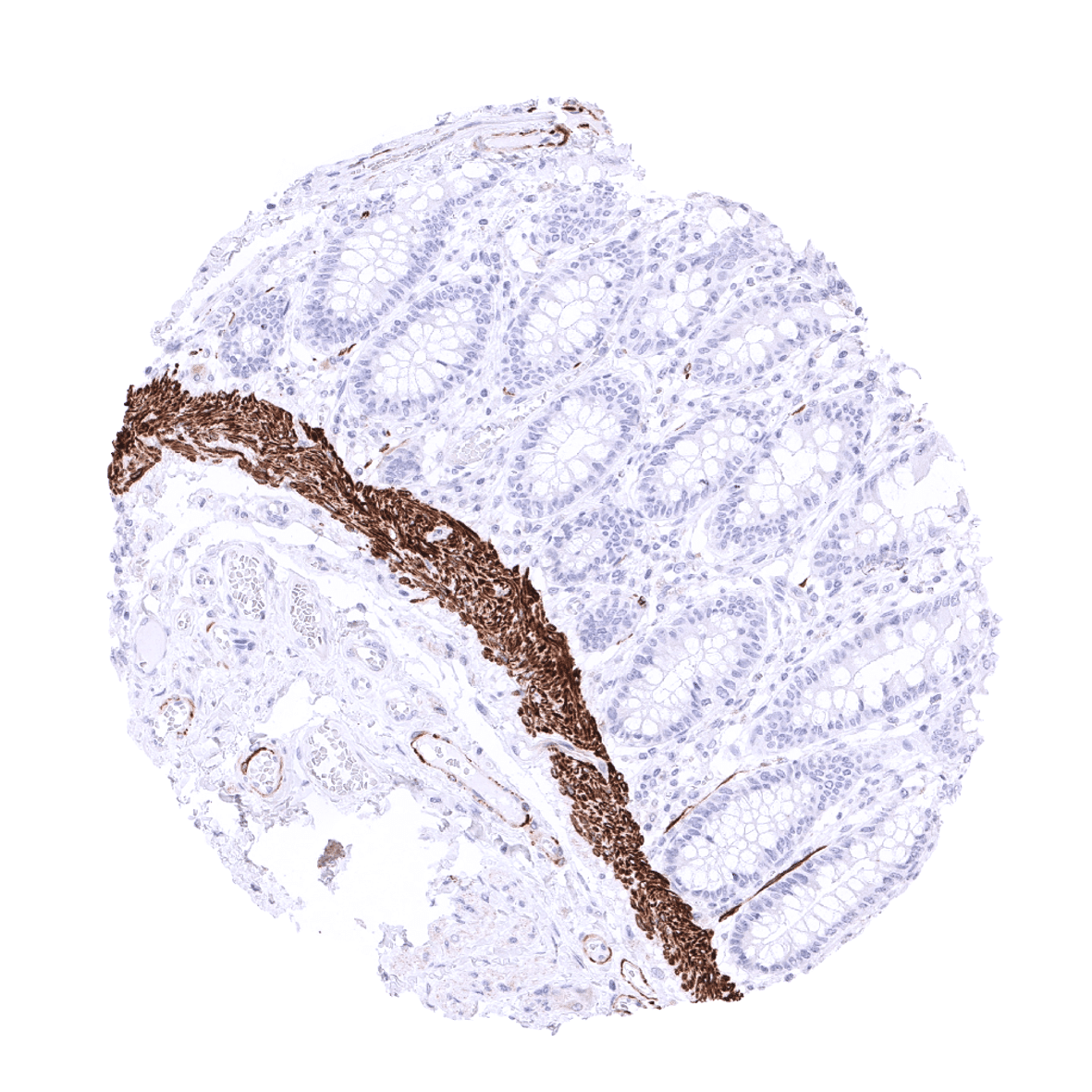
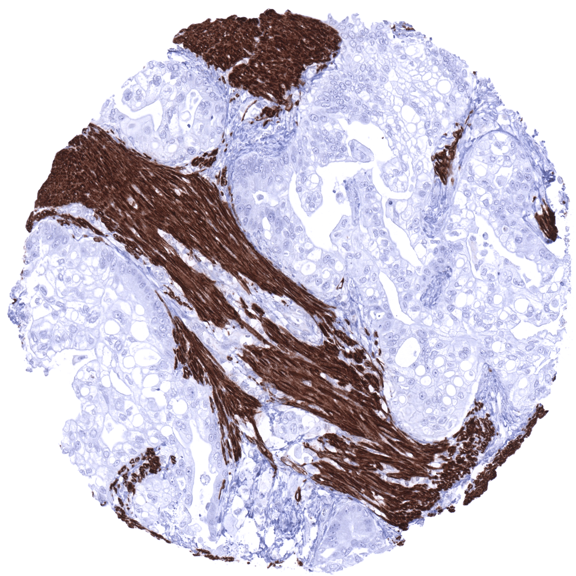
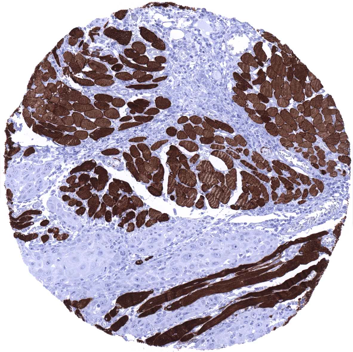
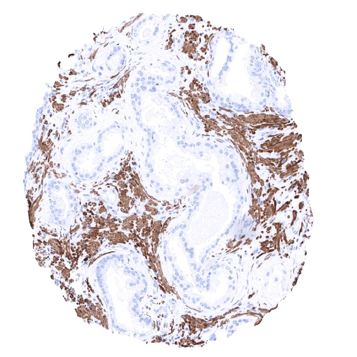
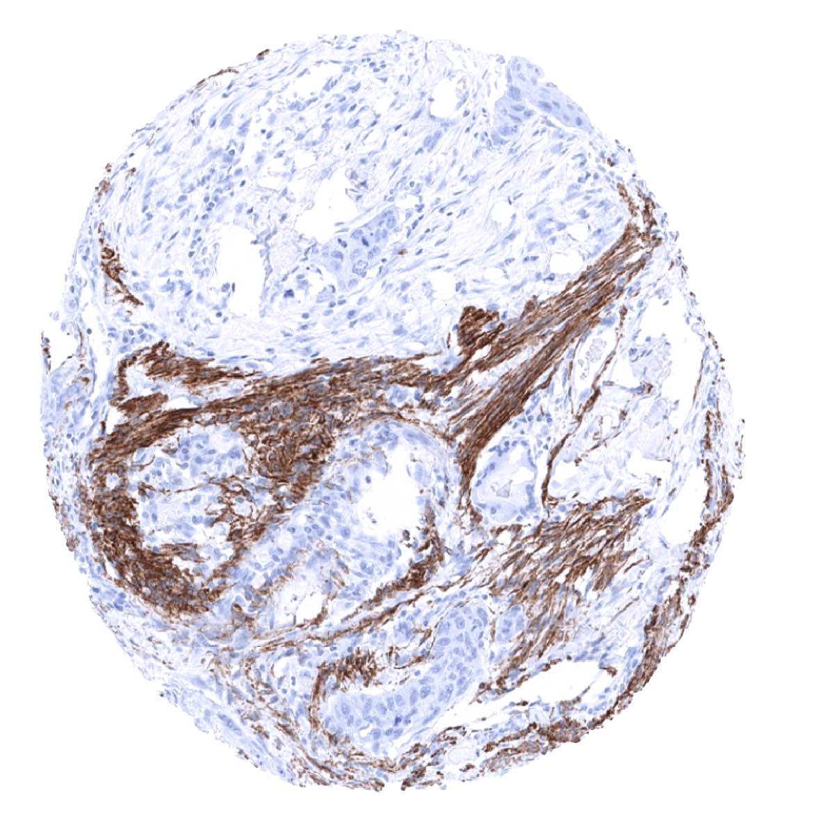
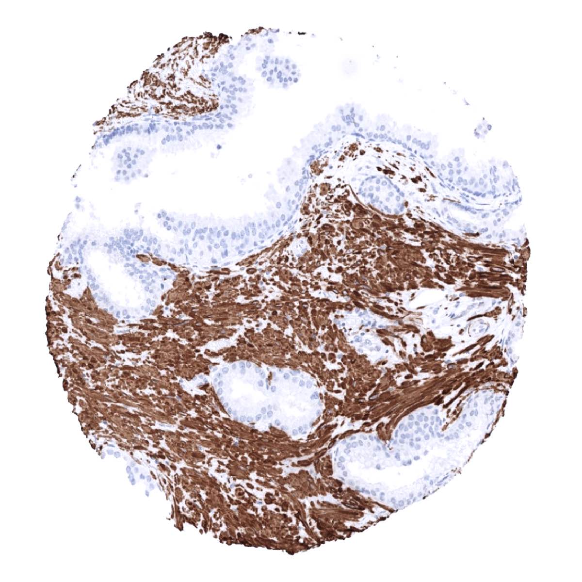
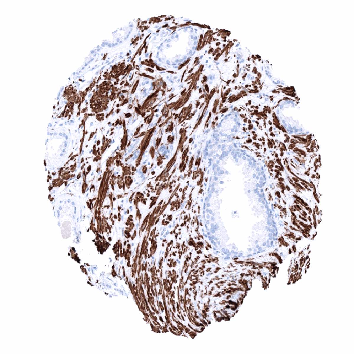
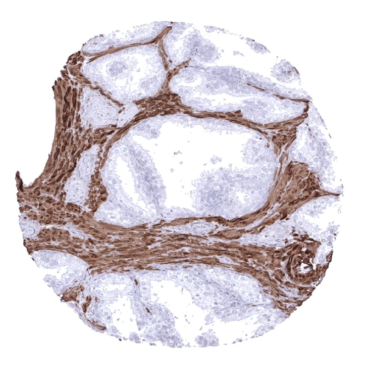
Desmin immunostaining is seen in smooth muscle, skeletal muscle and in heart muscle. In the heart, an accentuation of staining is seen at the intercalated disks. Smooth muscle staining is diminished in arteriolar blood vessels, where it is often limited to a subset of fibres. Desmin immunostaining is also seen in myofibroblasts and in placenta stroma cells.
These findings are largely comparable to the RNA and protein data described in the Human Protein Atlas (Tissue expression Desmin) with the exception of placenta stroma cell staining. RNA data in the protein atlas do not suggest an RNA expression in the placenta. However, placenta stroma cell staining is confirmed by immunostaining with the antibody CAB000034 shown in the protein atlas.
Positive control: Appendix: Smooth muscle cells in the lamina muscularis mucosae and muscularis propria must show a strong cytoplasmic staining while staining should be at least moderate in most smooth muscle cells of blood vessels and in dispersed myofibroblasts.
Negative control: Appendix: Desmin staining should not be seen in lymphocytes and epithelial cells.


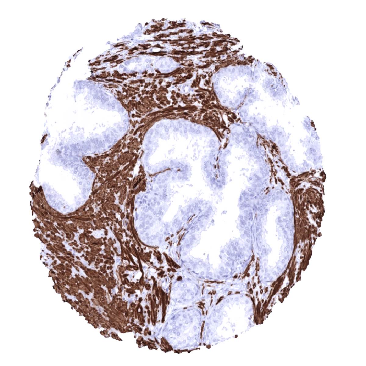
Staining Pattern in Relevant Tumor Types
Desmin is almost always expressed in tumors of myogenic origin such as leiomyoma, leiomyosarcoma and rhabdomyosarcoma. Poorly differentiated myogenic tumours may loose desmin expression. Triton tumor, glomus tumour, alveolar soft part sarcoma, and PEComa are often desmin positive. Other malignant mesenchymal tumors such as fibrosarcoma, liposarcoma and carcinosarcoma often show focal desmin positivity reflecting focal myogenic differentiation. A few non-myogenic tumors such as lung adenocarcinoma, malignant melanoma and gliomas have been reported to occasionally express desmin. Gastrointestinal stromal tumour (GIST) is almost always desmin negative.
The TCGA findings on Desmin RNA expression in different tumor categories have been summarized in the Human Protein Atlas.

Compatibility of Antibodies
No data available at the moment
Protocol Recommendations
IHC users have different preferences on how the stains should look like. Some prefer high staining intensity of the target stain and even accept some background. Others favor absolute specificity and lighter target stains. Factors that invariably lead to more intense staining include higher concentration of the antibody and visualization tools, longer incubation time, higher temperature during incubation, higher temperature and longer duration of the heat induced epitope retrieval (slide pretreatment). The impact of the pH during slide pretreatment has variable effects and depends on the antibody and the target protein.
All images and data shown here and in our image galleries are obtained by the manual protocol described below. Other protocols resulting in equivalent staining are described as well.
Manual protocol
Freshly cut sections should be used (less than 10 days between cutting and staining). Heat-induced antigen retrieval for 5 minutes in an autoclave at 121°C in pH 9,0 Target Retrieval Solution buffer. Apply MSVA-651M at a dilution of 1:150 at 37°C for 60 minutes. Visualization of bound antibody by the EnVision Kit (Dako, Agilent) according to the manufacturer’s directions.


Agilent / Dako – Autostainer Link 48
Pretreatment in PT-Link for 30 minutes at 95°C (pH high); FLEX peroxidase blocking for 5 minutes (room temperature), MSVA-651M 1:150 for 20 minutes (room temperature), FLEX+ mouse/rabbit (LINKER) for 15 minutes (room temperature), horseradish peroxidase (HRP) for 20 minutes (room temperature), FLEX DAB+Sub-Chromo for 10 minutes (room temperature), FLEX hematoxylin for 5 minutes (room temperature).

These images reflect stainings by the protocol described above. It is of note that a comparable staining result can also be obtained by different protocols. In general, a longer pretreatment, a longer incubation time of the primary antibody, a higher antibody concentration, and a longer incubation time of FLEX+LINKER result in stronger staining, potentially at the cost of more background staining. Modifications of the protocol with a strengthening effect on staining intensity in combination with changes of other parameters that result in lower staining intensity can result in a comparable result as shown above.
Leica – BOND RX
Dewax at 72°C for 30 seconds; Pretreatment in Bond Epitope Retrieval Solution (ER2 – EDTA pH9) for 20 minutes at 100°C; Peroxidase blocking for 5 minutes (room temperature), MSVA-651M 1:150 for 15 minutes (room temperature), Post primary (rabbit anti mouse) for 8 minutes (room temperature), Polymer (goat anti rabbit) for 8 minutes (room temperature), mixed DAB refine for 10 minutes (room temperature), hematoxylin for 5 minutes (room temperature).

These images reflect stainings by the protocol described above. It is of note that a comparable staining result can also be obtained by different protocols. In general, a longer pretreatment, a longer incubation time of the primary antibody, a higher antibody concentration, a higher temperature during incubation, and a longer incubation time of Post primary and or the Polymer result in stronger staining, potentially at the cost of more background staining. Modifications of the protocol with a strengthening effect on staining intensity in combination with changes of other parameters that result in lower staining intensity can result in a comparable result as shown above.
Roche – Ventana Discovery ULTRA
Pretreatment for 64 minutes at 100°C (pH 8,4); CM peroxidase blocking for 12 minutes (room temperature), MSVA-651M 1:50 for 40 minutes at 36°C, secondary antibody (anti-mouse HQ) for 12 minutes at 36°C, anti-HQ HRP for 12 minutes at room temperature, DAB at room temperature, hematoxylin II at room temperature for 8 minutes, bluing reagent at room temperature for 4 minutes.


These images depict staining results obtained by the protocol described above. It is of note, that the Ventana machines generally require higher antibody concentrations than other commonly used autostainers because the antibodies are automatically diluted during the procedure. Various other protocols can result in an identical result as shown above. A longer pretreatment, a longer incubation time of the primary antibody, a higher antibody concentration, a higher temperature during incubation, and a longer incubation time of secondary antibody and or the anti-HQ HRP result in stronger staining, potentially at the cost of more background staining.
Impact of pH
The strongest Desmin staining by MSVA-651M is obtained at a pH 9,0. However, pH 7,8 results in only a slight reduction of the staining intensity as compared to pH9.
Potential Research Applications
- Desmin plays a pivotal role in muscular function, especially in the heart. The role of desmin in heart failure is under investigation.
- The prevalence and role of desmin expression in non-myogenic tumors has not been sufficiently clarified.
Evidence for Antibody Specificity in IHC
There are two ways how the specificity of antibodies can be documented for immunohistochemistry on formalin fixed tissues. These are: 1. comparison with a second independent method for target expression measurement across a large number of different tissue types (orthogonal strategy), and 2. Comparison with one or several independent antibodies for the same target and showing that all positive staining results are also seen with other antibodies for the same target (independent antibody strategy).
Orthogonal validation: For the antibody MSVA-651M specificity is suggested by the strong concordance of the immunostaining with RNA expression data derived from the Human Protein Atlas (HPA) RNA-seq tissue dataset, the FANTOM5 project, and the Genotype-Tissue Expression (GTEx) project which are summarized in the Human Protein Atlas (Tissue expression Desmin). Immunostaining by using MSVA-651M was almost exclusively detected in organs with documented RNA expression. In addition, positive immunostaining was also found in placenta stroma cells. These structures constitute small subsets of the total amount of cells in these organs and may have been largely underrepresented in RNA analyses.
Comparison of antibodies: True desmin expression in placental stroma cells is suggested by similar findings obtained by the use of the antibody CAB000034.
