295,00 € – 995,00 €
Product details
Synonyms = G protein-coupled receptor class C group 5 member A , GPCR5A , PEIG-1 , RAI3 , RAIG1 , TIG1
Antibody type = Recombinant Rabbit monoclonal / IgG
Clone = HMV4717
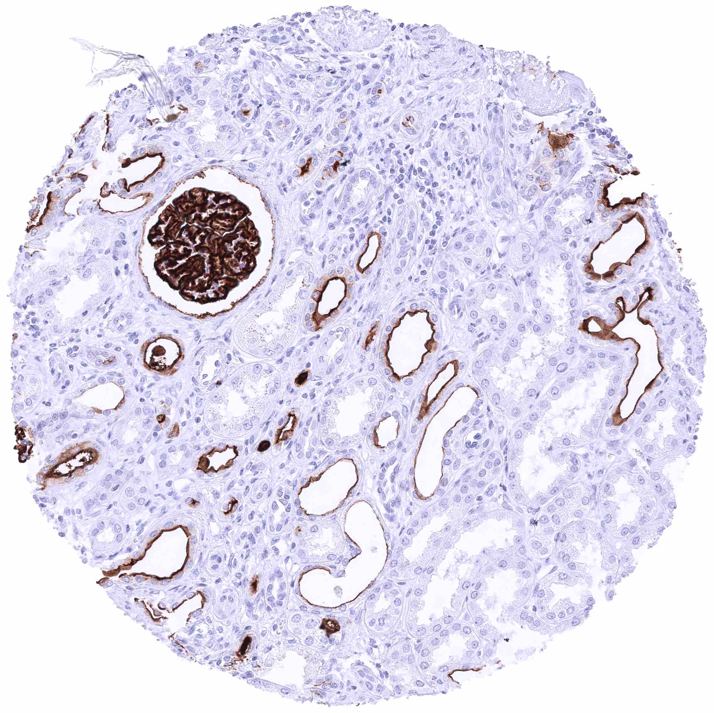
Positive control = Kidney: GRRC5A staining must be intense in glomeruli, strong at the surface membranes of a fraction of tubuli, and moderate at the parietal layer of the Bowman’s capsule while GPRC5A is absent in another subset of tubuli.
Negative control = Tonsil: GRRC5A staining should be completely absent in lymphocytes while it is seen in a subset of epithelial cells.
Cellular localization = Membraneous
Reactivity = Human
Application = Immunohistochemistry
Dilution = 1:100 – 1:200
Intended Use = Research Use Only
Relevance of Antibody
GPRC5A is a surface protein with aberrant expression in cancer.
Biology Behind
G Protein-coupled receptor class C group 5 member A (GPRC5A) is a member of the GPCR superfamily, which constitutes one of the largest potentially druggable family of membrane proteins. The transmembranous protein is coded by the GPRC5A gene on chromosome 12p13.1. Its molecular function is not well known. GPRC5A knock-out mice have altered lung morphology and suffer from increased susceptibility to tumor formation, especially in the lung. Altered function of GPRC5A has been linked to several disease types such as diabetic nephropathy, endotoxin-induced acute lung injury, and chronic airway inflammation, but most of the current interest in GPRC5A comes from studies implicating a role in cancer. The GPRC5A promoter region contains binding sites for several important cancer genes such as p53, MYC, and BRCA1. Moreover, GPRC5A expression has been suggested to promote BRCA1 expression and to inhibit NF-κB, Gsα, MCM2 and CCND1 expression although exact mechanisms have not been revealed and conclusions are partly based on indirect evidence. Both reduced expression and upregulation of GPRC5A has been described in several cancer types and aberrant GPRC5A levels have been linked to cancer aggressiveness and chemoresistance. Because of its membranous location, GPRC5A constitutes a potential drug target.
Staining Pattern in Normal Tissues
GPRC5A staining predominates in epithelial cell types. It is strong in renal glomeruli, chorion cells of the placenta, and alveolar pneumocytes. In many epithelial cell types, the staining only occurs at lumina/apical surfaces. GPRC5A staining is also seen in mesothelial cells.
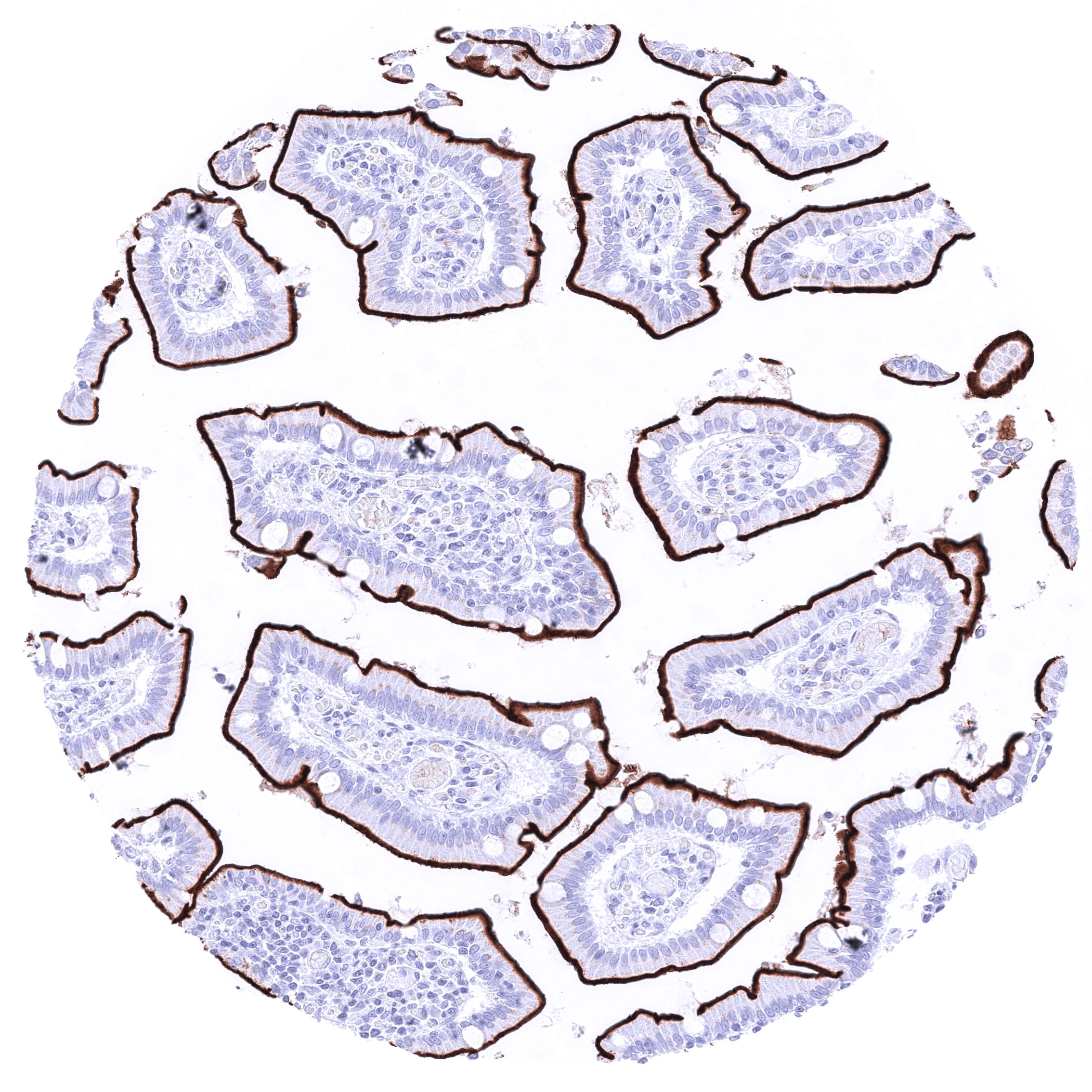
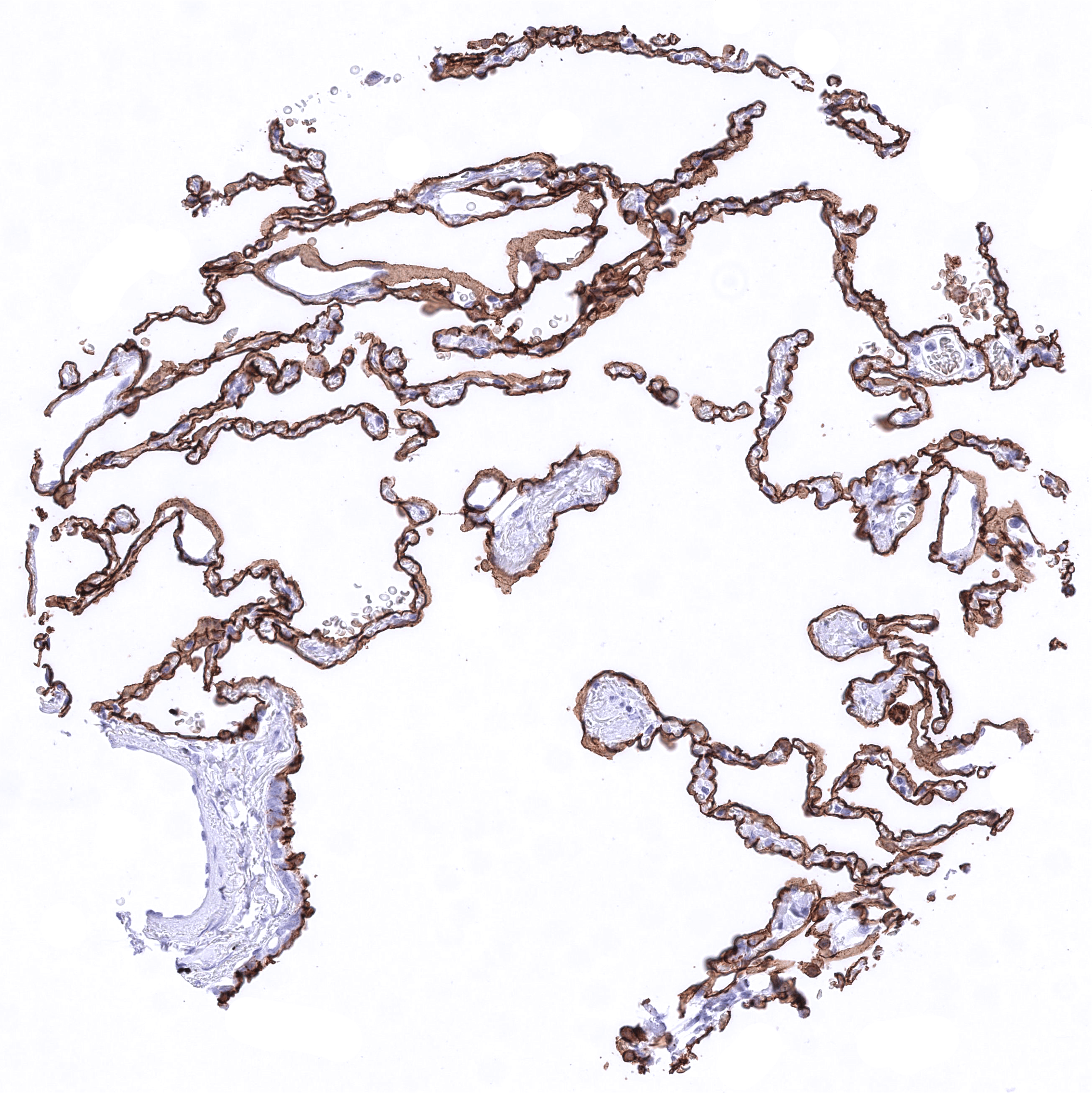
Images describing the GPM6A staining pattern in normal tissues obtained by the antibody HMV4717 are shown in our “Normal Tissue Gallery”.
| Brain | Cerebrum | Negative. |
| Cerebellum | Negative. | |
| Endocrine Tissues | Thyroid | Strong GPRC5A staining of the luminal membranes of thyreocytes. |
| Parathyroid | Negative. | |
| Adrenal gland | Distinct membranous GPRC5A staining of groups of cells (not in all samples). | |
| Pituitary gland | Negative. | |
| Respiratory system | Respiratory epithelium | Membranous GPRC5A staining of basal cells and moderate GPRC5A staining of luminal membranes in the respiratory epithelium of the lung. |
| Lung | Strong membranous GPRC5A staining of all alveolar pneumocytes. | |
| Gastrointestinal Tract | Salivary glands | Strong GPRC5A staining of luminal membranes of excretory duct cells. A less intensive staining is also seen in variable subsets of glands. |
| Esophagus | Variable patterns of membranous GPRC5A staining ranging from limited staining of the superficial layers to staining of all cell layers of the squamous epithelium. | |
| Stomach | Strong GPRC5A membranous staining, largely limited to the luminal membranes. The staining intensity is higher at the surface than in glands. Especially in deep glands, the membranous GPRC5A appears to be incomplete. | |
| Duodenum | Strong GPRC5A membranous staining, largely limited to the luminal membranes. The staining intensity slightly decreases from the surface to the glands. Strong GPRC5A staining, predominantly at the luminal membranes of Brunner gland cells. | |
| Small intestine | Strong GPRC5A membranous staining, largely limited to the luminal membranes. The staining intensity slightly decreases from the surface to the glands. | |
| Appendix | Strong GPRC5A membranous staining, largely limited to the luminal membranes. The staining intensity slightly decreases from the surface to the glands. | |
| Colon | Strong GPRC5A membranous staining, largely limited to the luminal membranes. The staining intensity slightly decreases from the surface to the glands. | |
| Rectum | Strong GPRC5A membranous staining, largely limited to the luminal membranes. The staining intensity slightly decreases from the surface to the glands. | |
| Liver | Negative. | |
| Gallbladder | Variable, weak to strong membranous GPRC5A staining limited to the luminal membranes of a subset of cells. | |
| Pancreas | Weak to moderate GPRC5A staining of luminal membranes of cells from small excretory ducts. A focal strong GPRC5A staining can also occur in very small subset of possible acinar cells. | |
| Genitourinary | Kidney | Strong GPRC5A staining of podocytes and membranes of the Bowman capsule. A moderate to strong luminal membranous GPRC5A staining also occurs in a fraction of tubuli (predominantly distal) and collecting ducts. |
| Urothelium | Variable GPRC5A staining patterns ranging from a strong membranous positivity of all cells to a predominant staining of the basal/suprabasal cells accompanied by a strong positivity of the luminal membrane of the umbrella cell layer. | |
| Male genital | Prostate | A GPRC5A staining of variable intensity (weak to strong) can be seen on (mostly small) subsets of luminal and basal cells. It preferably occurs at luminal membranes in areas of atrophy. |
| Seminal vesicles | Moderate to strong membranous GPRC5A staining of luminal membranes of a subset of epithelial cells. | |
| Testis | A variable, faint to moderate, predominantly membranous staining of some Leydig cells can occur. | |
| Epididymis | Negative. | |
| Female genital | Breast | Strong GPRC5A staining of apical membranes of luminal cells. |
| Uterus, myometrium | Negative. | |
| Uterus, ectocervix | Variable patterns of membranous GPRC5A staining ranging from absence of staining to a distinct staining of the superficial (top 35%) layers to staining of the squamous epithelium. | |
| Uterus endocervix | Strong GPRC5A staining of (predominantly) apical membranes of endocervical cells. | |
| Uterus, endometrium | Strong GPRC5A staining of apical membranes of endometrium gland cells. GPRC5A This staining may be more intense in proliferative than in secreting endometrium. During pregnancy the staining may also be lower than in proliferating glands, and here is variability between glands. Variable intensity membranous positivity of some decidua cells. | |
| Fallopian Tube | Variable, weak to strong membranous GPRC5A staining limited to the luminal membranes of a subset of epithelial cells. | |
| Ovary | Stroma cells show a moderate to strong perinuclear “dot-like” GPRC5A staining and a faint membranous GPRC5A staining of subsets of cells. A dot-like” GPRC5A staining also occurs in theca interna cells around follicular cysts. | |
| Placenta early | Negative. | |
| Placenta mature | A distinct membranous staining of the luminal membrane of syncytiotrophoblast cells can focally be seen in areas of placenta tissue damage (not in all samples). | |
| Amnion | Strong GPRC5A staining of amnion cells, predominantly at the luminal membrane. | |
| Chorion | Strong, predominantly membranous GPRC5A staining of chorion cells. | |
| Skin | Epidermis | A faint membranous GPRC5A staining of a fraction of basal/suprabasal squamous epithelial cells. |
| Sebaceous glands | Negative. | |
| Muscle/connective tissue | Heart muscle | Negative. |
| Skeletal muscle | Negative. | |
| Smooth muscle | Negative. | |
| Vessel walls | Negative. | |
| Fat | Negative. | |
| Stroma | Negative. | |
| Endothelium | Negative. | |
| Bone marrow/ lymphoid tissue | Bone marrow | Negative. |
| Lymph node | Negative. | |
| Spleen | Negative. | |
| Thymus | GPRC5A staining is limited to corpuscles of Hassall’s. | |
| Tonsil | GPRC5A staining is limited to squamous epithelial cells. | |
| Remarks | Distinct membranous GPRC5A staining of the luminal membrane of mesothelial cells. |
The findings described above are thus consistent with the RNA data described in the Human Protein Atlas (Tissue expression GPRC5A).
Positive control = Kidney: GRRC5A staining must be intense in glomeruli, strong at the surface membranes of a fraction of tubuli, and moderate at the parietal layer of the Bowman’s capsule while GPRC5A is absent in another subset of tubuli.
Negative control = Tonsil: GRRC5A staining should be completely absent in lymphocytes while it is seen in a subset of epithelial cells.
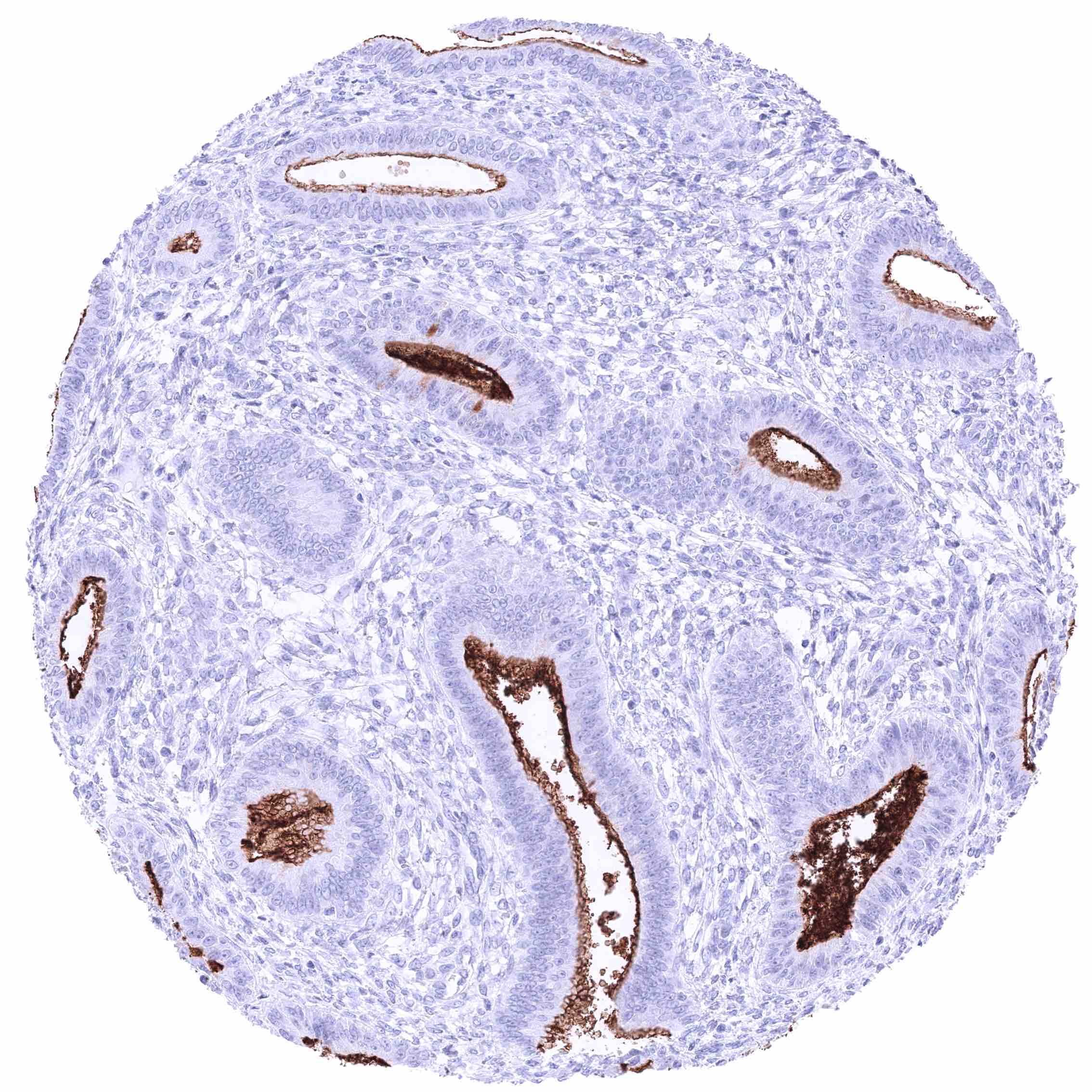
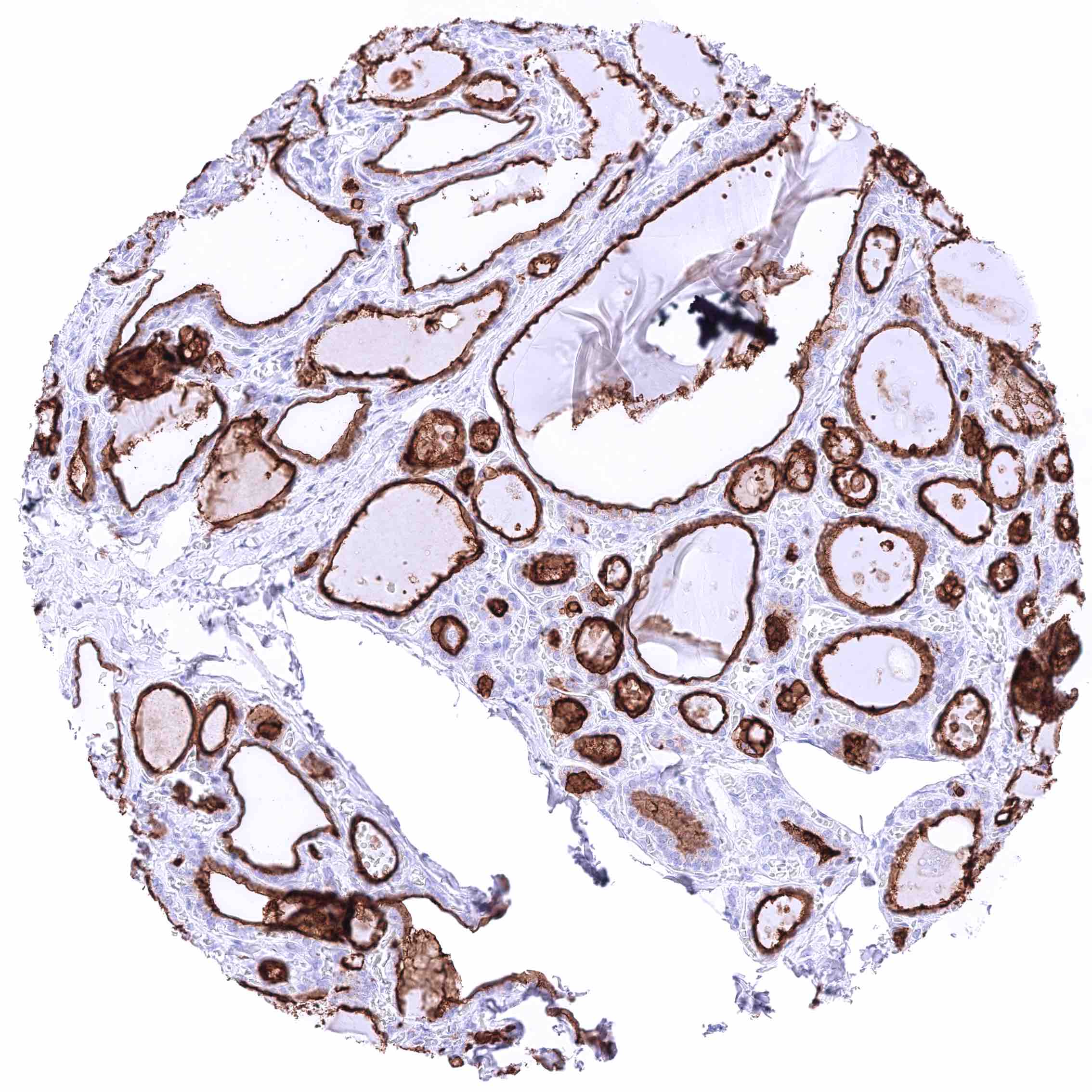
Staining Pattern in Relevant Tumor Types
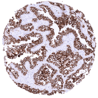
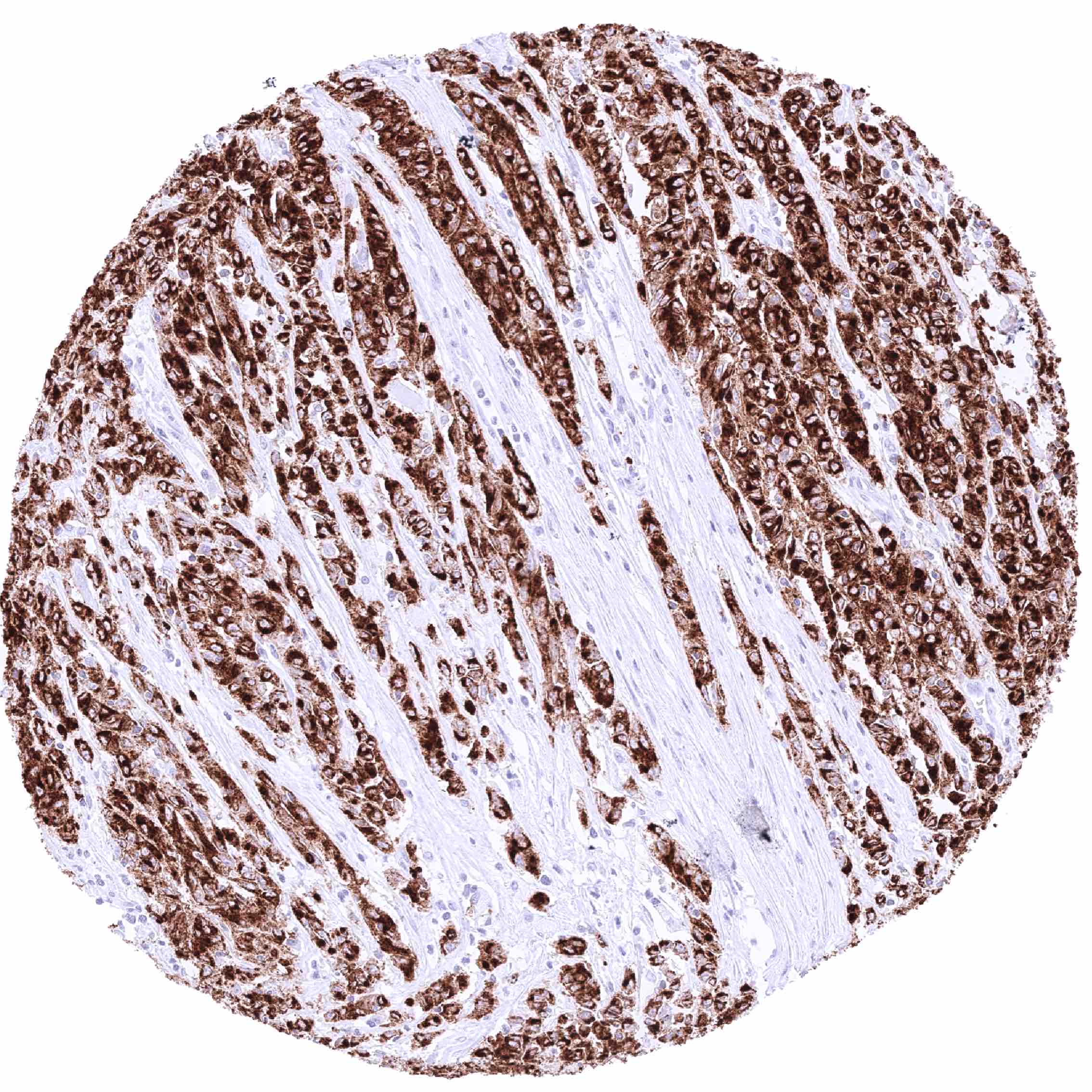
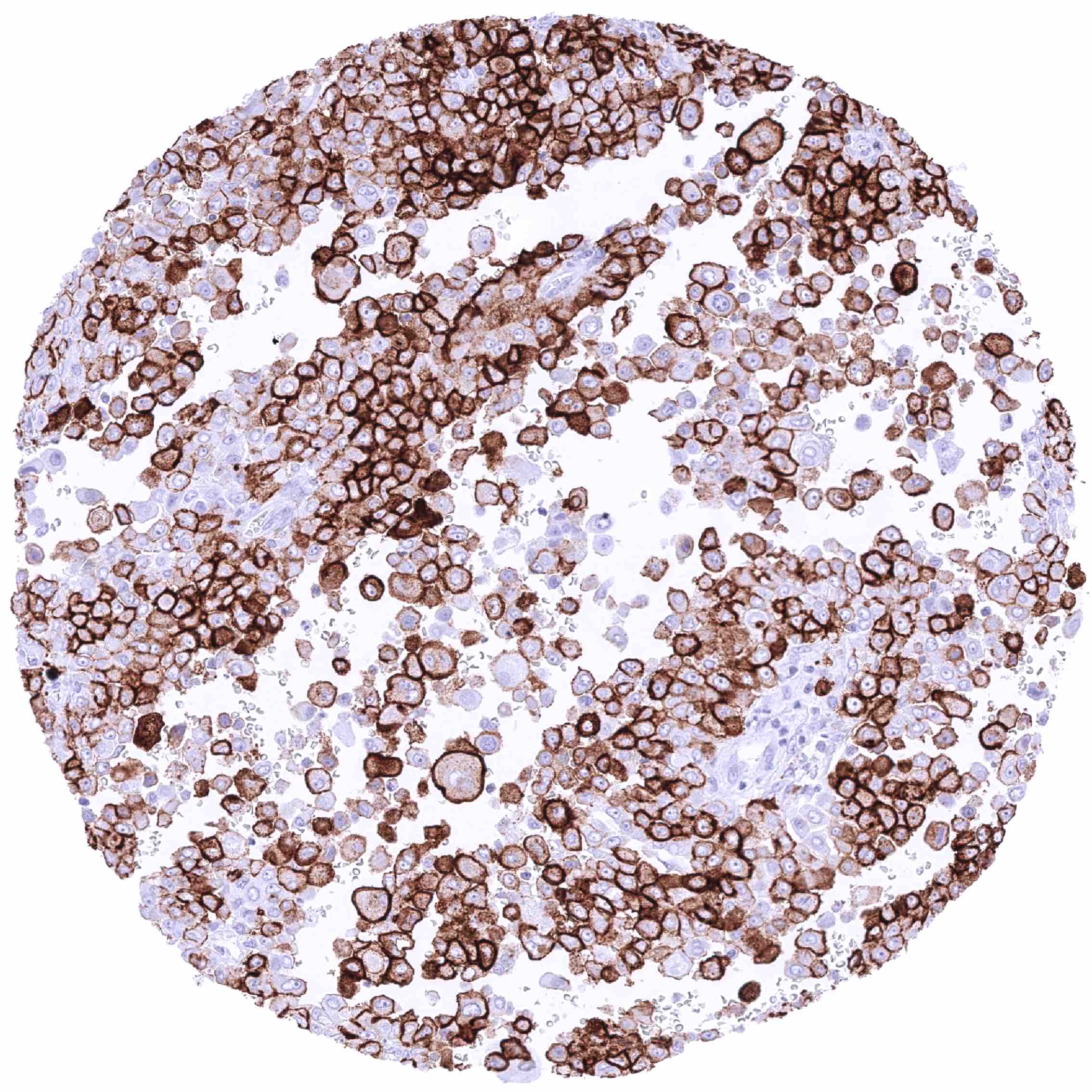
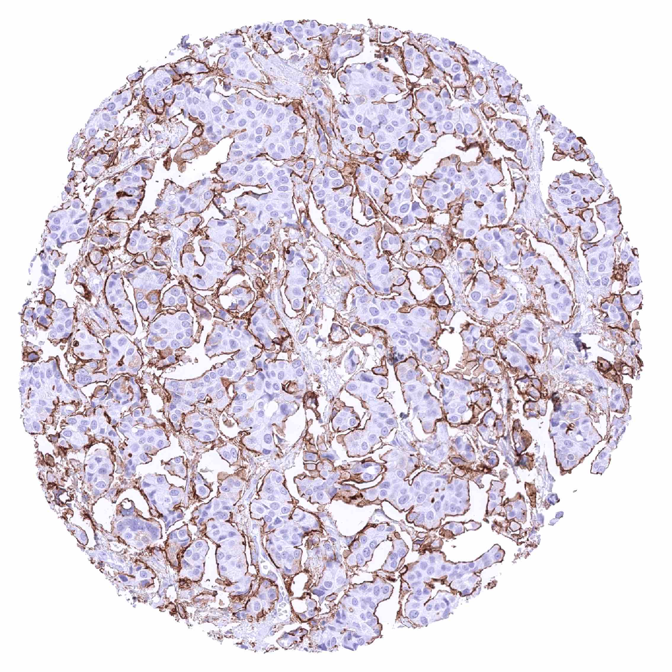
Based on RNA expression data, GRRC5A expression occurs in many different cancer types. It might be particularly common in pancreatic and gastrointestinal cancers.
The TCGA findings on GPRC5A RNA expression in different tumor categories have been summarized in the Human Protein Atlas.
Compatibility of Antibodies
No data available at the moment
Protocol Recommendations
IHC users have different preferences on how the stains should look like. Some prefer high staining intensity of the target stain and even accept some background. Others favor absolute specificity and lighter target stains. Factors that invariably lead to more intense staining include higher concentration of the antibody and visualization tools, longer incubation time, higher temperature during incubation, higher temperature and longer duration of the heat induced epitope retrieval (slide pretreatment). The impact of the pH during slide pretreatment has variable effects and depends on the antibody and the target protein.
All images and data shown here and in our image galleries are obtained by the manual protocol described below. Other protocols resulting in equivalent staining are described as well.
Manual protocol
Freshly cut sections should be used (less than 10 days between cutting and staining). Heat-induced antigen retrieval for 5 minutes in an autoclave at 121°C in pH 7,8 Target Retrieval Solution buffer. Apply HMV4717 at a dilution of 1:150 at 37°C for 60 minutes. Visualization of bound antibody by the EnVision Kit (Dako, Agilent) according to the manufacturer’s directions.
Potential Research Applications
- Comprehensive data on prevalence of GRRC5A expression in cancer and its clinical significance are lacking.
- The predictive role of GRRC5A expression for response to chemotherapy in cancer needs to be further evaluated.
- The molecular mechanisms underlying GPRC5A’s role in tumor suppression and promotion need to be further clarified?
- Potential approaches to therapeutically target GPRC5A have to be evaluated.
Evidence for Antibody Specificity in IHC
There are two ways how the specificity of antibodies can be documented for immunohistochemistry on formalin fixed tissues. These are: 1. Comparison with a second independent method for target expression measurement across a large number of different tissue types (orthogonal strategy), and 2. Comparison with one or several independent antibodies for the same target and showing that all positive staining results are also seen with other antibodies for the same target (independent antibody strategy).
Orthogonal validation: For the antibody HMV4717 specificity is supported by the good concordance of the immunostaining data with data from three independent RNA screening studies, including the Human Protein Atlas (HPA) RNA-seq tissue dataset, the FANTOM5 project, and the Genotype-Tissue Expression (GTEx) project, which are all summarized in the Human Protein Atlas (Tissue expression GPRC5A). GPRC5A positivity by HMV4717 is particularly strong in the lung, the tissue with the highest GPRC5A RNA expression. Moreover, immunostaining by using HMV4717 was absent in most tissues lacking RNA expression (spleen, lymph node, bone marrow, brain, epididymis, parathyroid, pituitary gland, liver) although rare cell types were IHC positive despite a lack of documented RNA expression in pancreas (small excretory ducts), seminal vesicle (subset of epithelial cells), testis (subset of Leydig cells), and the thymus (corpuscle of Hassall’s), The value of orthogonal limitation is generally limited, however, in case of ubiquitously expressed proteins.
Comparison of antibodies: True expression of GPRC5A in all cell types found GPRC5A positive by HMV4717 is corroborated by identical staining patterns obtained by another commercially available independent antibody (termed “validation antibody”) although the staining by the validation antibody was always less intense.