Login / Register
MS Validated Antibodies - Logo
  • About us
  • Products
    • Primary antibodies
    • Antibody Validation Services
    • Gallery
  • Distributors
  • Validation
    • Antibody Validation Services
    • Tissue Microarrays
  • Publications
    • Large Scale Studies
    • Further Publications
    • Events
    • Quiz
  • Contact
  • About us
  • Products
    • Primary antibodies
    • Antibody Validation Services
    • Gallery
  • Distributors
  • Validation
    • Antibody Validation Services
    • Tissue Microarrays
  • Publications
    • Large Scale Studies
    • Further Publications
    • Events
    • Quiz
  • Contact
×

Normal Tissue Gallery GPRC5A

Path Created with Sketch.
Go to GPRC5A Cancer Tissue GalleryGo back to Product Page
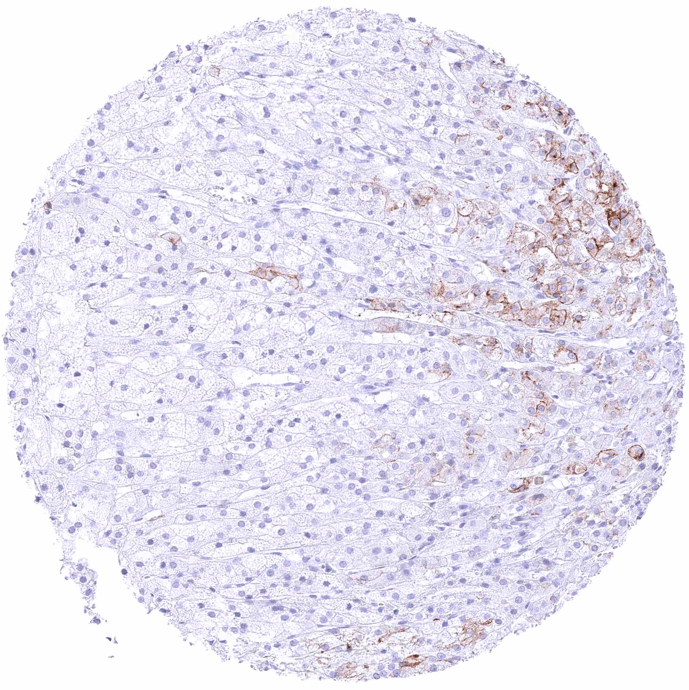
Adrenal gland – Distinct membranous GPRC5A staining of groups of cells (not in all samples).
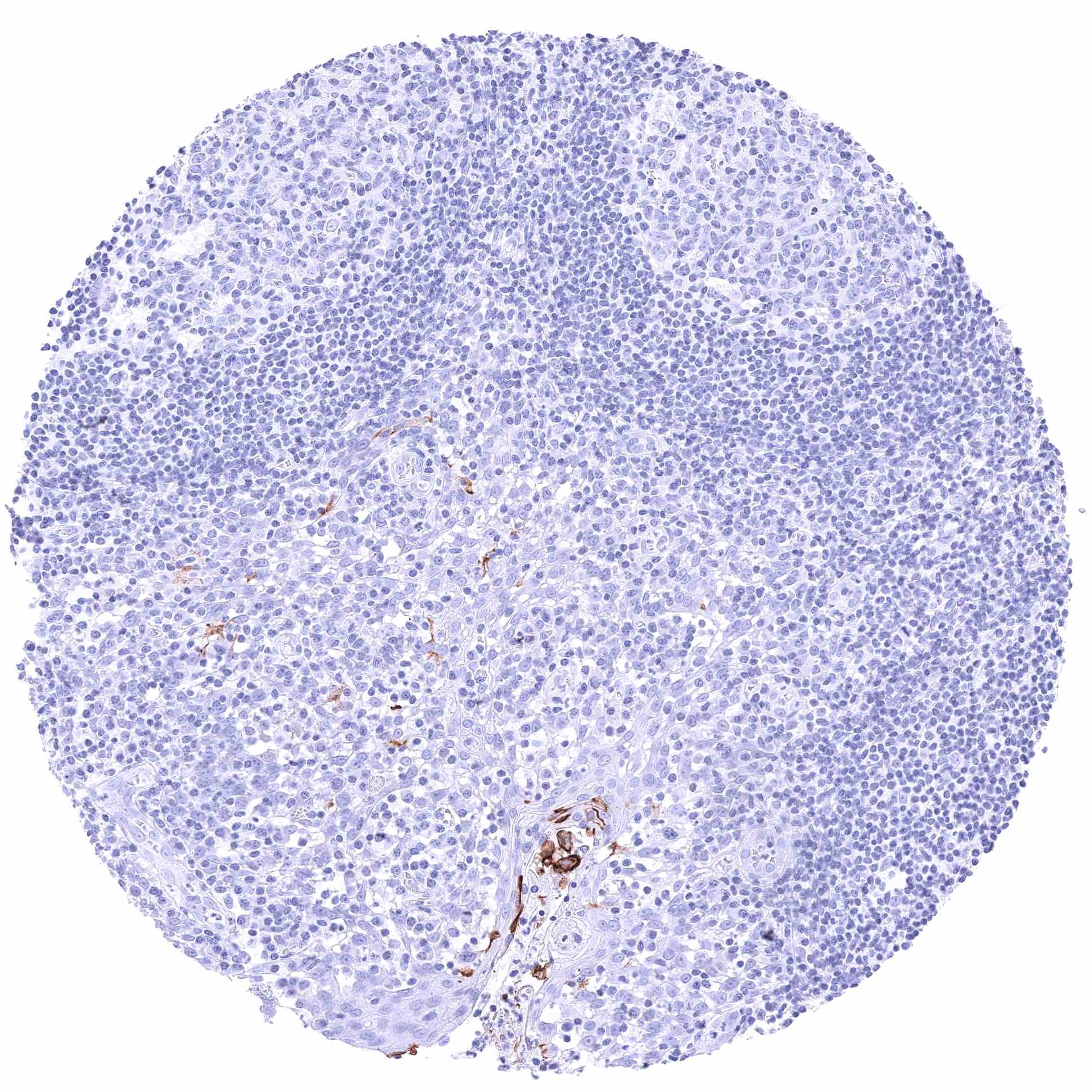
Aorta, media
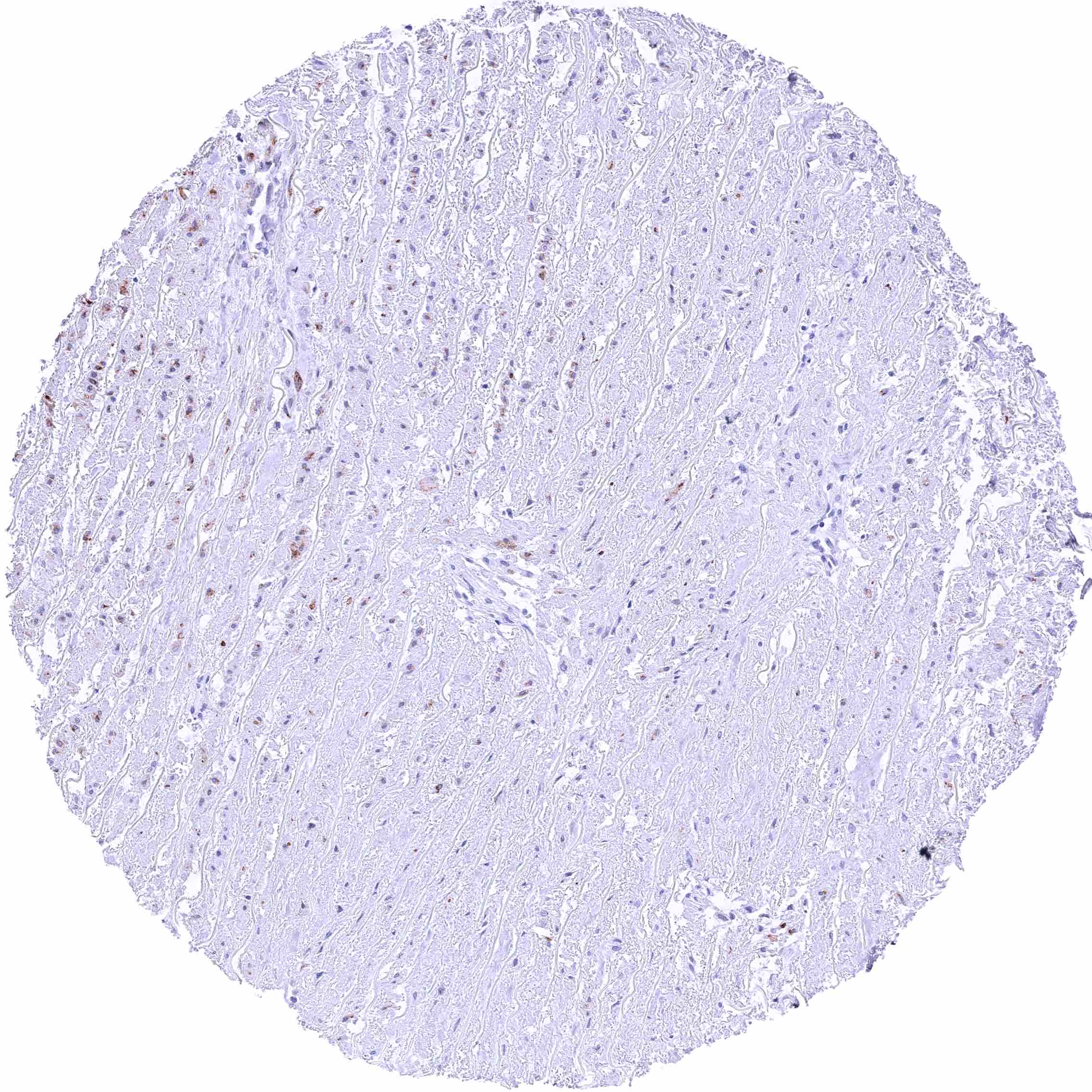
Aorta, media
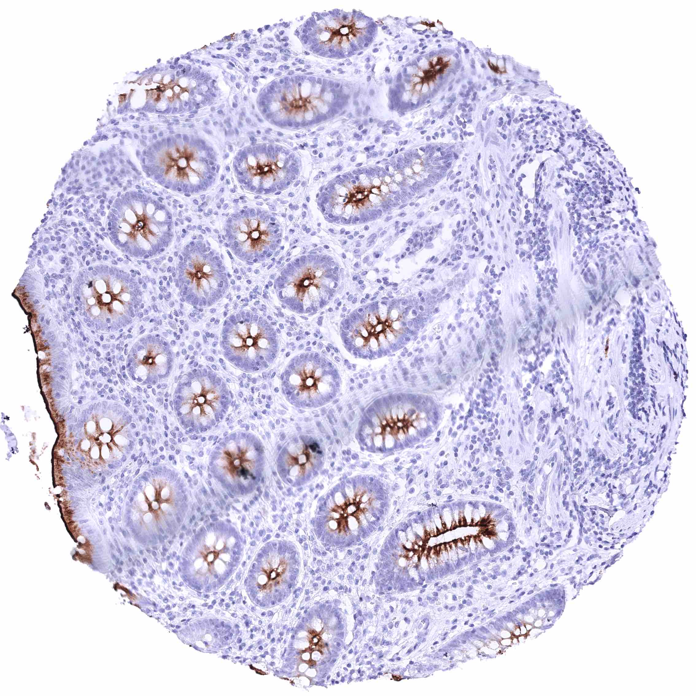
Appendix, mucosa – Strong GPRC5A membranous staining, largely limited to the luminal membranes. The staining intensity slightly decreases from the surface to the glands.
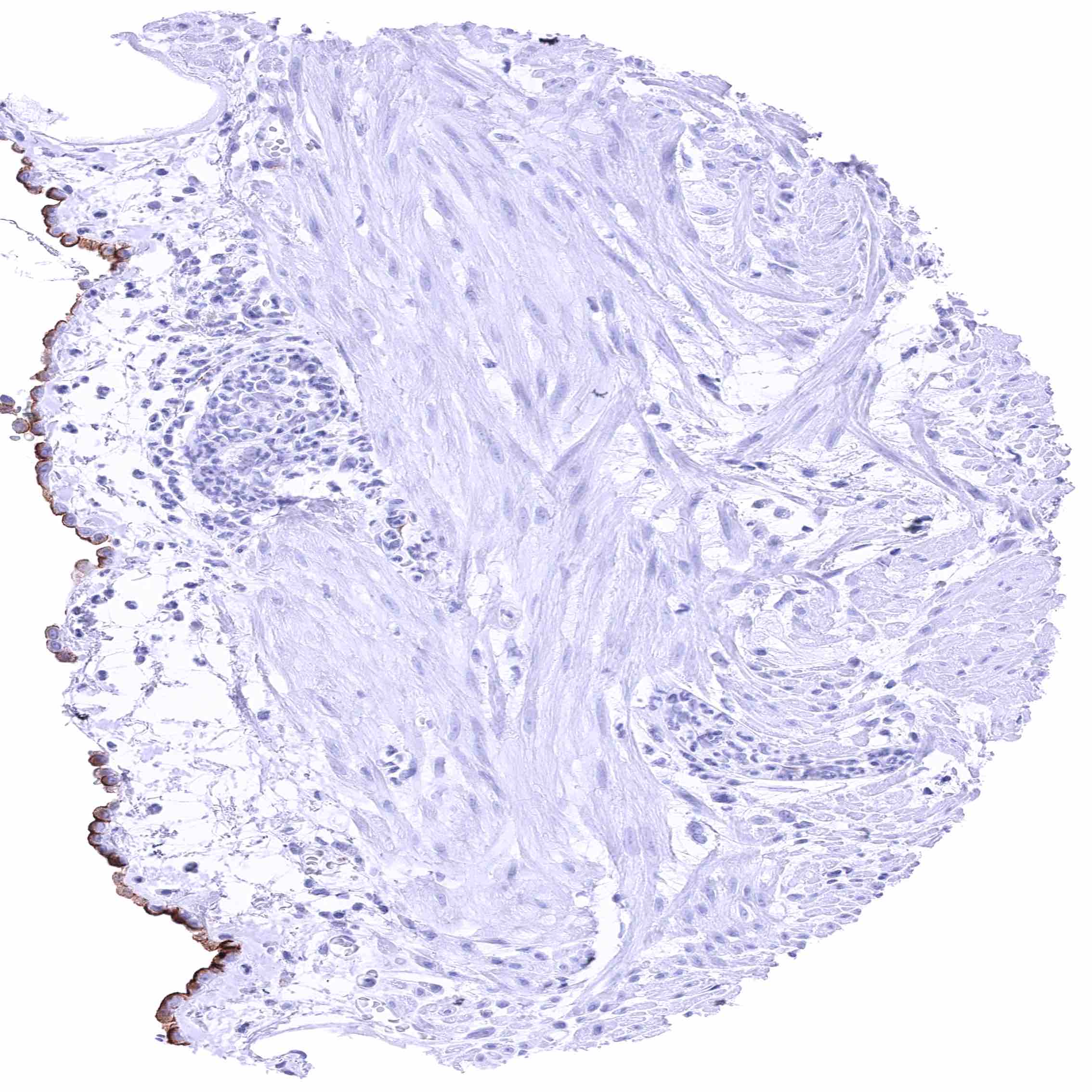
Appendix, muscular wall – Distinct membranous GPRC5A staining of the luminal membrane of mesothelial cells.
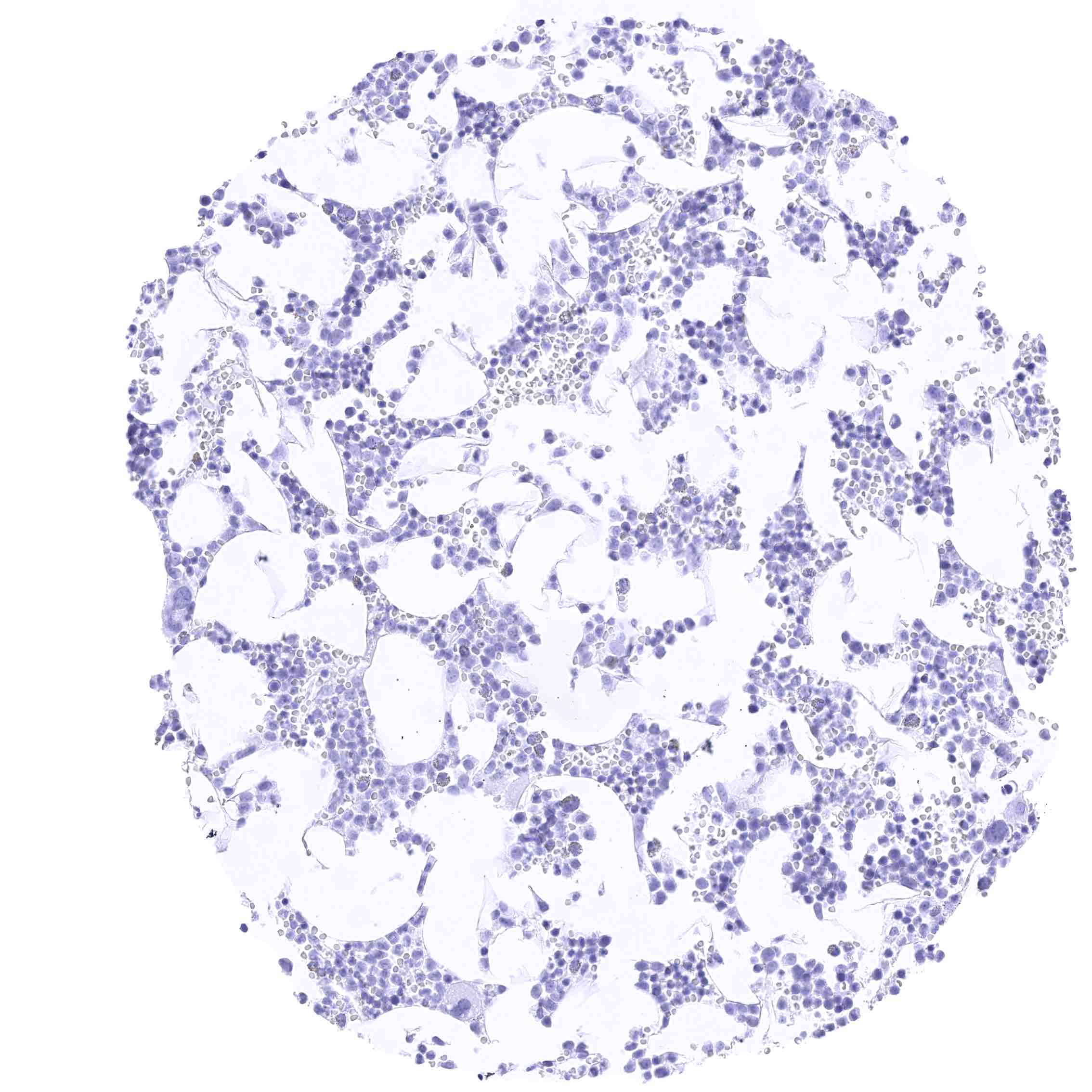
Bone marrow
Bone marrow
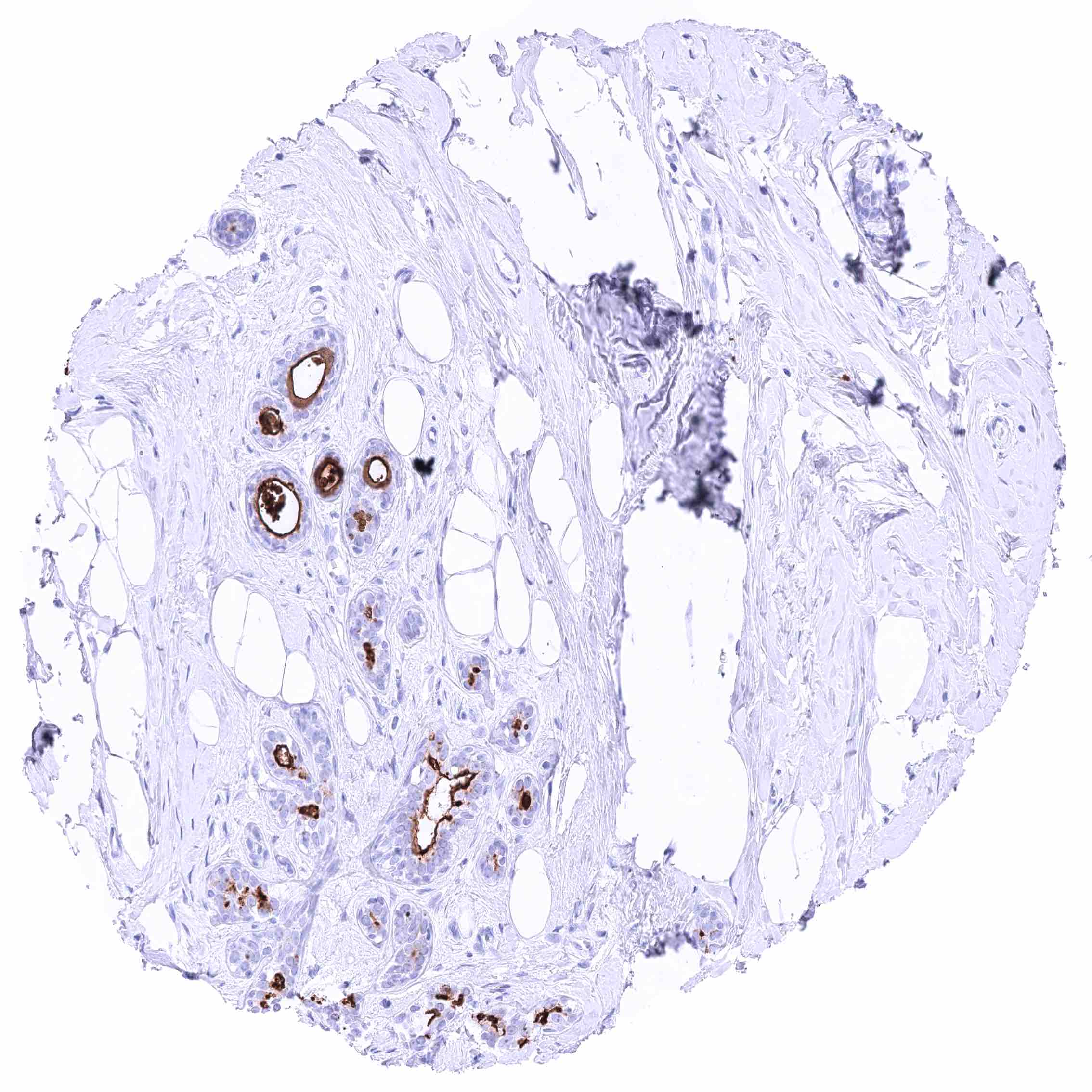
Breast – Strong GPRC5A staining of apical membranes of luminal cells.
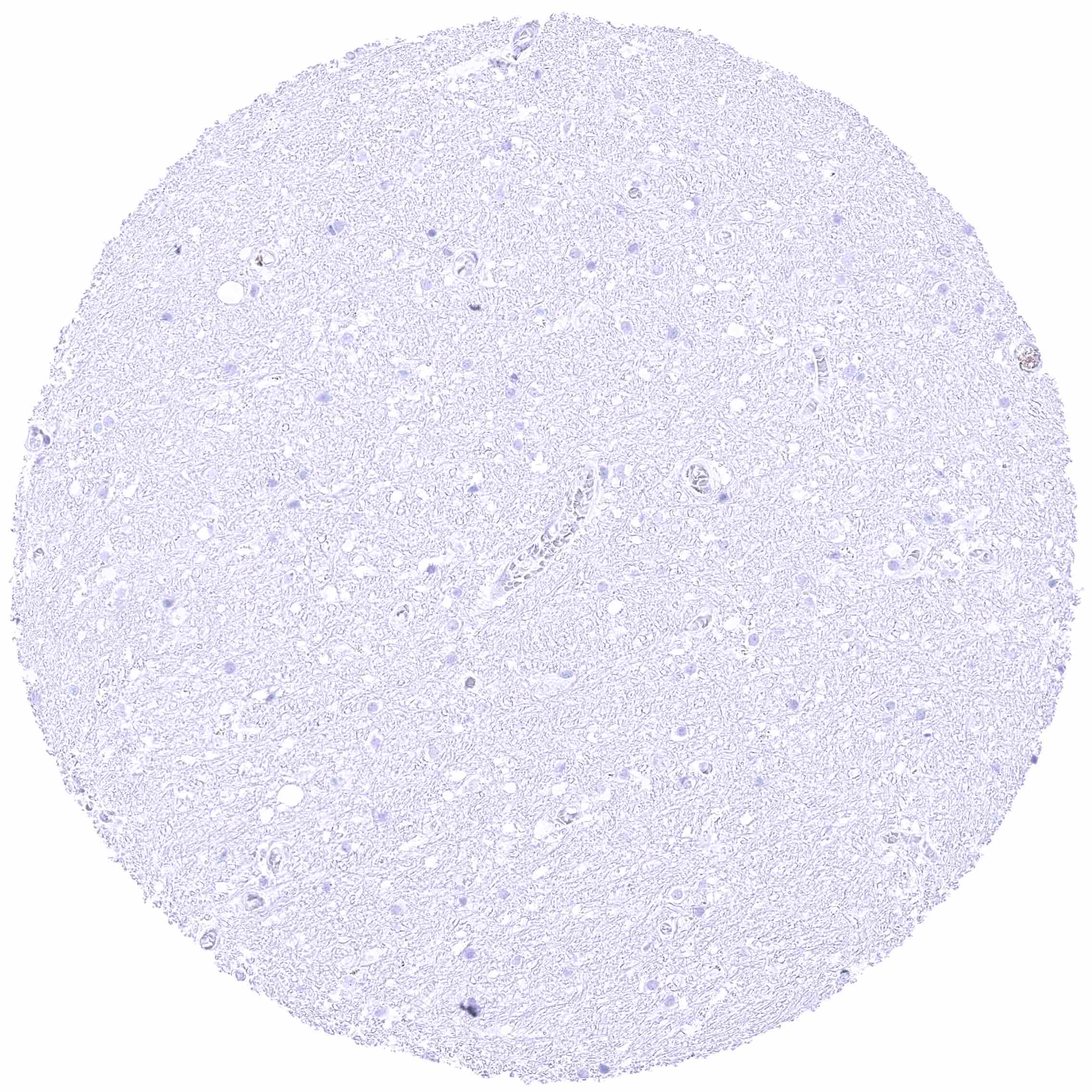
Cerebellum (white matter)
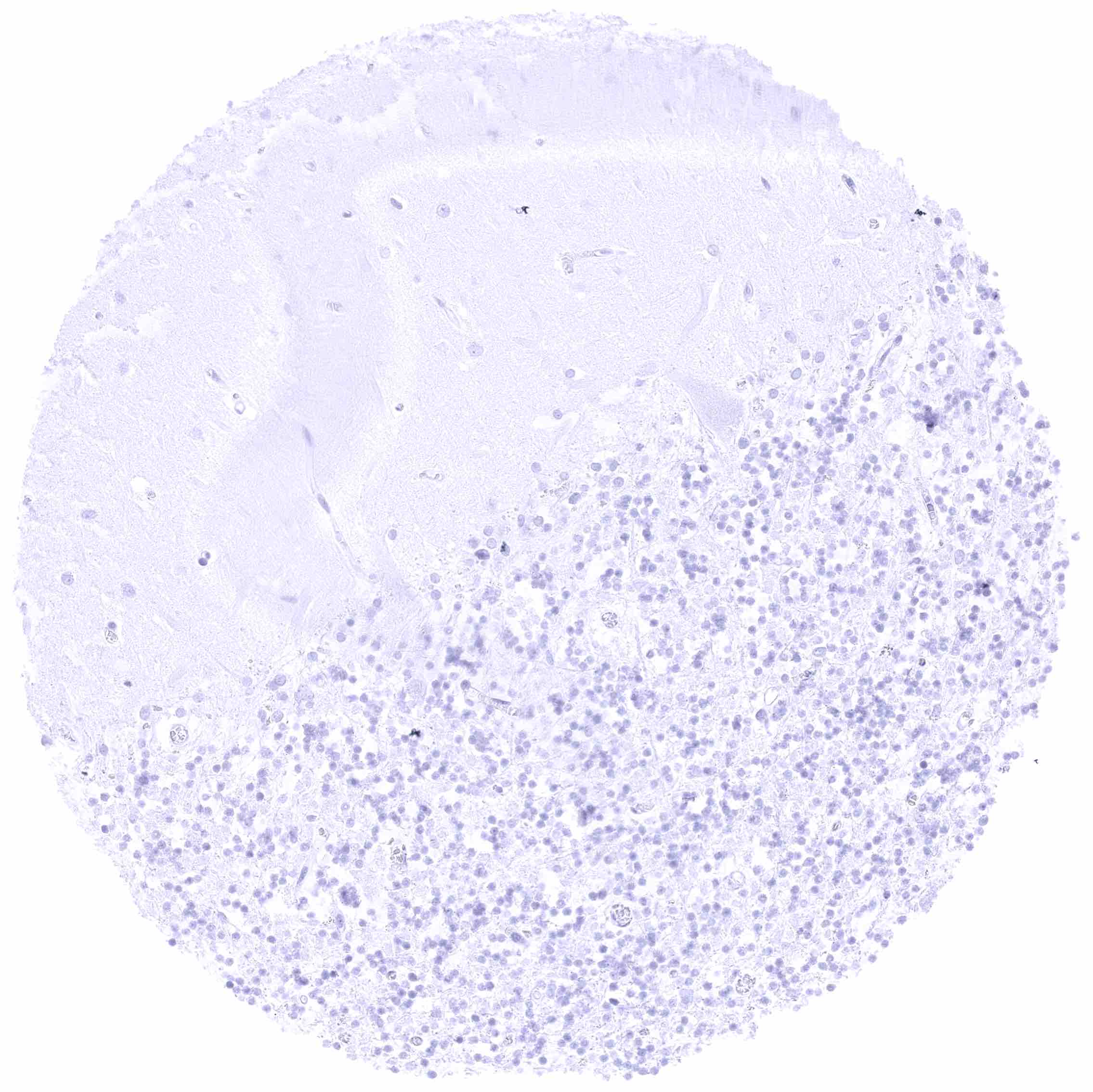
Cerebellum, cortex (grey)
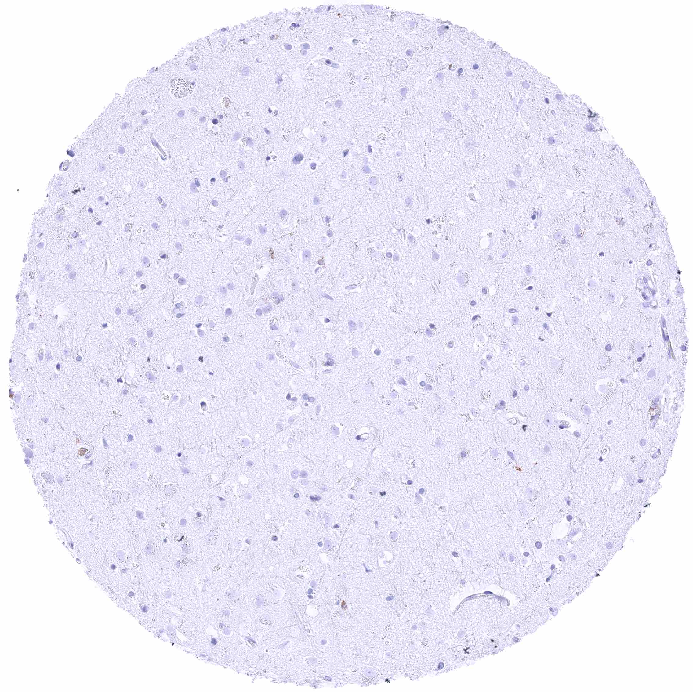
Cerebrum (grey matter)
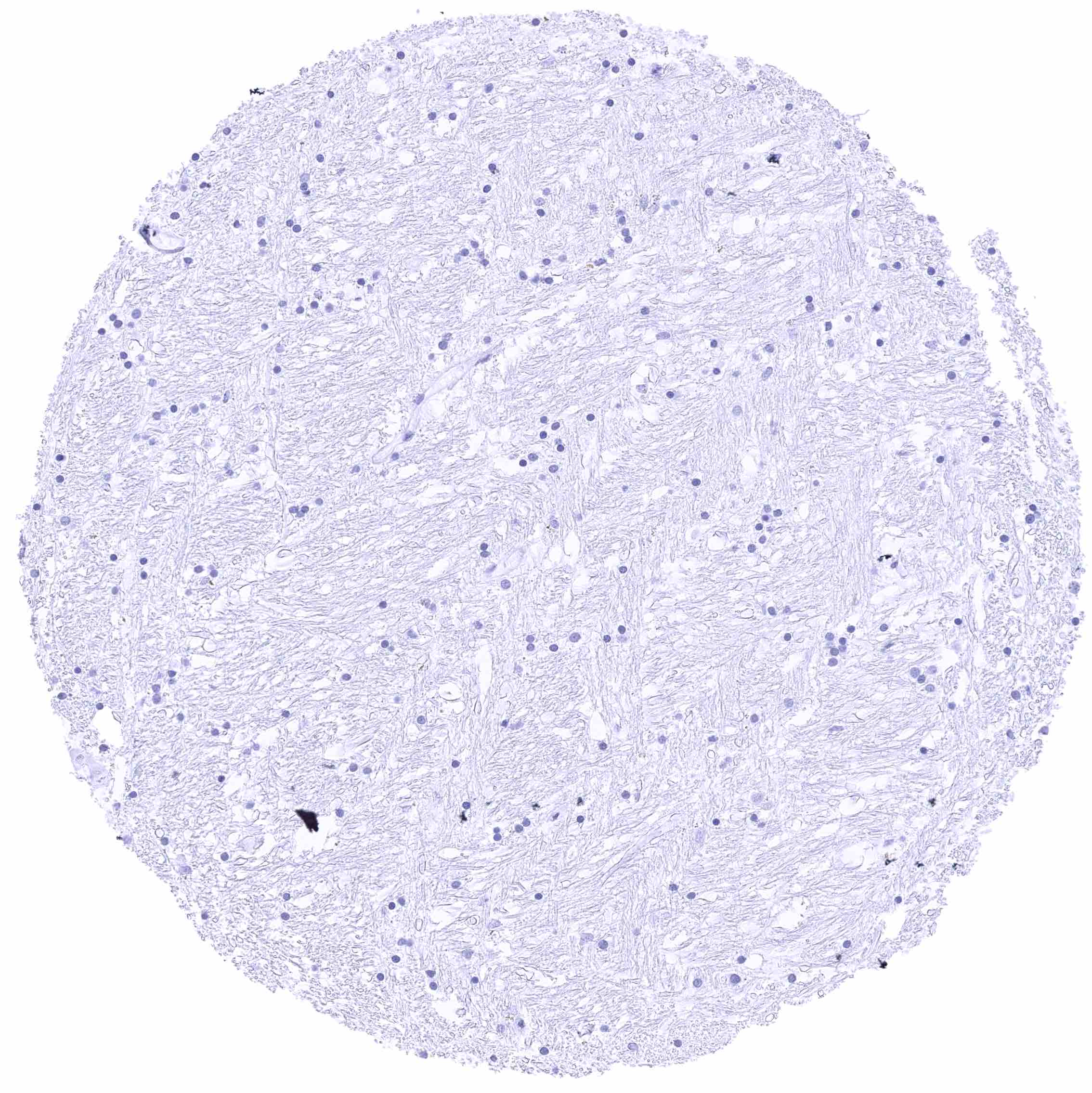
Cerebrum (white matter)
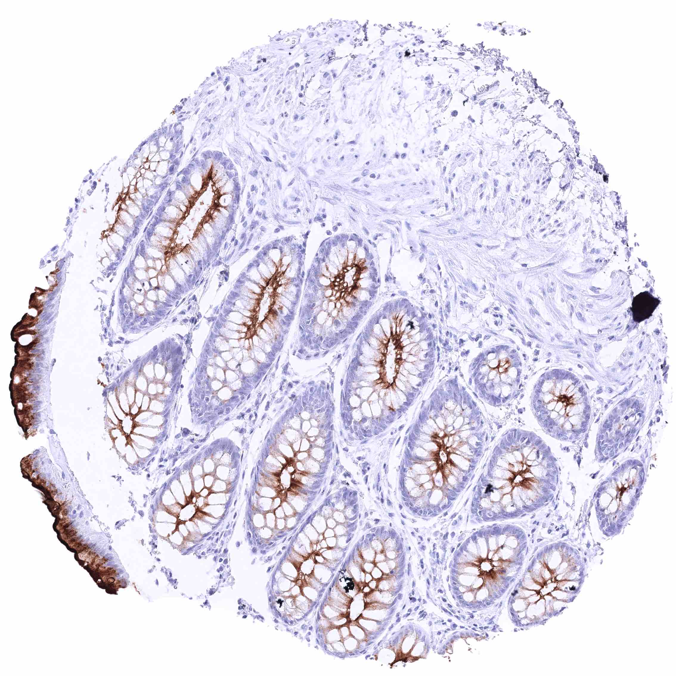
Colon descendens, mucosa – Strong GPRC5A membranous staining, largely limited to the luminal membranes. The staining intensity slightly decreases from the surface to the glands.
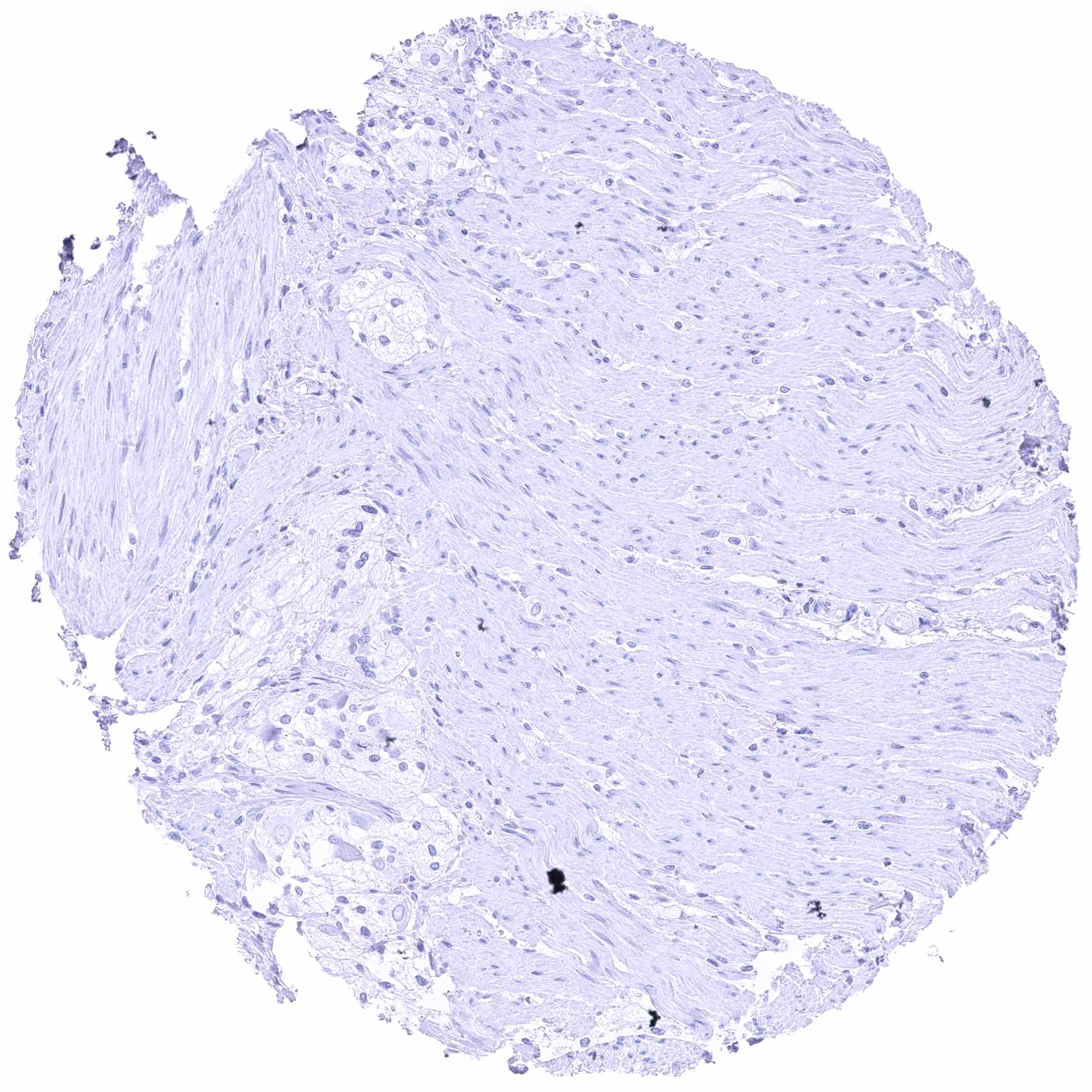
Colon descendens, muscular wall
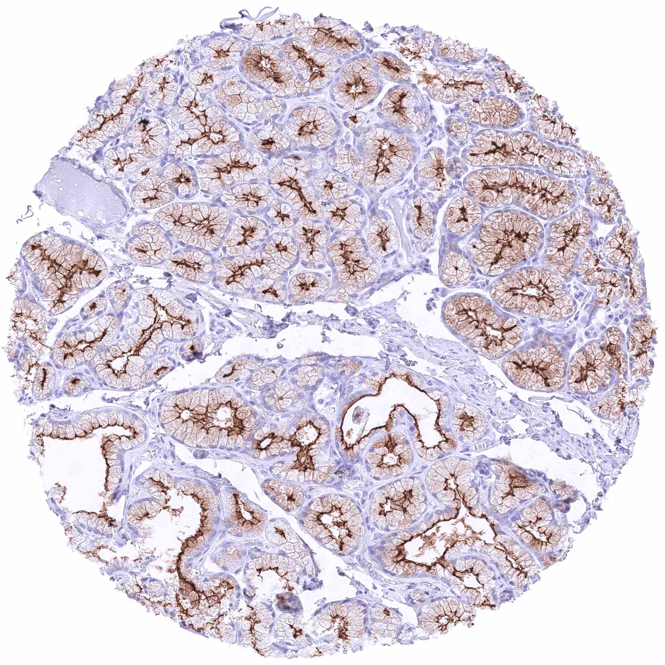
Duodenum, Brunner gland – Strong GPRC5A staining, predominantly at the luminal membranes of Brunner gland cells.
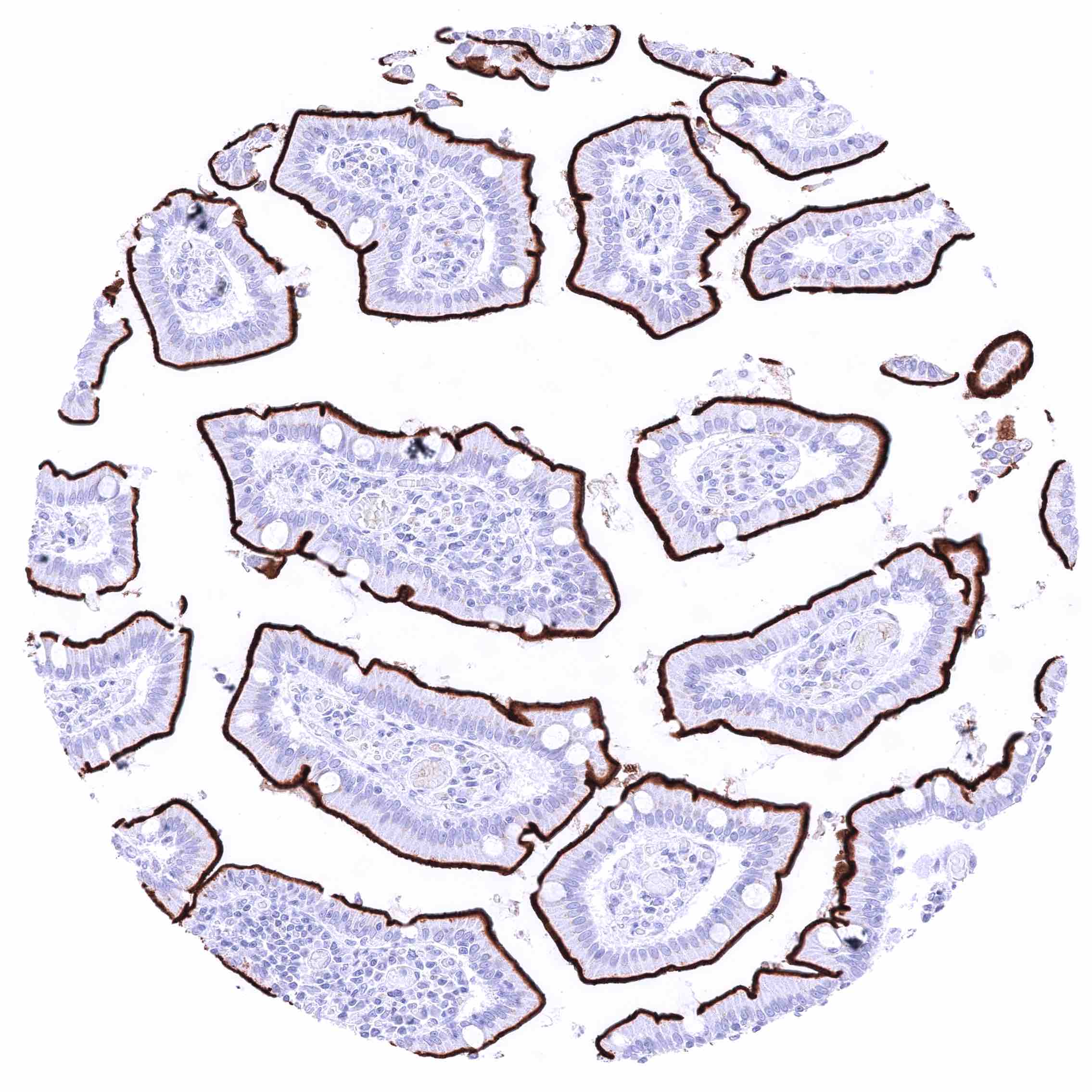
Duodenum, mucosa – Strong GPRC5A membranous staining, largely limited to the luminal membranes.
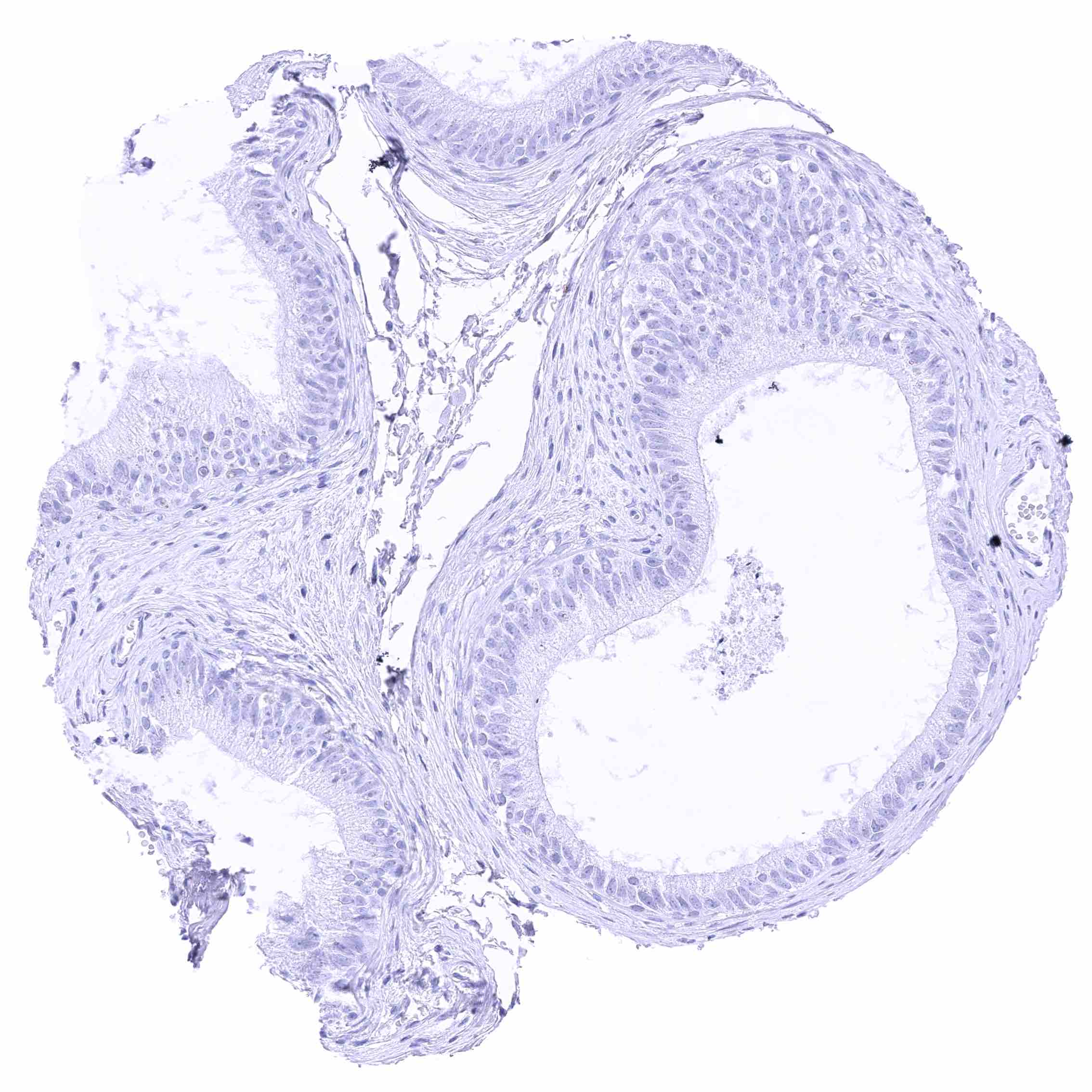
Epididymis (Caput)
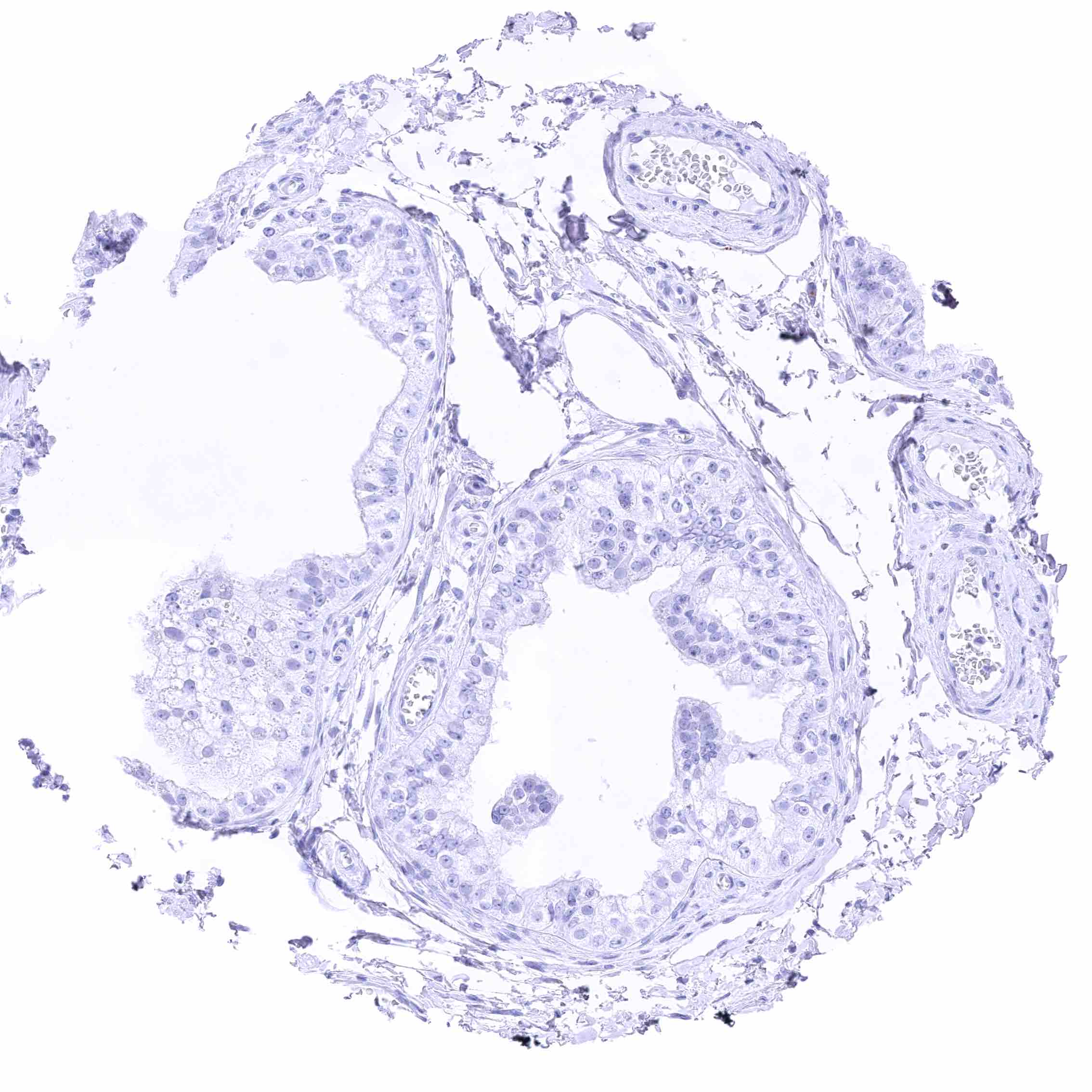
Epididymis (Caput)
Epididymis (Cauda)
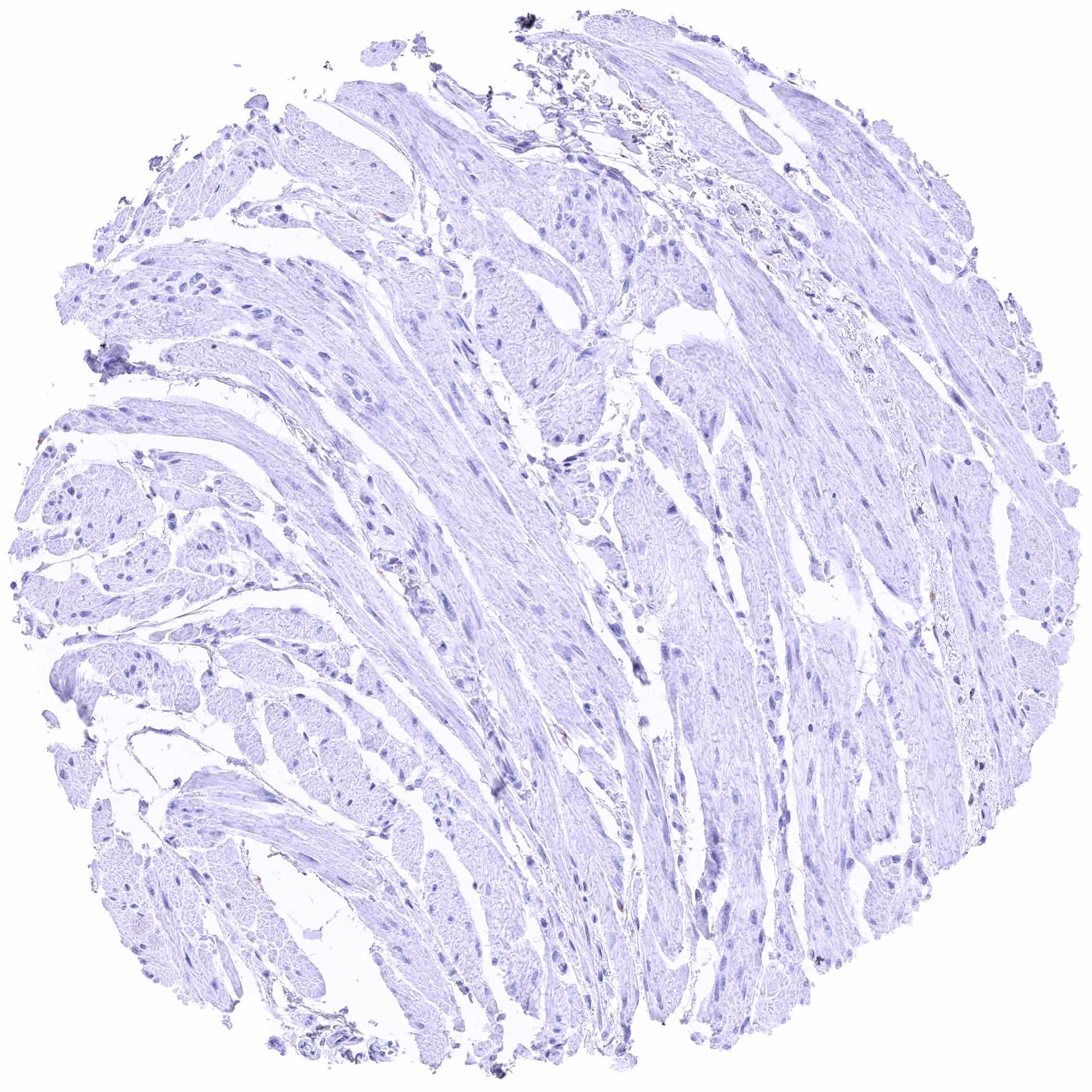
Epididymis (Cauda)
Esophagus, muscular wall
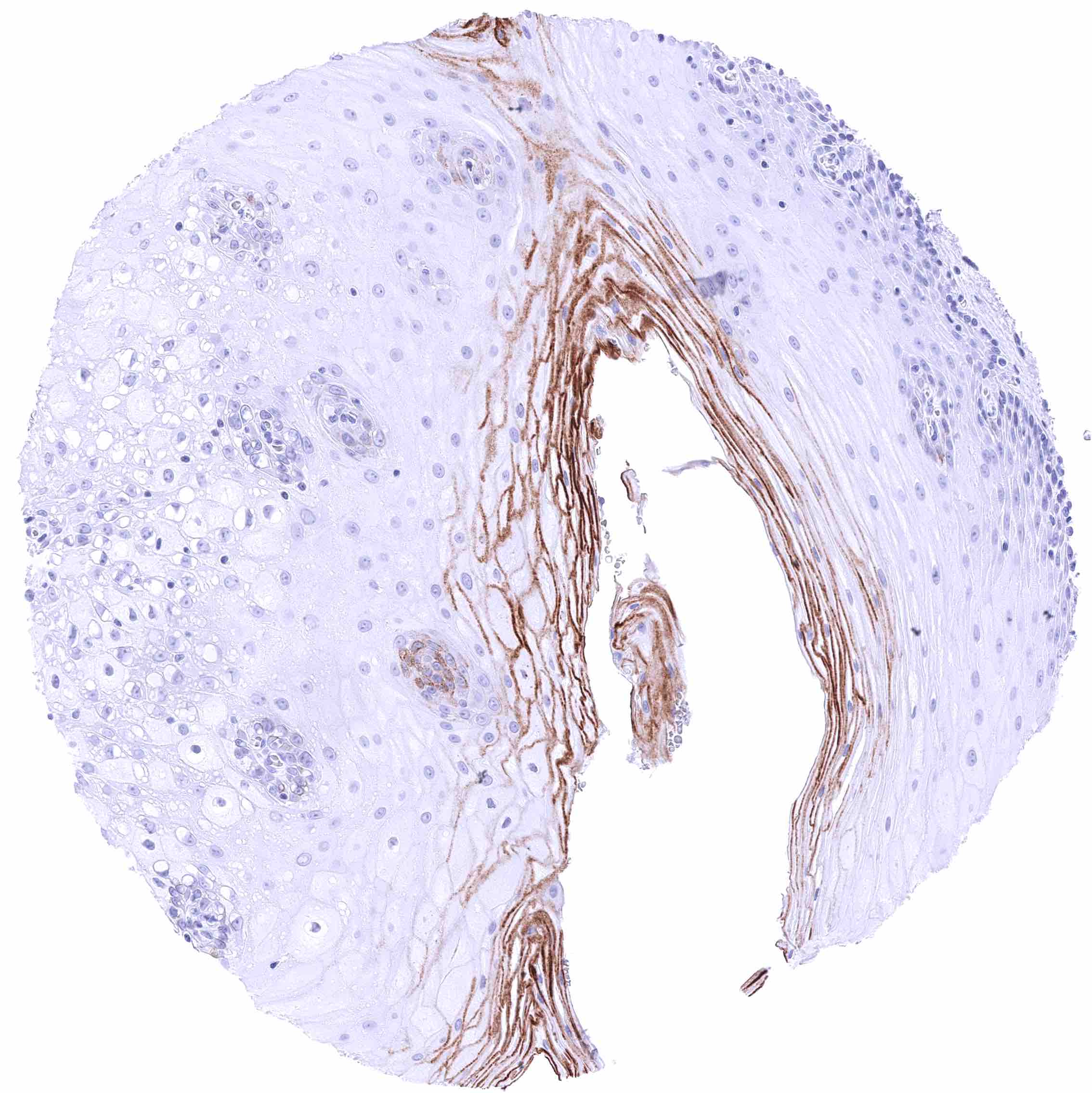
Esophagus, squamous epithelium – Moderate membranous GPRC5A staining of the superficial layers of squamous epithelial cells.
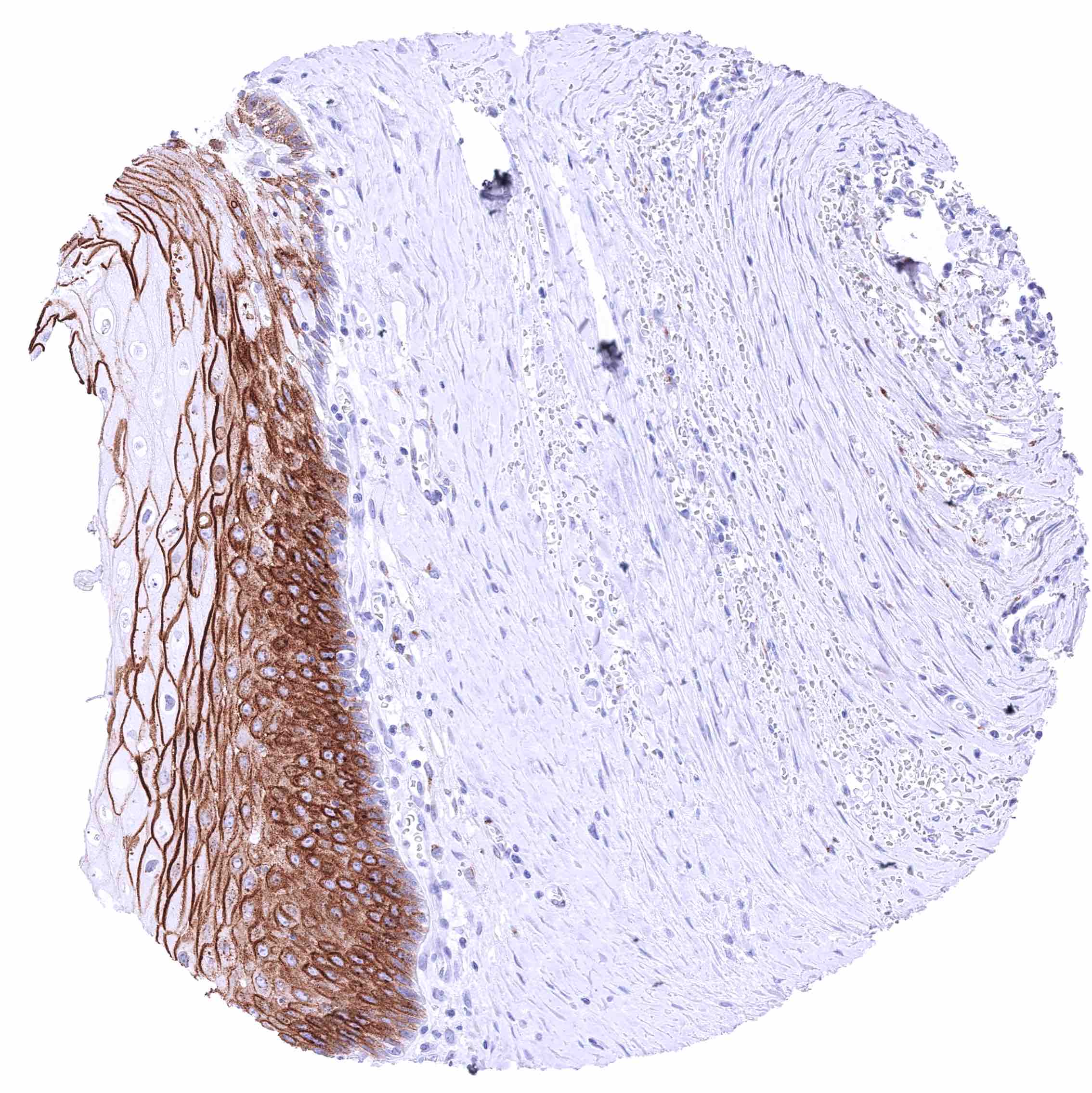
Esophagus, squamous epithelium – Strong, predominantly membranous GPRC5A staining all layers of the squamous epithelium.
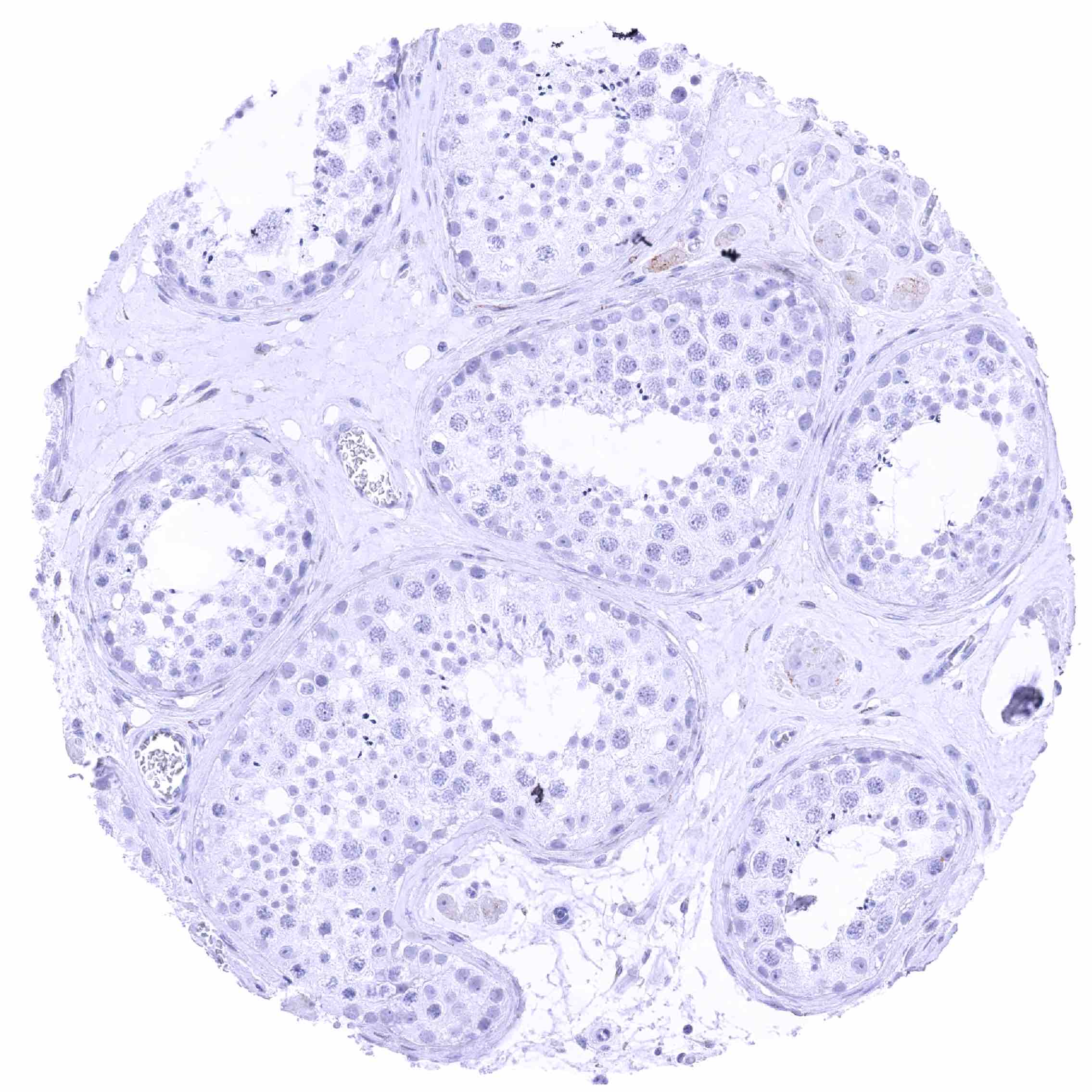
Faint membranous GPRC5A staining of some Leydig cells.
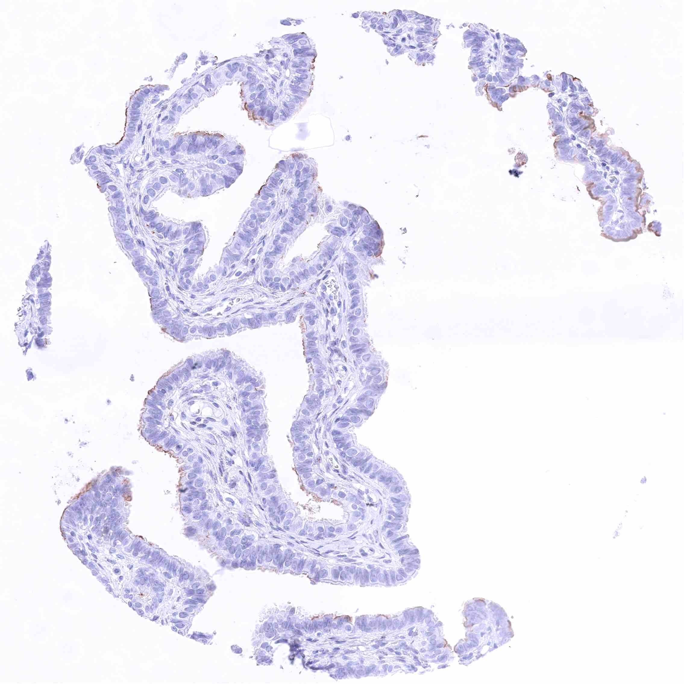
Fallopian tube, mucosa
Fat
Fat
Gallbladder, epithelium – Variable, weak to moderate membranous GPRC5A staining limited to the luminal membranes of a subset of cells.
Heart muscle
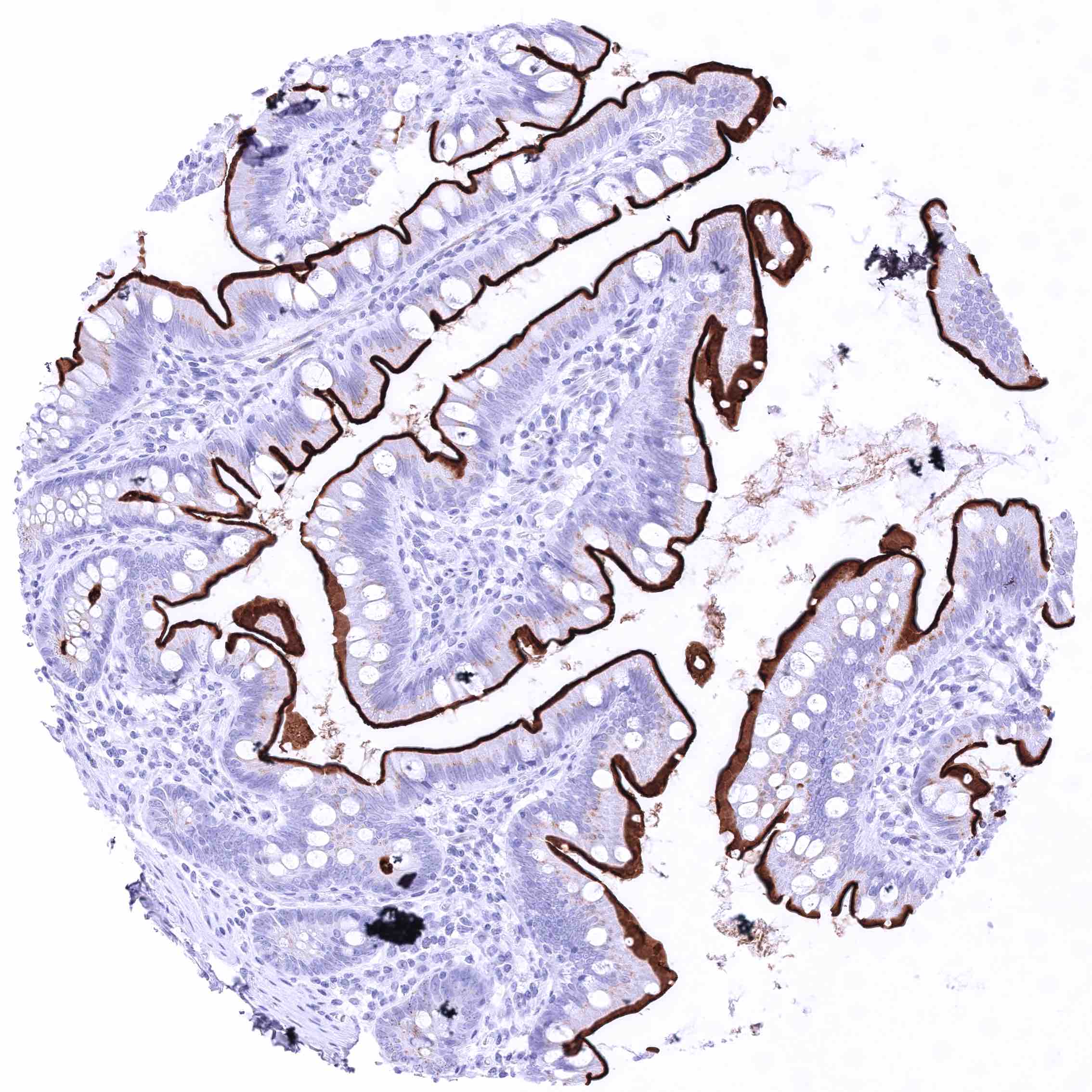
Heart muscle
Ileum, mucosa – Strong GPRC5A membranous staining, largely limited to the luminal membranes.
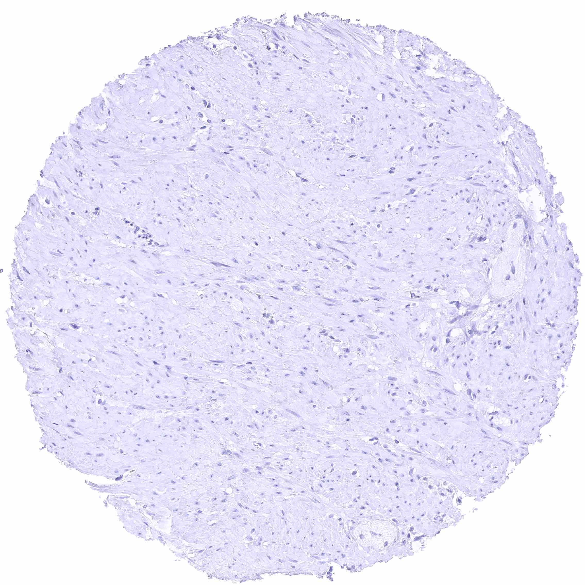
Ileum, muscular wall
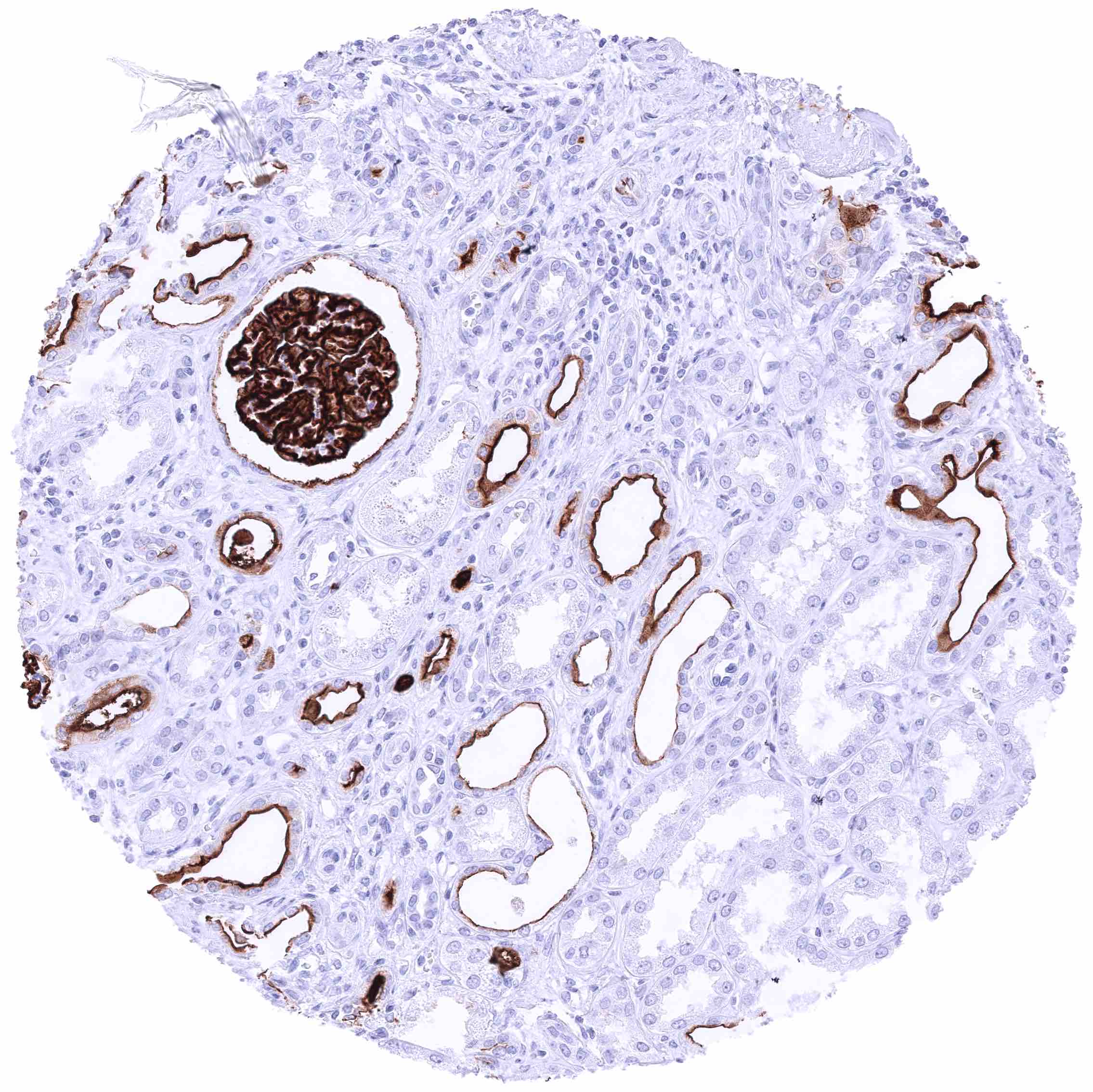
Kidney, cortex – Strong GPRC5A staining of podocytes and membranes of the Bowman capsule. Moderate to strong luminal membranous GPRC5A staining of a fraction of tubuli (predominantly distal) and collecting ducts
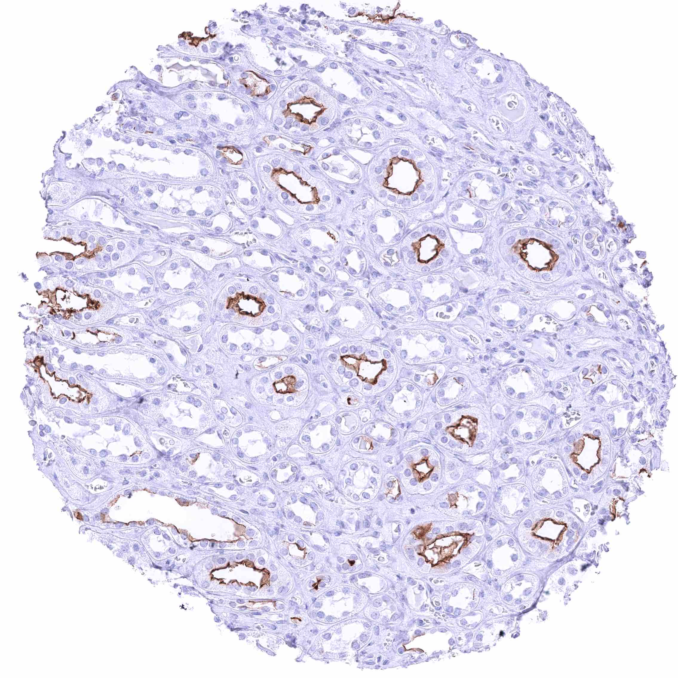
Kidney, medulla – Strong luminal membranous GPRC5A staining of a fraction of collecting ducts.
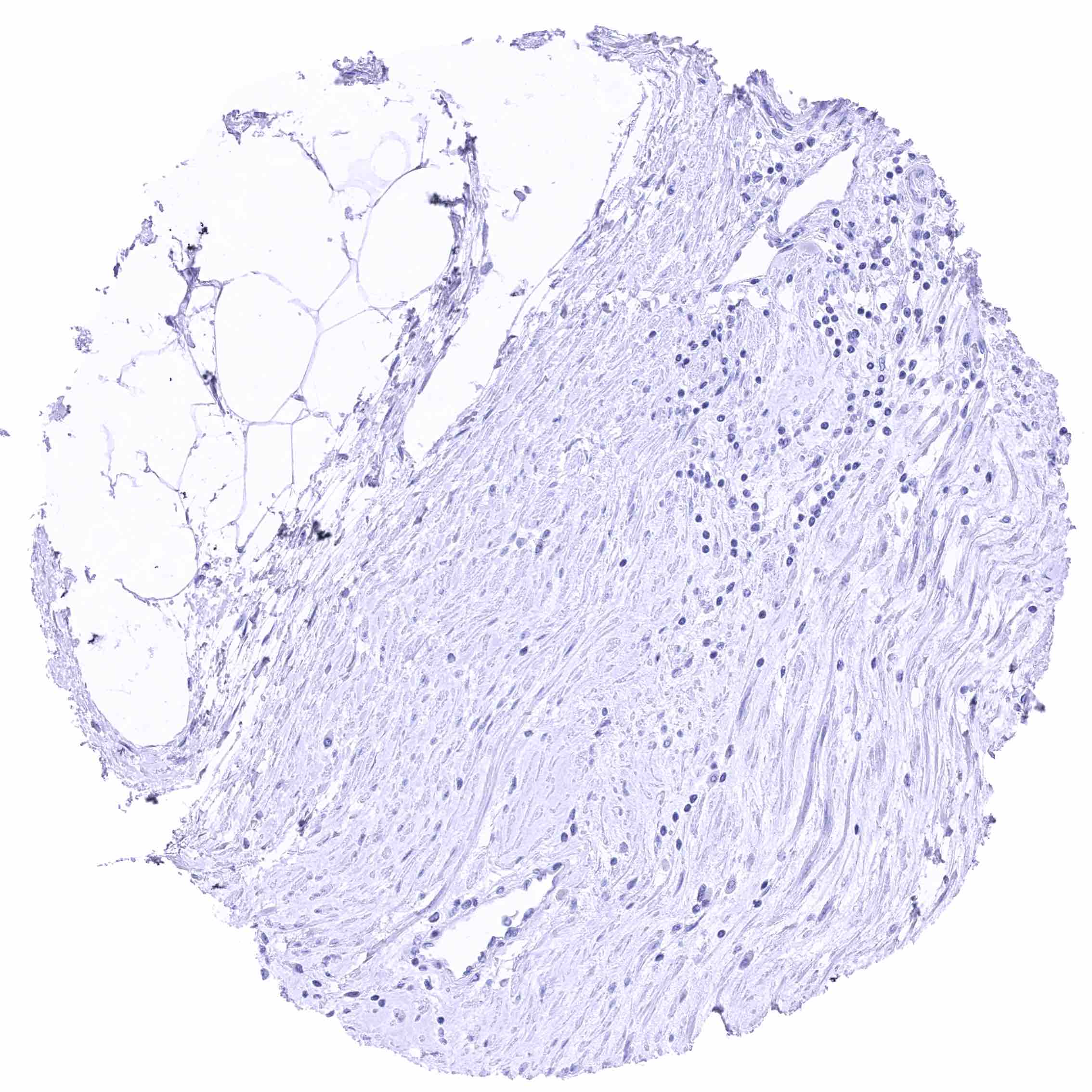
Kidney, pelvis, muscular wall
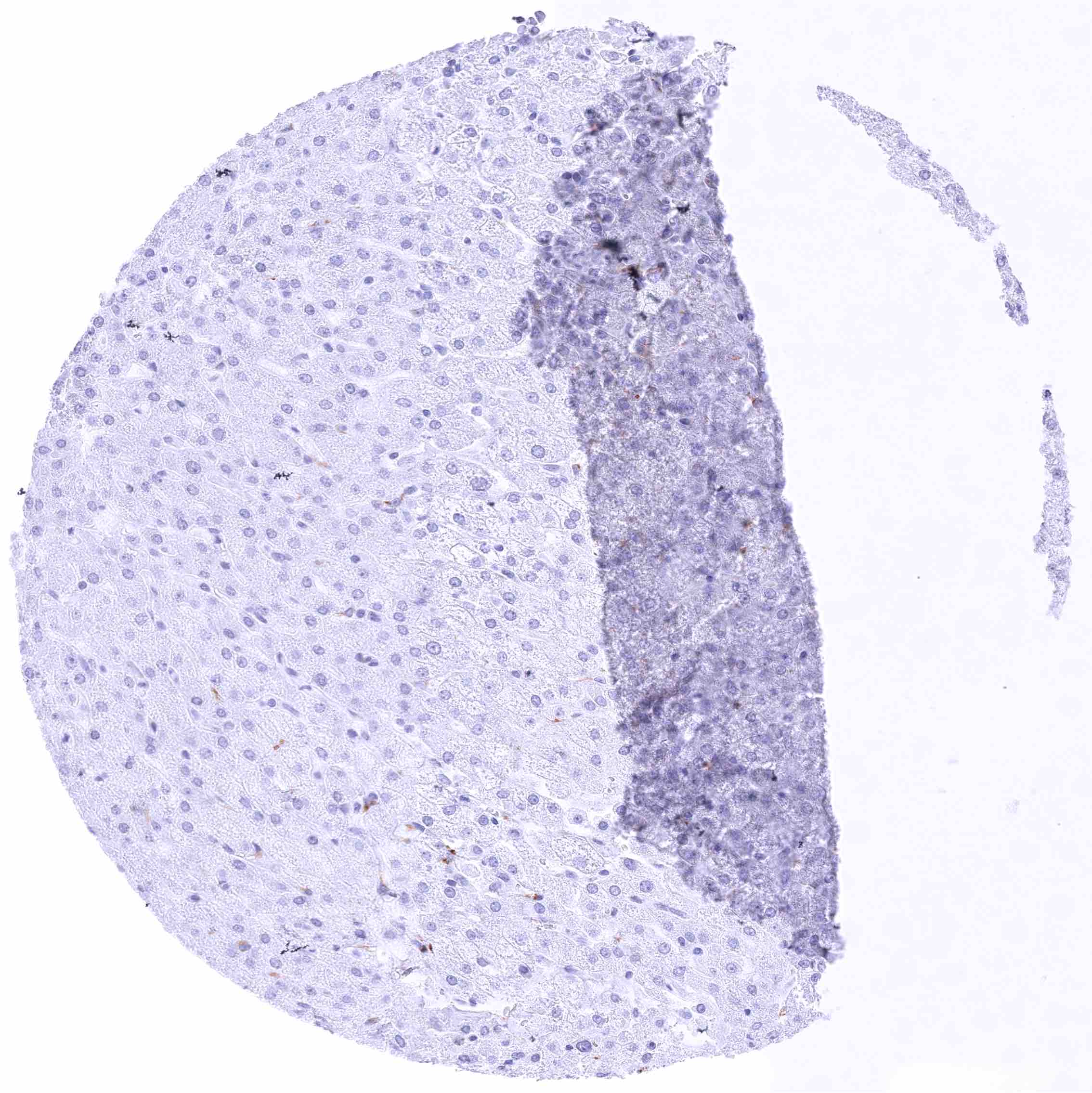
Liver
Liver
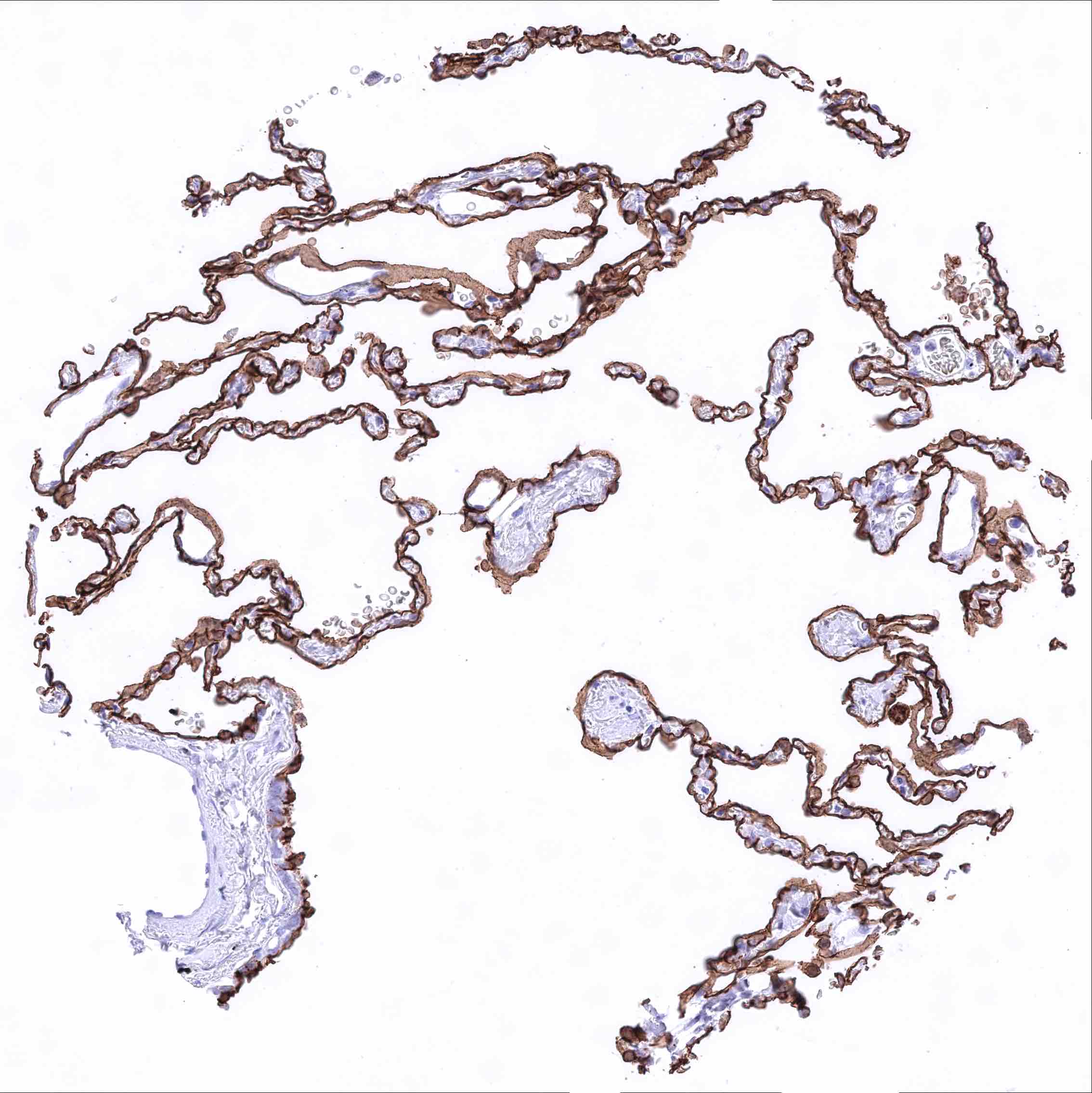
Lung – Strong membranous GPRC5A staining of all alveolar pneumocytes
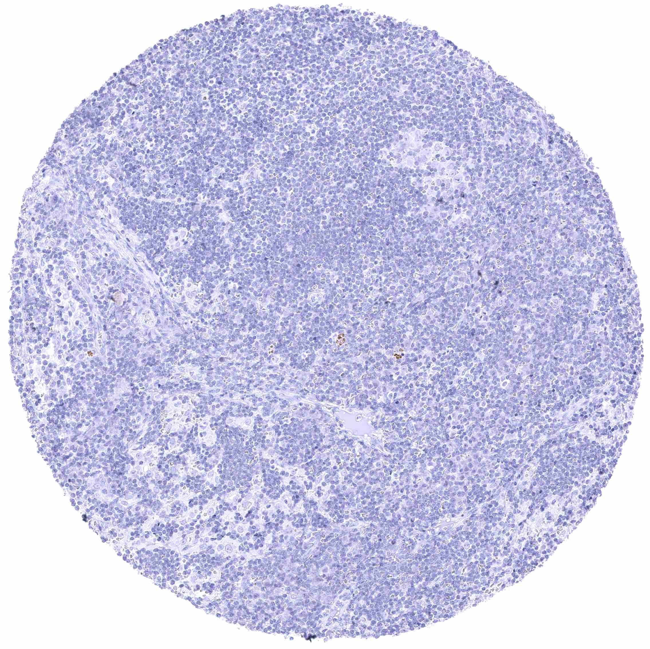
Lymph node
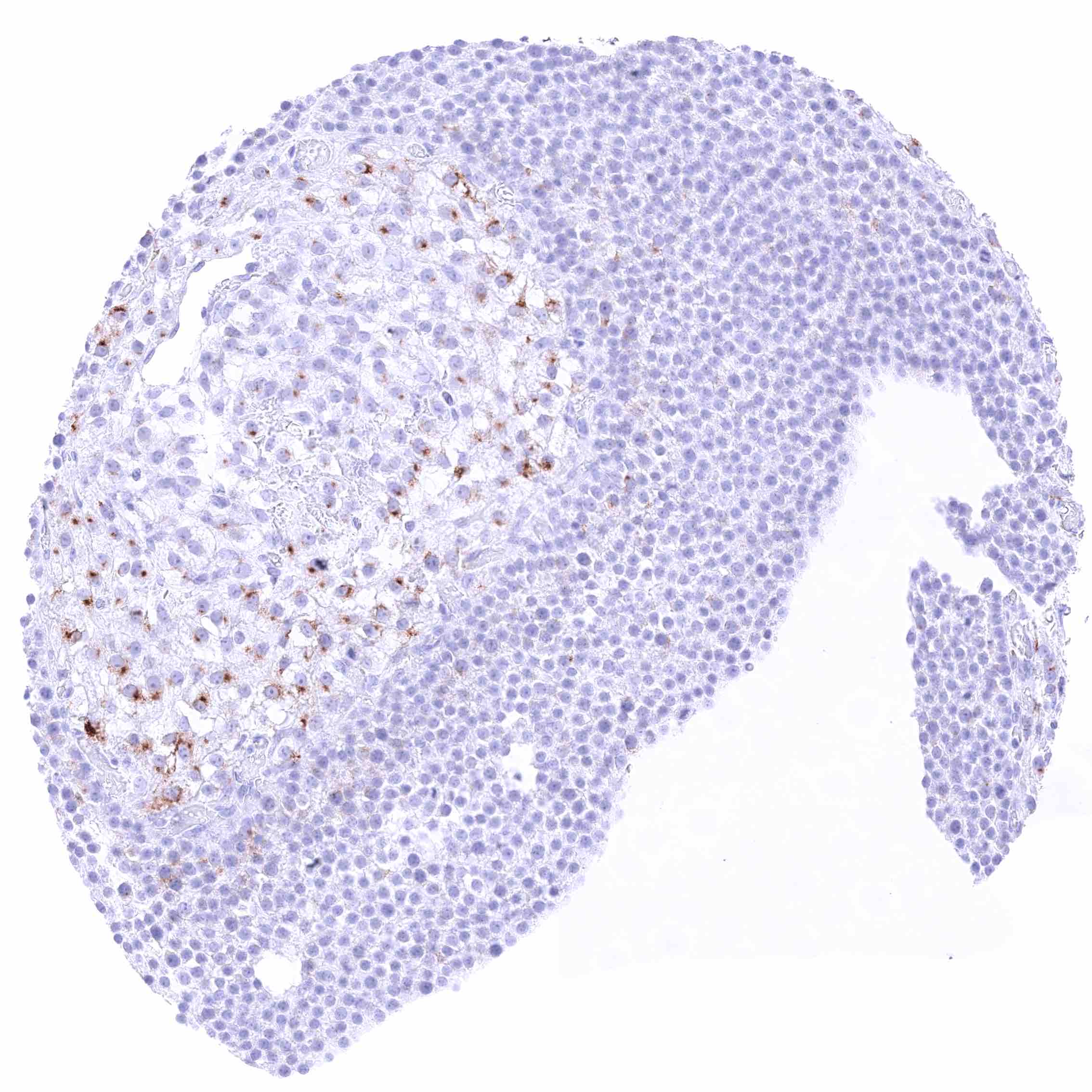
Lymph node
Ovary, follicular cyst – Strong „dot-like“ perinuclear staining of theca interna cells around a follicular cyst.
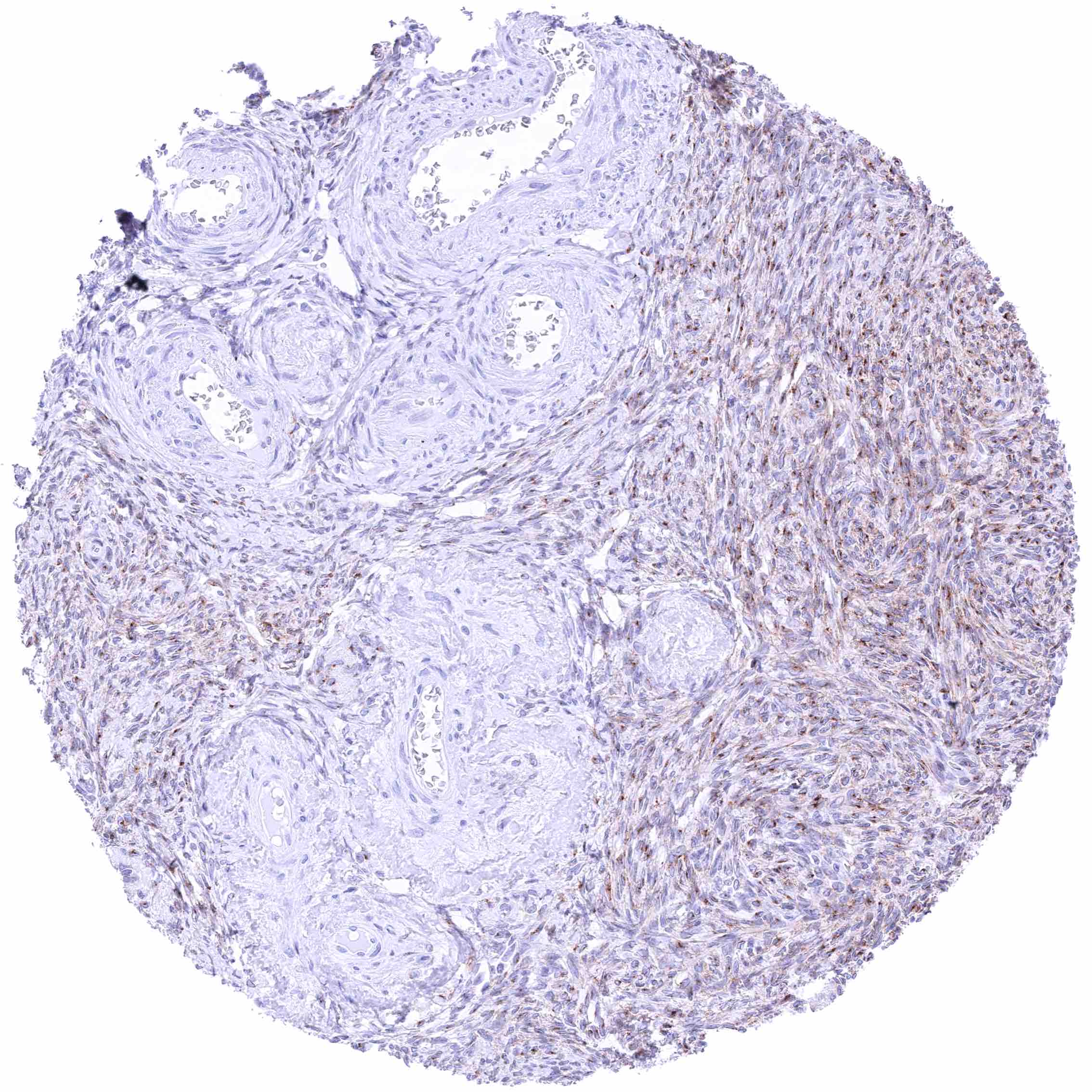
Ovary, stroma – Moderate to strong perinuclear “dot-like” GPRC5A staining and faint membranous GPRC5A staining of subsets of stroma cells.
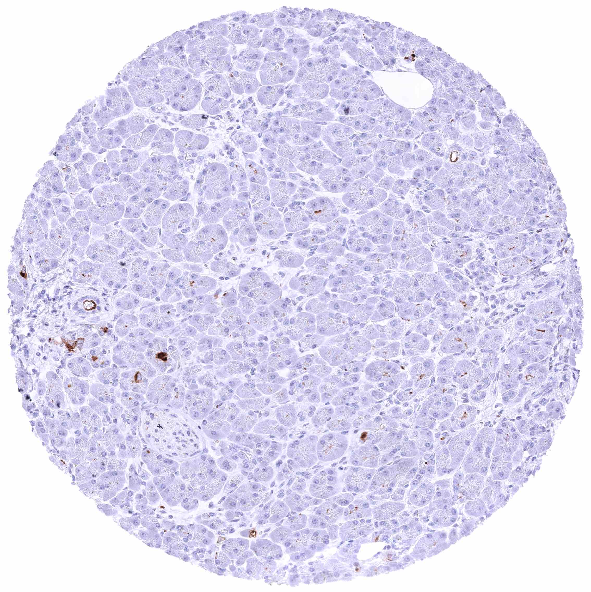
Pancreas – Weak to moderate GPRC5A staining of luminal membranes of cells from small excretory ducts. A focal strong GPRC5A staining can also occur in very small subset of possible acinar cells.
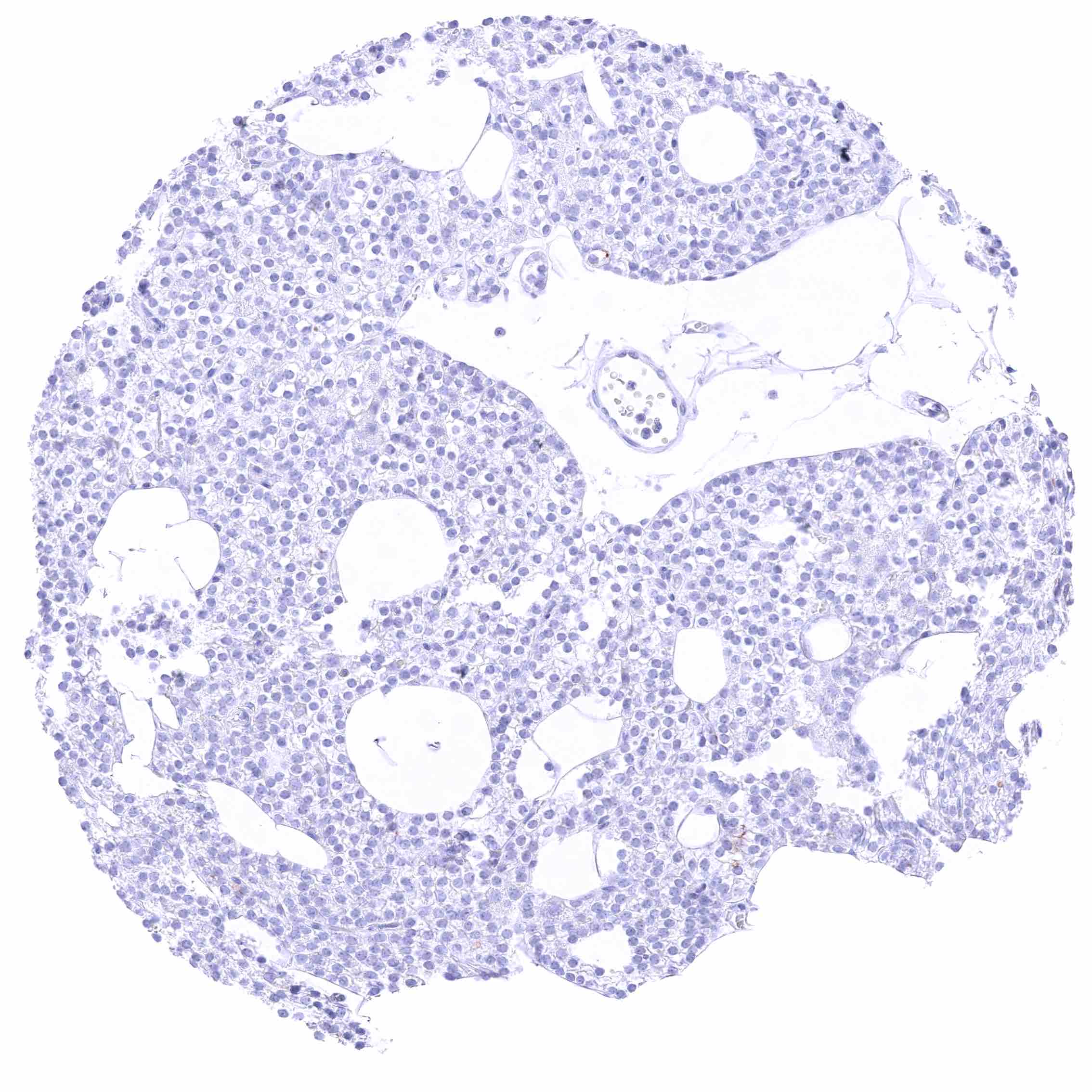
Parathyroid gland
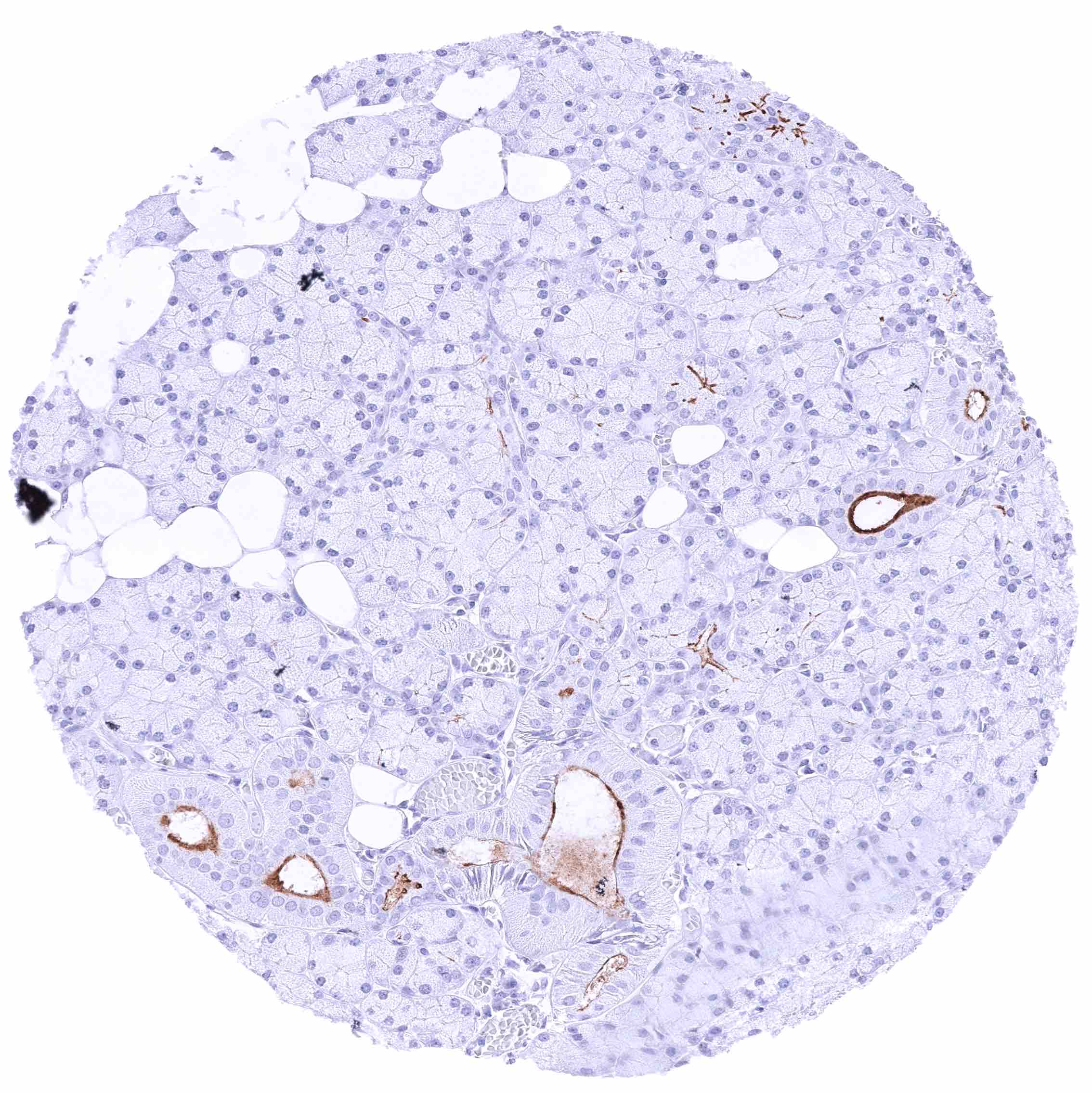
Parathyroid gland
Parotid gland – Strong GPRC5A staining of luminal membranes of excretory duct cells. A less intensive staining is also seen in variable subsets of glands.
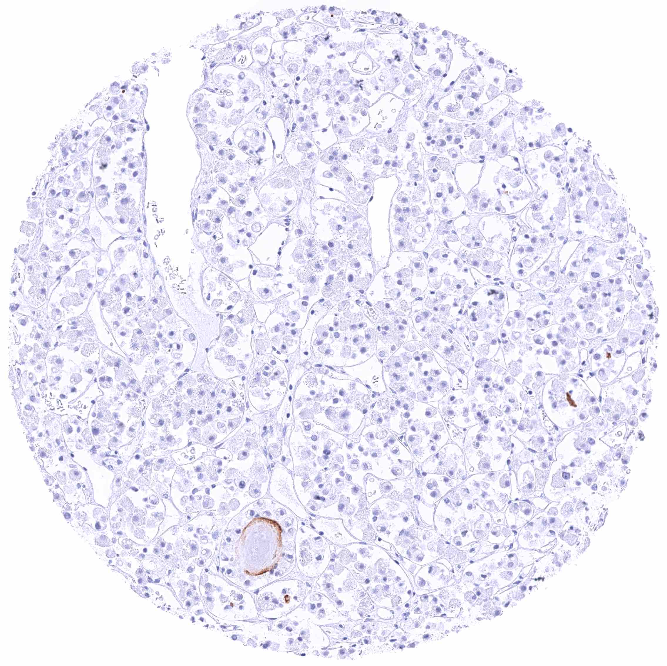
Pituitary gland, anterior lobe
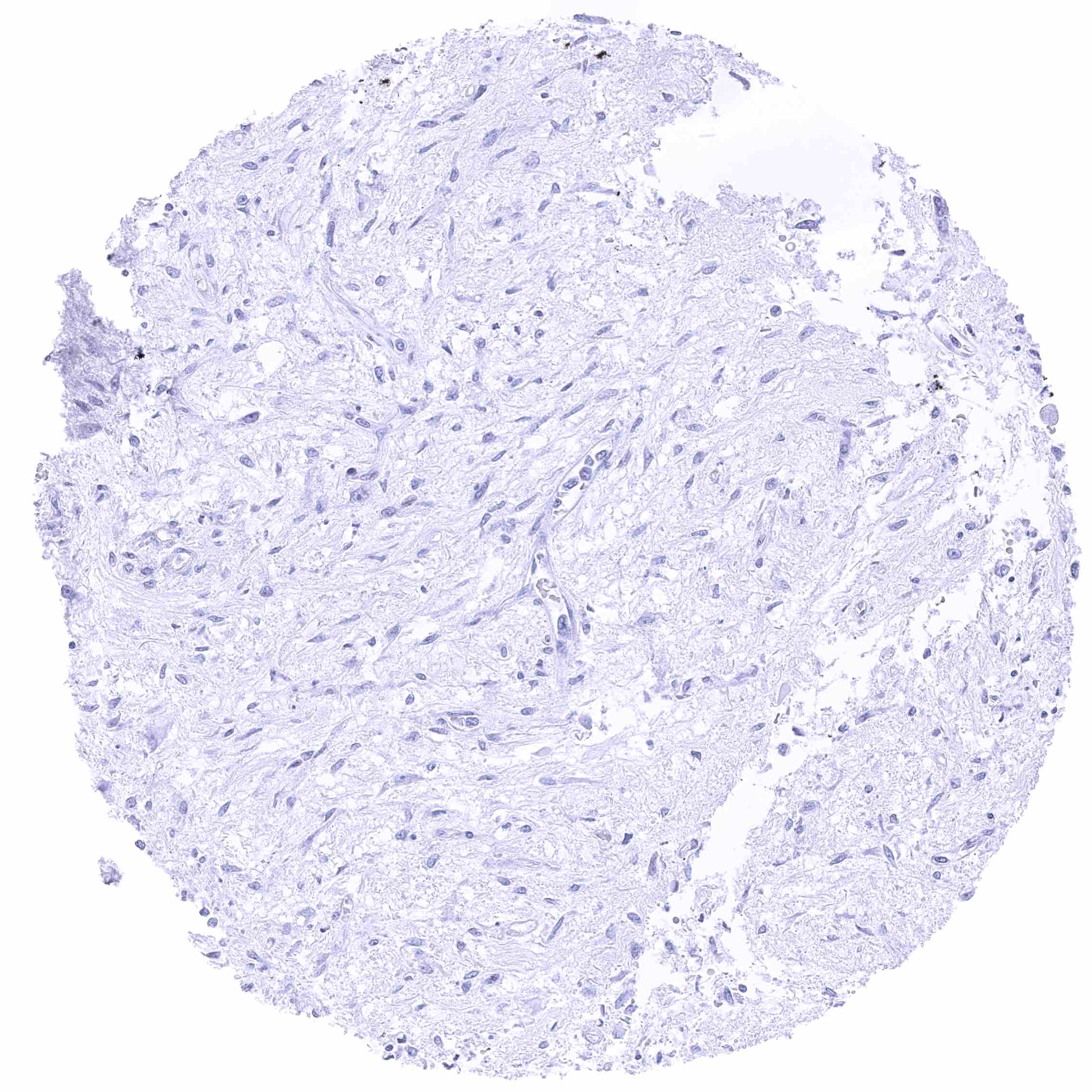
Pituitary gland, posterior lobe
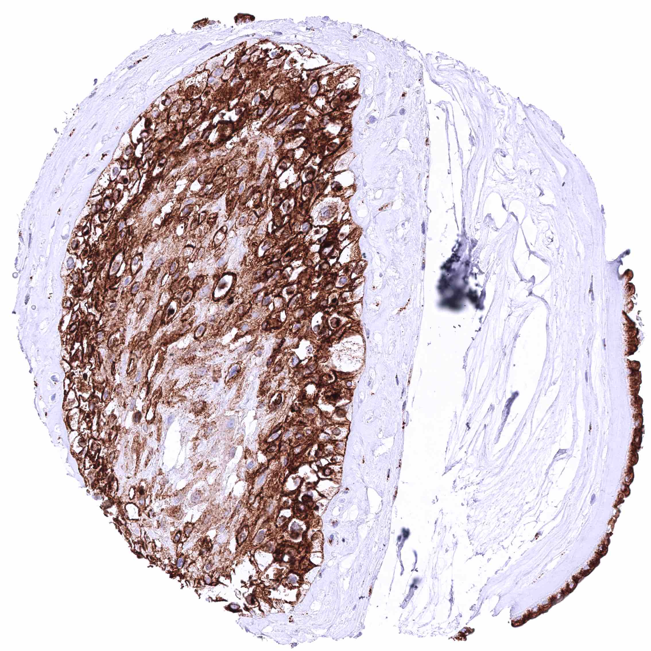
Placenta (amnion and chorion)
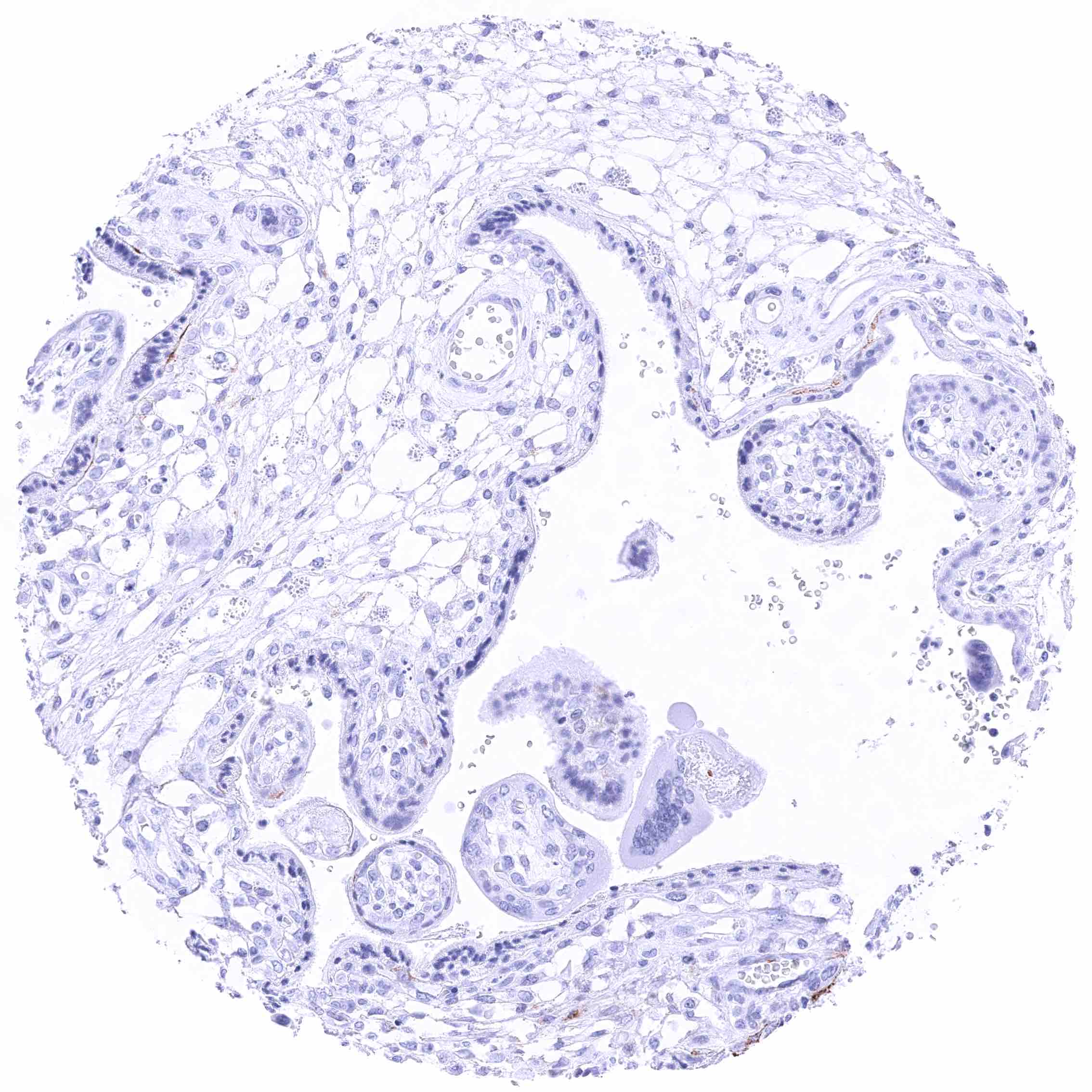
Placenta, early
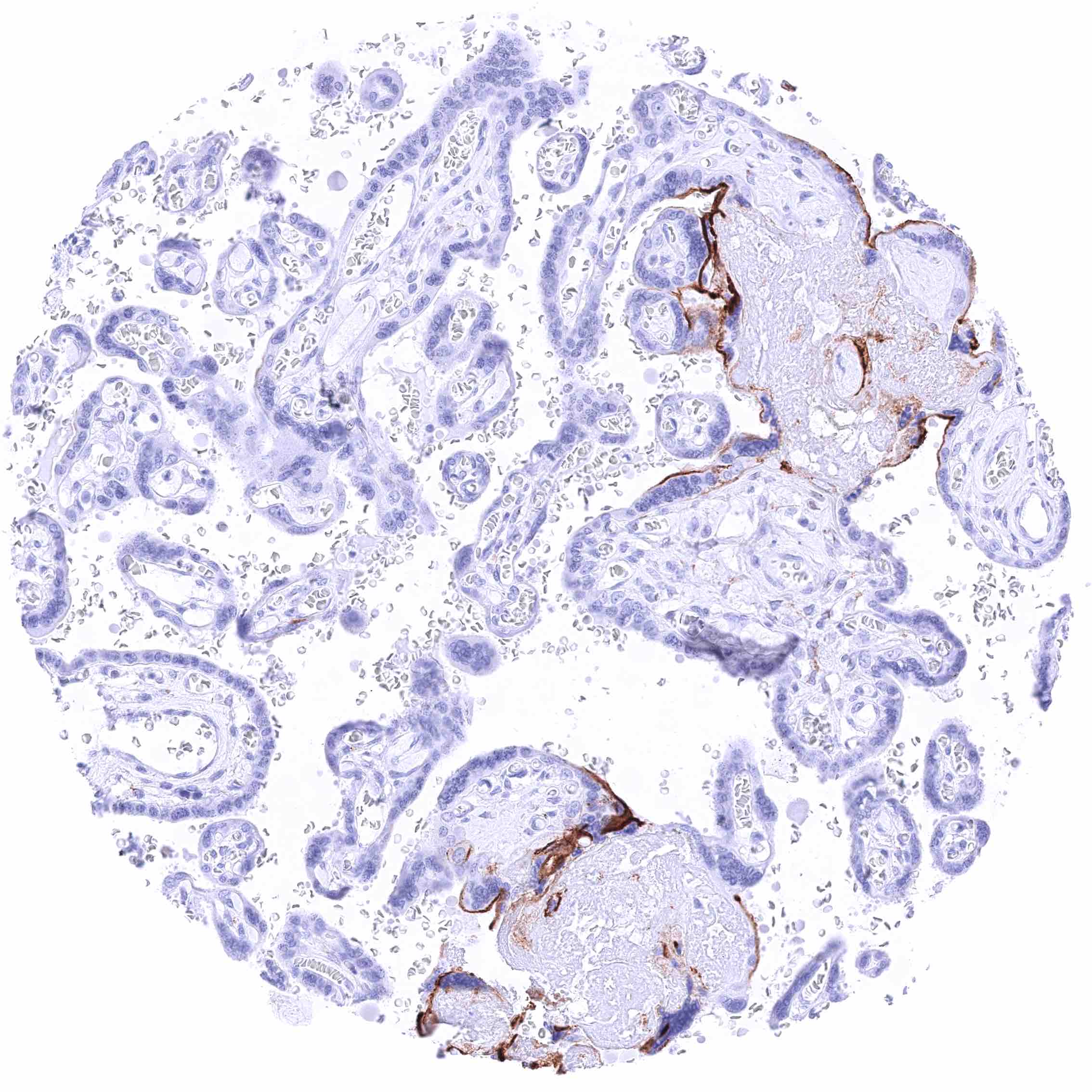
Placenta, early
Placenta, mature – Distinct membranous staining of the luminal membrane of syncytiotrophoblast cells in areas of placenta tissue damage.
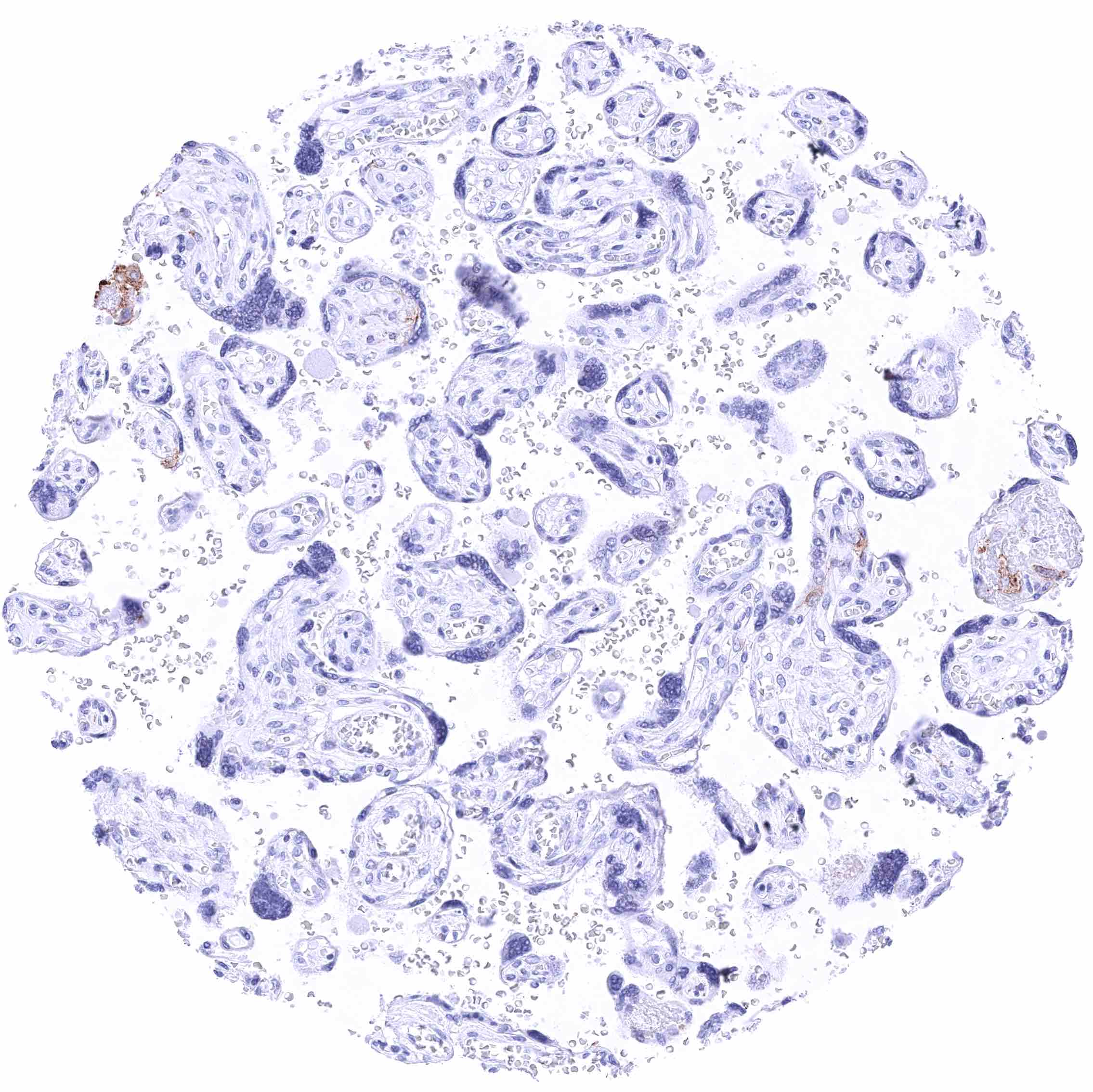
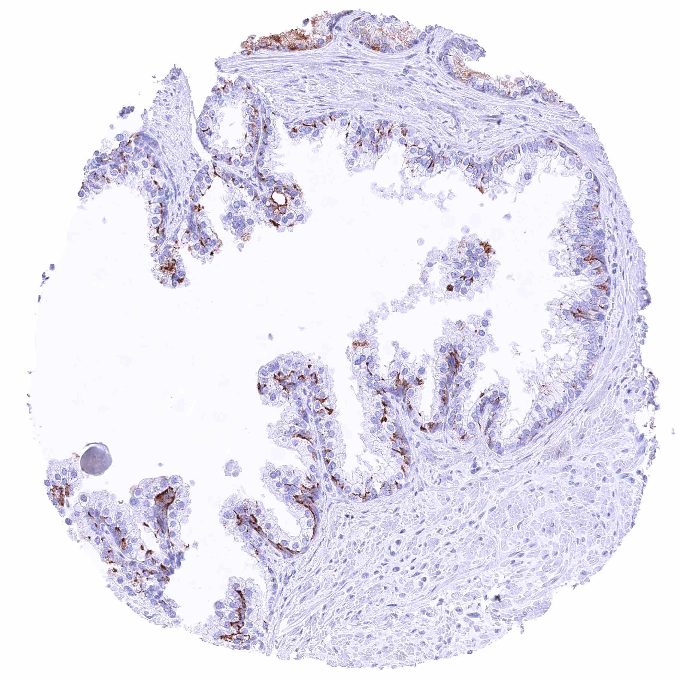
Placenta, mature
Placenta, mature
Prostate – A GPRC5A staining of variable intensity (weak to strong) can be seen on (mostly small) subsets of luminal and basal cells. It preferably occurs at luminal membranes.
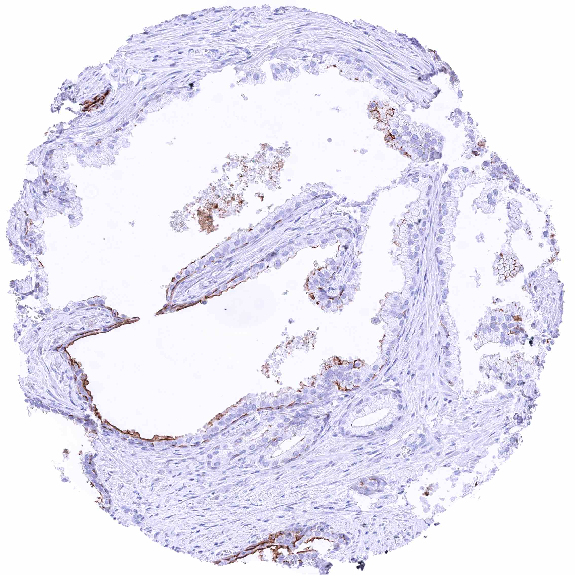
Prostate – GPRC5A staining of subsets of luminal and basal cells in an area of atrophy.
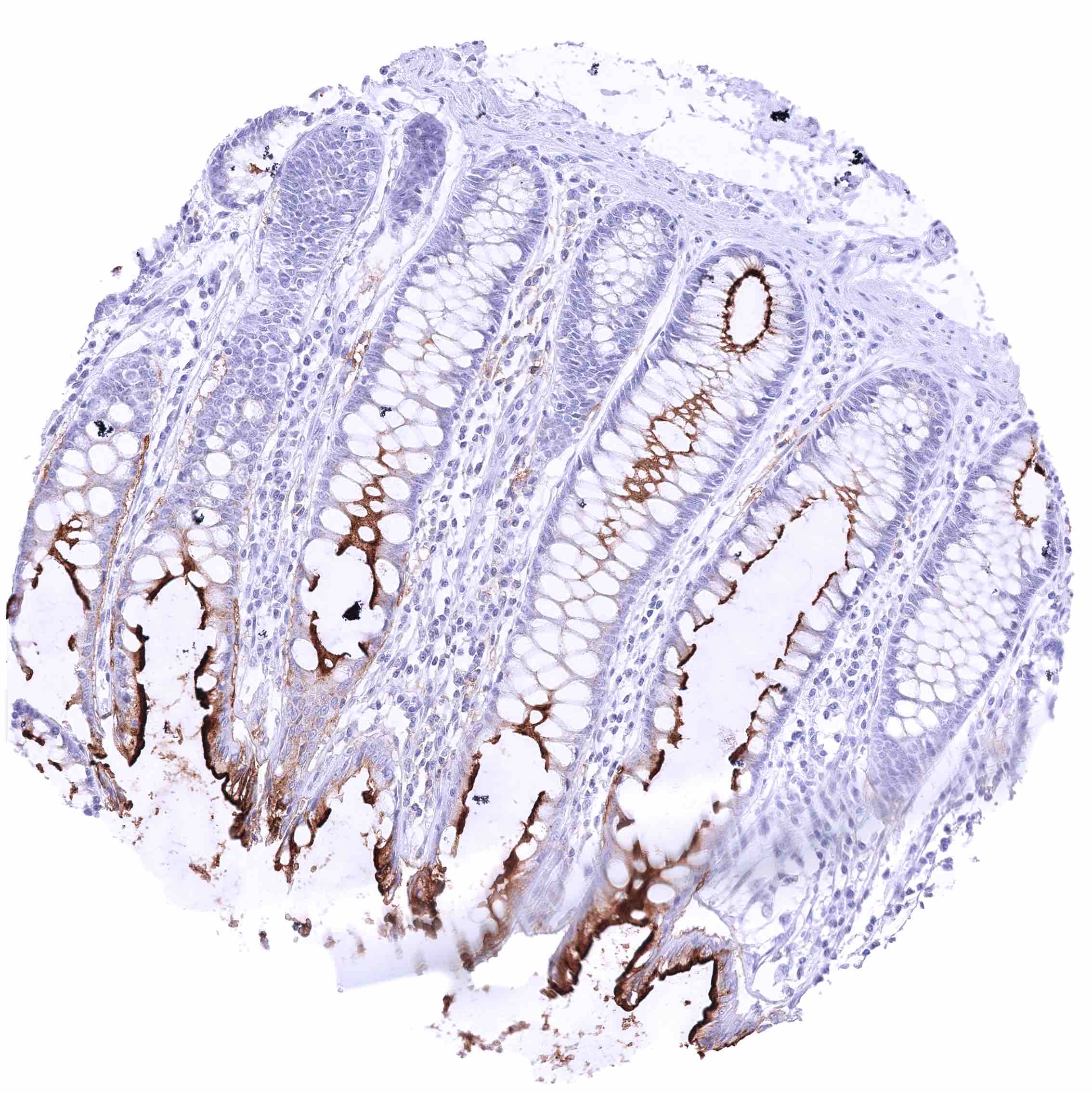
Rectum, mucosa – Strong GPRC5A membranous staining, largely limited to the luminal membranes. The staining intensity slightly decreases from the surface to the glands.
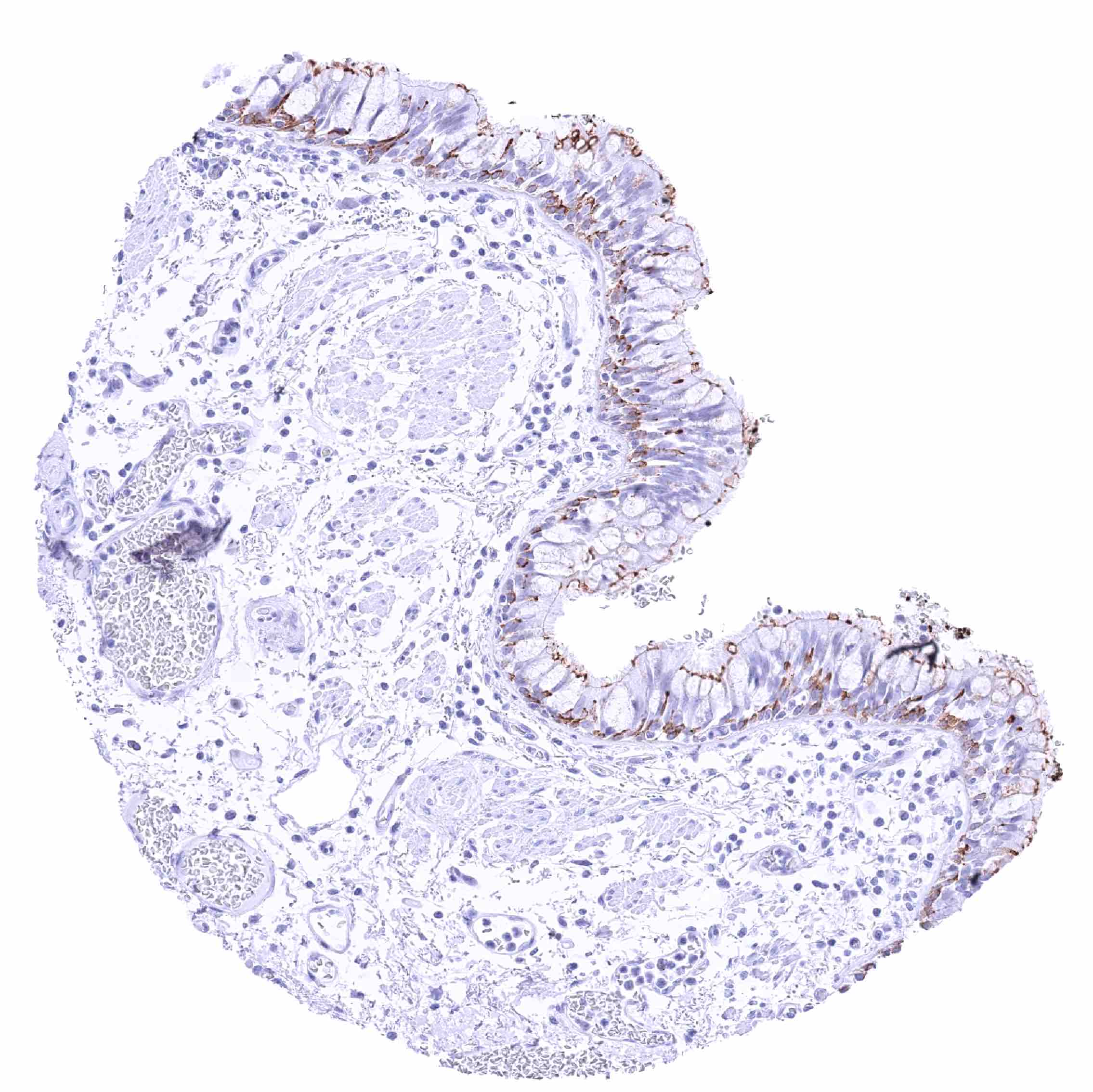
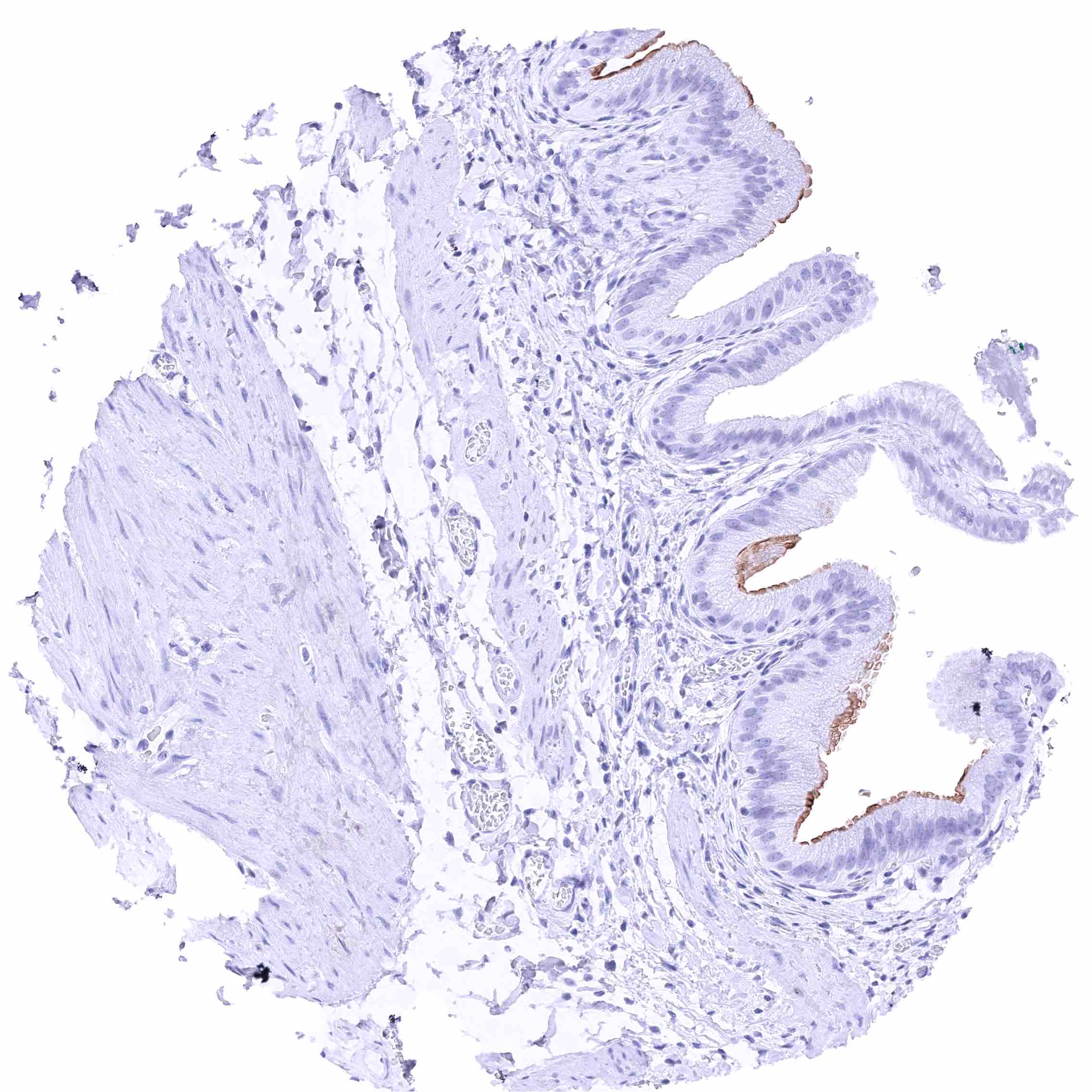
Respiratory epithelium with membranous GPRC5A staining of basal cells and amoderate GPRC5A staining of luminal membranes.
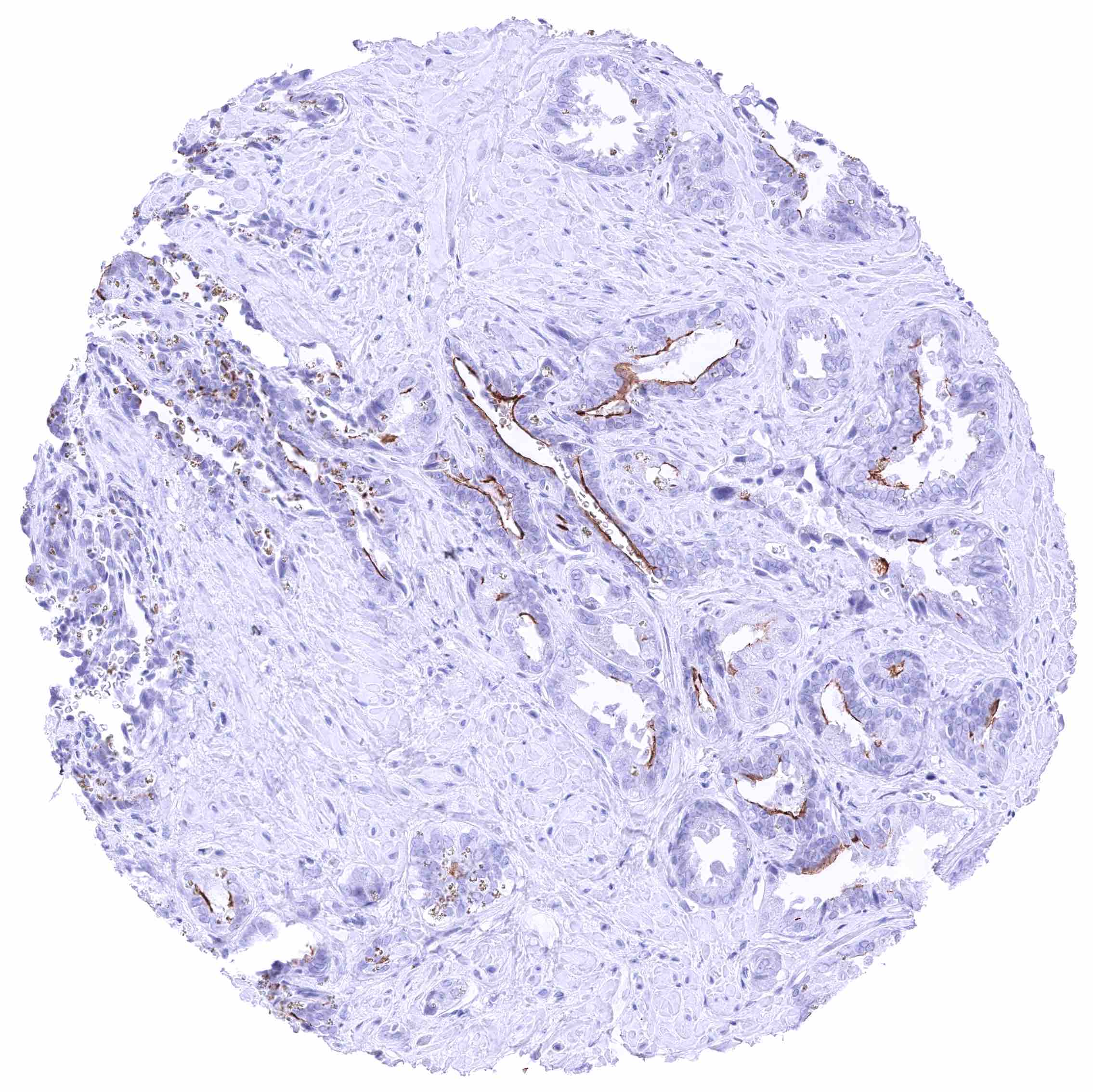
Seminal vesicle – Moderate to strong membranous GPRC5A staining of luminal membranes of a subset of epithelial cells.jpeg
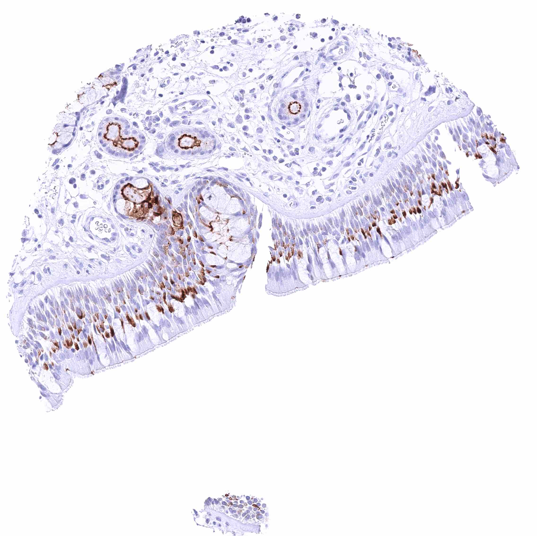
Seminal vesicle – Moderate to strong membranous GPRC5A staining of luminal membranes of a subset of epithelial cells
Sinus paranasales
Sinus paranasales
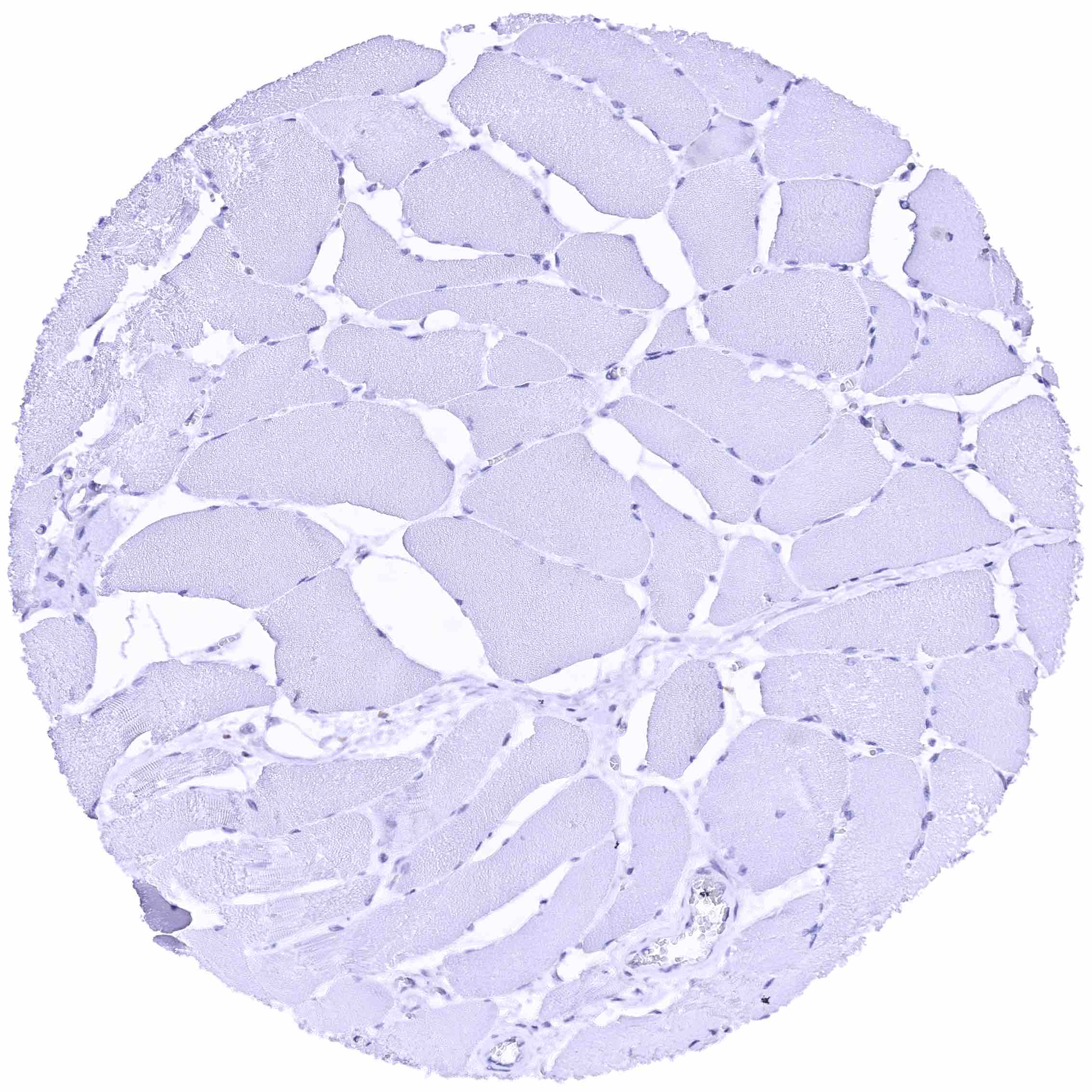
Skeletal muscle
Skeletal muscle
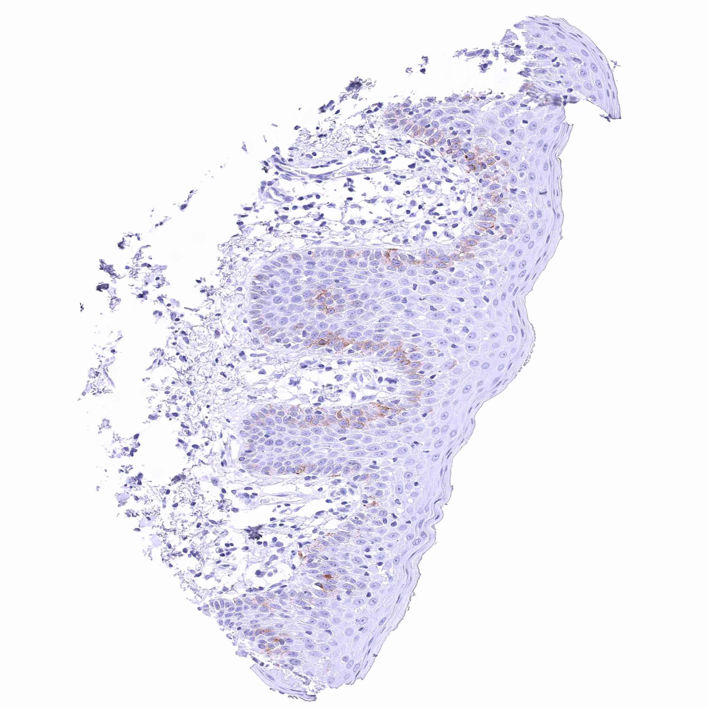
Skin – Faint membranous GPRC5A staining of a fraction of basal-suprabasal squamous epithelial cells in this sample.
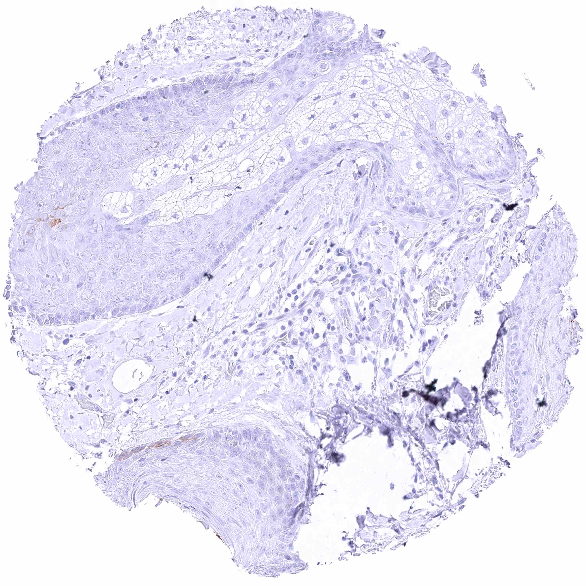
Skin, hairfollicel and sebaceous glands – Faint membranous GPRC5A staining of few basal-suprabasal squamous epithelial cells.
Spleen
Spleen
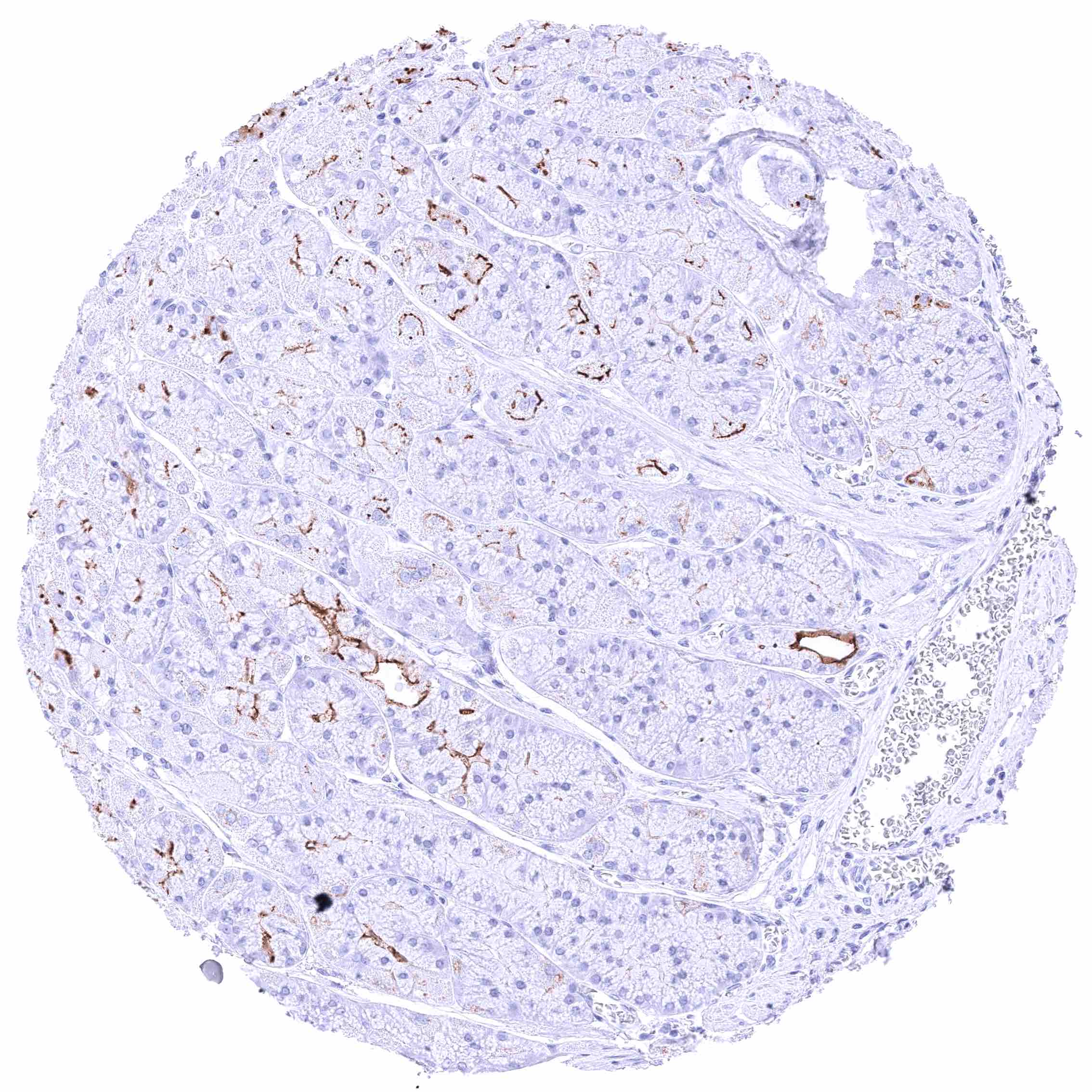
Stomach, antrum – Strong GPRC5A membranous staining, largely limited to the luminal membranes. Especially in deep glands, the membranous GPRC5A appears to be incomplete.
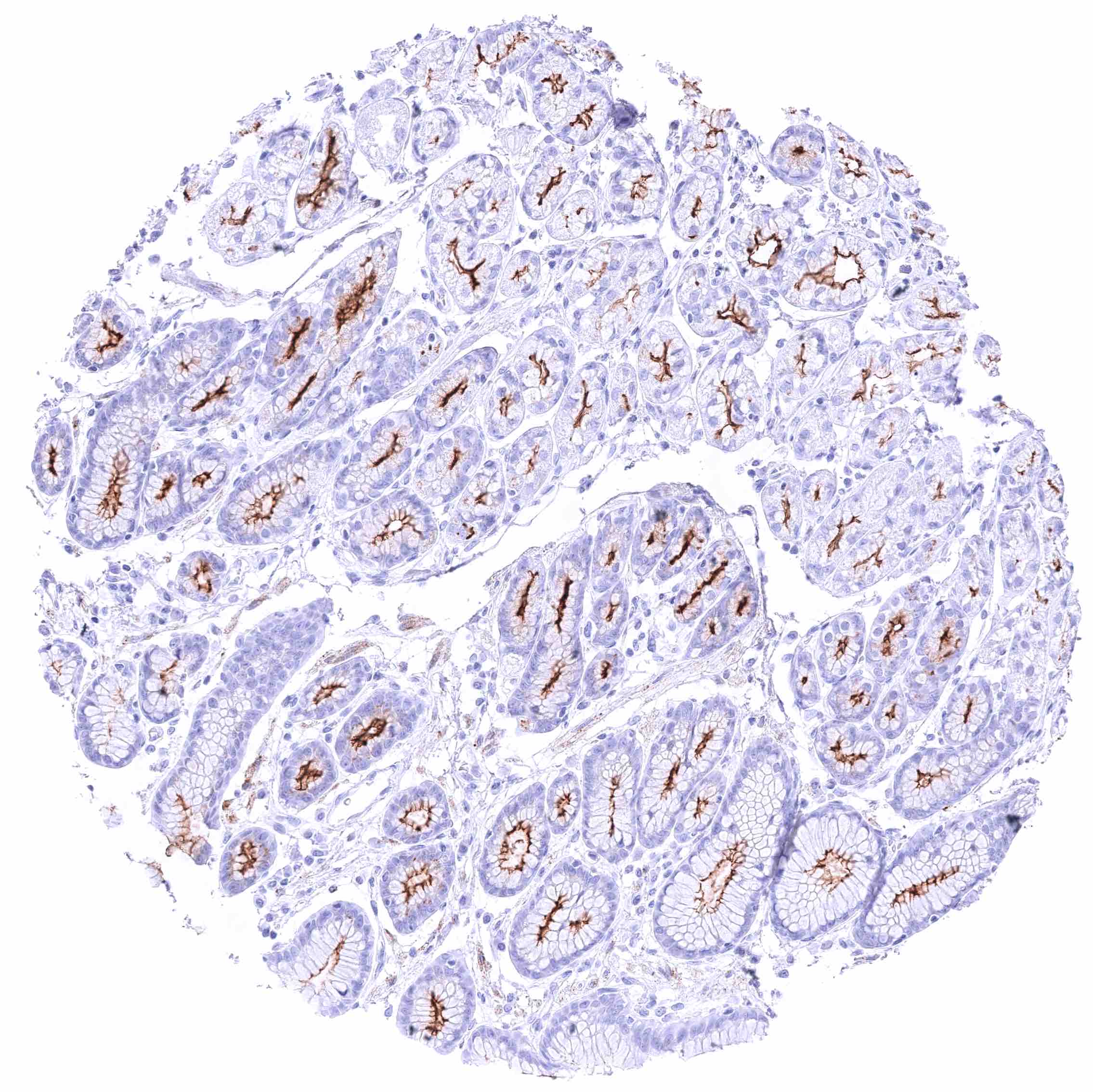
Stomach, antrum – Strong GPRC5A membranous staining, largely limited to the luminal membranes. The staining intensity is higher at the surface than in glands.
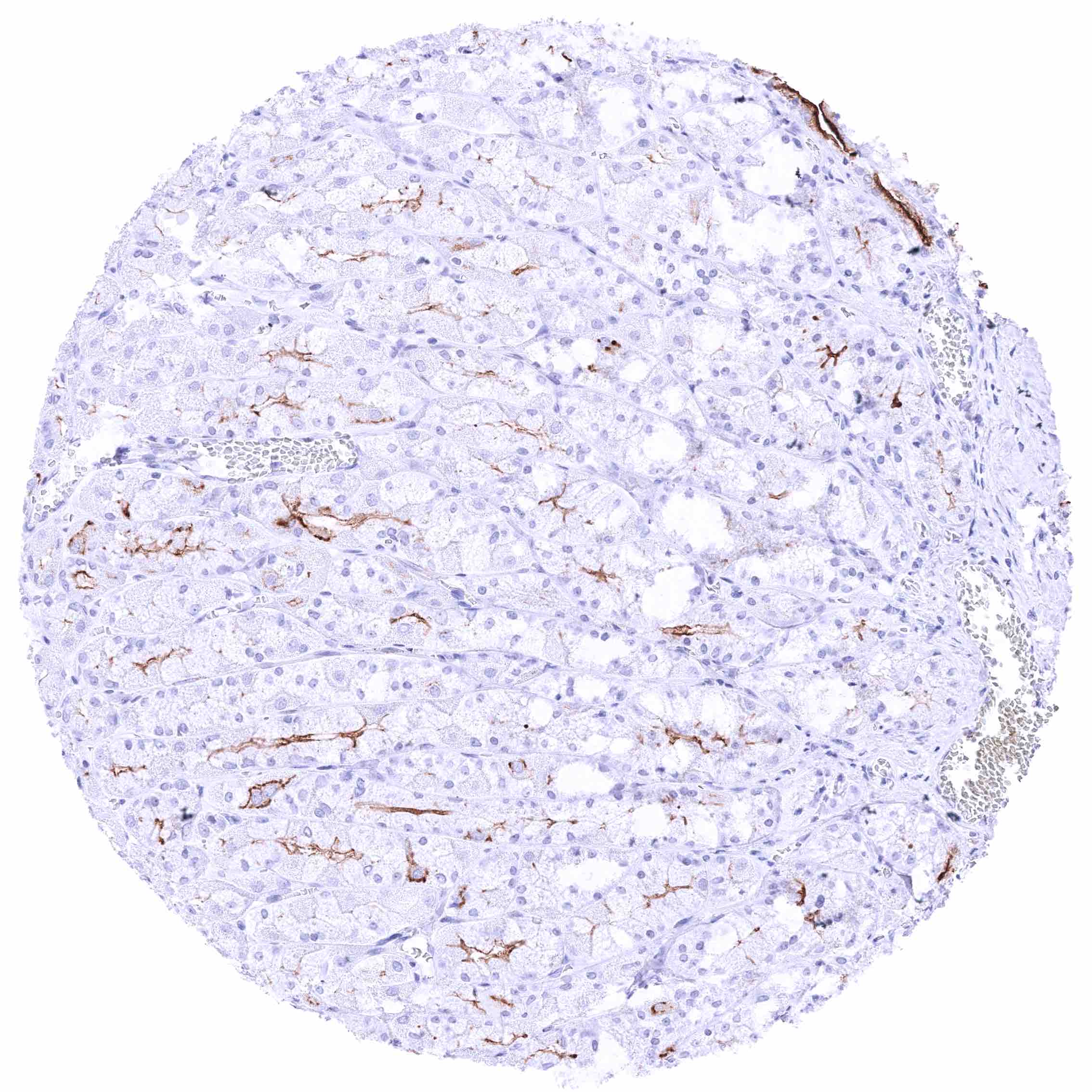
Stomach, corpus – Strong GPRC5A membranous staining, largely limited to the luminal membranes. Especially in deep glands, the membranous GPRC5A appears to be incomplete.
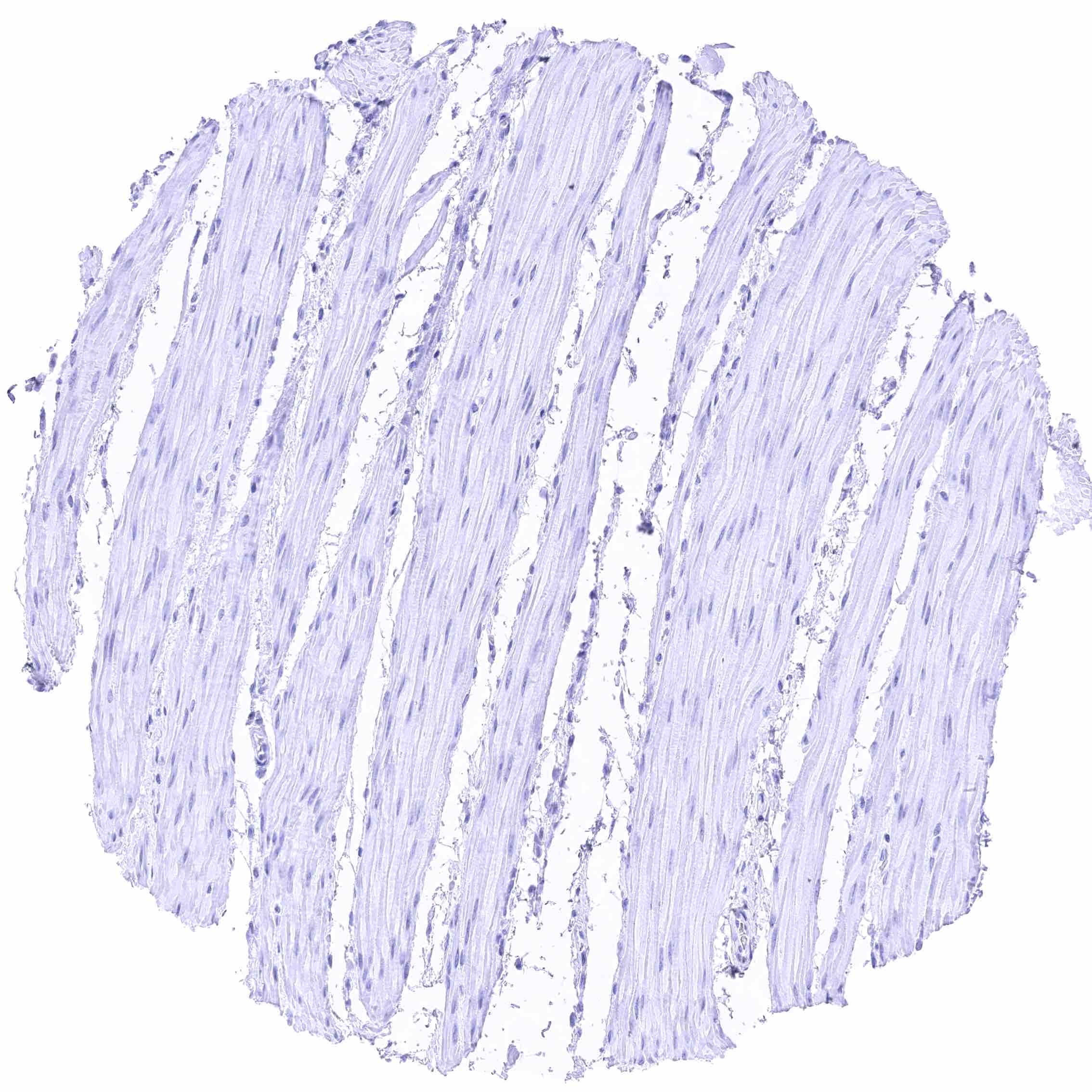
Stomach, muscular wall
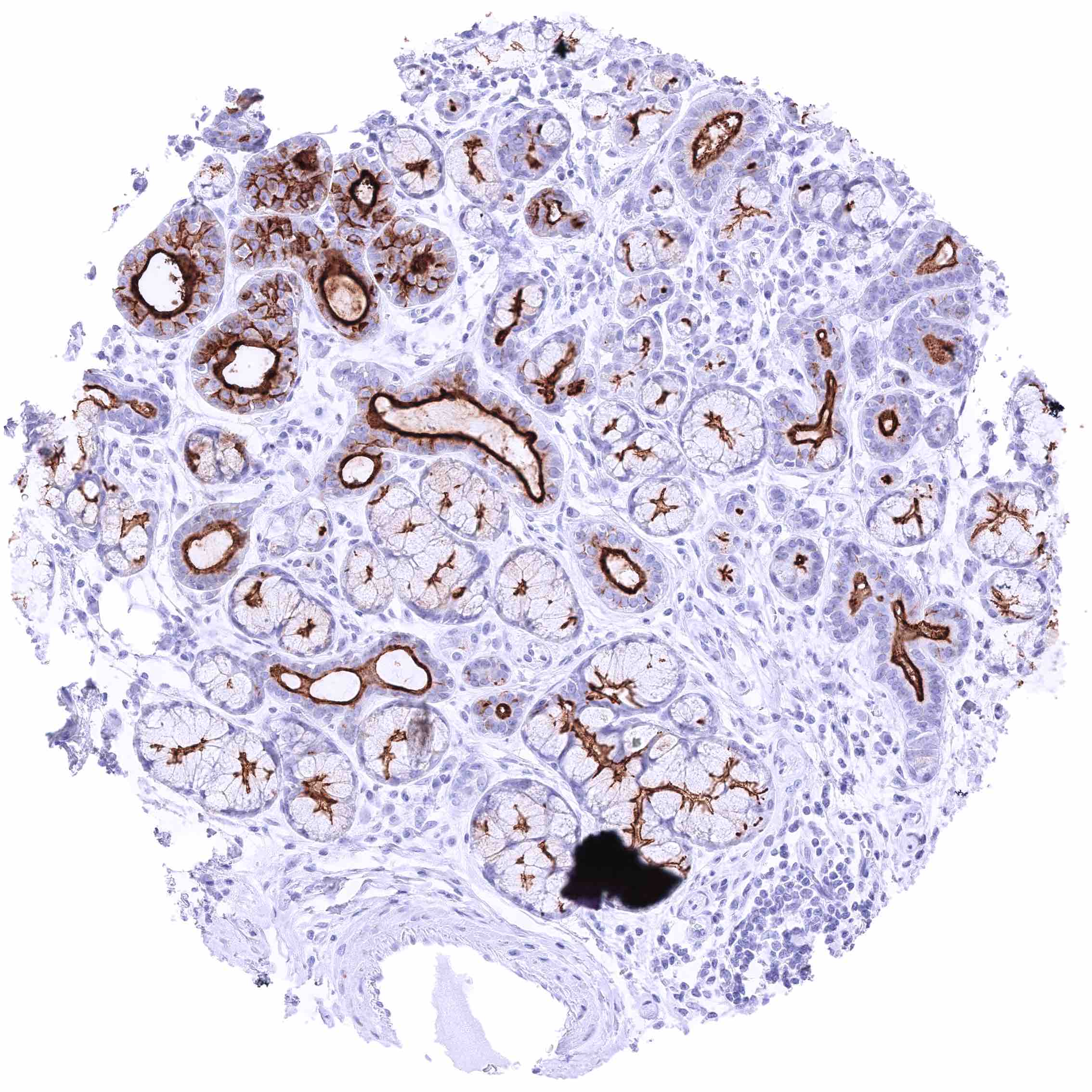
Sublingual gland – Intense GPRC5A staining of luminal membranes of excretory duct cells. GPRC5A staining is less intensive in glands.
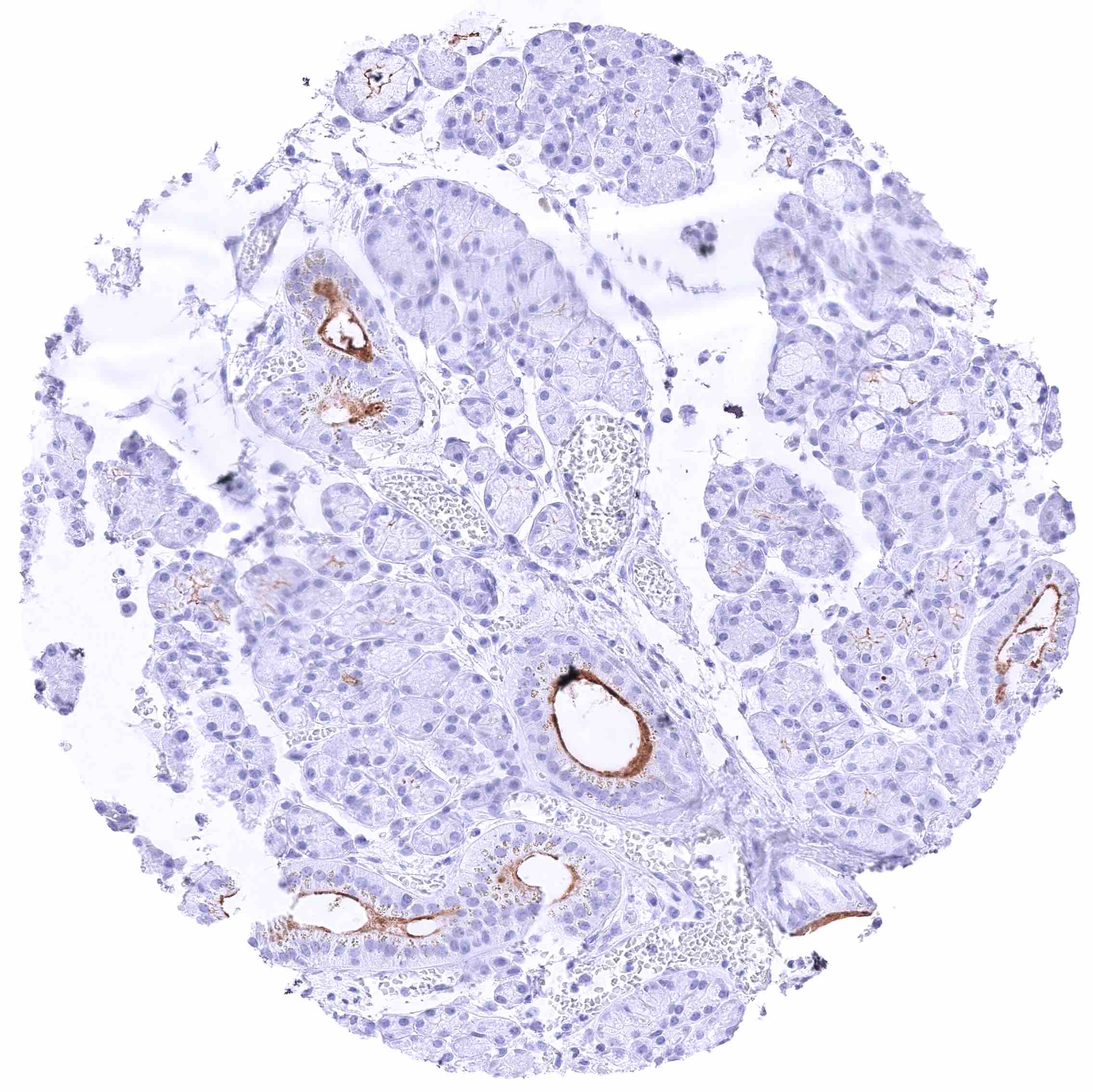
Submandibular gland – Strong GPRC5A staining of luminal membranes of excretory duct cells. A less intensive staining is also seen in variable subsets of glands.
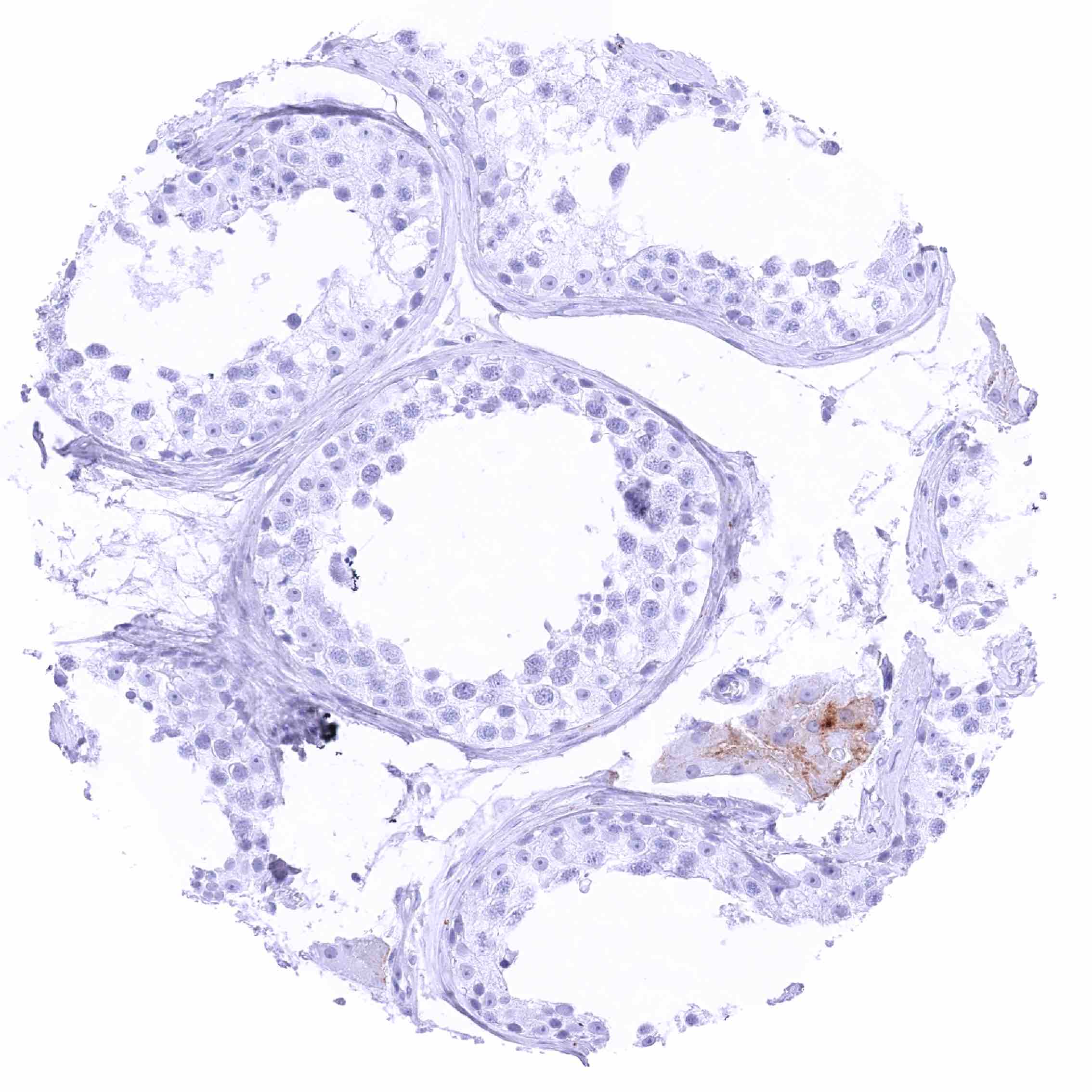
Testis – Weak, predominantly membranous GPRC5A staining of Leydig cells.
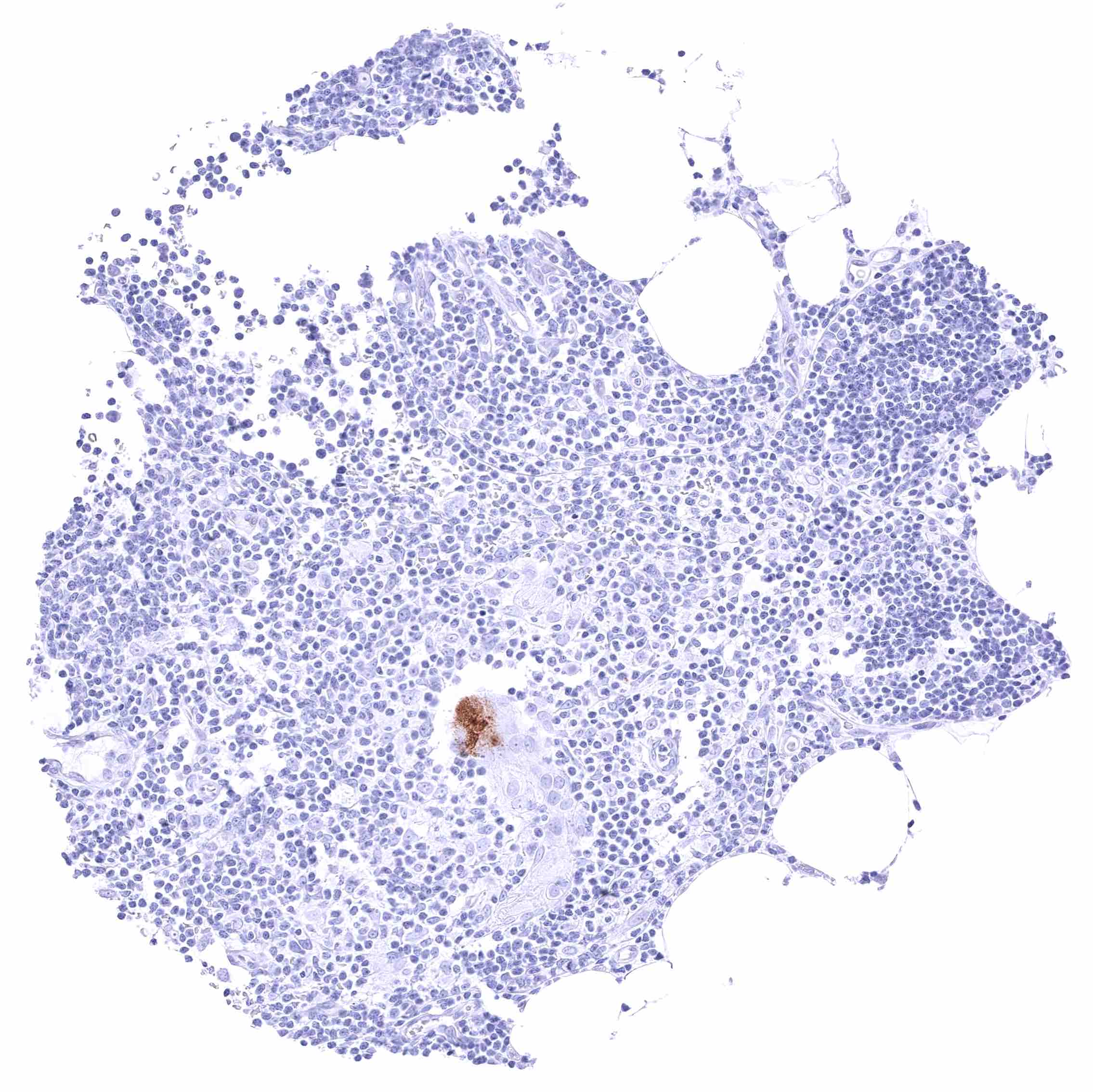
Thymus – GPRC5A staining is limited to a corpuscle of Hassall’s.
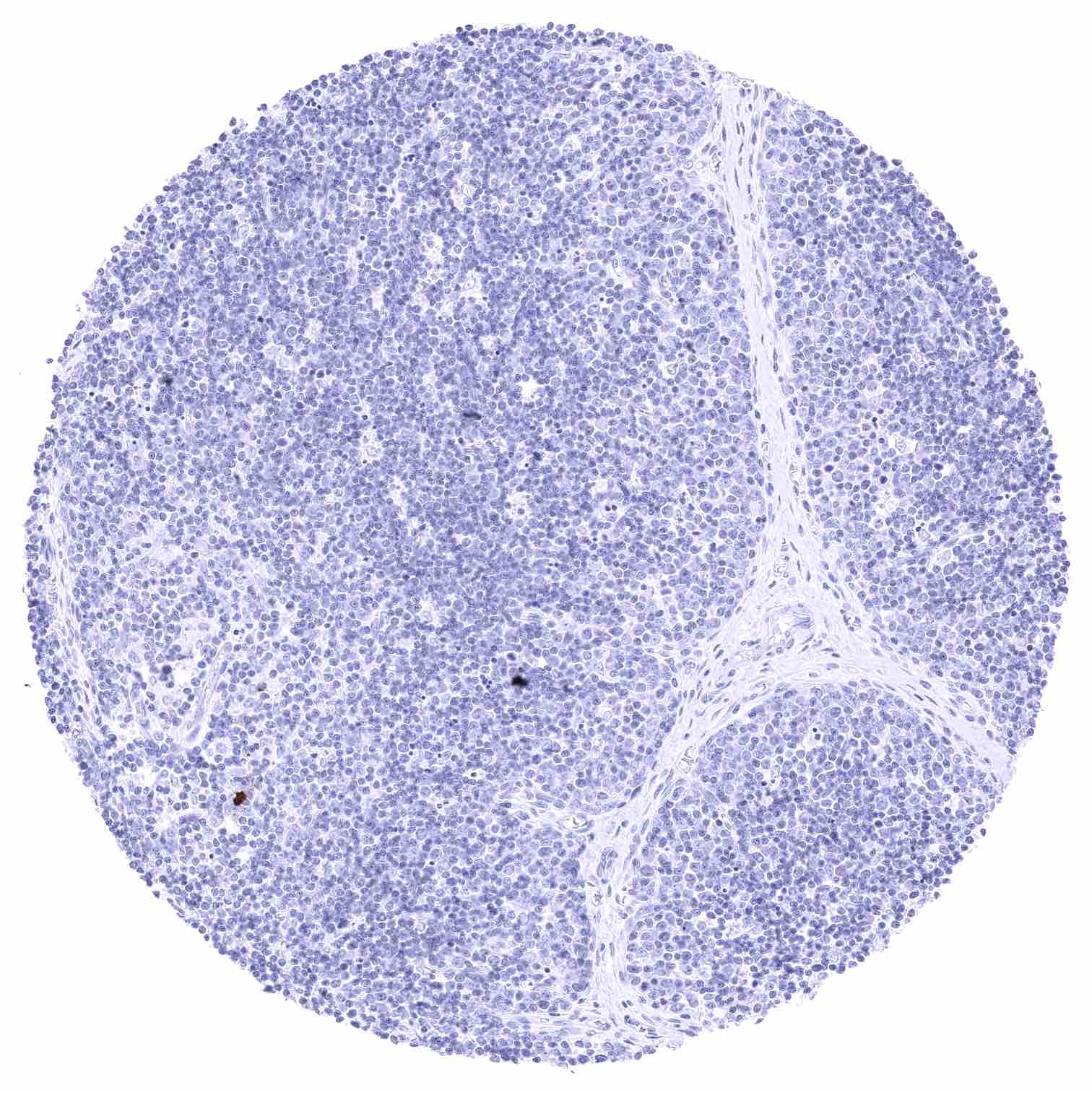
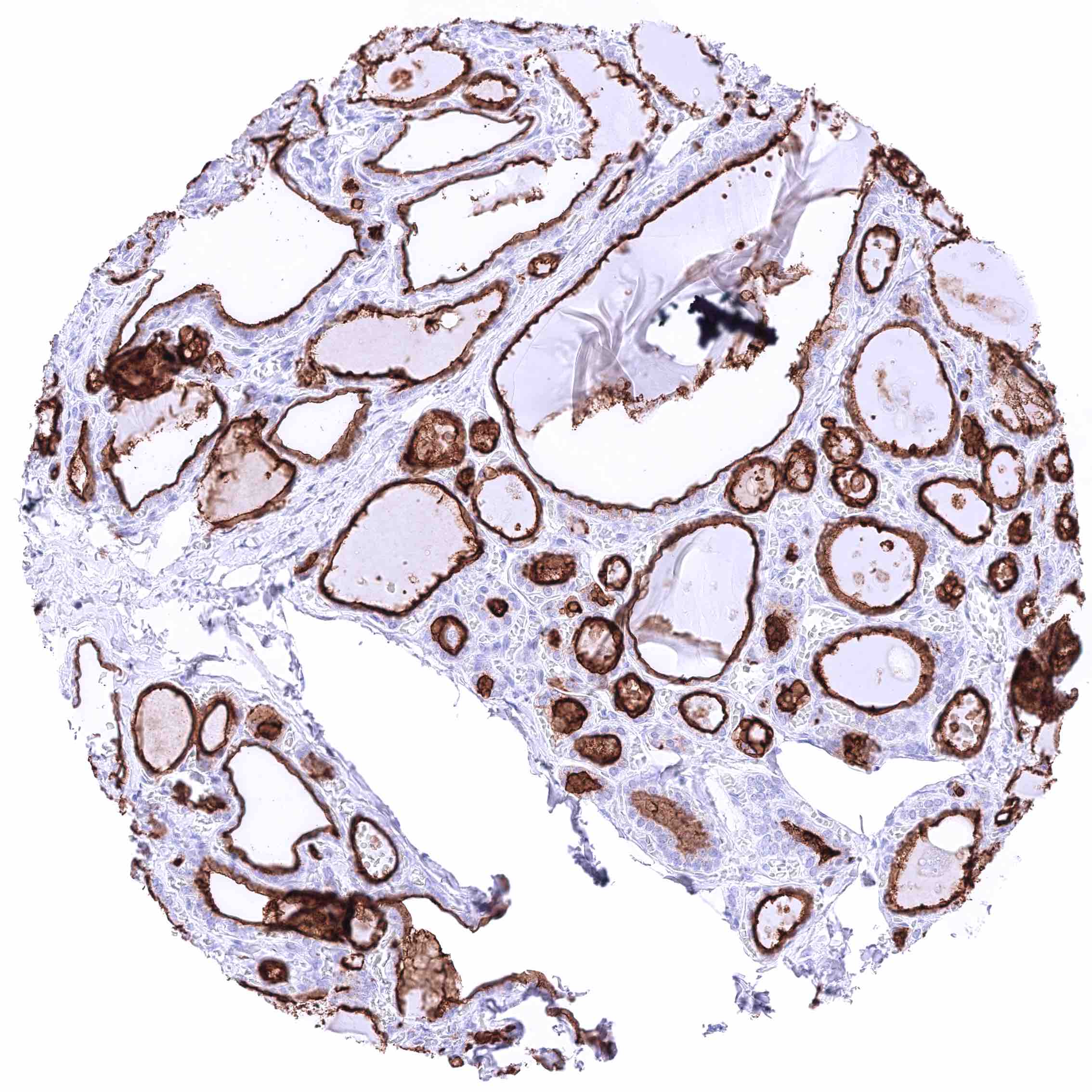
Thymus
Thymus
Thyroid gland
Thyroid gland
Tonsil – GPRC5A staining is limited to few squamous epithelial cells.
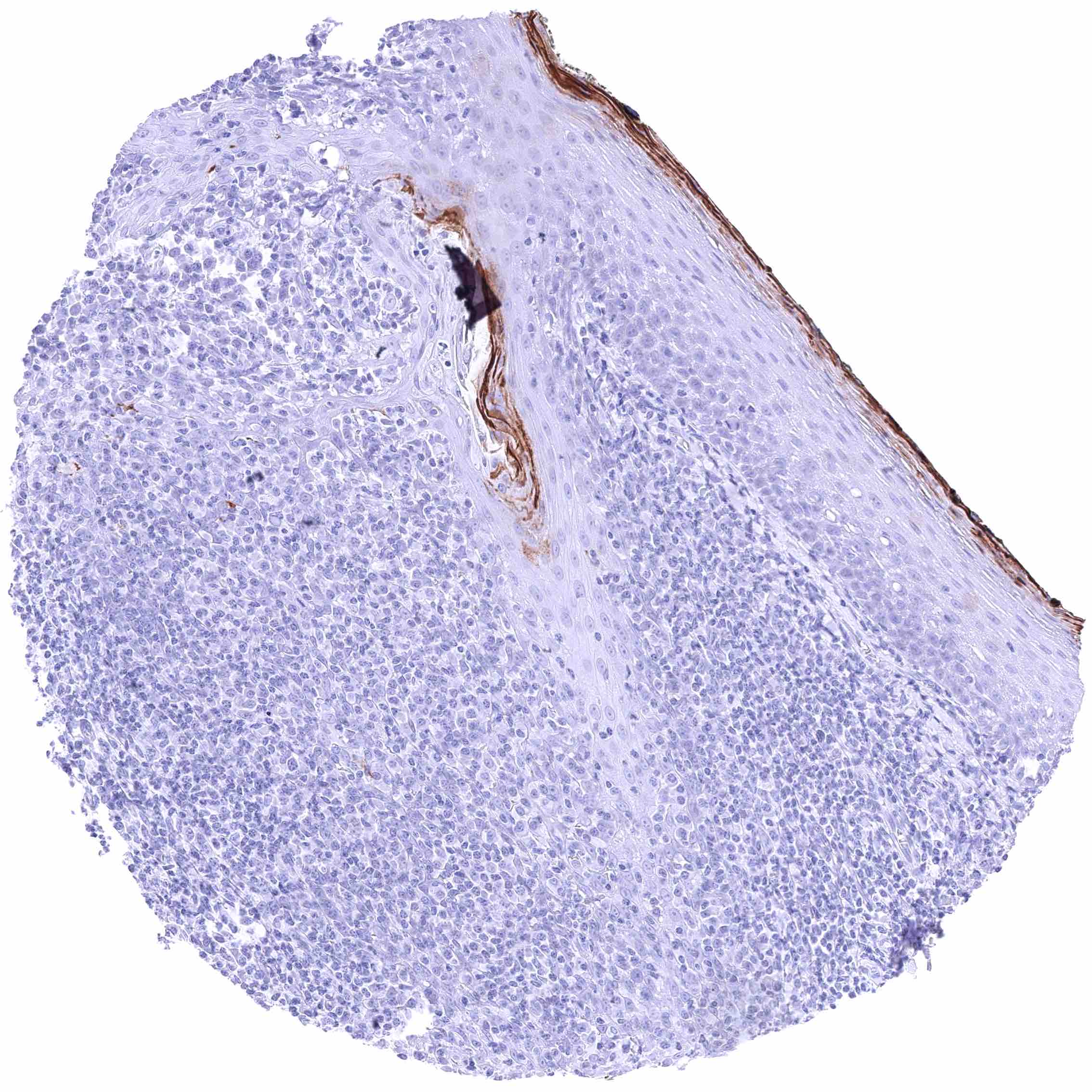
Tonsil, surface epithelium – Strong membranous and cytoplasmic GPRC5A staining of the superficial layers of squamous epithelial cells.
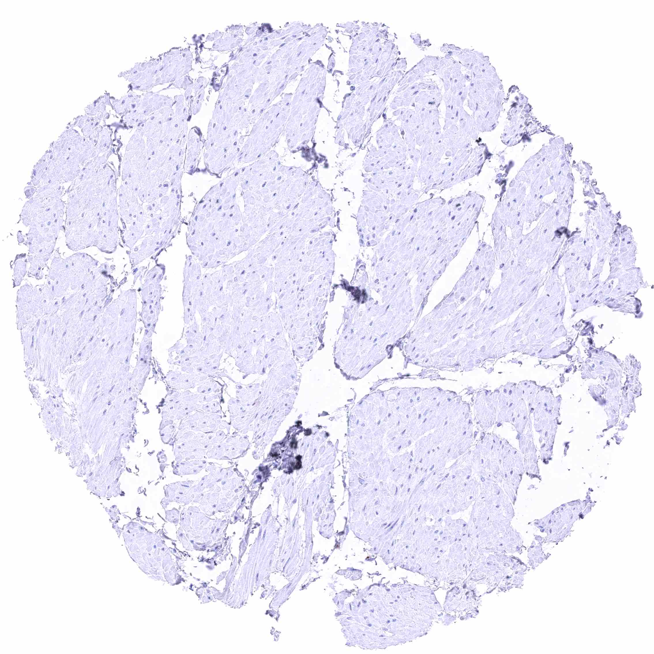
Urinary bladder, muscular wall
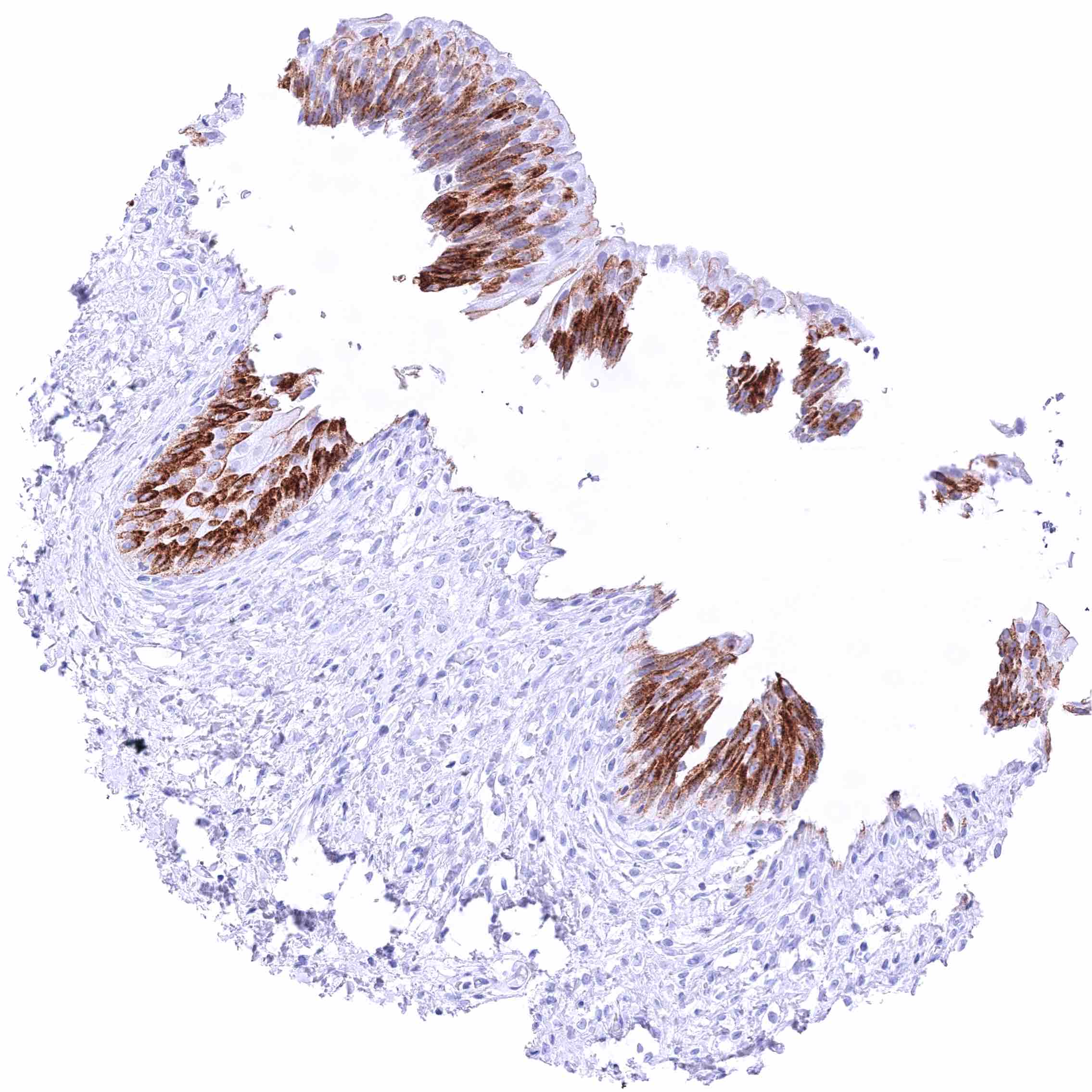
Urinary bladder, urothelium
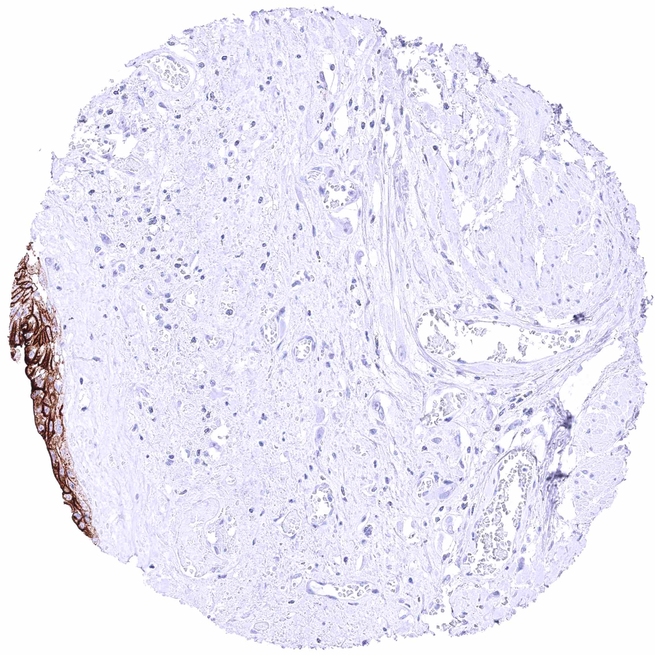
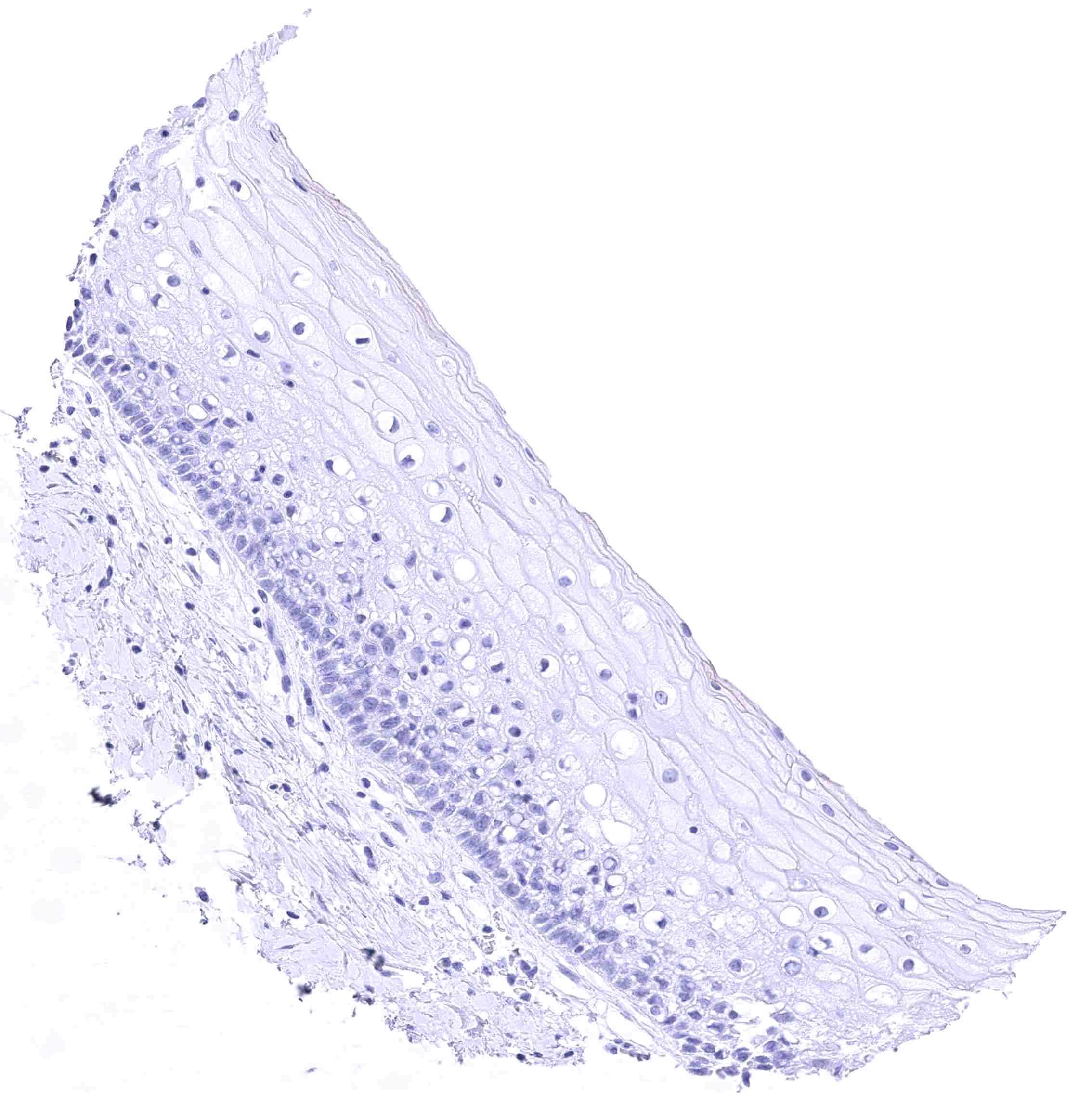
Uterus, ectocervix – Absence of GPRC5A staining in this sample.
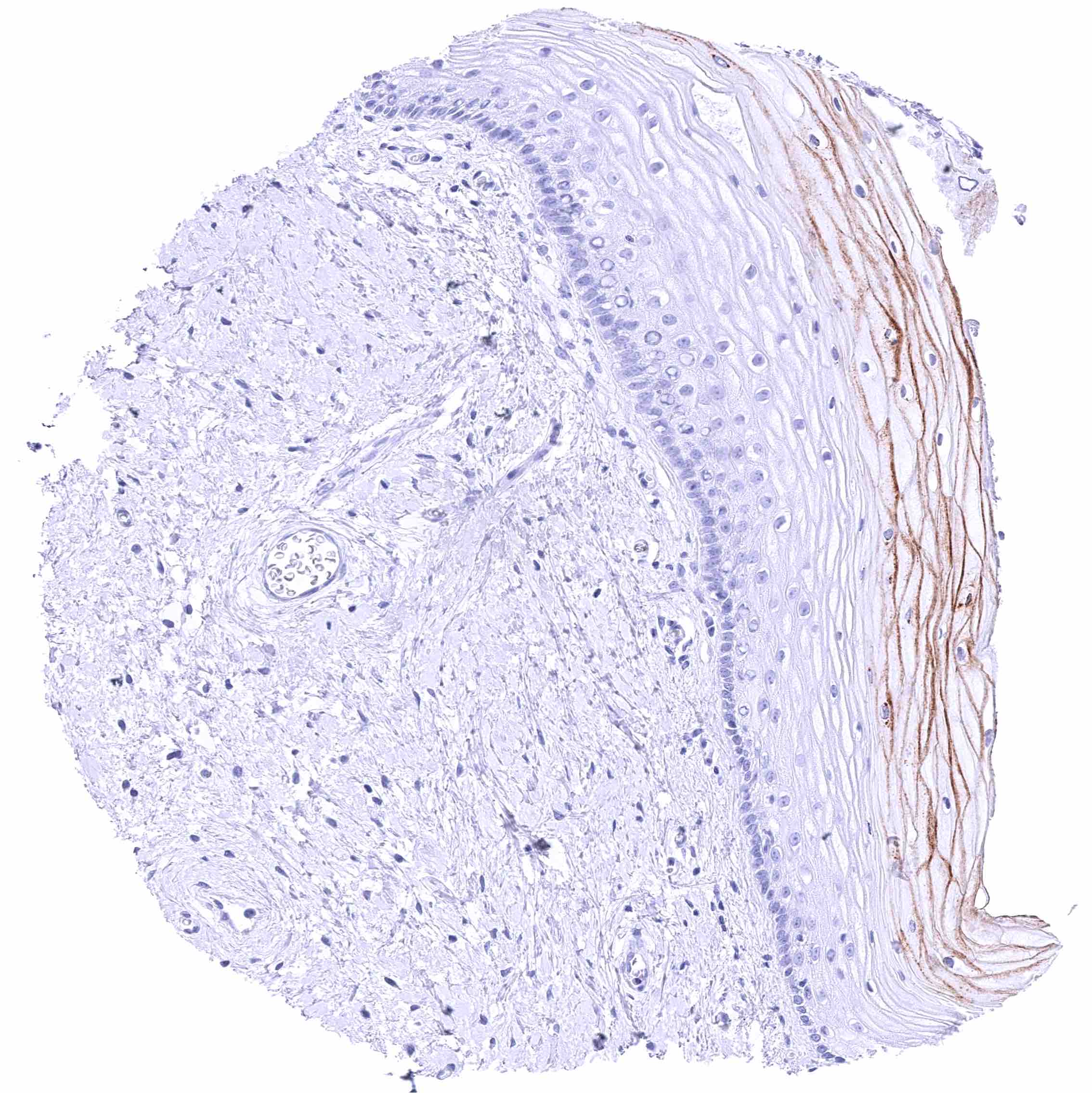
Uterus, ectocervix – Moderate membranous GPRC5A staining of the superficial layers of squamous epithelial cells.
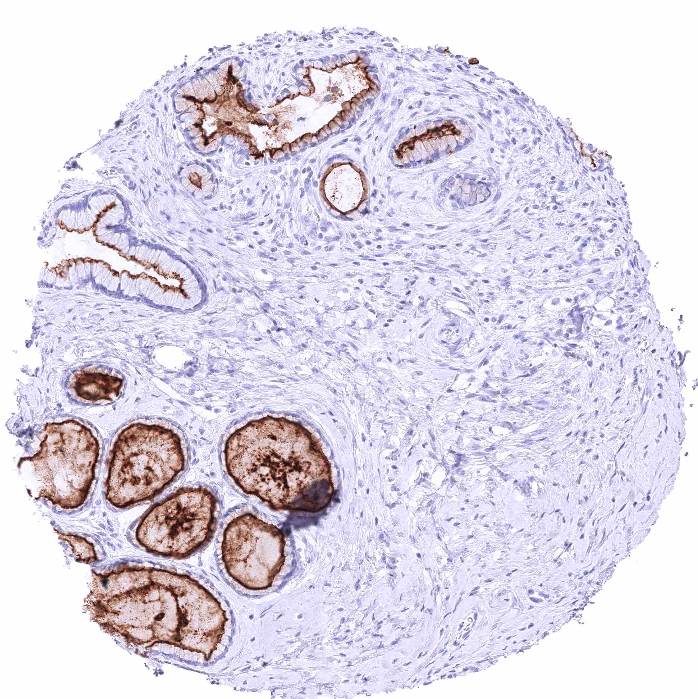
Uterus, endocervix – Strong GPRC5A staining of (predominantly) apical membranes of endocervical cells.
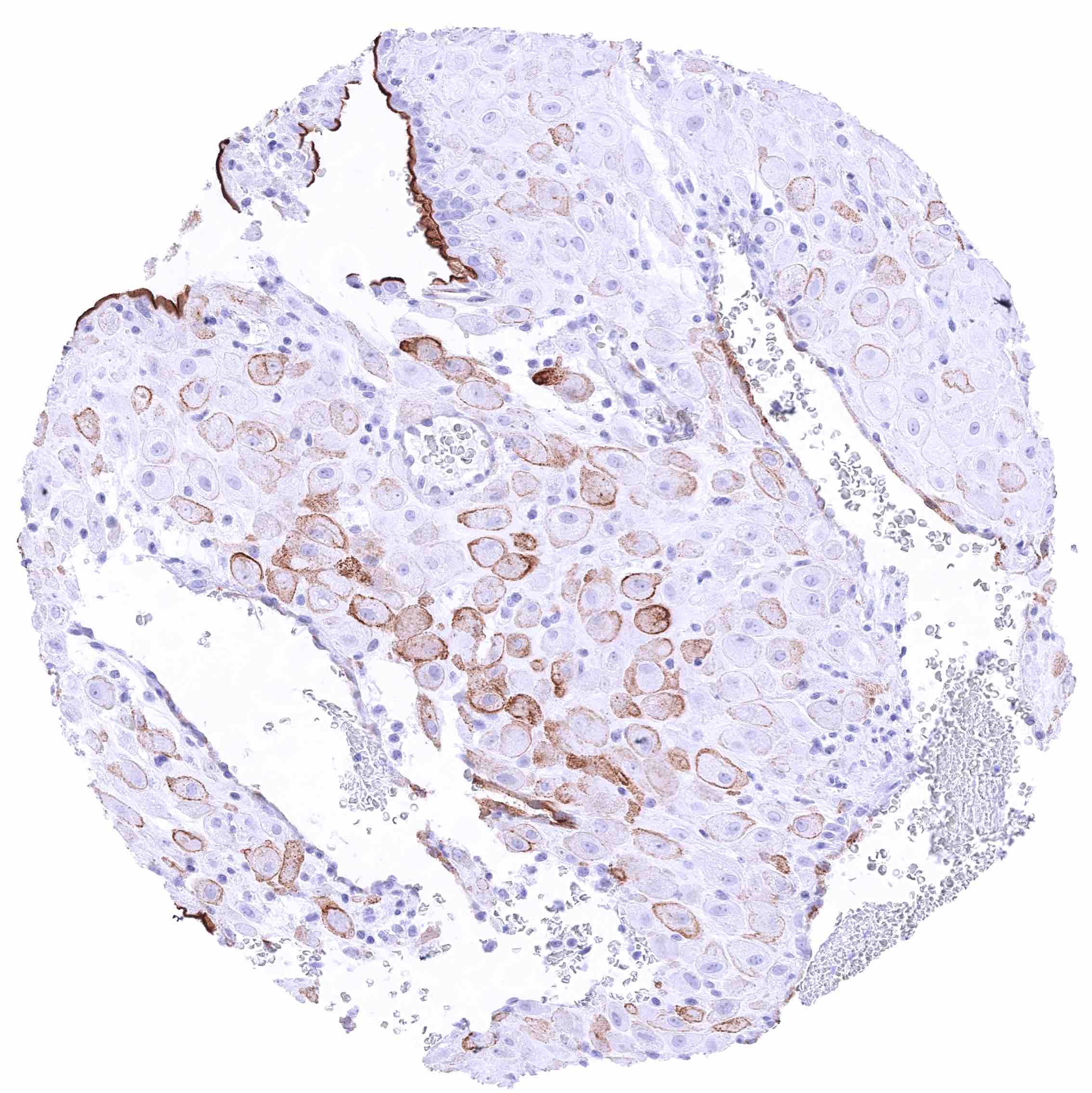
Uterus, endometrium (pregnancy) – GPRC5A staining of apical membranes of some but not all endometrium gland cells (variable intensity). A subset of decidua cells shows membranous GPRC5A positivity.
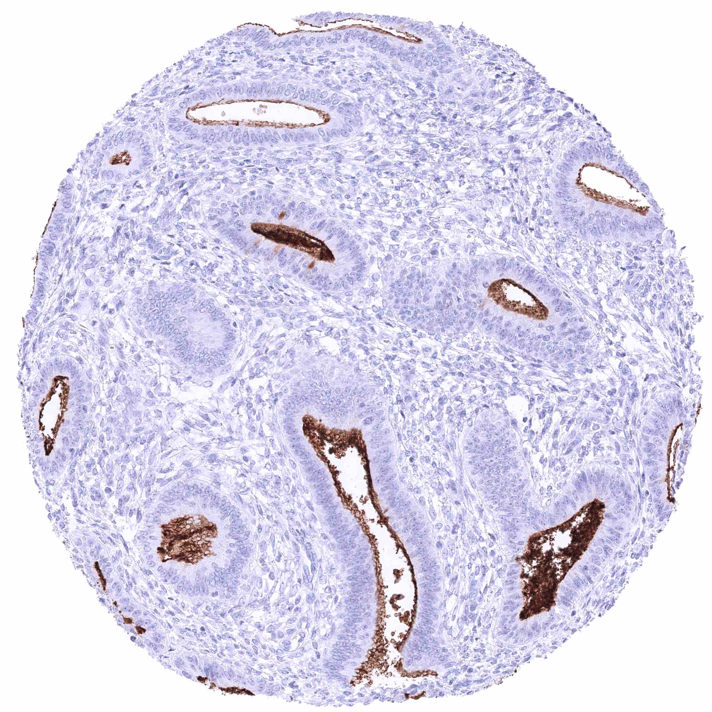
Uterus, endometrium (proliferation) – Strong GPRC5A staining of apical membranes of endometrium gland cells.
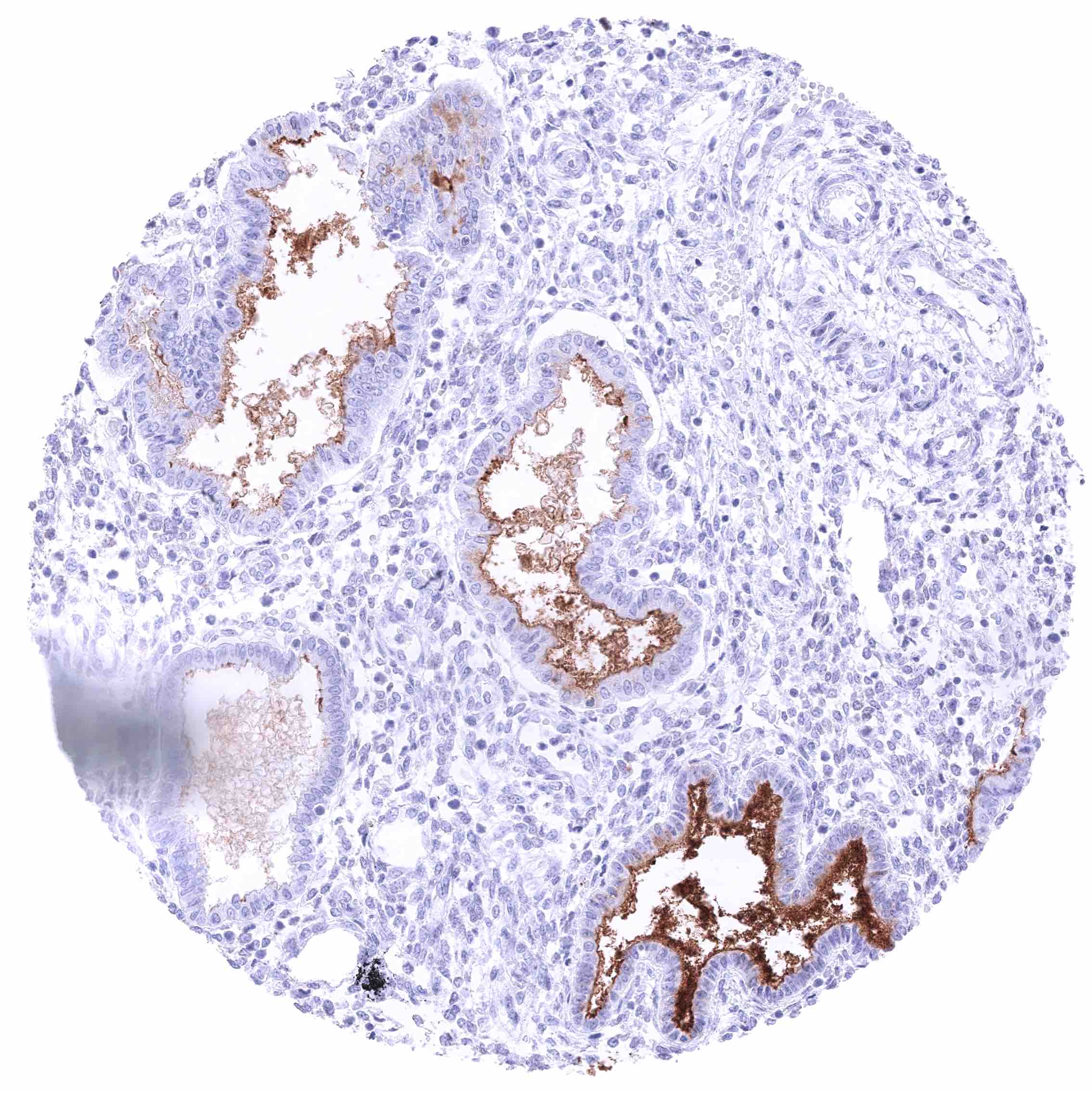
Uterus, endometrium (secretion) – Distinct GPRC5A staining of apical membranes of endometrium gland cells. The intensity of staining varies between different glands.
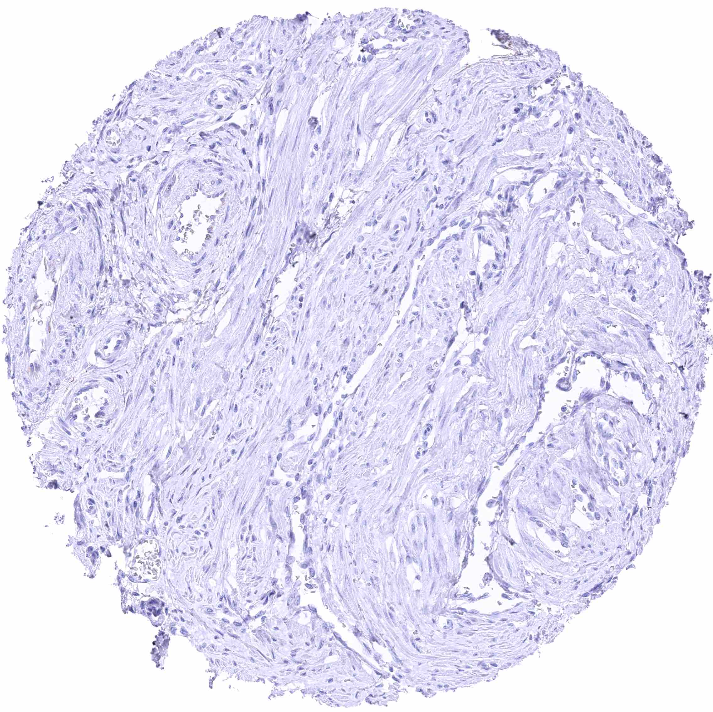
Uterus, myometrium
Uterus, myometrium
  • Linkedin
  • My Account
  • Search
    ×
  • Cart 0
© 2026 MS Validated Antibodies
  • Imprint
  • Privacy Policy
  • Distributors
  • Quality