295,00 € – 995,00 €
Product details
Synonyms = forkhead box A1 , HNF3A , TCF3A
Antibody type = Recombinant Rabbit monoclonal / IgG
Clone = HMV4726
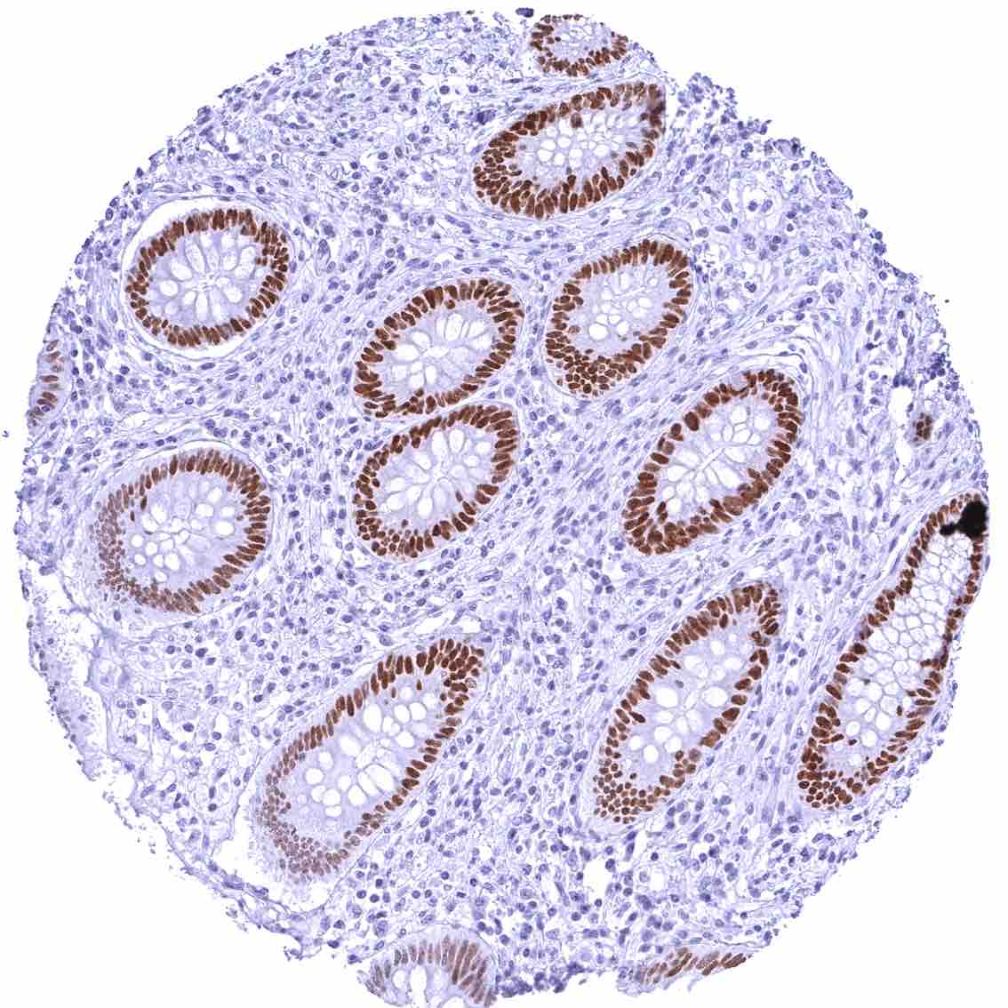
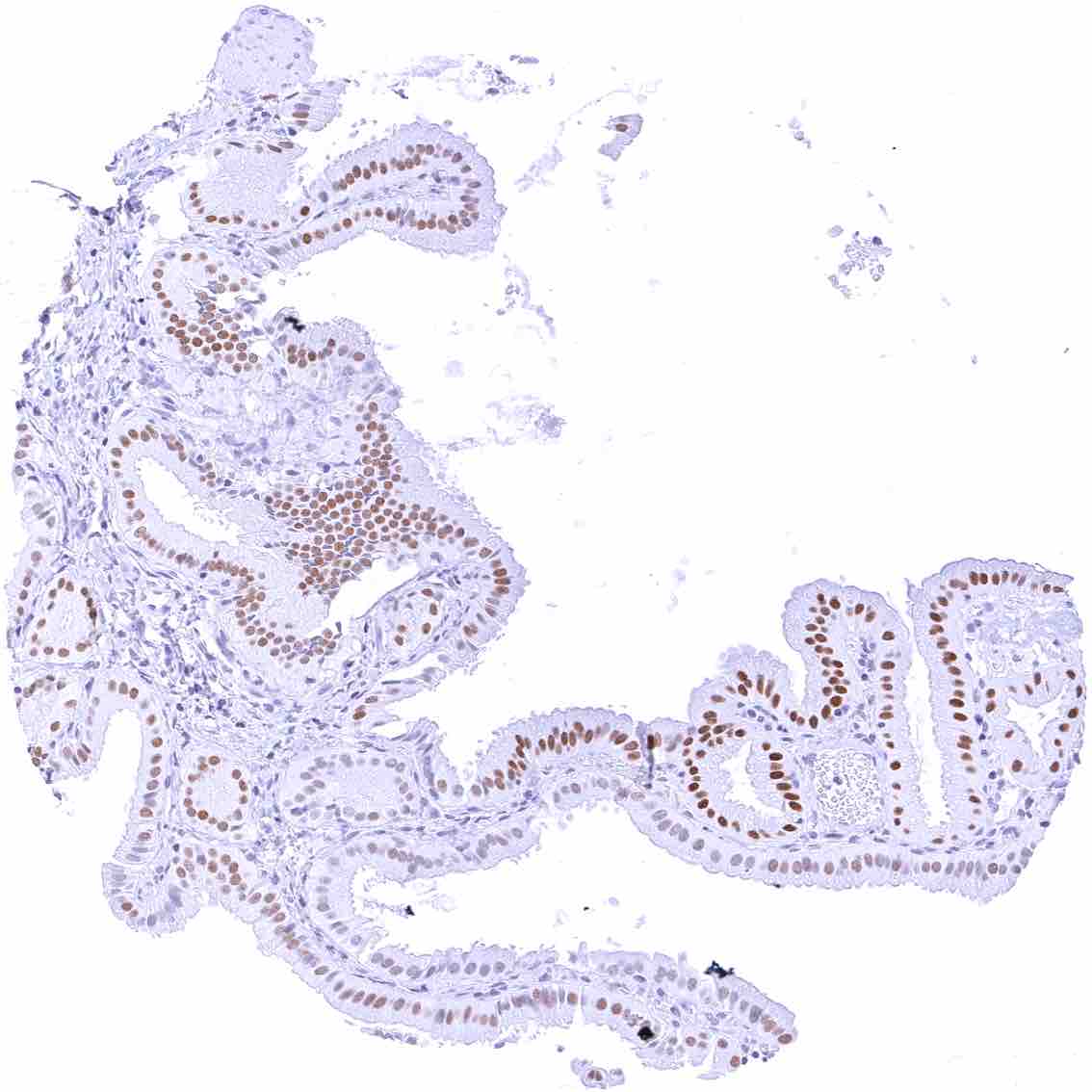
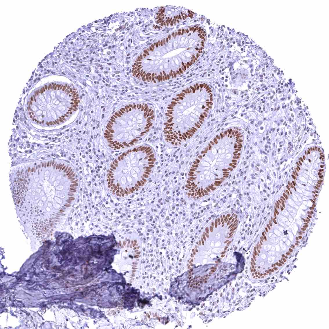
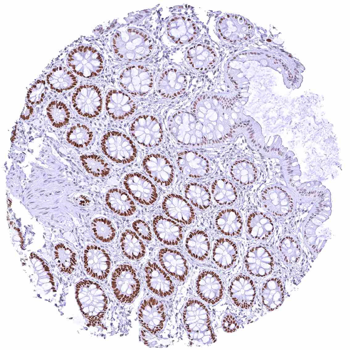
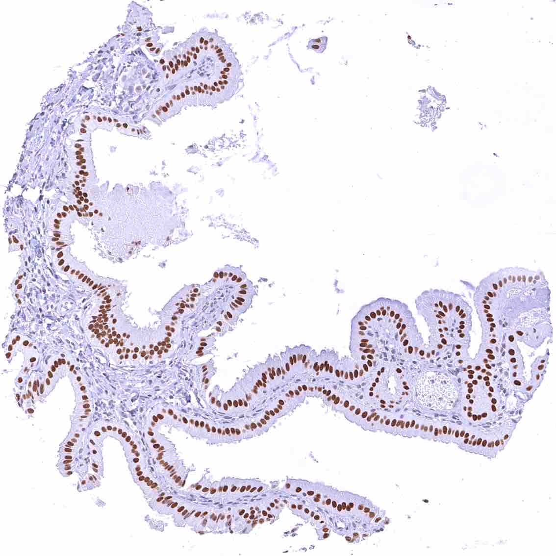
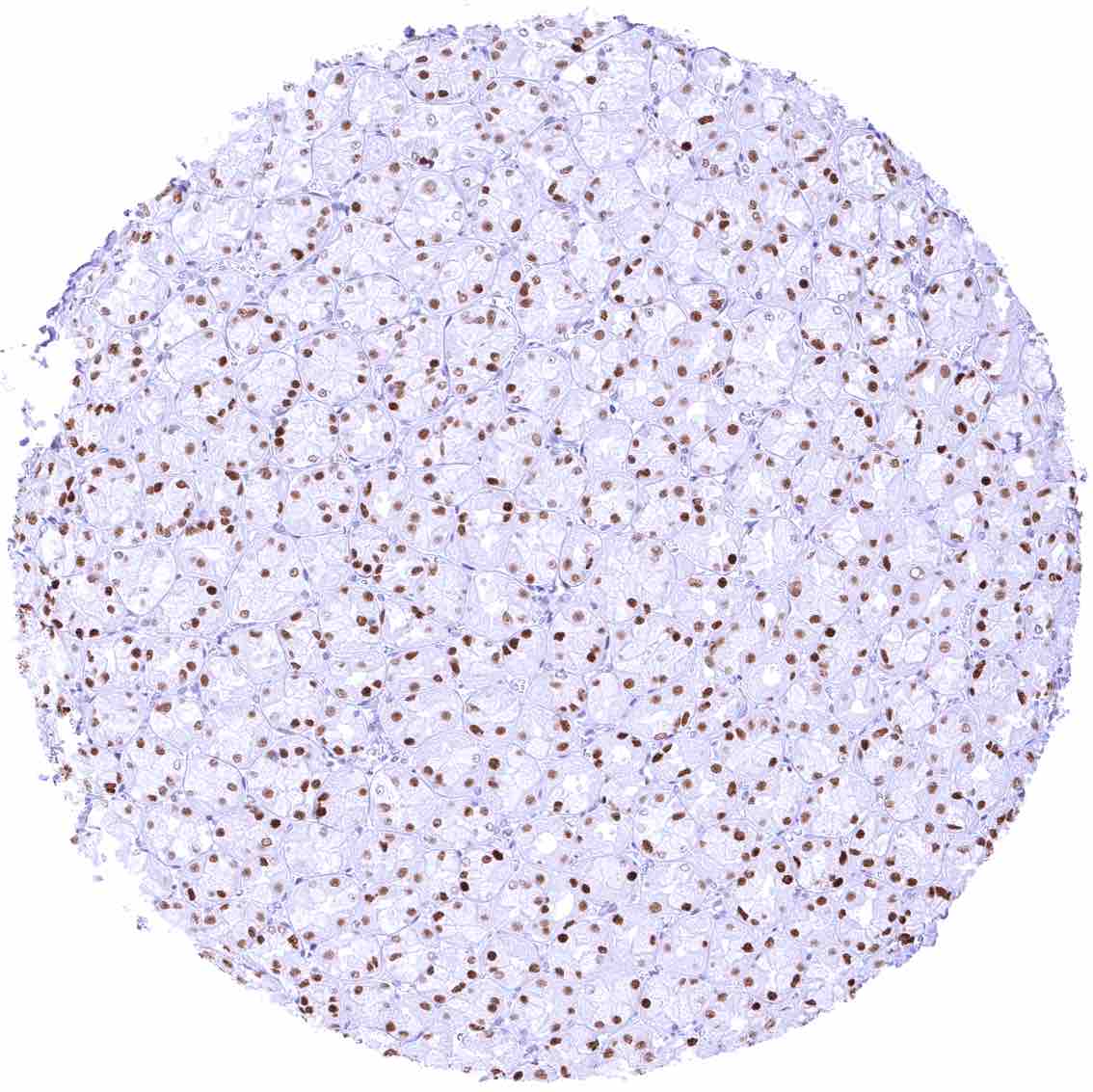
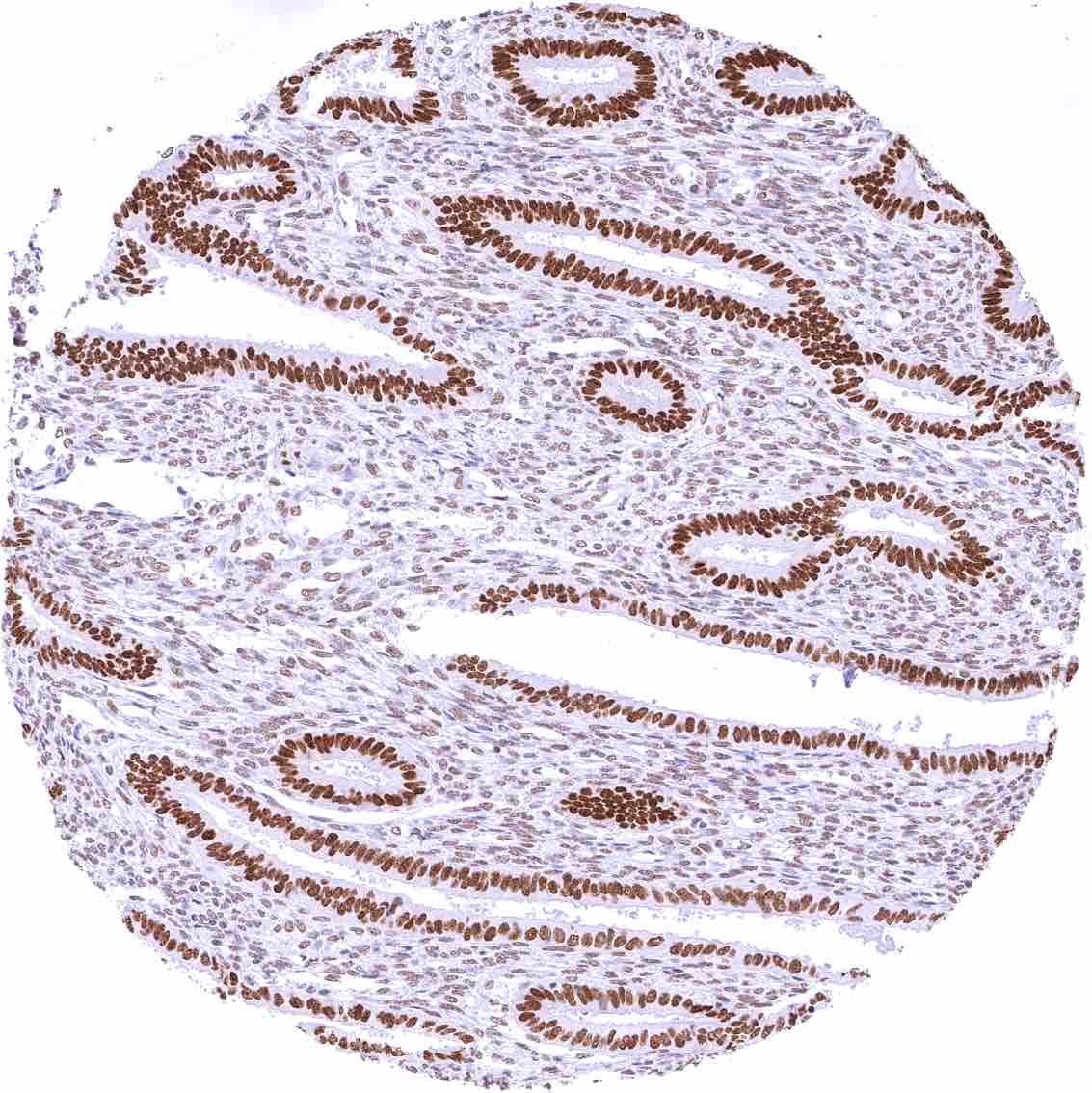
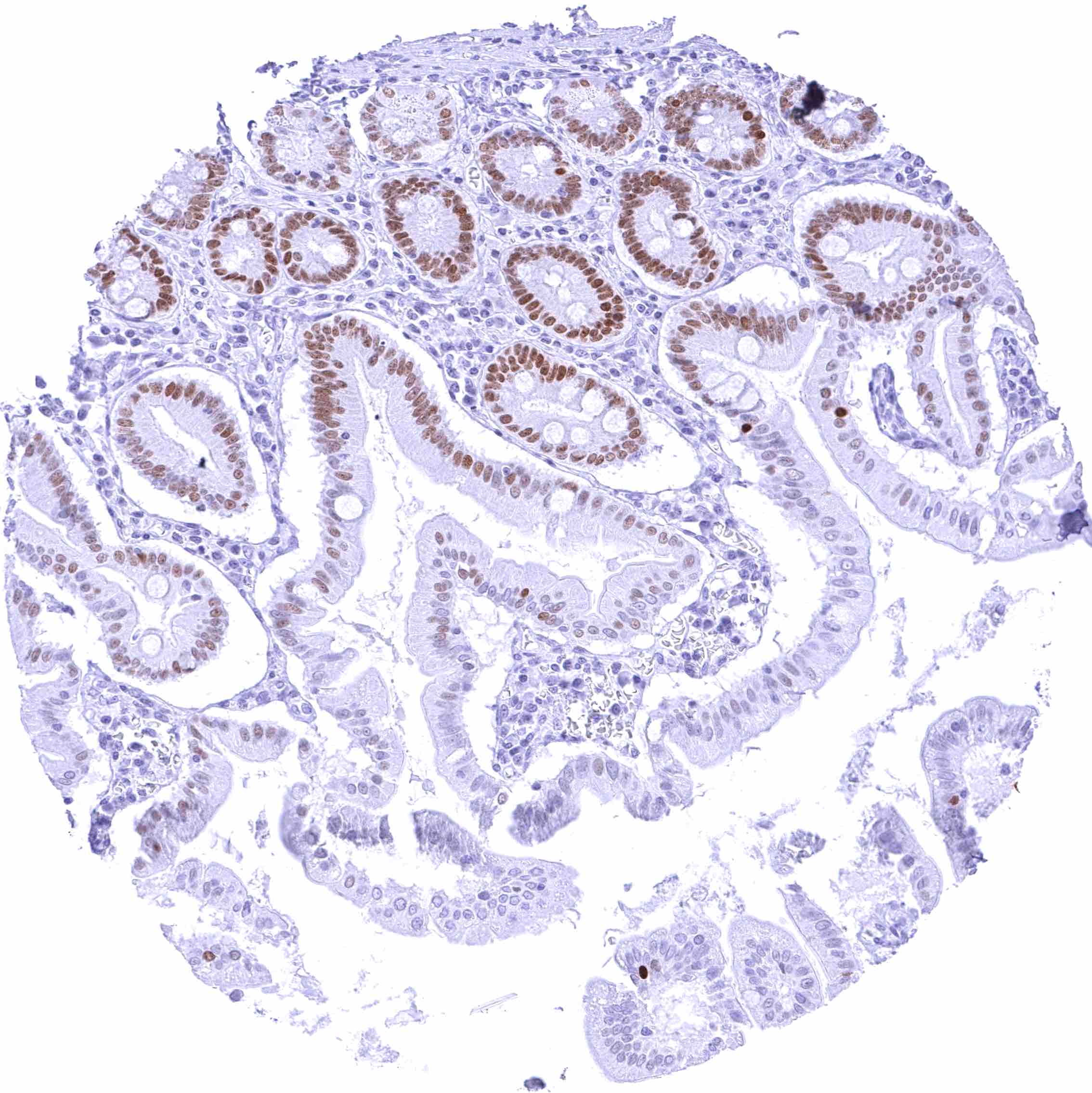
Positive control = Colon: A strong nuclear FOXA1 staining should be seen in crypt cells the mucosa. The FOXA1 staining intensity should gradually decrease towards the surface epithelium.
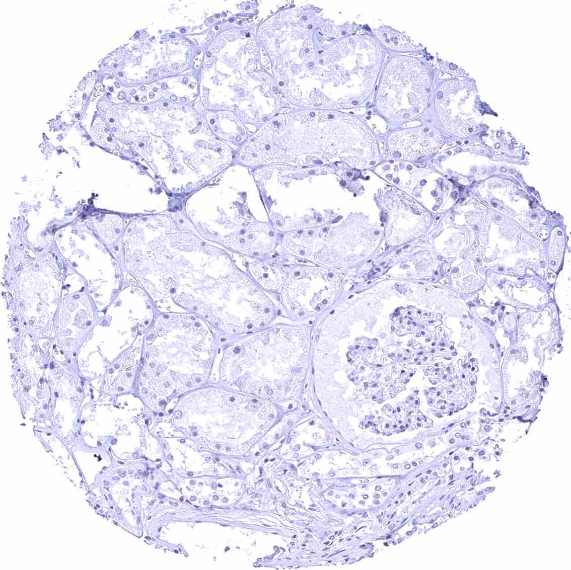
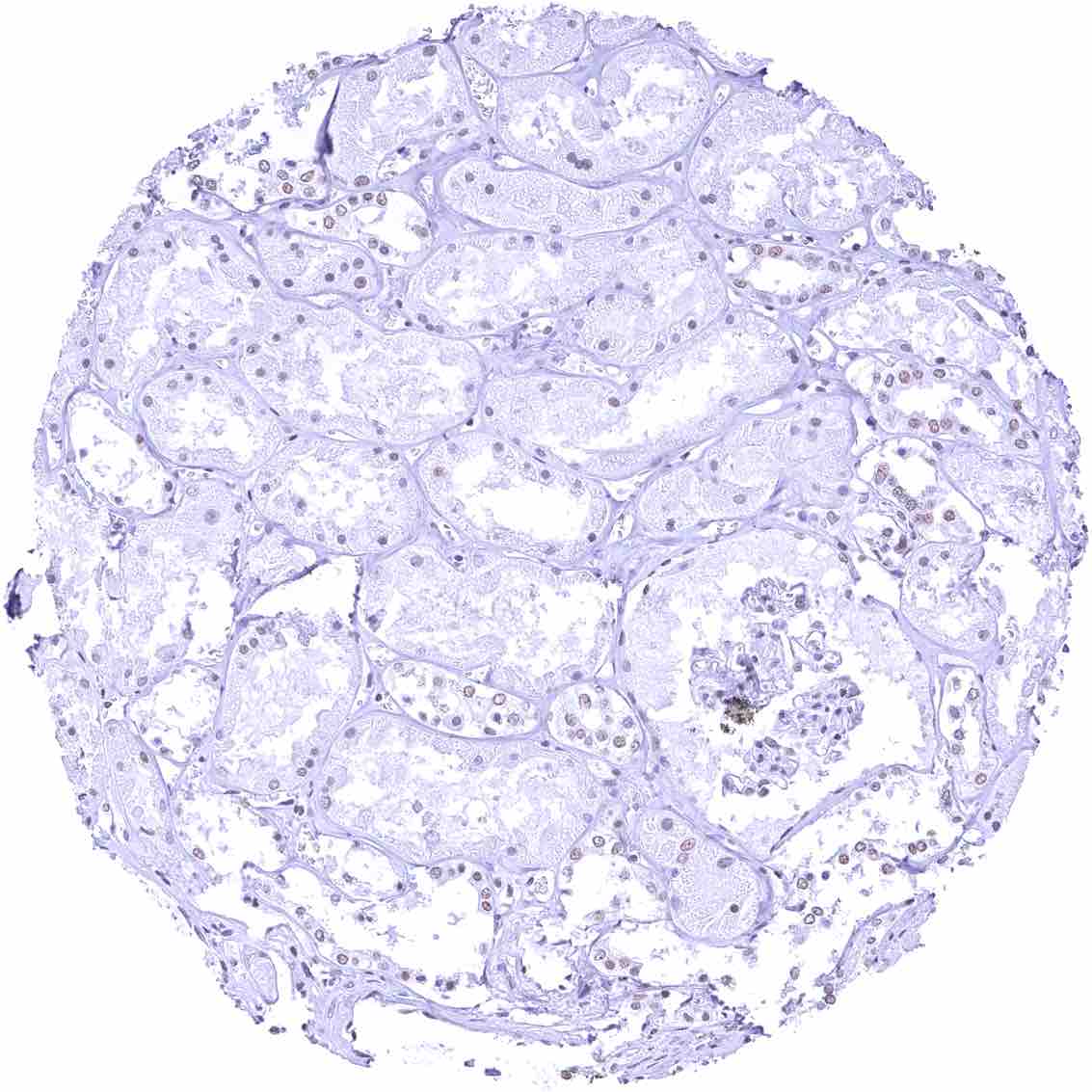
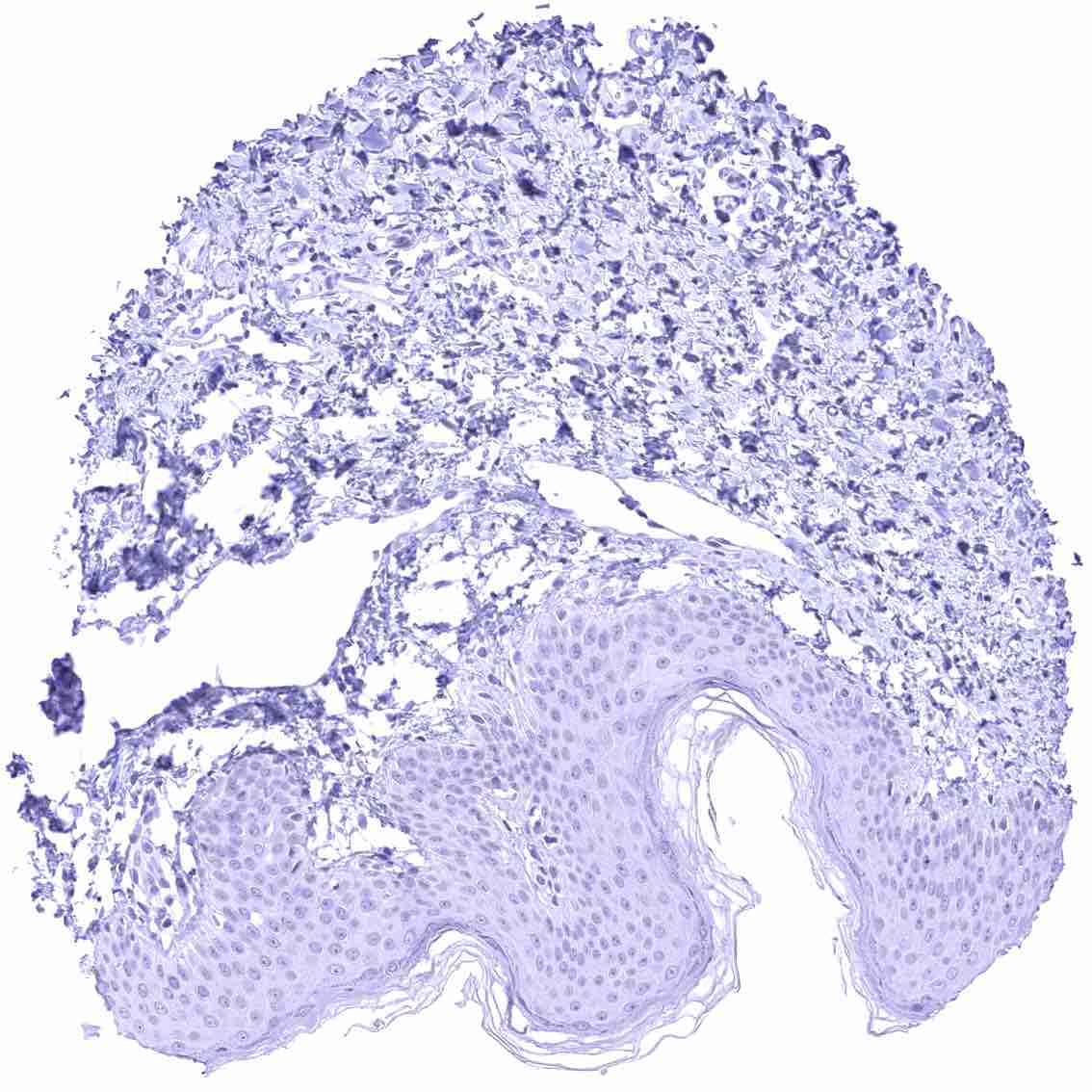
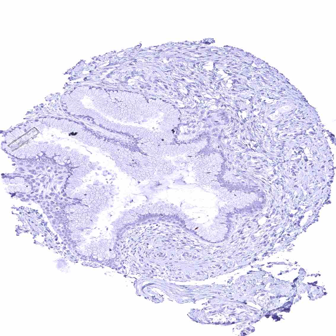
Negative control = Kidney: FOXA1 staining must not be seen in any cell type of the kidney.
Cellular localization = Nuclear
Reactivity = Human
Application = Immunohistochemistry
Dilution = 1:100 – 1:200
Intended Use = Research Use Only
Relevance of Antibody
FOXA1 is a master regulator of chromatin structure
Biology Behind
The FOXA1 (Forkhead Box A1) gene, located at chromosome 14q21.1 codes for a critical “pioneer factor” protein. It binds to nucleosomal DNA, opens local chromatin, and recruits or stabilizes other transcription factors, several of which are hormone receptors or related proteins such as for example estrogen receptor, androgen receptor or GATA3. FOXA1 thereby establishes lineage-specific transcriptional programs in endoderm-derived organs (liver, lung, pancreas, prostate, mammary gland) and in luminal mammary epithelial cells. FOXA1 is essential for the development of several organs including pancreas, liver, prostate, and the lung. FOXA1 knockout mice survive embryogenesis but die postnatally with hypoglycaemia – due to a lack of glucagon – and other metabolic defects. FOXA1 germline mutations are not known to occur in humans. In cancer, FOXA1 shows context‑dependent roles. Somatic FOXA1 mutations, focal amplifications, and overexpression are recurrent in prostate, breast, lung, bladder and other cancers. The functional and clinical consequences of these alterations are not fully understood. Depending on the tumor type, both reduced and increased expression has been found to be related to aggressive cancer phenotype.
Staining Pattern in Normal Tissues
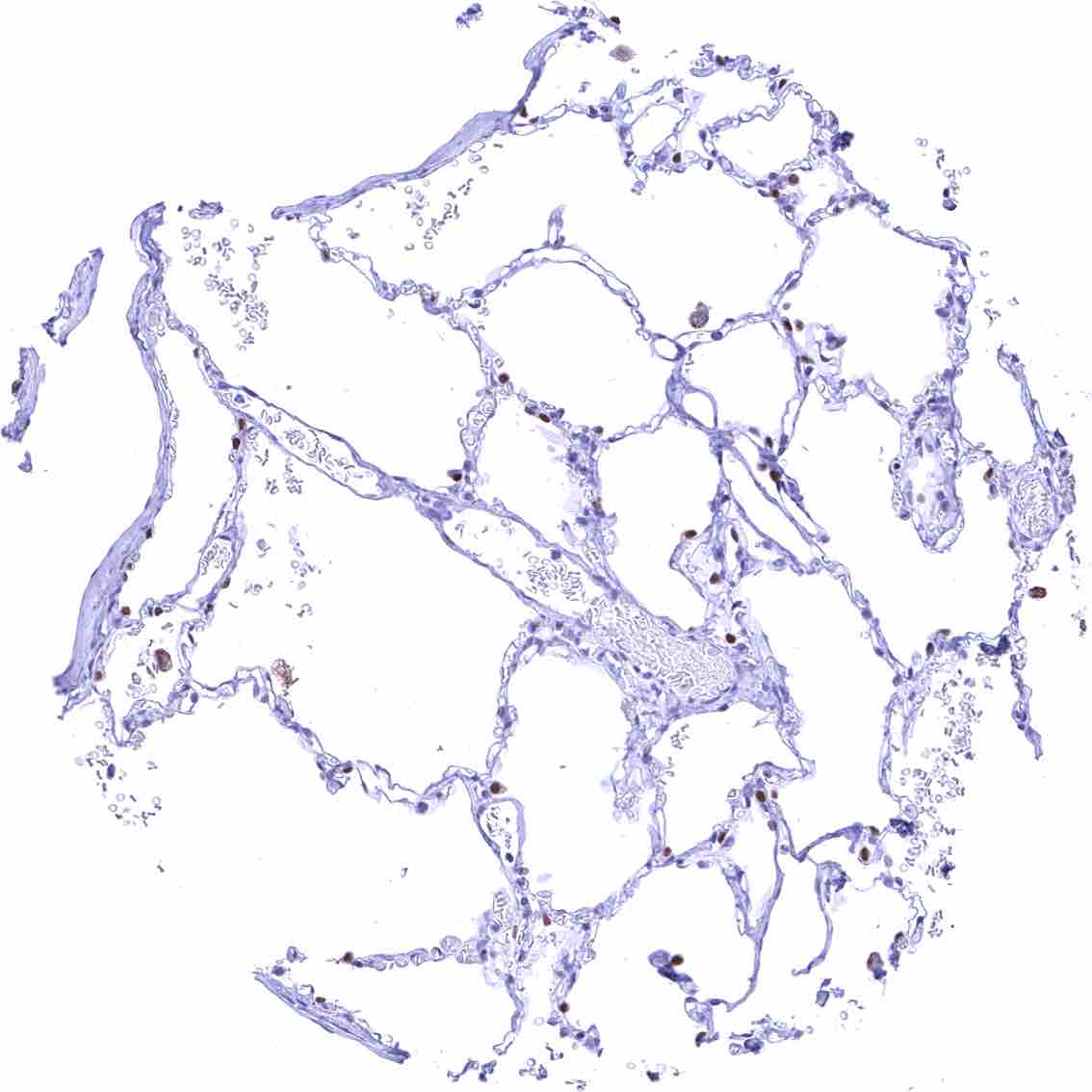
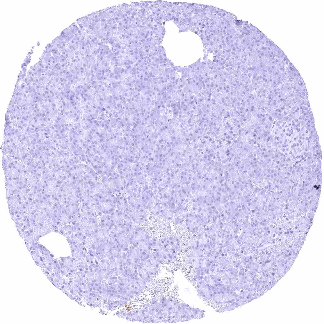
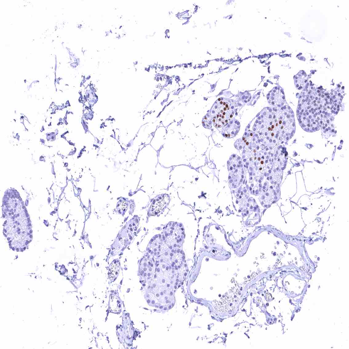
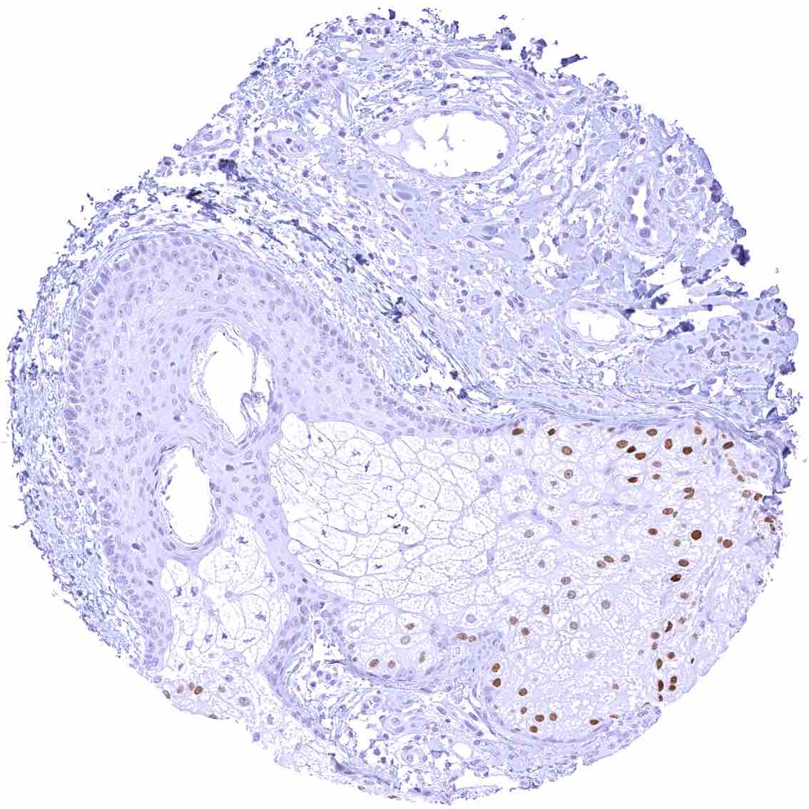
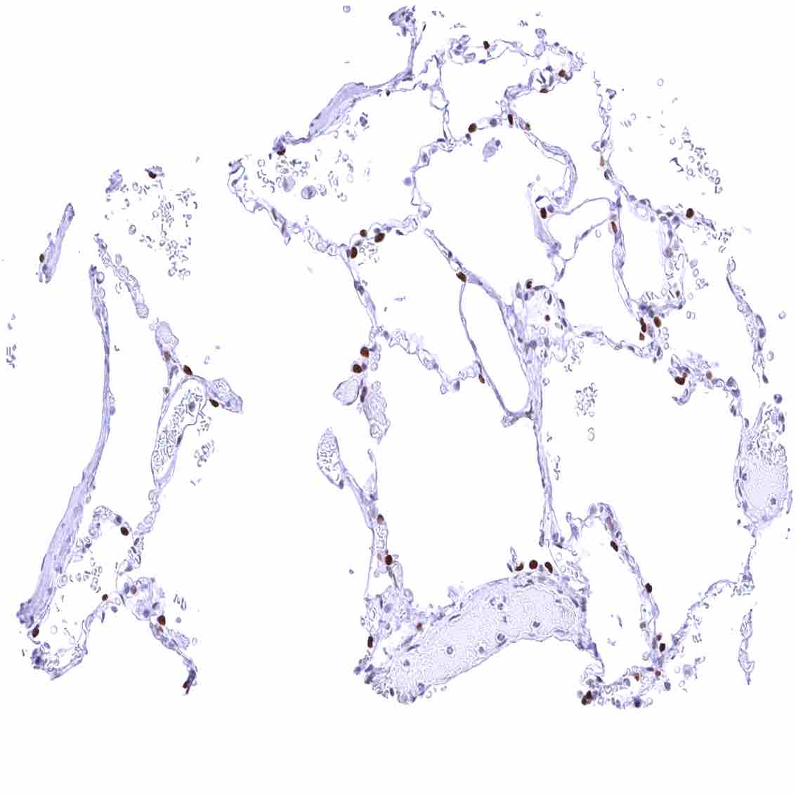
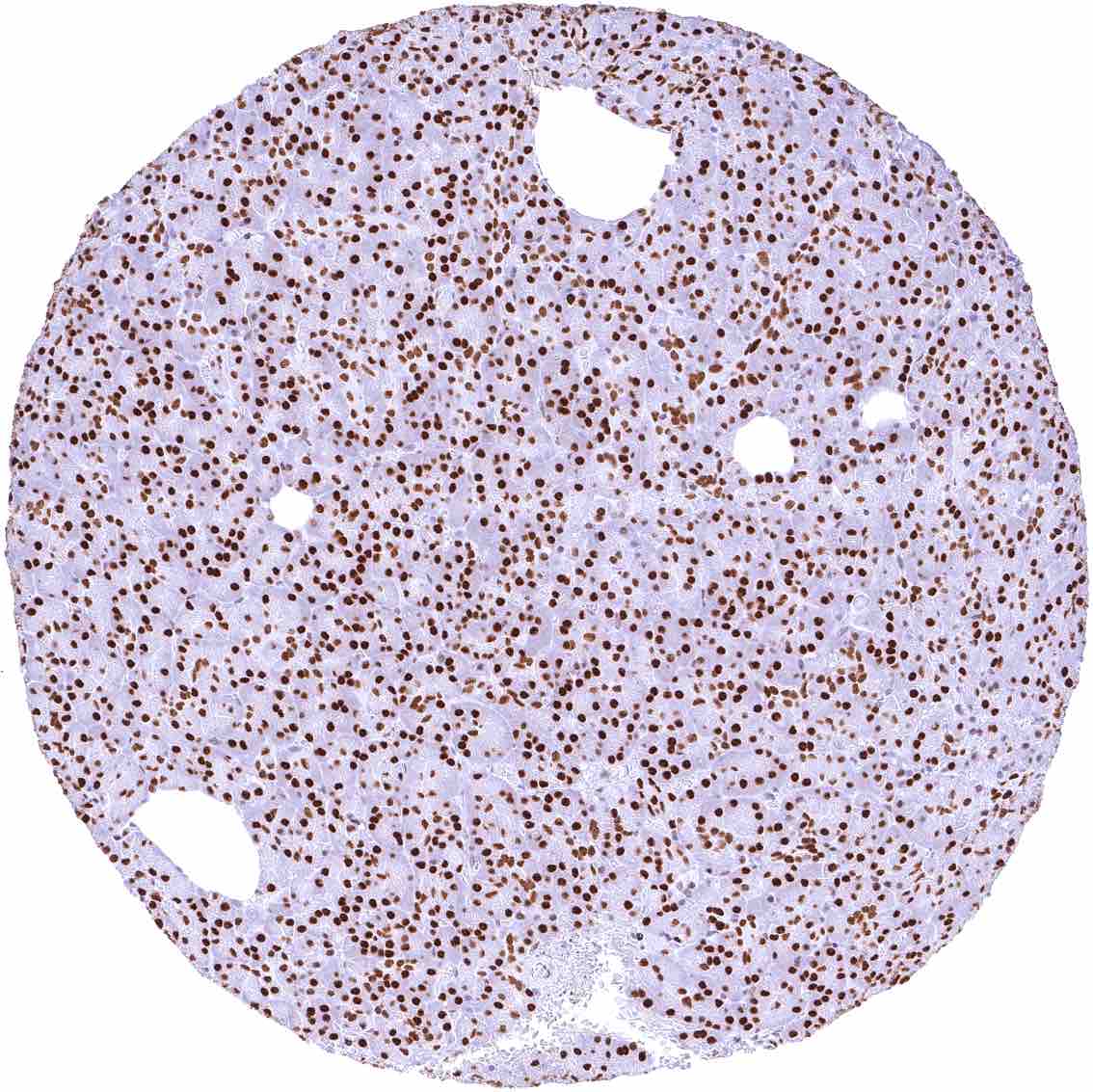
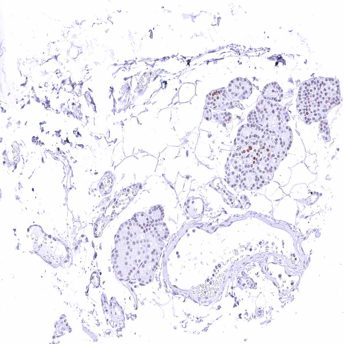
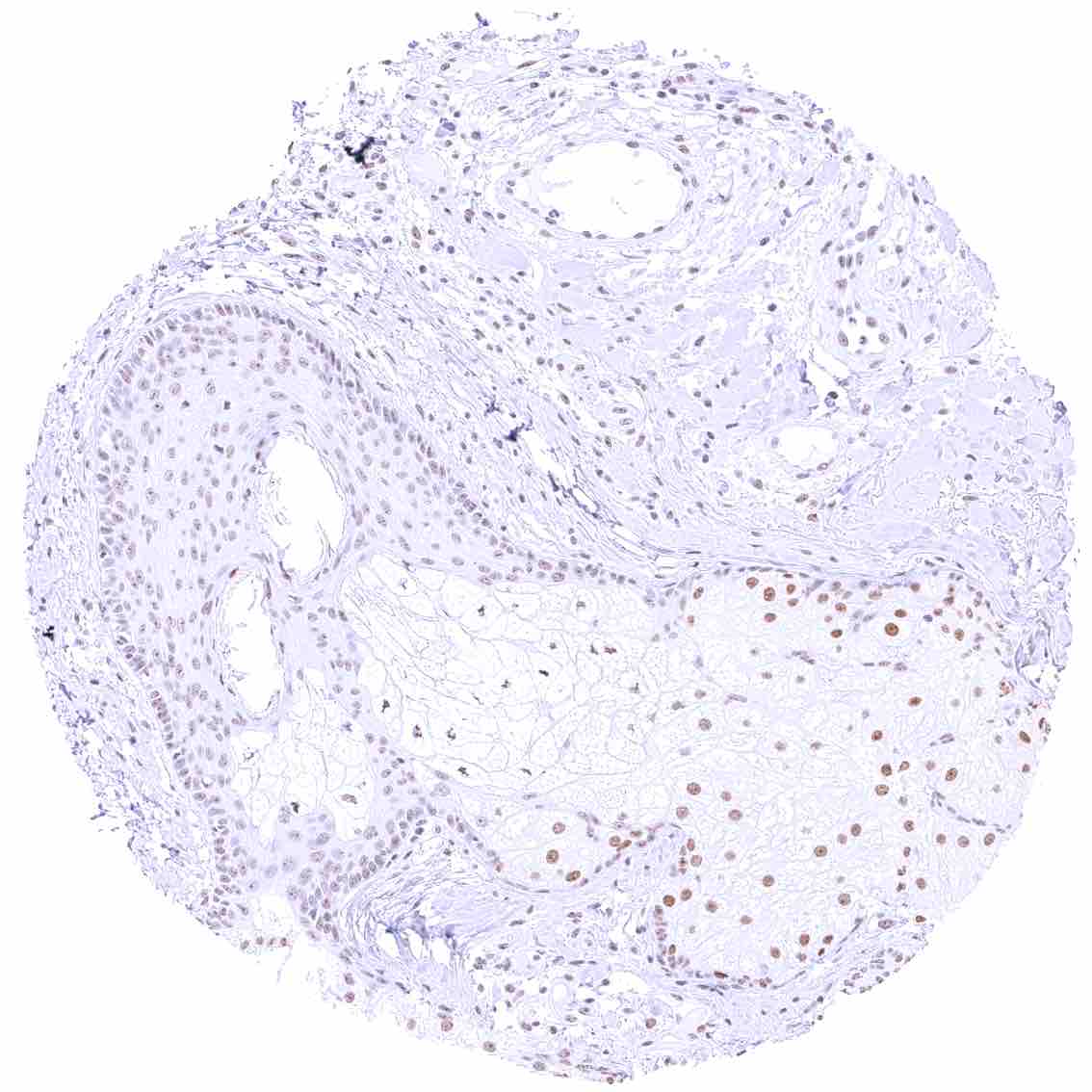
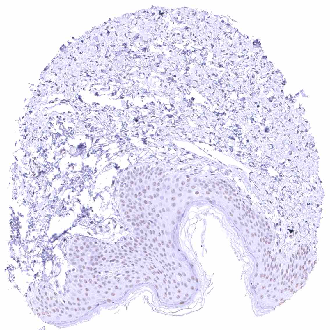
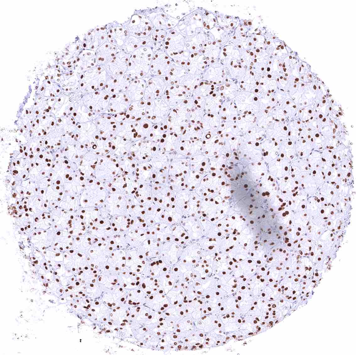
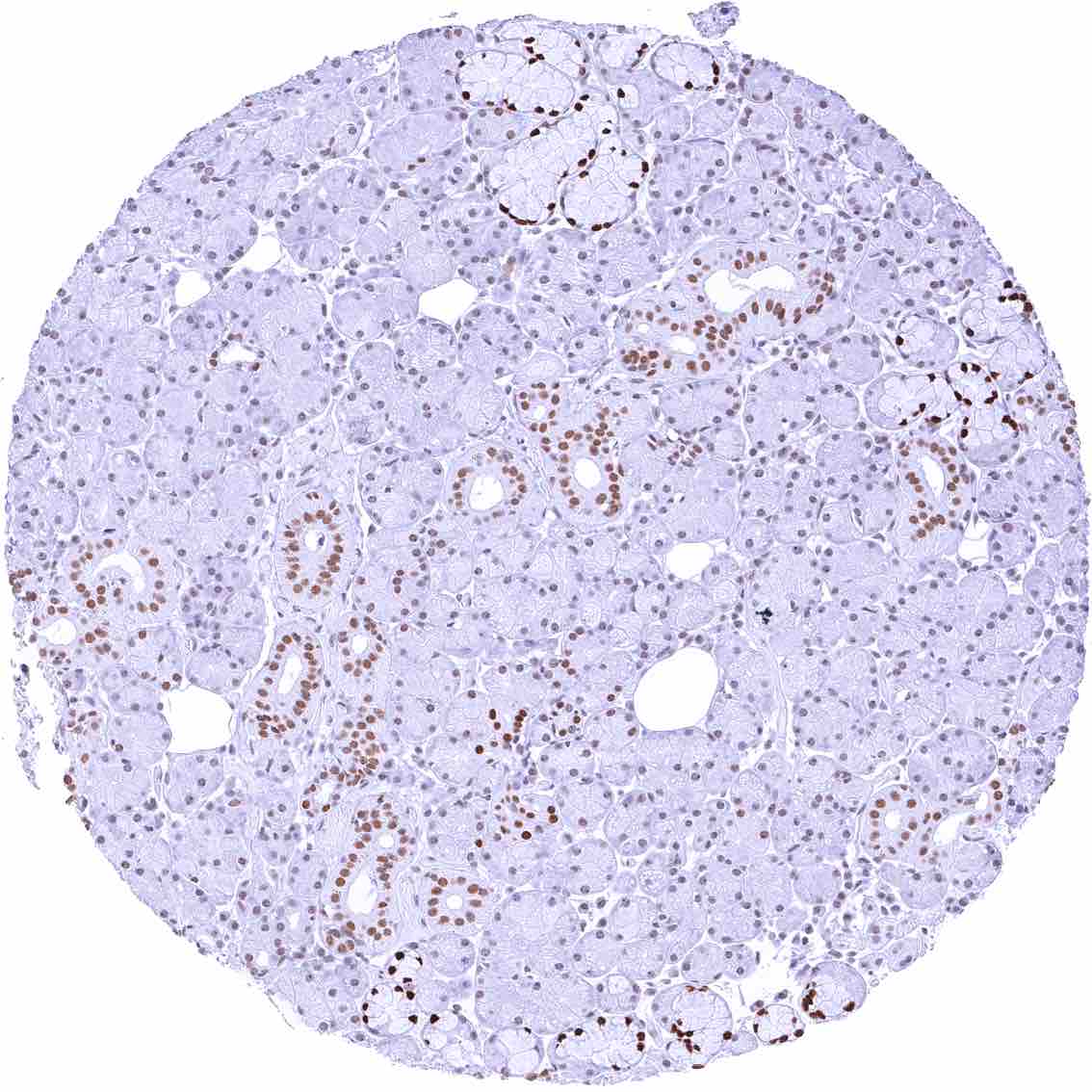
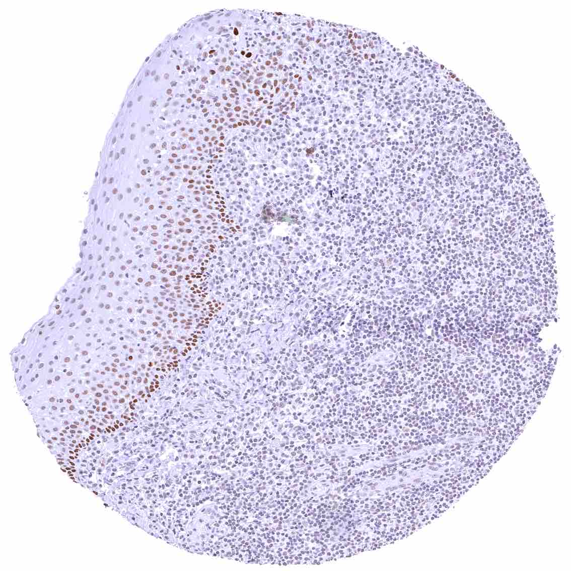
Images describing the EGFRvIII staining pattern in normal tissues obtained by the antibody HMV4726 are shown in our “Normal Tissue Gallery”.
| Brain | Cerebrum | Negative. |
| Cerebellum | Negative. | |
| Endocrine Tissues | Thyroid | Negative. |
| Parathyroid | Distinct nuclear FOXA1 staining of a small subset of epithelial cells (not in all samples). | |
| Adrenal gland | Negative. | |
| Pituitary gland | Negative. | |
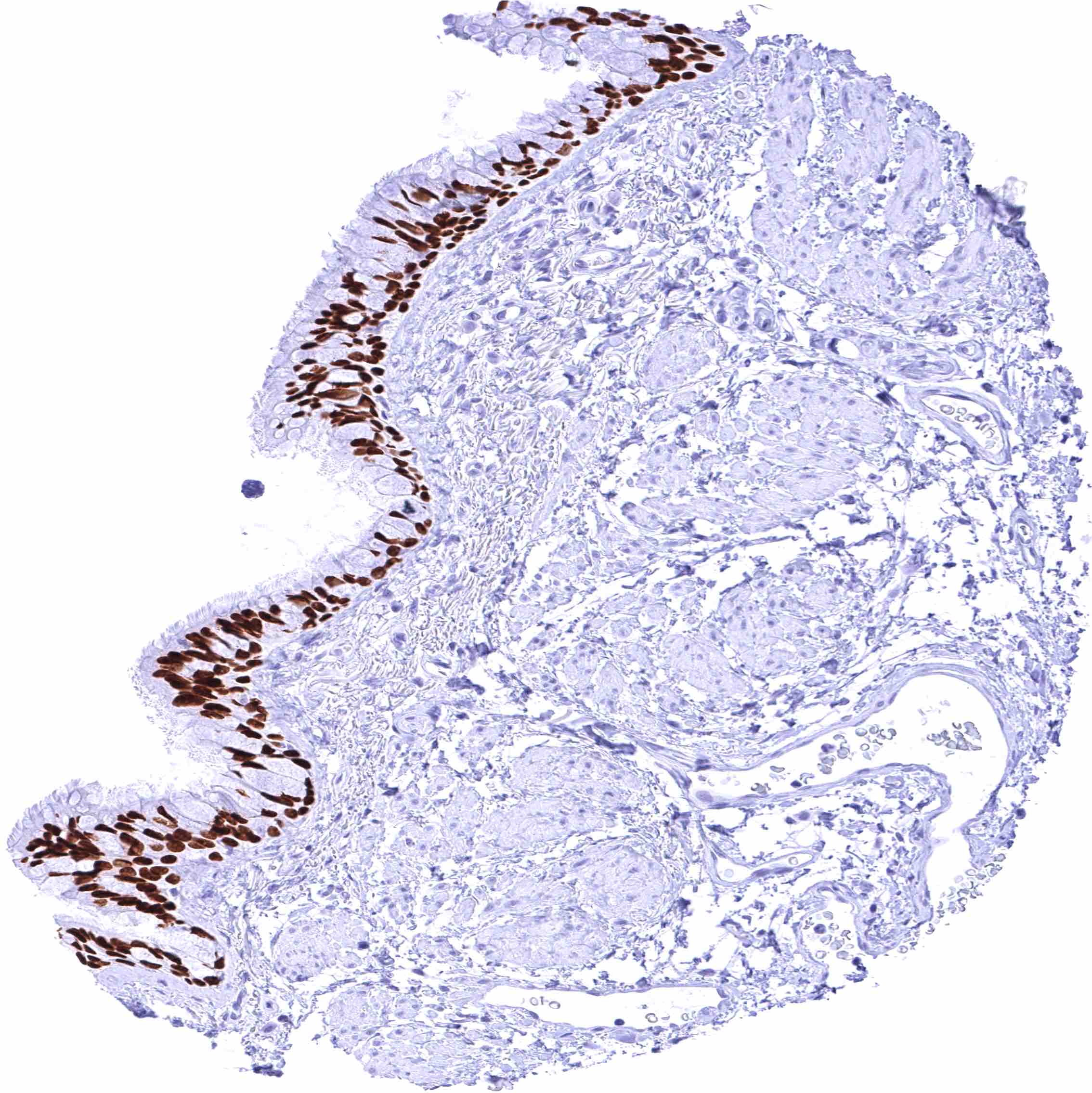
| Respiratory system | Respiratory epithelium | Intense nuclear FOXA1 staining of all epithelial cells of the respiratory epithelium – and of bronchial glands. |
| Lung | Weak to moderate nuclear FOXA1 staining of at least a subset of alveolar pneumocytes. | |
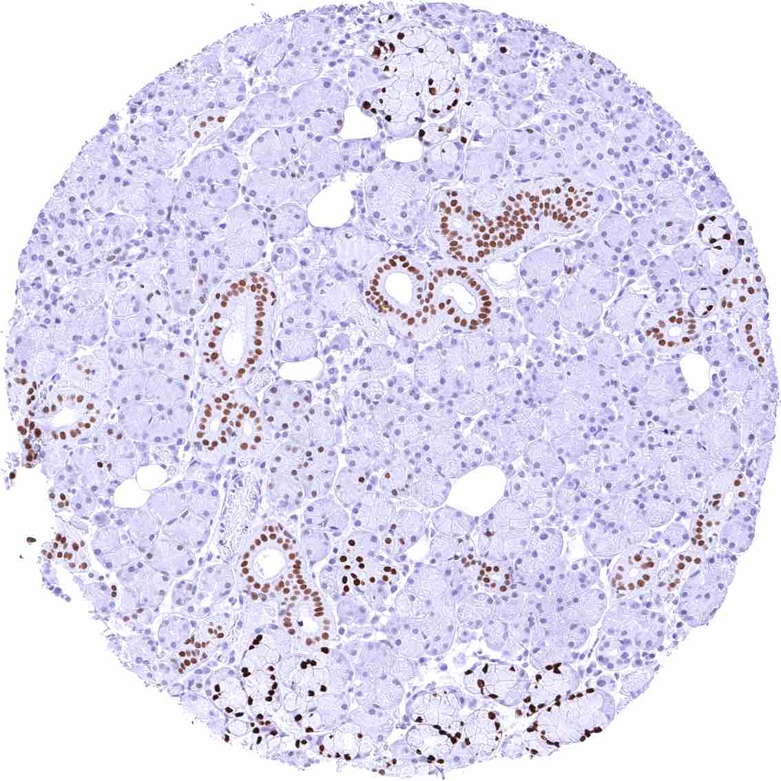
| Gastrointestinal Tract | Salivary glands | Strong nuclear FOXA1 staining of excretory ducts and of mucinous glands cells while serous glands are only faintly stained or FOXA1 negative. |
| Esophagus | Distinct nuclear FOXA1 staining of the basal and suprabasal layers of the squamous epithelium. The staining intensity markedly decreases towards the more superficial cell layers. | |
| Stomach | Moderate to strong nuclear FOXA1 staining of glandular cells. The staining intensity gradually decreases towards the surface epithelium. | |
| Duodenum | Moderate to strong nuclear FOXA1 staining of crypt epithelial cells. The staining intensity gradually decreases towards the surface epithelium and the peaks of the tufts. Distinct nuclear FOXA1 staining of mucinous glandular cells of Brunner glands. | |
| Small intestine | Moderate to strong nuclear FOXA1 staining of crypt epithelial cells. The staining intensity gradually decreases towards the surface epithelium and the peaks of the villi. | |
| Appendix | Strong nuclear FOXA1 staining of epithelial cells in the crypts. The staining intensity gradually decreases towards the surface epithelium. | |
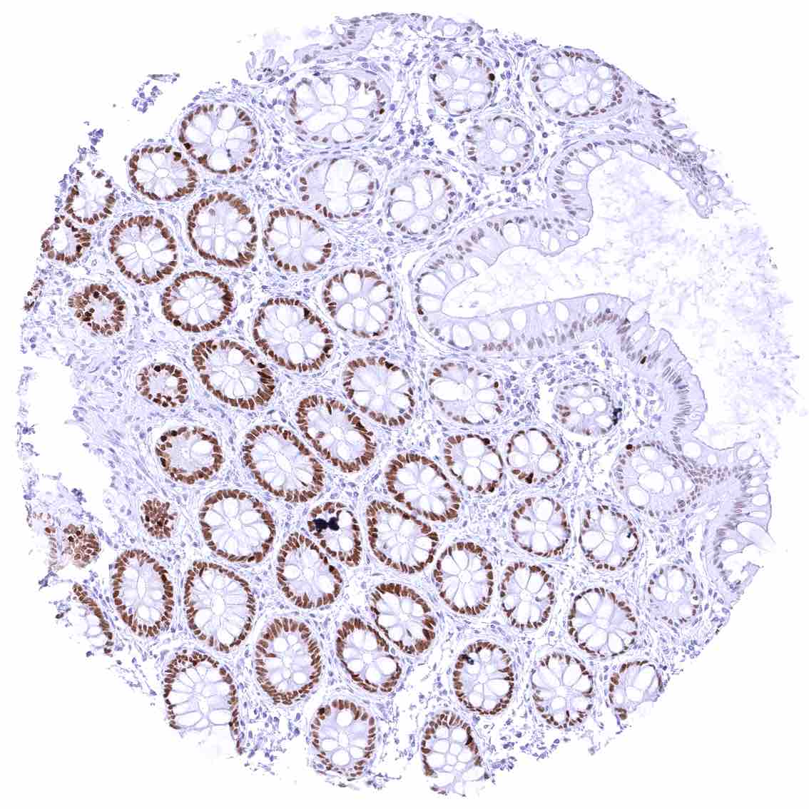
| Colon | Strong nuclear FOXA1 staining of epithelial cells in the crypts. The staining intensity gradually decreases towards the surface epithelium. | |
| Rectum | Strong nuclear FOXA1 staining of epithelial cells in the crypts. The staining intensity gradually decreases towards the surface epithelium. | |
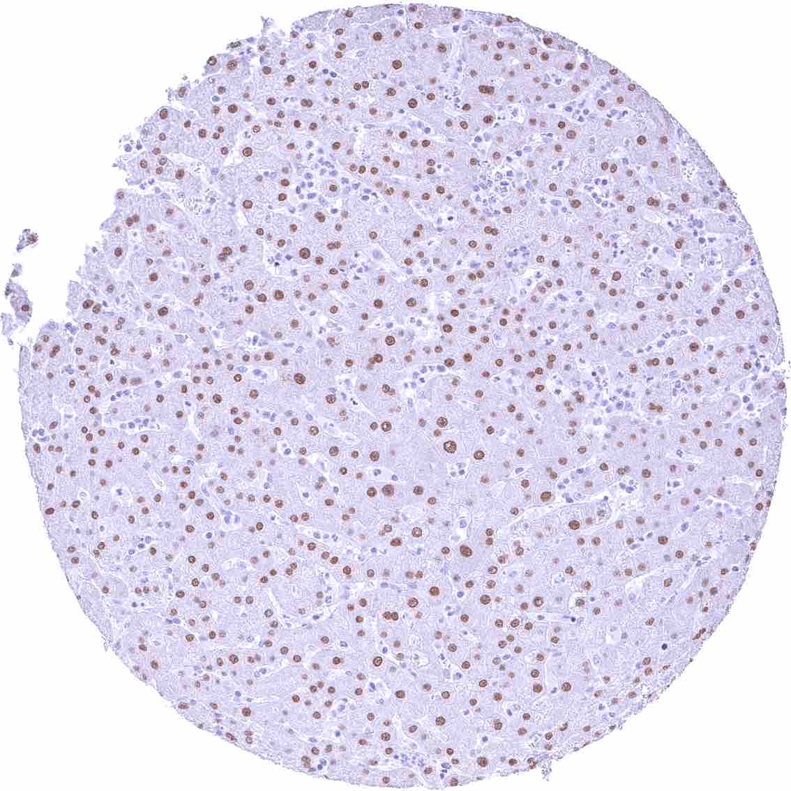
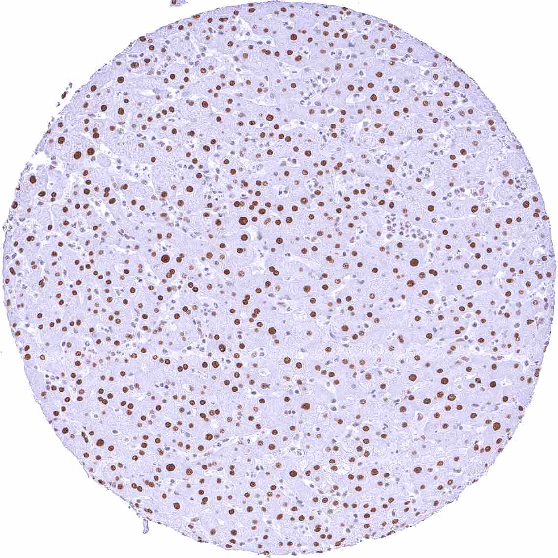
| Liver | The intensity of nuclear FOXA1 staining varies between samples and ranges from negative to strong in hepatocytes. | |
| Gallbladder | Nuclear FOXA1 staining of variable intensity – ranging from negative to strong – in epithelial cells (distinct differences between individual cells and cell groups).. | |
| Pancreas | Negative. | |
| Genitourinary | Kidney | Negative. |
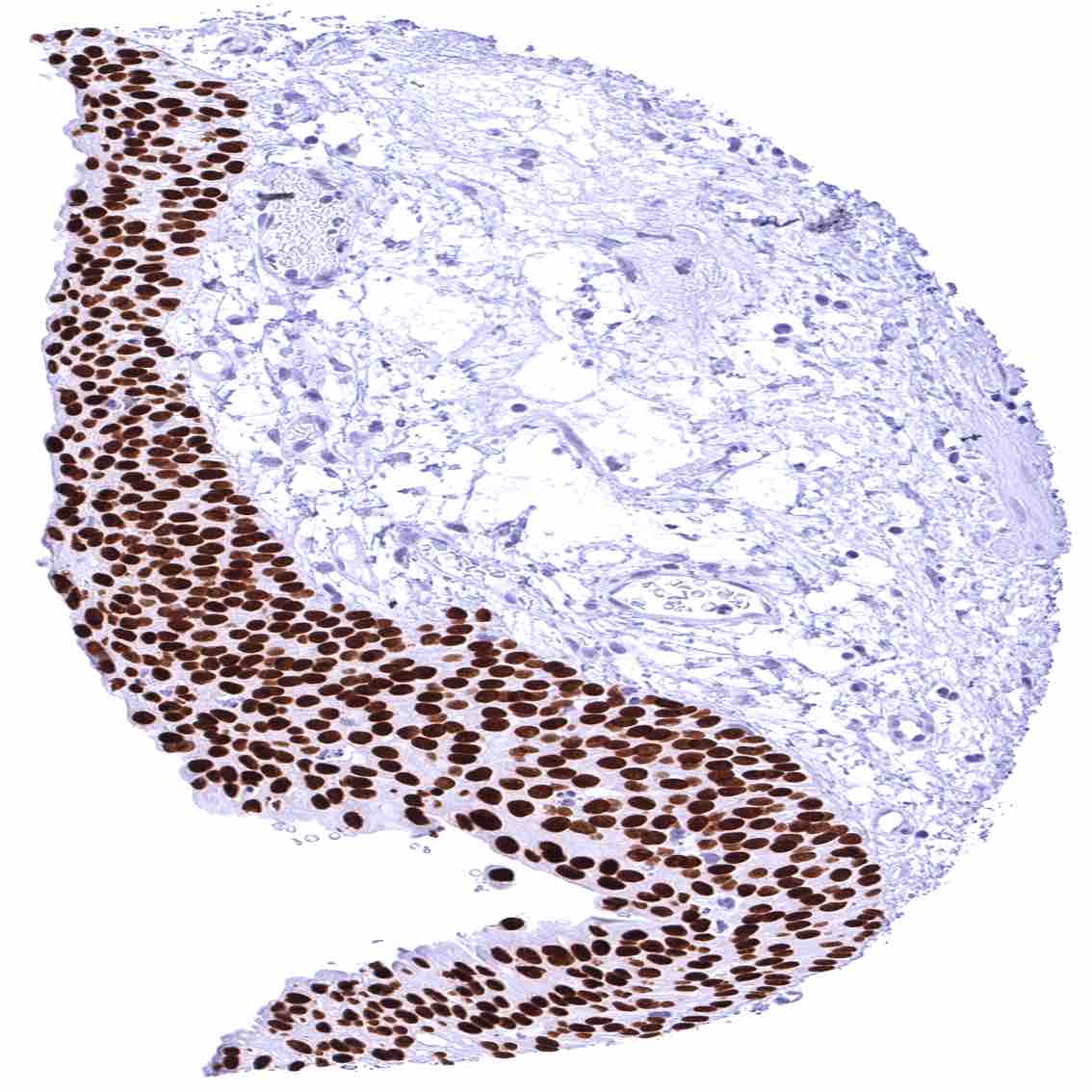
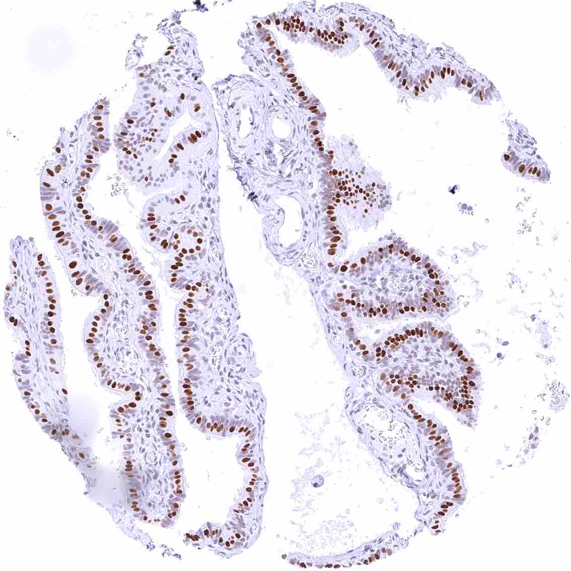
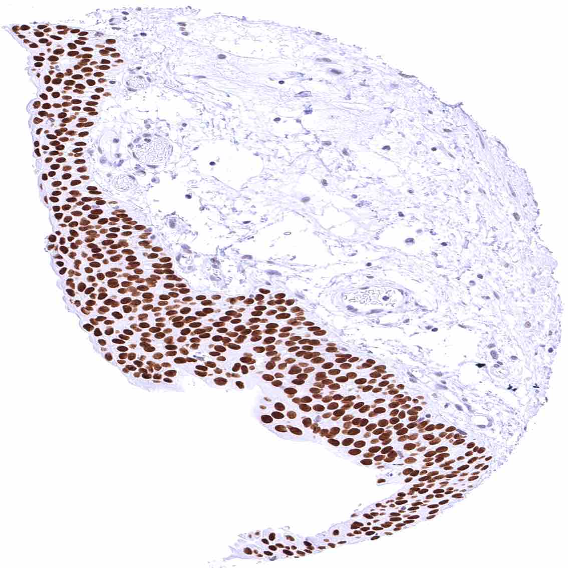
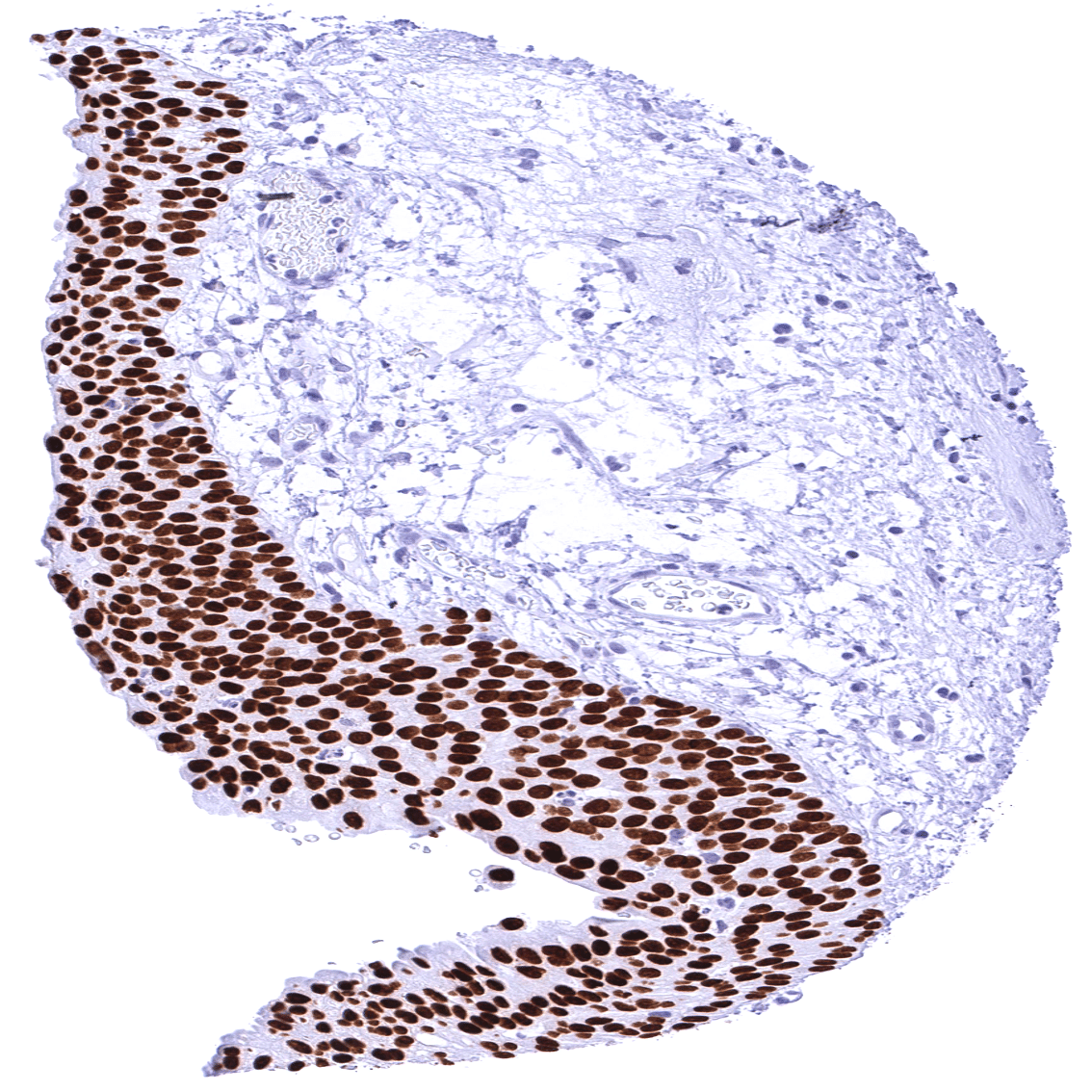
| Urothelium | Intense nuclear FOXA1 staining of urothelial cells. | |
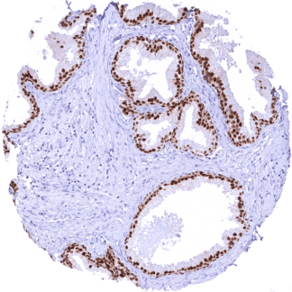
| Male genital | Prostate | Intense nuclear FOXA1 staining of all epithelial cells. |
| Seminal vesicles | Negative. | |
| Testis | Negative. | |
| Epididymis | Negative. | |
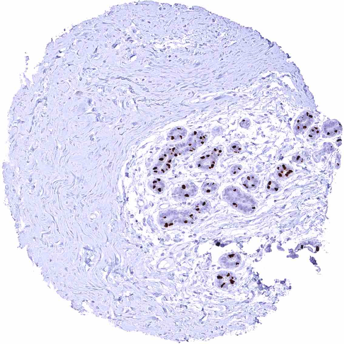
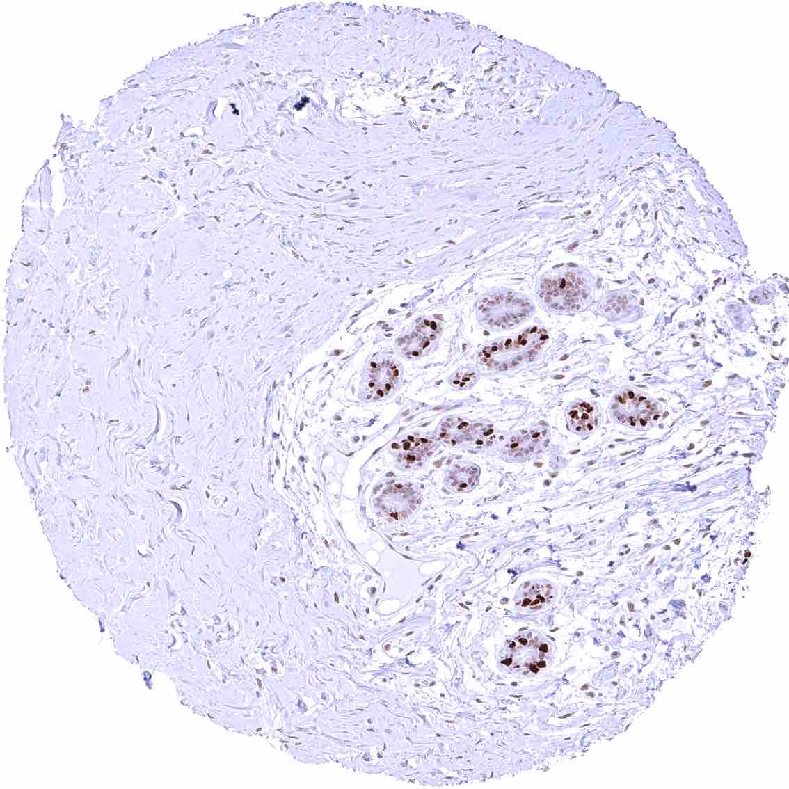
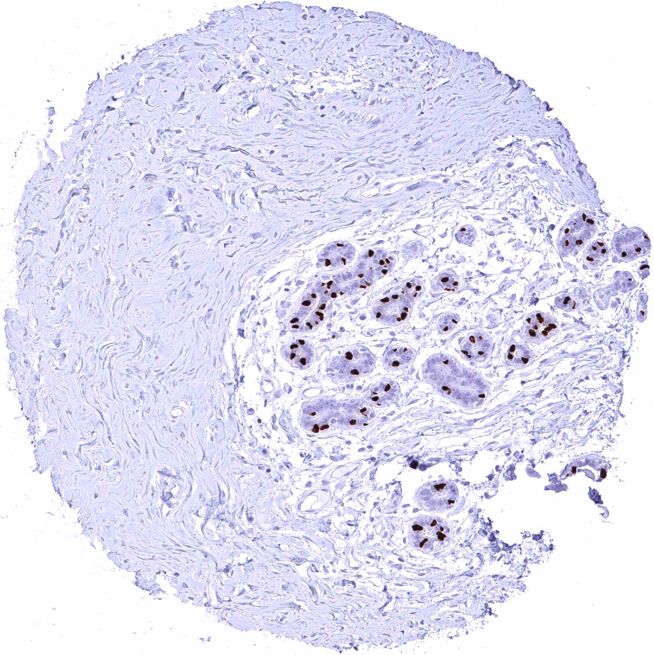
| Female genital | Breast | Intense nuclear FOXA1 staining of a subset of luminal epithelial cells. |
| Uterus, myometrium | Negative. | |
| Uterus, ectocervix | Distinct nuclear FOXA1 staining of the basal and suprabasal layers of the squamous epithelium. The staining intensity markedly decreases towards the more superficial cell layers. | |
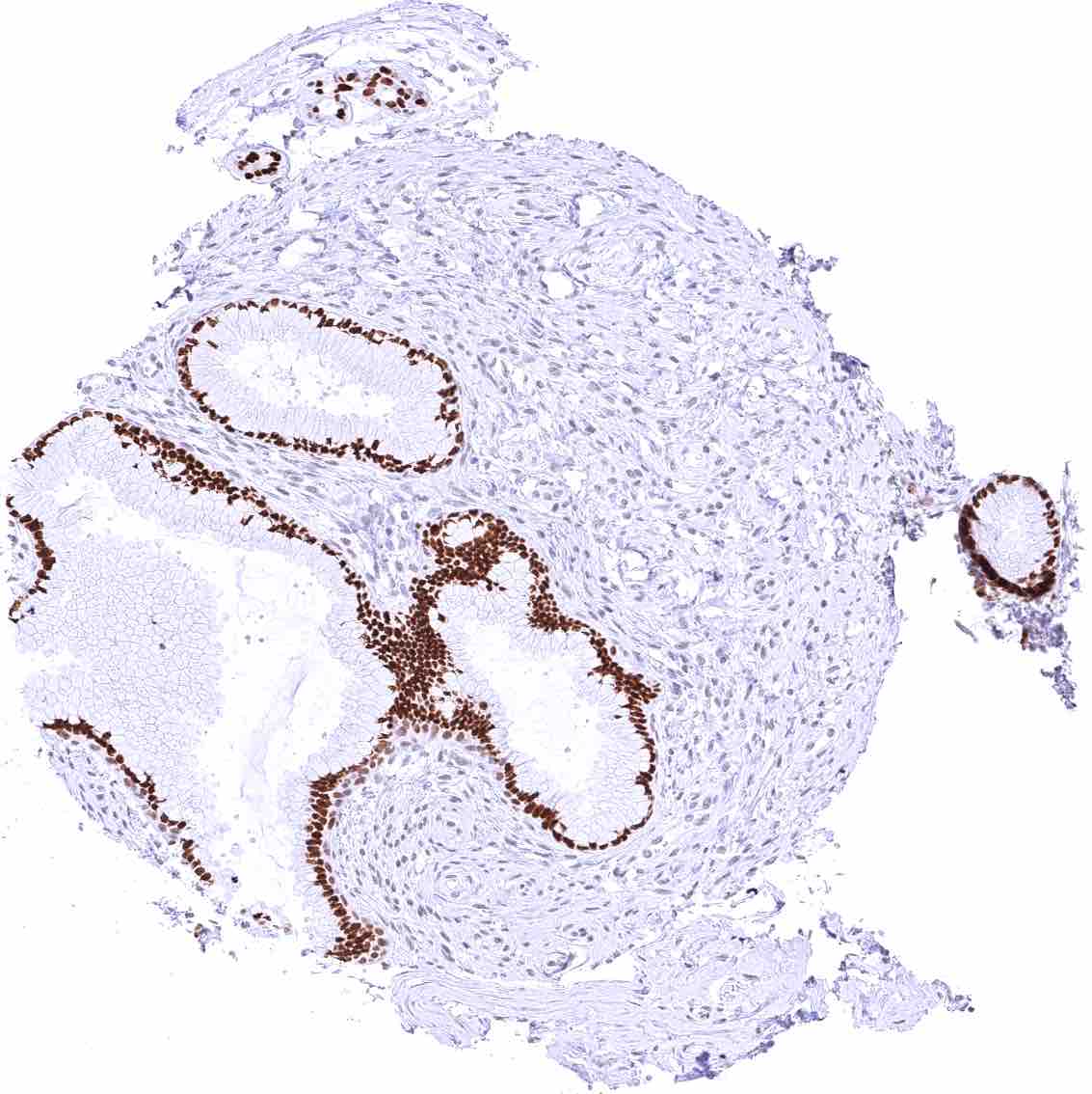
| Uterus endocervix | Endocervical glandular epithelium is FOXA1 negative. Basal cells at the transition from squamous epithelium to glandular epithelium are FOXA1 positive. | |
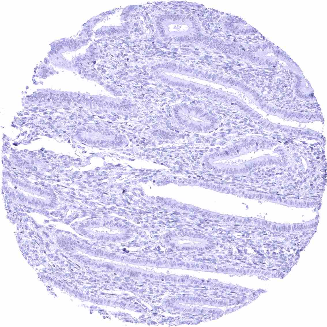
| Uterus, endometrium | Negative. | |
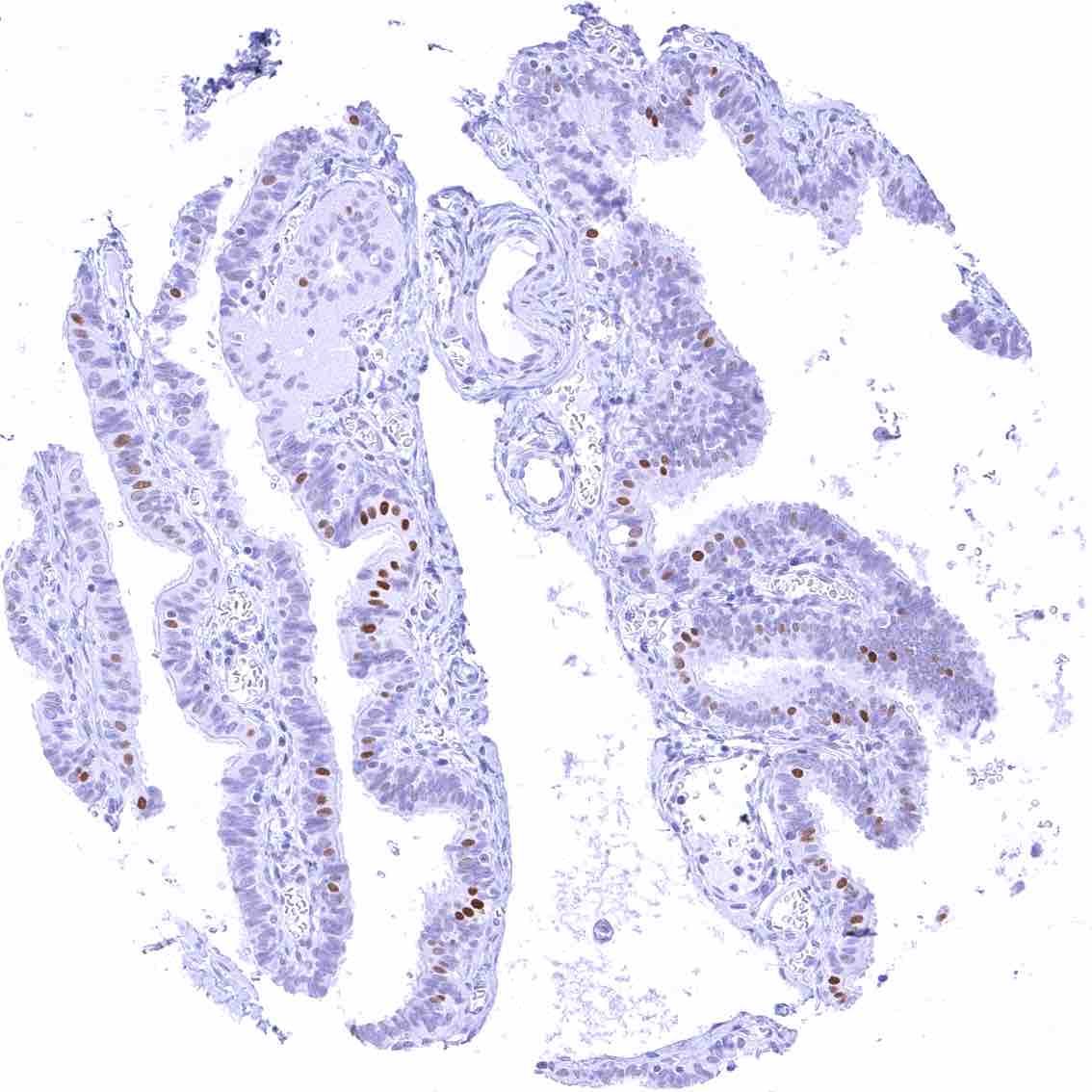
| Fallopian Tube | Distinct nuclear FOXA1 staining of a subset of epithelial cells while others are FOXA1 negative. | |
| Ovary | Negative (stroma, granulosa cells, theca cells, corpus luteum). | |
| Placenta early | Negative. | |
| Placenta mature | Negative. | |
| Amnion | Negative. | |
| Chorion | Negative. | |
| Skin | Epidermis | Negative. |
| Sebaceous glands | Distinct nuclear FOXA1 staining of a subset of sebaceous epithelial cells. | |
| Muscle/connective tissue | Heart muscle | Negative. |
| Skeletal muscle | Negative. | |
| Smooth muscle | Negative. | |
| Vessel walls | Negative. | |
| Fat | Negative. | |
| Stroma | Negative. | |
| Endothelium | Negative. | |
| Bone marrow/ lymphoid tissue | Bone marrow | Negative. |
| Lymph node | Negative. | |
| Spleen | Negative. | |
| Thymus | Negative. | |
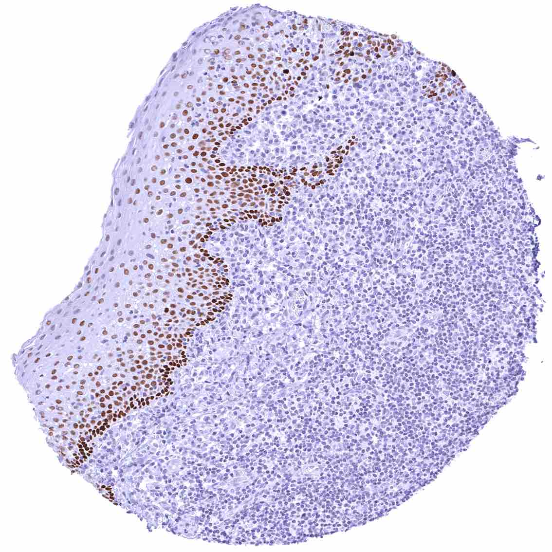
| Tonsil | Distinct nuclear FOXA1 staining of a subset of squamous epithelial cells. The staining intensity gradually decreases towards the surface epithelium. Lymphocytes are FOXA1 negative. | |
| Remarks | Most cells are either strongly positive or negative for FOXA1. Gradual differences in FOXA1 staining intensity exist in intestinal and non-keratinizing squamous epithelia. |
In normal tissues, FOXA1 staining is most intense in prostatic epithelium, urothelium, respiratory epithelium, and in a subset of breast epithelial cells. FOXA1 positivity also occurs in hepatocytes, intestinal epithelium, salivary glands, gallbladder epithelium, pneumocytes, fallopian tube, sebaceous cells of the skin, and in non-keratinizing squamous epithelium. These findings are largely consistent with the RNA data described in the Human Protein Atlas (Tissue expression FOXA1).
Positive control = Colon: A strong nuclear FOXA1 staining should be seen in crypt cells the mucosa. The FOXA1 staining intensity should gradually decrease towards the surface epithelium.
Negative control = Kidney: FOXA1 staining must not be seen in any cell type of the kidney.
Staining Pattern in Relevant Tumor Types
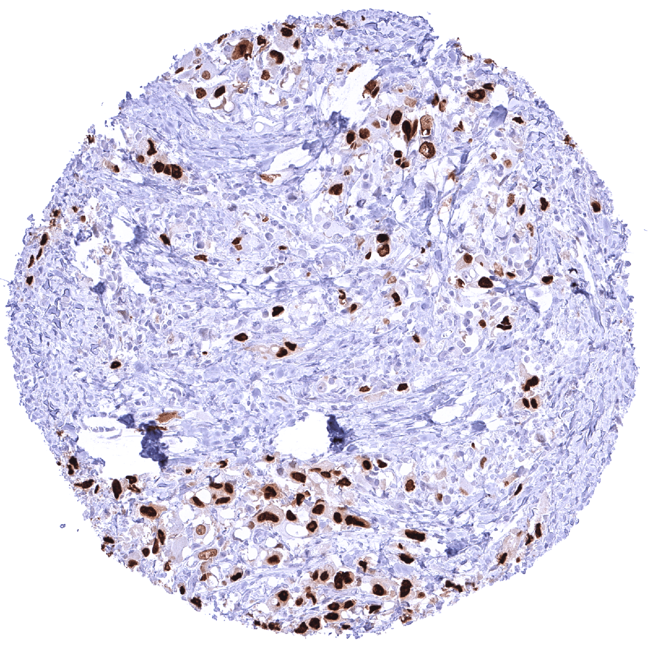
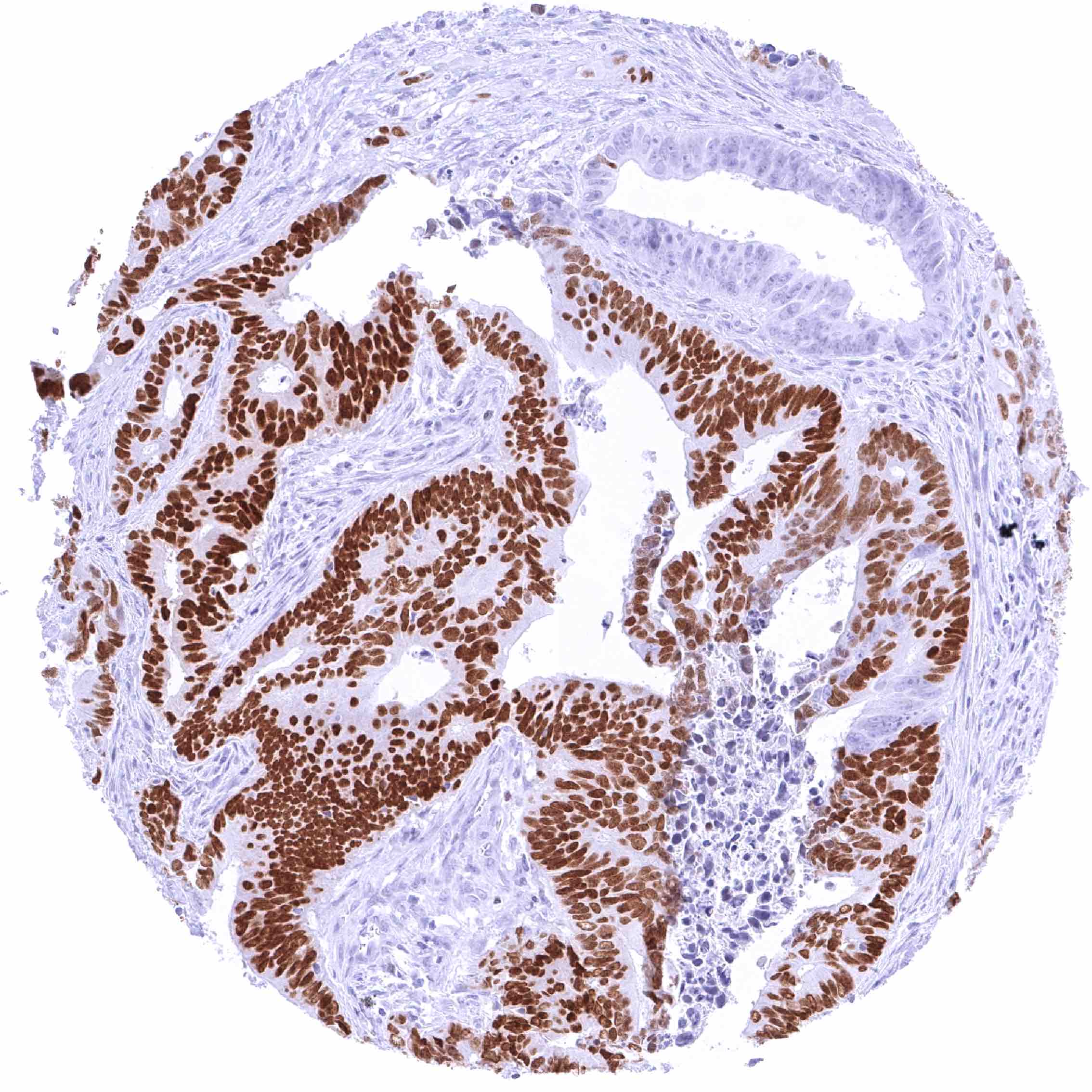
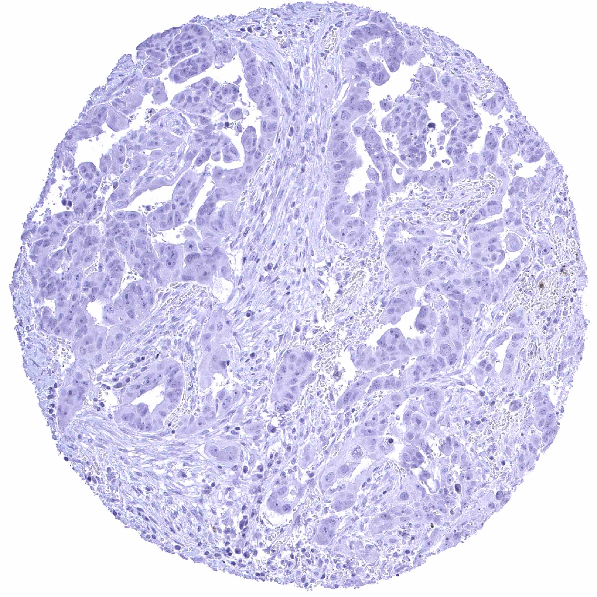
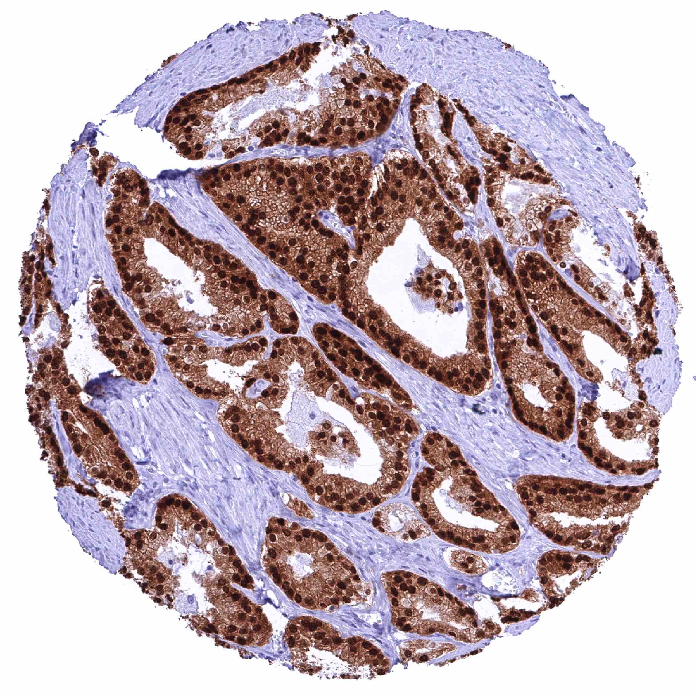
High FOXA1 expression is mostly seen in prostate and breast cancer but it can also occur – at lower prevalence and often also at lower level – in various other cancer types.
The TCGA findings on FOXA1 RNA expression in different tumor categories have been summarized in the FOXA1 RNA expression in different tumor categories.
Protocol Recommendations
IHC users have different preferences on how the stains should look like. Some prefer high staining intensity of the target stain and even accept some background. Others favor absolute specificity and lighter target stains. Factors that invariably lead to more intense staining include higher concentration of the antibody and visualization tools, longer incubation time, higher temperature during incubation, higher temperature and longer duration of the heat induced epitope retrieval (slide pretreatment). The impact of the pH during slide pretreatment has variable effects and depends on the antibody and the target protein.
All images and data shown here and in our image galleries are obtained by the manual protocol described below. Other protocols resulting in equivalent staining are described as well.
Manual protocol
Freshly cut sections should be used (less than 10 days between cutting and staining). Heat-induced antigen retrieval for 5 minutes in an autoclave at 121°C in pH 7,8 Target Retrieval Solution buffer. Apply HMV4726 at a dilution of 1:150 at 37°C for 60 minutes. Visualization of bound antibody by the EnVision Kit (Dako, Agilent) according to the manufacturer’s directions.
Potential Research Applications
- What is the prevalence and clinical significance of aberrant FOXA1 expression in different cancer types?
- Can FOXA1 IHC be used to distinguish clinically relevant tumor types?
- How do specific FOXA1 mutations mechanistically reprogram chromatin to drive therapy resistance in prostate and breast cancer?
- Which tumor contexts show FOXA1 as driver versus tumor suppressor, and what determines that switch?
Evidence for Antibody Specificity in IHC
There are two ways how the specificity of antibodies can be documented for immunohistochemistry on formalin fixed tissues. These are: 1. Comparison with a second independent method for target expression measurement across a large number of different tissue types (orthogonal strategy), and 2. Comparison with one or several independent antibodies for the same target and showing that all positive staining results are also seen with other antibodies for the same target (independent antibody strategy).
Orthogonal validation: For the antibody HMV4726 specificity is strongly supported by a comparison of its staining pattern with FOXA1 RNA expression data in normal tissues which were collected in three independent RNA screening studies, including the Human Protein Atlas (HPA) RNA-seq tissue dataset, the FANTOM5 project, and the Genotype-Tissue Expression (GTEx) project, and which are summarized in the Human Protein Atlas (Tissue expression FOXA1). In line with existing RNA data, strong/significant FOXA1 staining was highest in prostatic epithelium, urothelium, a subset of breast epithelial cells, hepatocytes, stomach glands, and it was also seen in intestinal epithelium, salivary glands, gallbladder, pneumocytes, fallopian tube, and in squamous epithelium. In line with absence of FOXA1 RNA, FOX1 immunostaining by HMV4726 was not seen in endometrium, placenta, ovary, muscle, testis, epididymis, seminal vesicle, kidney, pancreas, all endocrine tissues, and the brain.
Comparison of antibodies: True expression of FOXA1 in all cells with FOXA1 positivity by HMV4726 is corroborated by a confirmation of each individual staining seen by HMV4726 in 76 different normal tissue types by a commercially available independent second antibody (termed “validation antibody”). An additional nuclear staining of the reference antibody in several other epithelial tissues such as the endometrium, the pancreas, glia cells of the brain, corpus luteum of the ovary, endocervix, and chief cells of the epididymis was considered an antibody specific cross-reactivity of the validation antibody as it was not seen by HMV4726.