295,00 € – 995,00 €
Product details
Synonyms = Glycoprotein 2 (zymogen granule membrane); GP2; Pancreatic zymogen granule membrane associated protein GP2; Pancreatic zymogen granule membrane protein GP-2; ZAP75
Antibody type = Mouse monoclonal / IgG
Clone = MSVA-475M
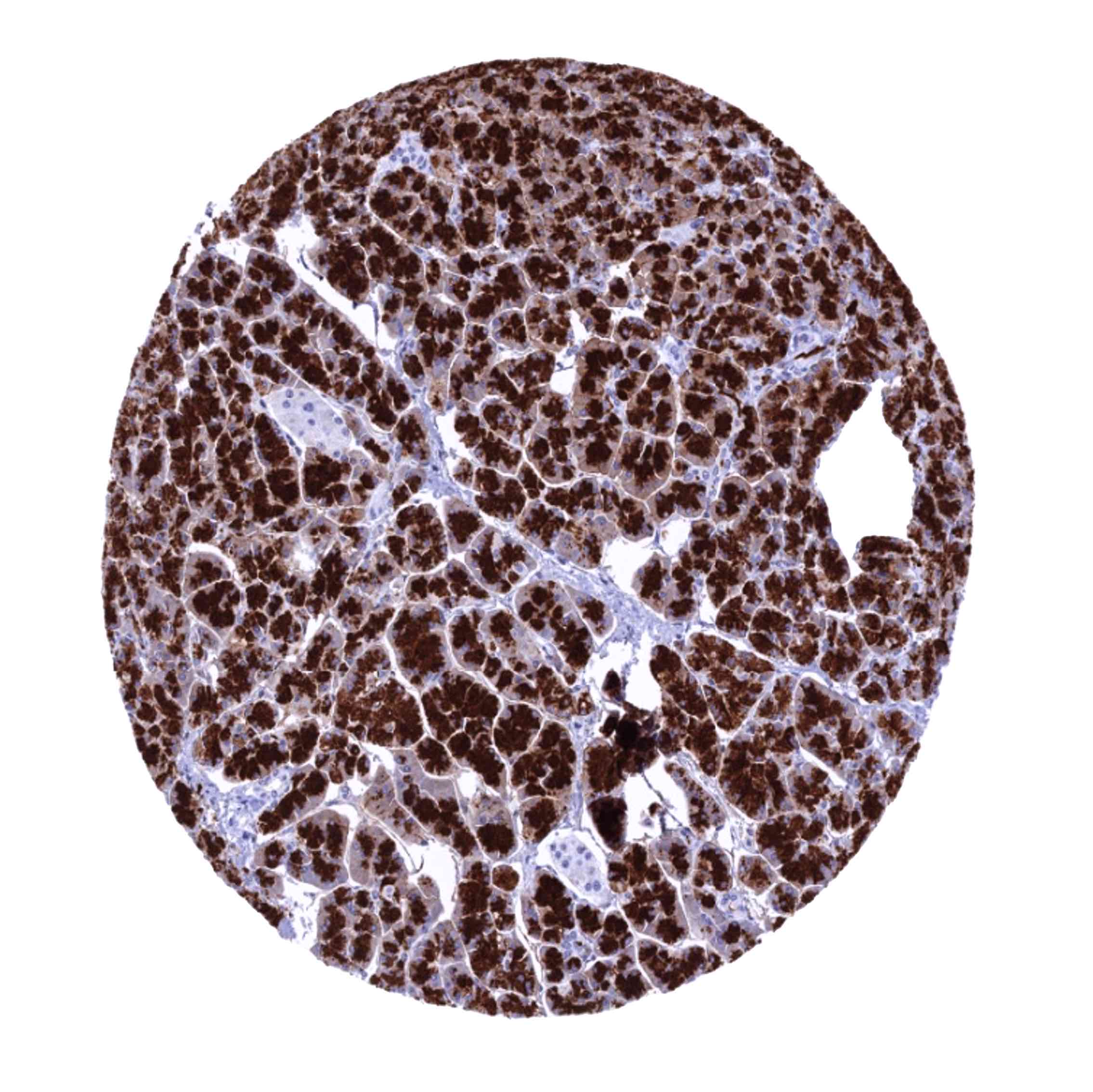
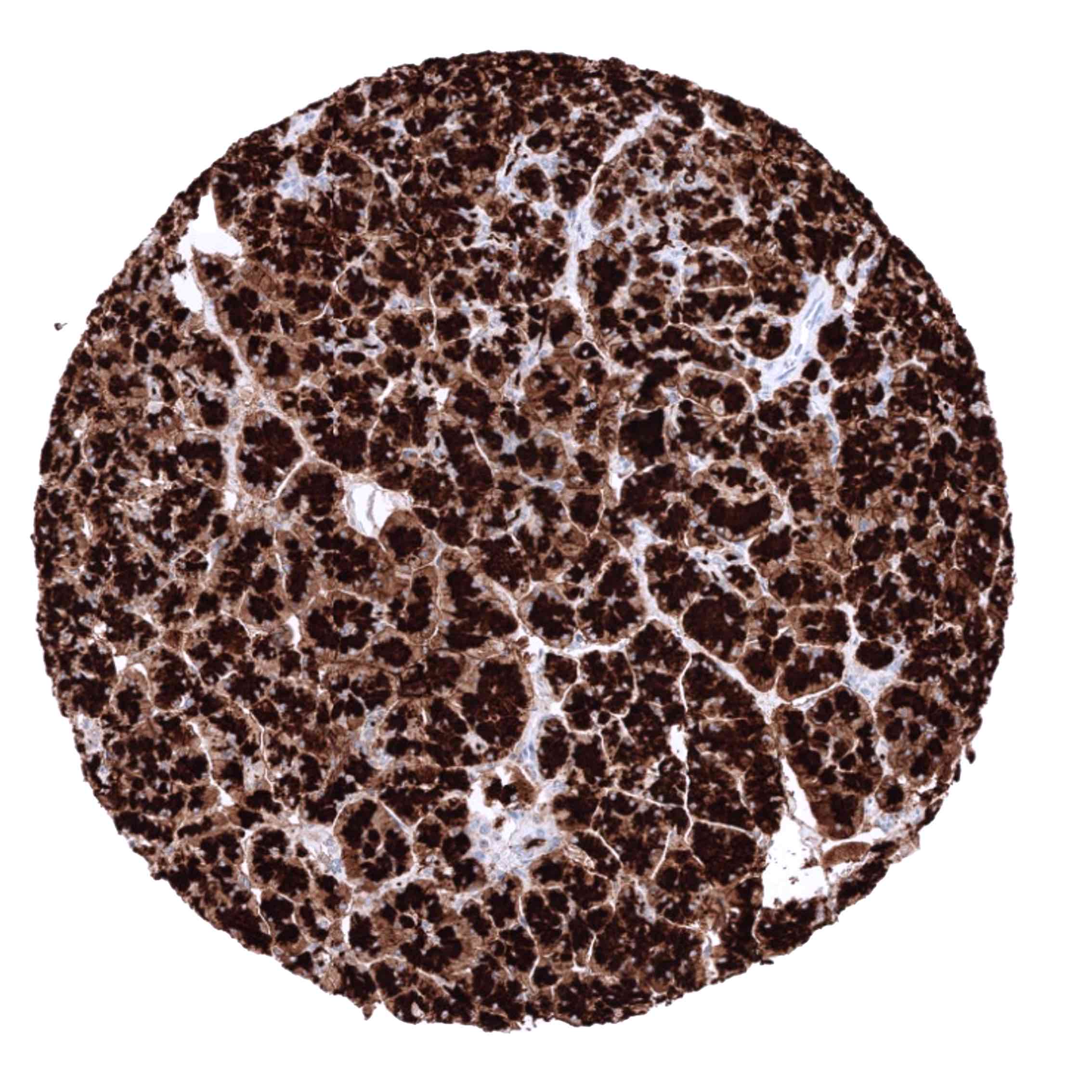
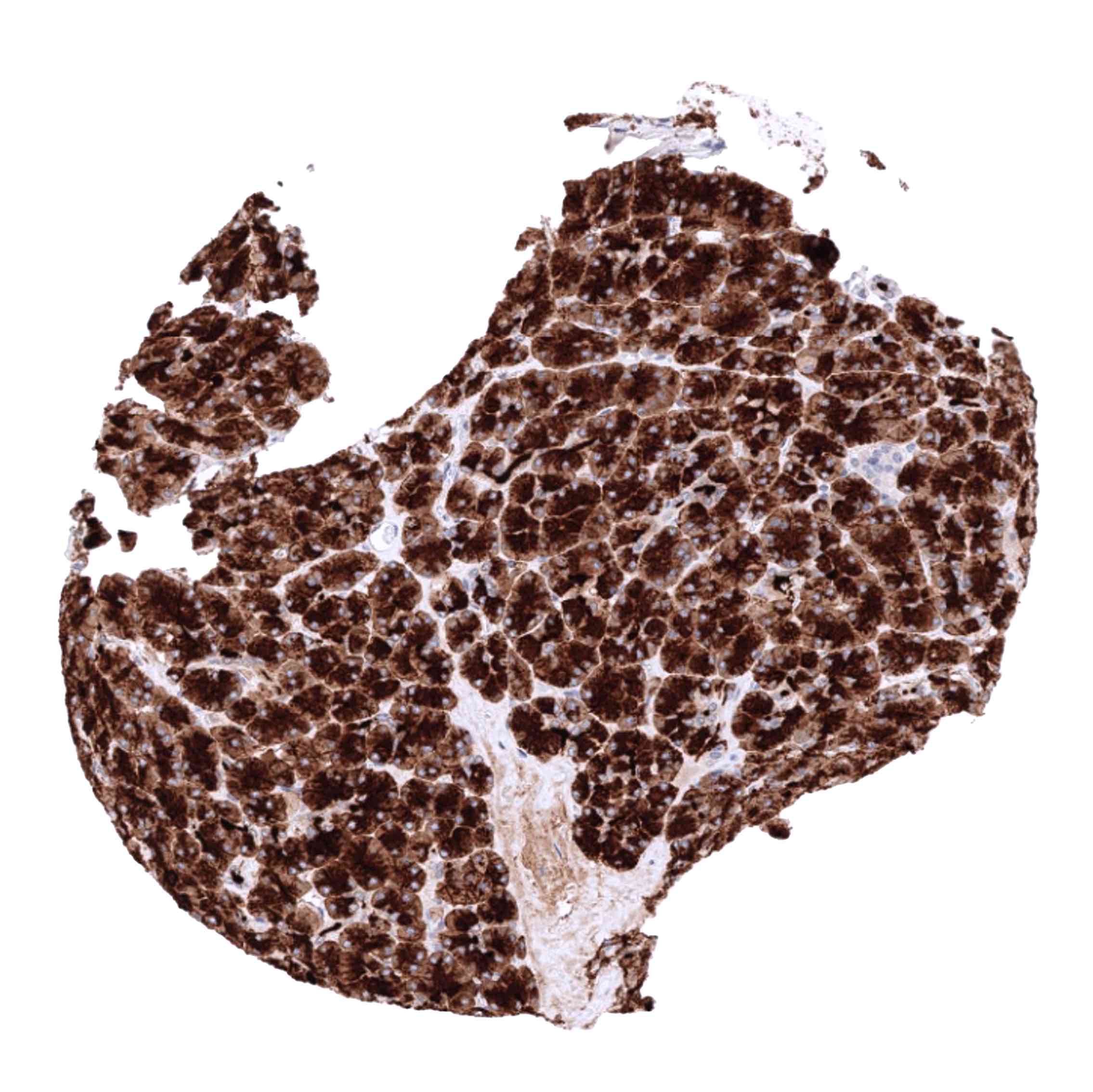
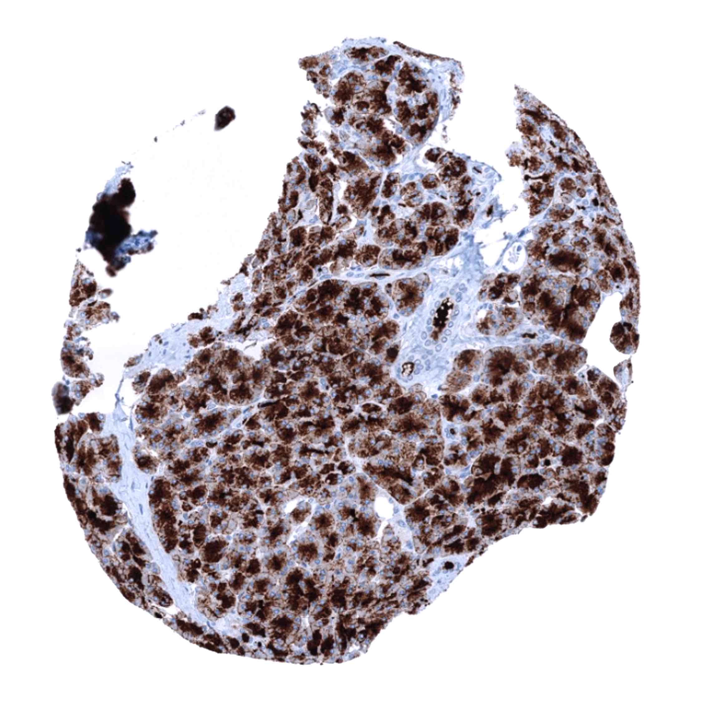
Positive control = Pancreas: Acinar cells should show a strong immunostaining with apical predominance. Duodenum: Glandular cells of Brunner glands should show a weak to moderate GP2 positivity.
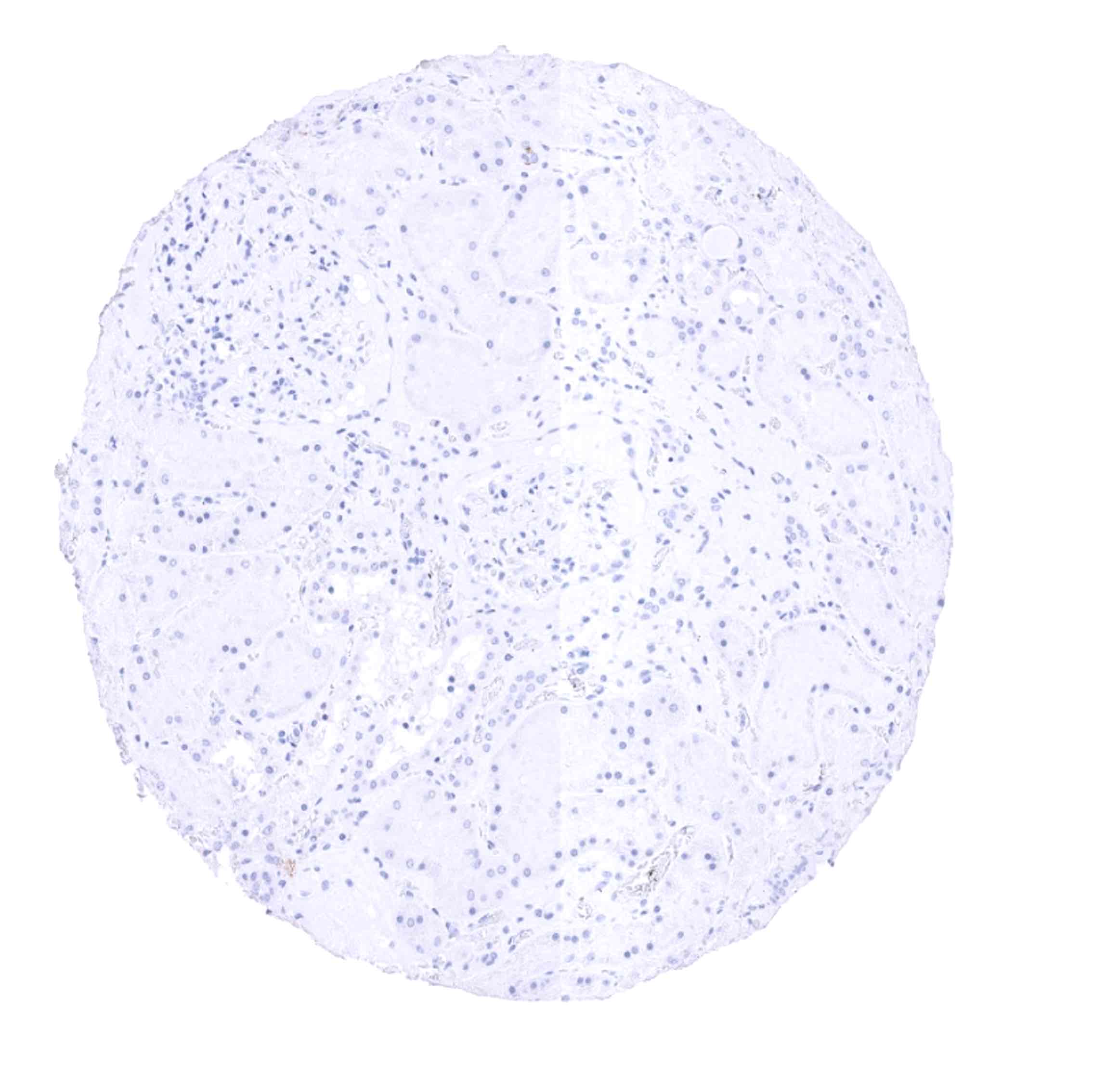
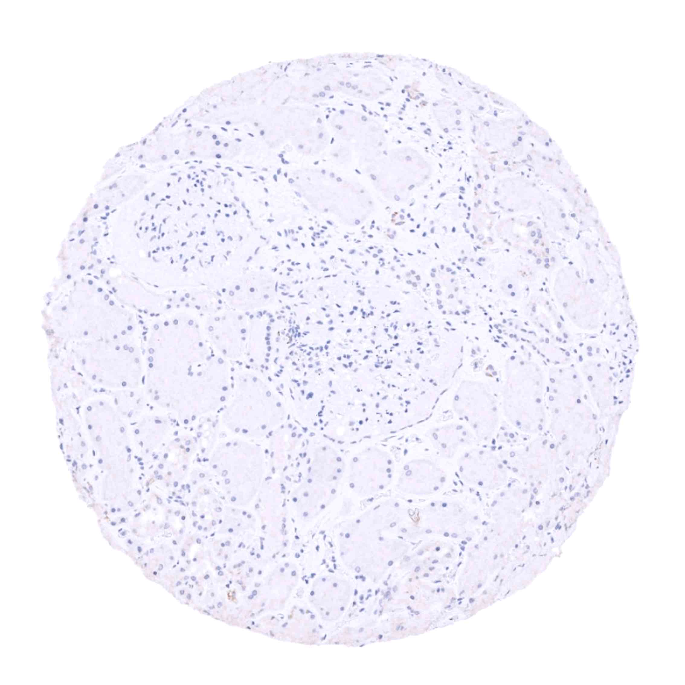
Negative control = Colon: GP2 staining should be completely absent in glandular and stromal cells.
Cellular localization = Cytoplasmic and cell Surface
Reactivity = Human
Application = Immunohistochemistry
Dilution = 1:100 – 1:200
Intended Use = Research Use Only
Relevance of Antibody
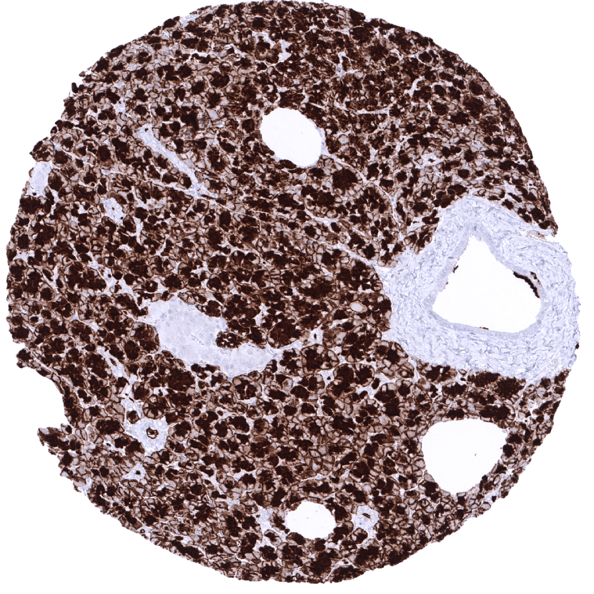
GP2 is expressed on normal and neoplastic pancreatic acinar cells.
Biology Behind
Pancreatic secretory granule membrane major glycoprotein (GP2) is a 68 kDa protein that in humans is encoded by the GP2 gene at chromosome 16p12.3. GP2 is the most abundant membrane protein found in zymogen granules of the pancreatic acinar cells, and is secreted together with digestive enzymes into the small intestine where it acts as an immunomodulator. The function of GP2 is poorly understood. GP2 knockout mice develop normally and do not have metabolic impairments, suggesting that this protein is not required for normal pancreas development and function. GP2 is also found on cells in various mucous glands in the murine digestive, respiratory, and genital tracts as well as on M-cells, an enterocyte derived cell-type specialized in the transport of luminal antigens and bacteria across the gut.
Staining Pattern in Normal Tissues
GP2 staining pattern in Normal Tissues with antibody MSVA-475M (images are shown in our “Normal Tissue Gallery”)
| Brain | Cerebrum | Negative. |
| Cerebellum | Negative. | |
| Endocrine Tissues | Thyroid | Negative. |
| Parathyroid | Negative. | |
| Adrenal gland | Negative. | |
| Pituitary gland | Negative. | |
| Respiratory system | Respiratory epithelium | Weak to moderate cytoplasmic GP2 staining of few serous cells in bronchial glands (not all samples). |
| Lung | Negative. | |
| Gastrointestinal Tract | Salivary glands | Negative. |
| Esophagus | Negative. | |
| Stomach | Weak to moderate cytoplasmic GP2 staining of some glandular cells in glands of the stomach antrum (not all samples). | |
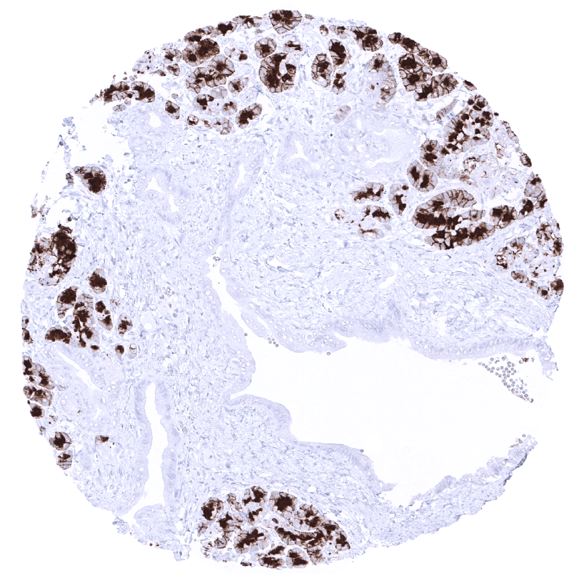
| Duodenum | Weak to moderate cytoplasmic GP2 staining predominating at the luminal membrane of duodenal Brunner gland cells. | |
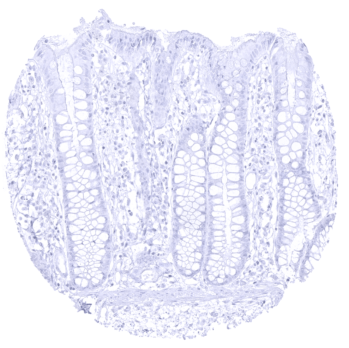
| Small intestine | Negative. | |
| Appendix | Negative. | |
| Colon | Negative. | |
| Rectum | Negative. | |
| Liver | Negative. | |
| Gallbladder | Weak to moderate cytoplasmic GP2 staining of some epithelial cells (not all samples). | |
| Pancreas | Strong cytoplasmatic and membranous GP2 staining of acinar cells. The staining often predominates at the apical membranes. | |
| Genitourinary | Kidney | Negative. |
| Urothelium | Negative. | |
| Male genital | Prostate | Negative. |
| Seminal vesicles | Negative. | |
| Testis | Negative. | |
| Epididymis | Negative. | |
| Female genital | Breast | Negative. |
| Uterus, myometrium | Negative. | |
| Uterus, ectocervix | Negative. | |
| Uterus endocervix | Negative. | |
| Uterus, endometrium | Weak to moderate cytoplasmic GP2 staining of few endometrial glands (not all samples). | |
| Fallopian Tube | Negative. | |
| Ovary | Negative. | |
| Placenta early | Negative. | |
| Placenta mature | Negative. | |
| Amnion | Negative. | |
| Chorion | Negative. | |
| Skin | Epidermis | Negative. |
| Sebaceous glands | Negative. | |
| Muscle/connective tissue | Heart muscle | Negative. |
| Skeletal muscle | Negative. | |
| Smooth muscle | Negative. | |
| Vessel walls | Negative. | |
| Fat | Negative. | |
| Stroma | Negative. | |
| Endothelium | Negative. | |
| Bone marrow/ lymphoid tissue | Bone marrow | Negative. |
| Lymph node | Negative. | |
| Spleen | Negative. | |
| Thymus | Negative. | |
| Tonsil | Negative. | |
| Remarks |
These findings are largely comparable to the RNA and protein data described in the Human Protein Atlas (Tissue expression GP2) even though the expression in Brunner glands, bronchial glands, gallbladder and stomach antrum glands is not described in the protein atlas. These structures constitute small subsets of the total amount of cells in their respective organs and may have been largely underrepresented in RNA analyses.
Positive control = Pancreas: Acinar cells should show a strong immunostaining with apical predominance. Duodenum: Glandular cells of Brunner glands should show a weak to moderate GP2 positivity.
Negative control = Colon: GP2 staining should be completely absent in glandular and stromal cells.
Staining Pattern in Relevant Tumor Types
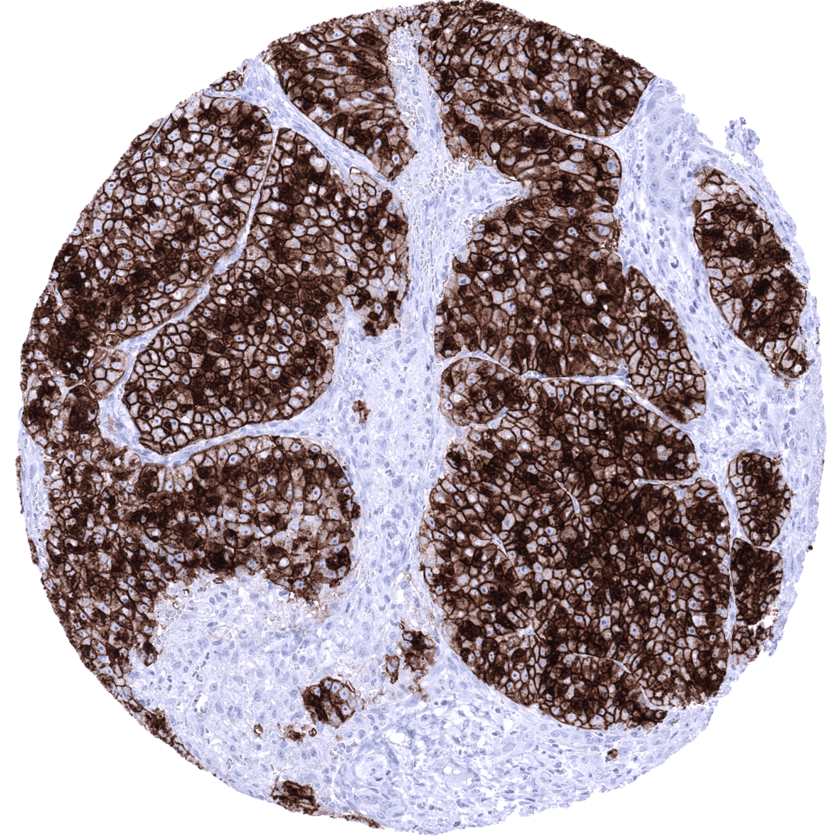
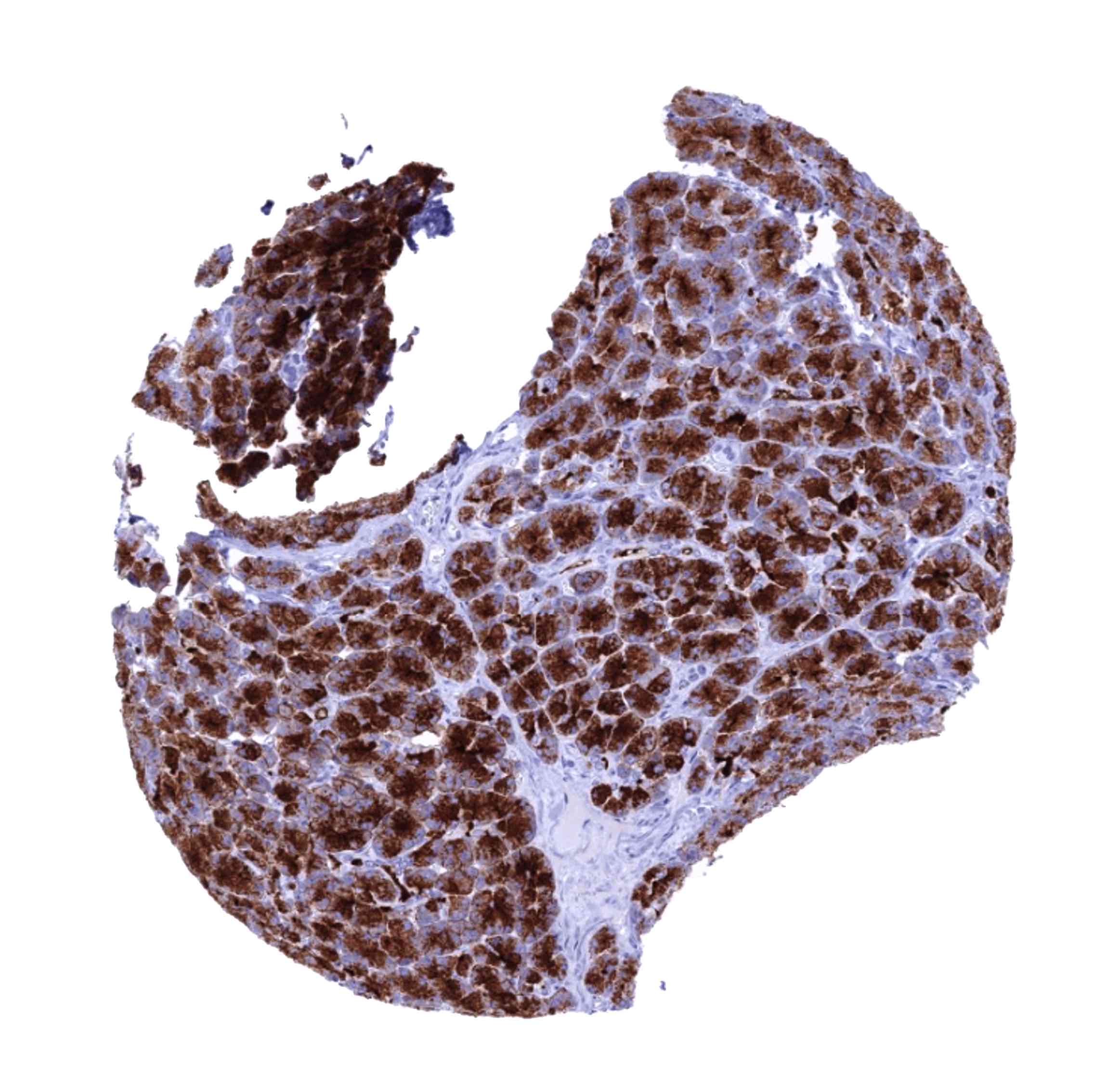
GP2 is expressed in the majority of acinar cell carcinomas of the pancreas. A lower level of GP2 expression may can rarely be seen in several other tumor entities.
The TCGA findings on GP2 RNA expression in different tumor categories have been summarized in the Human Protein Atlas.


Compatibility of Antibodies
GP2 (MSVA-475M) publication summary
Relevant publication: Uhlig et al.: “Diagnostic and prognostic role of pancreatic secretory granule membrane major glycoprotein 2 (GP2) immunohistochemistry: A TMA study on 27’681 tumors”. Published in Pathol Res Pract. 2022 Sep 15;238:154123. PMID: 36137400.
A total of 27’681 tumors were successfully analyzed from 132 different tumor categories by using the following protocol: Heat-induced antigen retrieval for 5 minutes in an autoclave at 121°C in pH7,8 Target Retrieval Solution buffer. MSVA-475M at a dilution of 1:150 at 37°C for 60 minutes. Visualization of bound antibody by the EnVision Kit (Dako, Agilent). This protocol was also used for all stainings depicted in our tumor and normal tissue galleries.
In this study, at least one GP2 positive case was seen in 25 (18,9%) of 132 tumor categories and 6 (4,5%) tumor categories included at least one case with strong positivity. Uhlig et al described the highest GP2 positivity rates in pancreatic acinar cell carcinomas (88%), followed by various categories of neuroendocrine neoplasms (14–59%), prostatic adenocarcinomas (8–19%), several other adenocarcinoma entities (0.1–8%) and several groups of salivary gland tumors (2–3 %). Strong GP2 positivity was only found in 6 tumor categories including 8 of 16 acinus cell carcinomas of the pancreas, 1 of 17 neuroendocrine tumors of the lung, 1 of 80 primary Gleason 4 + 4 and 1 of 181 recurrent prostate cancers, 1 of 133 pulmonary adenocarcinomas, and in 2 of 240 mucoepidermoid carcinomas of the salivary gland. The distribution of positive staining results is shown in an “organ-systematic” and in a “ranking order” figure below (images based on data from Uhlig et al.). Results on possible associations with histopathological and clinical parameters of tumor aggressiveness are also summarized below (table based on data from Uhlig et al.).
Authors conclusions on diagnostic utility of GP2 immunohistochemistry with respect to the distinction of neoplastic from non-neoplastic tissues (Uhlig et al.):
- Not applicable
Authors conclusions on diagnostic utility of GP2 immunohistochemistry with respect to the distinction of different tumor entities (Uhlig et al.):
- GP2 positivity in pancreatic tumor, especially if it is strong, argues for an acinar cell carcinoma. However, GP2 expression can (rarey) also occur in ductal adenocarcinoma and in neuroendocrine neoplasms of the pancreas!
Authors conclusions on prognostic/predictive role of GP2 expression (Uhlig et al.):
- GP2 positivity is strongly linked to unfavorable prognosis in prostate cancer.
Figure 1. GP2 staining in cancer (“organ-systematic”; according to Uhlig et al.)

Figure 2. GP2 staining in cancer (“ranking list”; according to Uhlig et al.)

Protocol Recommendations
IHC users have different preferences on how the stains should look like. Some prefer high staining intensity of the target stain and even accept some background. Others favor absolute specificity and lighter target stains. Factors that invariably lead to more intense staining include higher concentration of the antibody and visualization tools, longer incubation time, higher temperature during incubation, higher temperature and longer duration of the heat induced epitope retrieval (slide pretreatment). The impact of the pH during slide pretreatment has variable effects and depends on the antibody and the target protein.
All images and data shown here and in our image galleries are obtained by the manual protocol described below. Other protocols resulting in equivalent staining are described as well.
Manual protocol
Freshly cut sections should be used (less than 10 days between cutting and staining). Heat-induced antigen retrieval for 5 minutes in an autoclave at 121°C in pH 7,8 Target Retrieval Solution buffer. Apply MSVA-475M at a dilution of 1:150 at 37°C for 60 minutes. Visualization of bound antibody by the EnVision Kit (Dako, Agilent) according to the manufacturer’s directions.
Agilent / Dako – Autostainer Link 48
Pretreatment in PT-Link for 30 minutes at 95°C (pH high); FLEX peroxidase blocking for 5 minutes (room temperature), MSVA-475M 1:150 for 20 minutes (room temperature), FLEX+ mouse/rabbit (LINKER) for 15 minutes (room temperature), horseradish peroxidase (HRP) for 20 minutes (room temperature), FLEX DAB+Sub-Chromo for 10 minutes (room temperature), FLEX hematoxylin for 5 minutes (room temperature).

These images reflect stainings by the protocol described above. It is of note that a comparable staining result can also be obtained by different protocols. In general, a longer pretreatment, a longer incubation time of the primary antibody, a higher antibody concentration, and a longer incubation time of FLEX+LINKER result in stronger staining, potentially at the cost of more background staining. Modifications of the protocol with a strengthening effect on staining intensity in combination with changes of other parameters that result in lower staining intensity can result in a comparable result as shown above.
Leica – BOND RX
Dewax at 72°C for 30 seconds; Pretreatment in Bond Epitope Retrieval Solution (ER2 – EDTA pH9) for 20 minutes at 100°C; Peroxidase blocking for 5 minutes (room temperature), MSVA-475M 1:150 for 15 minutes (room temperature), Post primary (rabbit anti mouse) for 8 minutes (room temperature), Polymer (goat anti rabbit) for 8 minutes (room temperature), mixed DAB refine for 10 minutes (room temperature), hematoxylin for 5 minutes (room temperature).

These images reflect stainings by the protocol described above. It is of note that a comparable staining result can also be obtained by different protocols. In general, a longer pretreatment, a longer incubation time of the primary antibody, a higher antibody concentration, a higher temperature during incubation, and a longer incubation time of Post primary and or the Polymer result in stronger staining, potentially at the cost of more background staining. Modifications of the protocol with a strengthening effect on staining intensity in combination with changes of other parameters that result in lower staining intensity can result in a comparable result as shown above.
Roche – Ventana Discovery ULTRA
Pretreatment for 64 minutes at 100°C (pH 8,4); CM peroxidase blocking for 12 minutes (room temperature), MSVA-475M 1:150 for 20 minutes at 36°C, secondary antibody (anti-mouse HQ) for 12 minutes at 36°C, anti-HQ HRP for 12 minutes at room temperature, DAB at room temperature, hematoxylin II at room temperature for 8 minutes, bluing reagent at room temperature for 4 minutes.


These images depict staining results obtained by the protocol described above. It is of note, that the Ventana machines generally require higher antibody concentrations than other commonly used autostainers because the antibodies are automatically diluted during the procedure. Various other protocols can result in an identical result as shown above. A longer pretreatment, a longer incubation time of the primary antibody, a higher antibody concentration, a higher temperature during incubation, and a longer incubation time of secondary antibody and or the anti-HQ HRP result in stronger staining, potentially at the cost of more background staining.
Potential Research Applications
- The diagnostic utility of GP2 IHC should be investigated in a large cohort of tumors from different entities. In particular, its utility as a marker for pancreatic acinar carcinoma should be analyzed.
- The function of GP2 is largely unknown.
- The prognostic role of GP2 expression in cancers is unknown.
- Autoantibodies against GP2 occur in Crohn’s disease. The significance of this phenomenon is not understood.
Evidence for Antibody Specificity in IHC
There are two ways, how the specificity of antibodies can be documented for immunohistochemistry on formalin fixed tissues. These are: 1. comparison with a second independent method for target expression measurement across a large number of different tissue types (orthogonal strategy), and 2. Comparison with one or several independent antibodies for the same target and showing that all positive staining results are also seen with other antibodies for the same target (independent antibody strategy).
Orthogonal validation: For the antibody MSVA-475M specificity is documented by the strong concordance of the immunostaining with RNA expression data derived from the Human Protein Atlas (HPA) RNA-seq tissue dataset, the FANTOM5 project, and the Genotype-Tissue Expression (GTEx) project which are all summarized in the Human Protein Atlas (Tissue expression GP2) Immunostaining by using MSVA-475M was predominantly detected in the pancreas which was the only organ with documented RNA expression. In addition, a positive immunostaining was also found in Brunner glands, bronchial glands, and stomach antrum glands. These structures constitute small subsets of the total amount of cells in these organs and may have been largely underrepresented in RNA analyses.
Comparison of antibodies: True expression of GP2 in Brunner glands, bronchial glands, stomach antrum glands, gallbladder epithelium and endometrial glands was documented by an identical staining pattern with the unrelated antibody “MSVA-GP2 candidate F3g” raised against an identical GP2 epitope.
Also the stainings obtained by the antibody HPA016668 depicted in the human protein atlas were consistent with membranous staining of:
while images of Brunner glands and bronchial glands were not depicted in the protein atlas.
