395,00 € – 1.495,00 €
Product details
Synonyms = ASBABP2; Aspecific BCL2 ARE binding protein 2; Differentially placenta 1 expressed protein; DIPLA1; IGF-dependent; IGFBP4ase; Insulin-like growth factor-dependent IGF-binding protein 4 protease (IGFBP-4 protease); PAPA; PAPP A; PAPPA1; Pappalysin-1; Pregnancy Associated Plasma Protein A (PAPP-A)
Antibody type = Mouse monoclonal / IgG
Clone = MSVA-780M
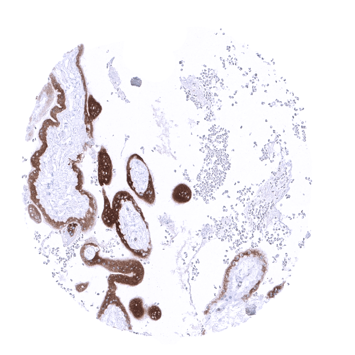
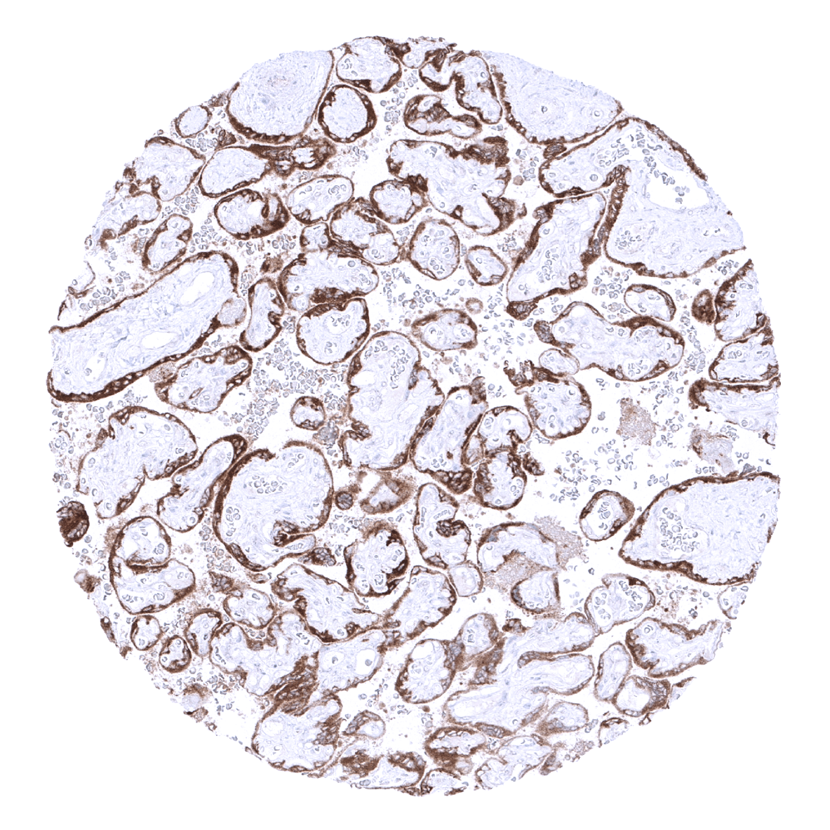
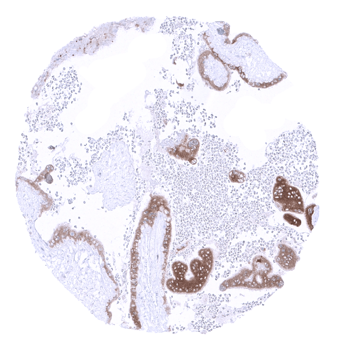
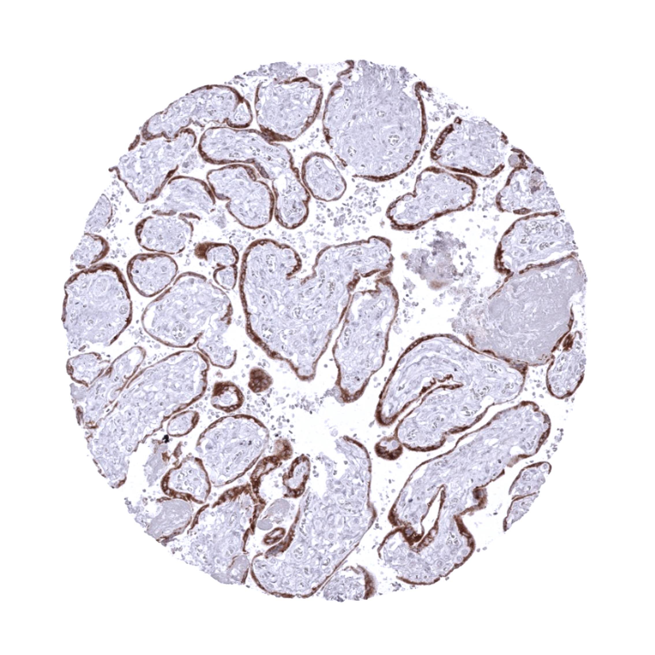
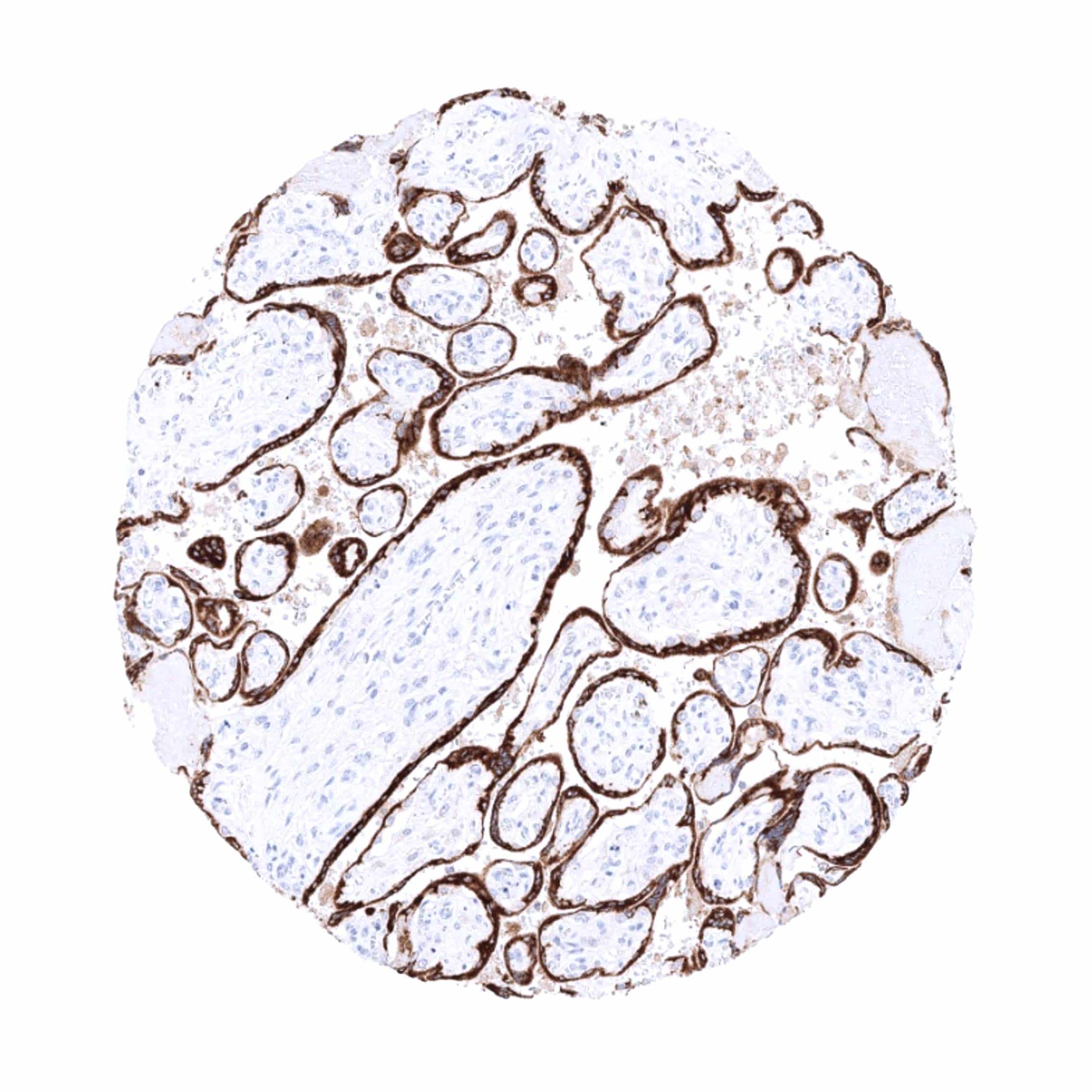
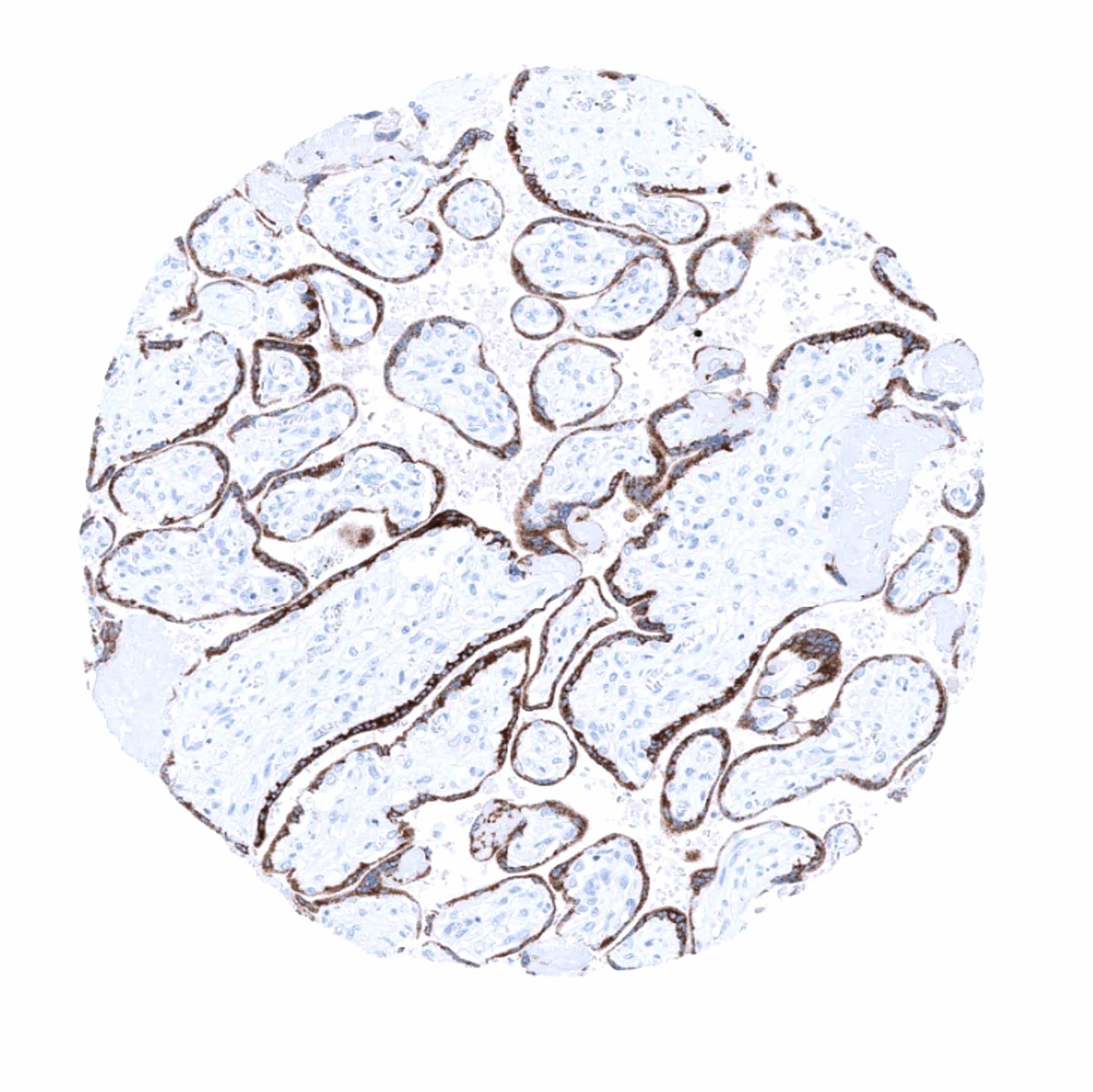
Positive control = Placenta: Trophoplast and chorion cells should show a strong cytoplasmic papp-A staining.
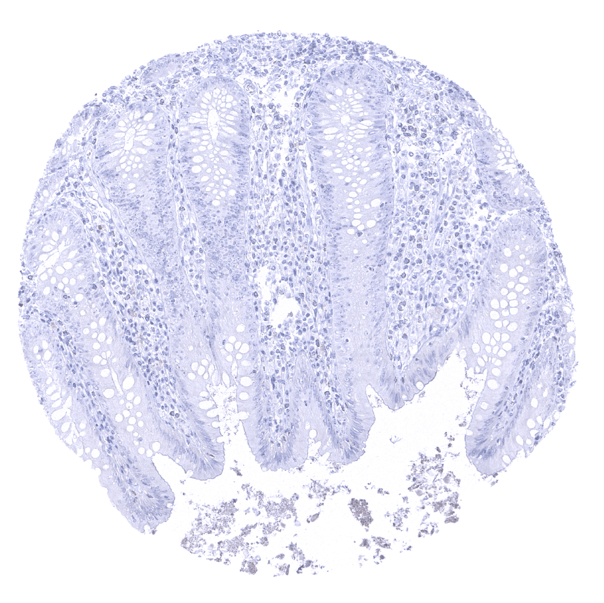
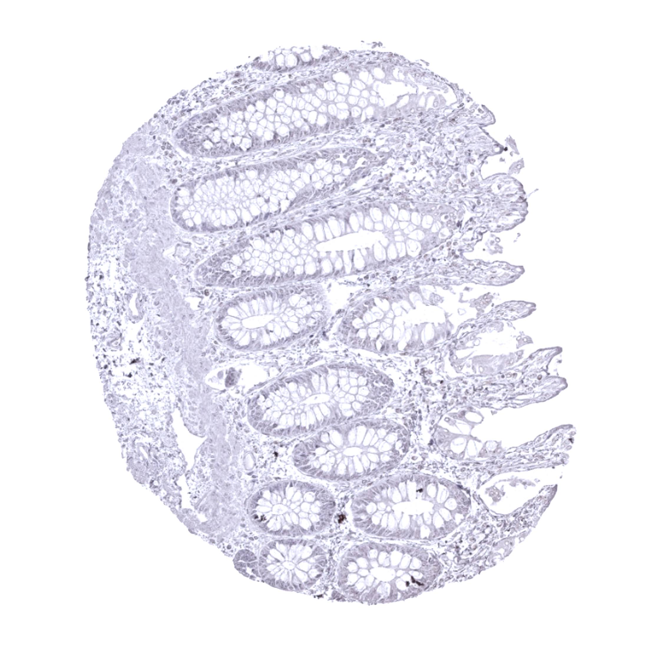
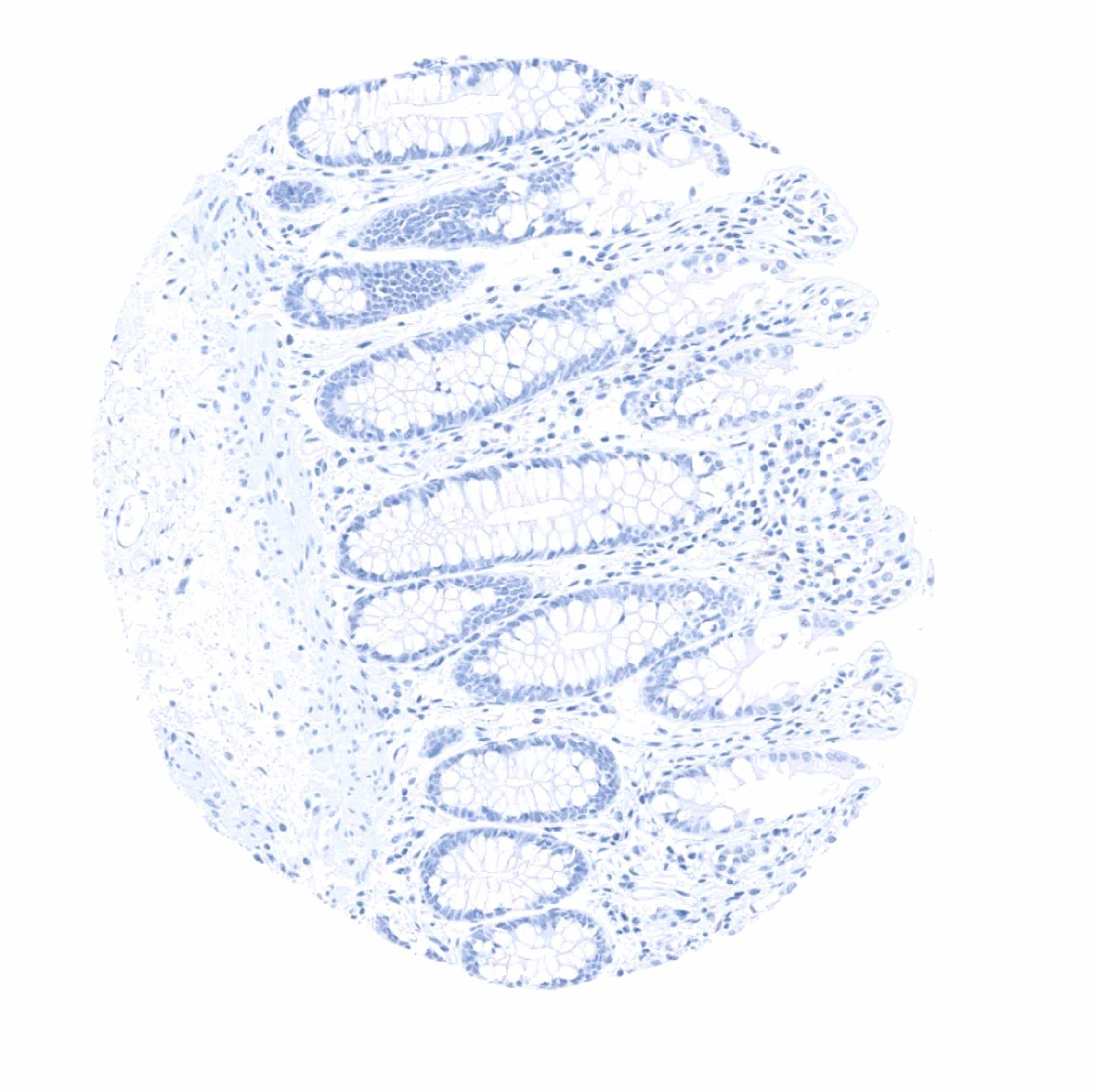
Negative control =Colon: Papp-A immunostaining should be completely absent in both stromal end epithelial cells.
Cellular localization = Cytoplasmic.
Reactivity = Human
Application = Immunohistochemistry
Dilution = 1:100
Intended Use = Research Use Only
Relevance of Antibody
PAPP-A is expressed in the placenta.
Biology Behind
Pregnancy-associated plasma protein A (Papp-A) is a metalloproteinase encoded by the PAPPA gene located at chromosome 9q33.2. The protein is secreted, activated upon collagen binding, and is thought to be involved in local proliferative processes such as wound healing and bone remodeling. Its main substrates are insulin-like growth factor binding proteins which are cleaved by papp-A. Papp-A is mainly produced in the placenta and has been connected with paracrine and autocrine control of trophoblast invasion of the decidua. Low maternal serum papp-A concentrations have been implicated with preeclampsia and Down syndrome.
Staining Pattern in Normal Tissues
Papp-A is a placenta specific protein which is seen both in the early and mature placenta. Strongest immunostaining is seen in syncytiotrophoblast and in chorion cells. Staining may be less or even absent in the cytotrophoblast.
These findings are largely comparable to the RNA and protein data described in the Human Protein Atlas (Tissue expression PAPP-A)
Positive control: Placenta: Trophoplast and chorion cells should show a strong cytoplasmic papp-A staining.
Negative control: Colon: Papp-A immunostaining should be completely absent in both stromal end epithelial cells.

Staining Pattern in Relevant Tumor Types
Papp-A is not known to be expressed in cancers.
The TCGA findings on PAPP-A RNA expression in different tumor categories have been summarized in the Human Protein Atlas.
Compatibility of Antibodies
No data available at the moment
Protocol Recommendations
IHC users have different preferences on how the stains should look like. Some prefer high staining intensity of the target stain and even accept some background. Others favor absolute specificity and lighter target stains. Factors that invariably lead to more intense staining include higher concentration of the antibody and visualization tools, longer incubation time, higher temperature during incubation, higher temperature and longer duration of the heat induced epitope retrieval (slide pretreatment). The impact of the pH during slide pretreatment has variable effects and depends on the antibody and the target protein.
All images and data shown here and in our image galleries are obtained by the manual protocol described below. Other protocols resulting in equivalent staining are described as well.
Manual protocol
Freshly cut sections should be used (less than 10 days between cutting and staining). Heat-induced antigen retrieval for 5 minutes in an autoclave at 121°C in pH 7,8 Target Retrieval Solution buffer. Apply MSVA-780M at a dilution of 1:100 at 37°C for 60 minutes. Visualization of bound antibody by the EnVision Kit (Dako, Agilent) according to the manufacturer’s directions.

Leica – BOND RX
Dewax at 72°C for 30 seconds; Pretreatment in Bond Epitope Retrieval Solution (ER2 – EDTA pH9) for 20 minutes at 100°C; Peroxidase blocking for 5 minutes (room temperature), MSVA-780M 1:100 for 15 minutes (room temperature), Post primary (rabbit anti mouse) for 8 minutes (room temperature), Polymer (goat anti rabbit) for 8 minutes (room temperature), mixed DAB refine for 10 minutes (room temperature), hematoxylin for 5 minutes (room temperature).


These images reflect stainings by the protocol described above. It is of note that a comparable staining result can also be obtained by different protocols. In general, a longer pretreatment, a longer incubation time of the primary antibody, a higher antibody concentration, a higher temperature during incubation, and a longer incubation time of Post primary and or the Polymer result in stronger staining, potentially at the cost of more background staining. Modifications of the protocol with a strengthening effect on staining intensity in combination with changes of other parameters that result in lower staining intensity can result in a comparable result as shown above.
Roche – Ventana Discovery ULTRA
Pretreatment for 64 minutes at 100°C (pH 8,4); CM peroxidase blocking for 12 minutes (room temperature), MSVA-780M 1:100 for 20 minutes at 36°C, secondary antibody (anti-mouse HQ) for 12 minutes at 36°C, anti-HQ HRP for 12 minutes at room temperature, DAB at room temperature, hematoxylin II at room temperature for 8 minutes, bluing reagent at room temperature for 4 minutes.

These images depict staining results obtained by the protocol described above. It is of note, that the Ventana machines generally require higher antibody concentrations than other commonly used autostainers because the antibodies are automatically diluted during the procedure. Various other protocols can result in an identical result as shown above. A longer pretreatment, a longer incubation time of the primary antibody, a higher antibody concentration, a higher temperature during incubation, and a longer incubation time of secondary antibody and or the anti-HQ HRP result in stronger staining, potentially at the cost of more background staining.
Potential Research Applications
- It is currently unknown, whether Papp-A expression can occur in cancer.
Evidence for Antibody Specificity in IHC
There are two ways how the specificity of antibodies can be documented for immunohistochemistry on formalin fixed tissues. These are: 1. comparison with a second independent method for target expression measurement across a large number of different tissue types (orthogonal strategy), and 2. Comparison with one or several independent antibodies for the same target and showing that all positive staining results are also seen with other antibodies for the same target (independent antibody strategy).
Orthogonal validation: For the antibody MSVA-780M specificity is suggested by the full concordance of the immunostaining with RNA expression data derived from the Human Protein Atlas (HPA) RNA-seq tissue dataset, the FANTOM5 project, and the Genotype-Tissue Expression (GTEx) project which are all summarized in the protein atlas. Immunostaining by using MSVA-780M was exclusively detected in the placenta, the only organ with documented RNA expression.
Comparison of antibodies: True expression of Papp-A in the placenta is also suggested by similar staining patterns obtained by the use of the antibody HPA001667. The respective images are depicted in Human Protein Atlas (Tissue expression PAPP-A)
