Adrenal gland
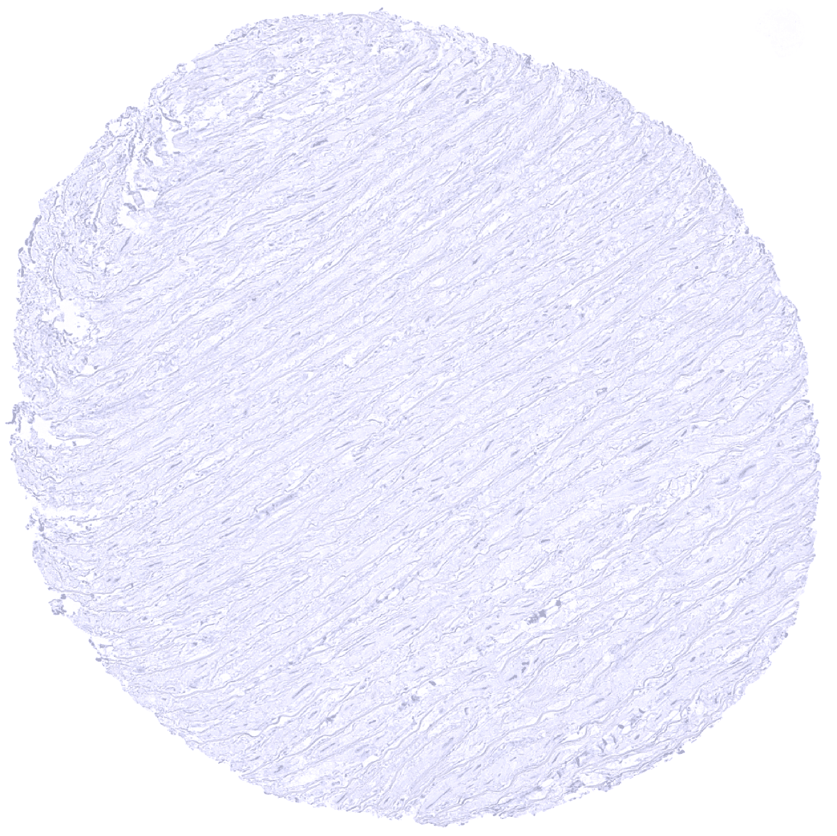
Aorta, media
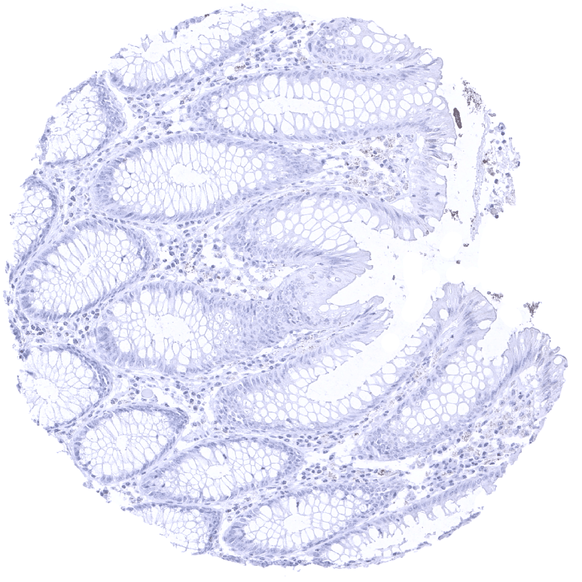
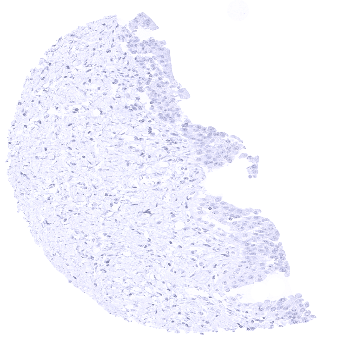
Appendix, mucosa

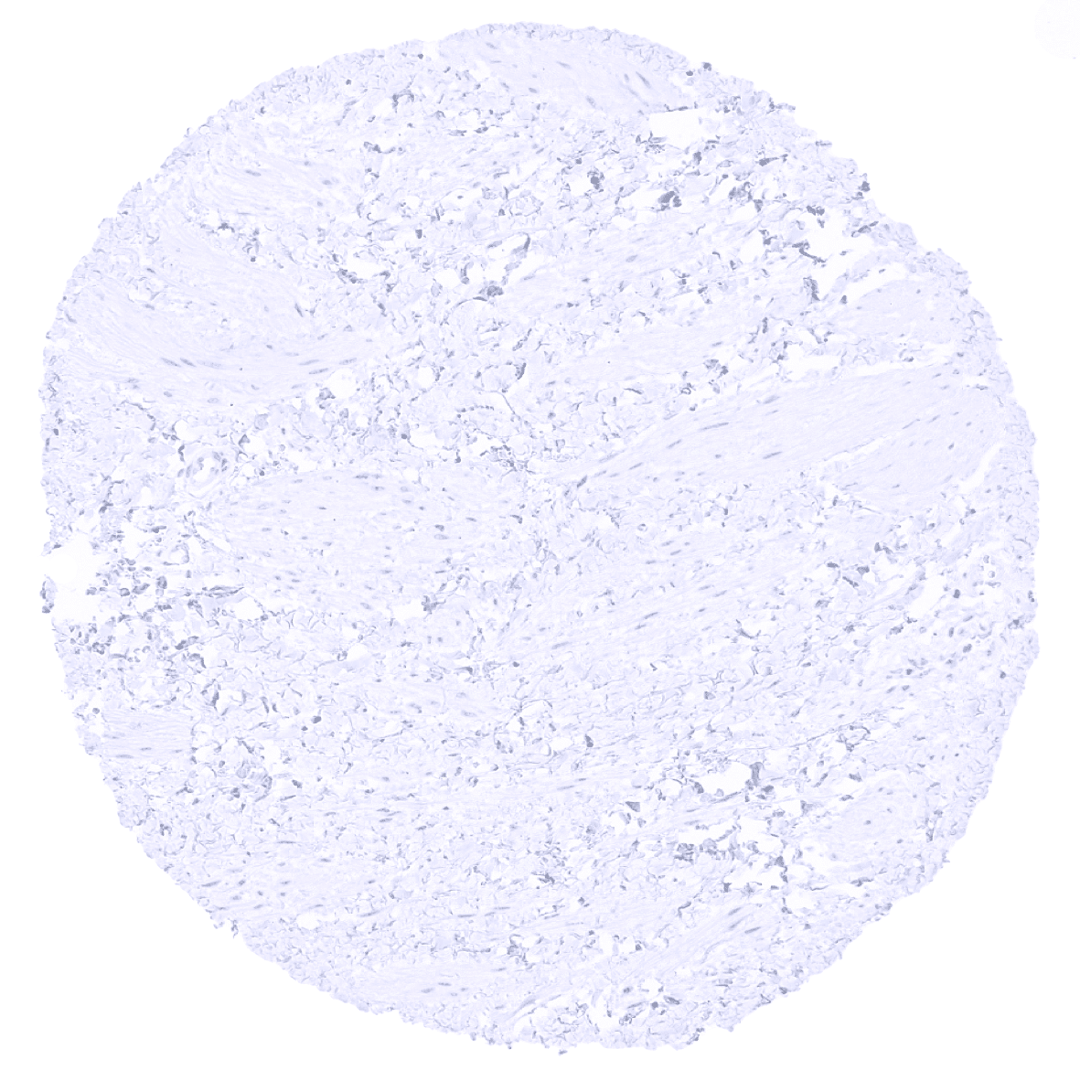
Appendix, muscular wall
Bone marrow
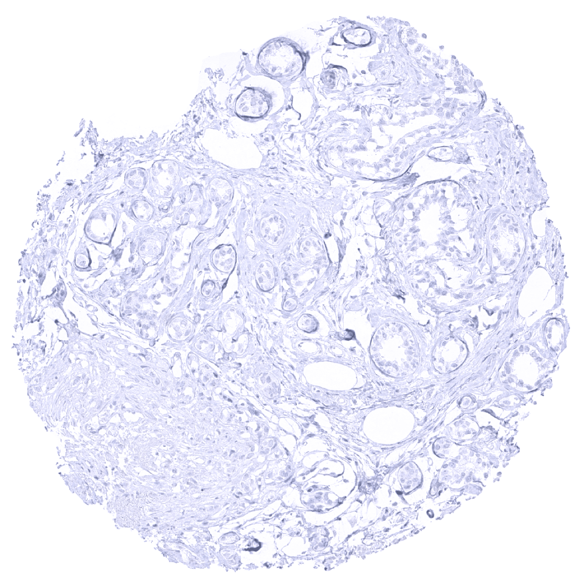
Breast
Bronchus, bronchial glands - A weak to moderate GP2 staining occurs in selected cells of bronchial glands. The staining is cytoplasmic but shows a distinct apical predominance.
Bronchus, mucosa
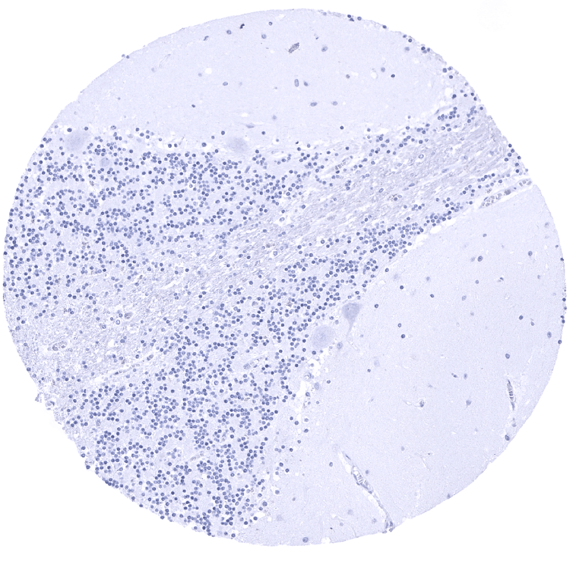
Cerebellum (molecular layer, Purkinje cell layer, granule cell layer, white matter)
Cerebellum (white matter)
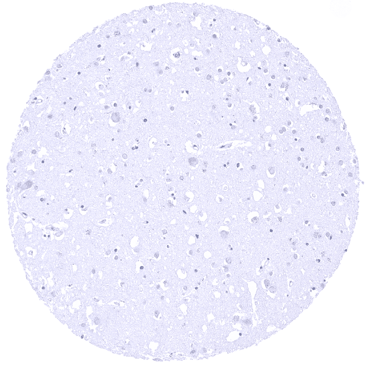
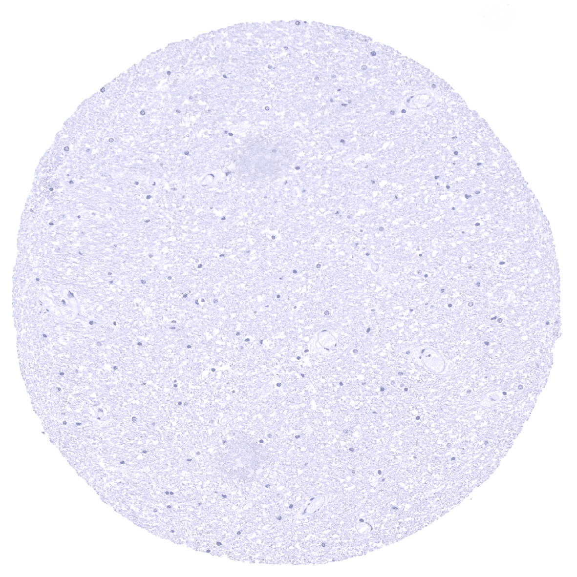
Cerebrum, grey matter
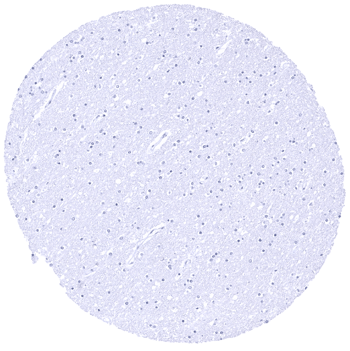
Cerebrum, white matter
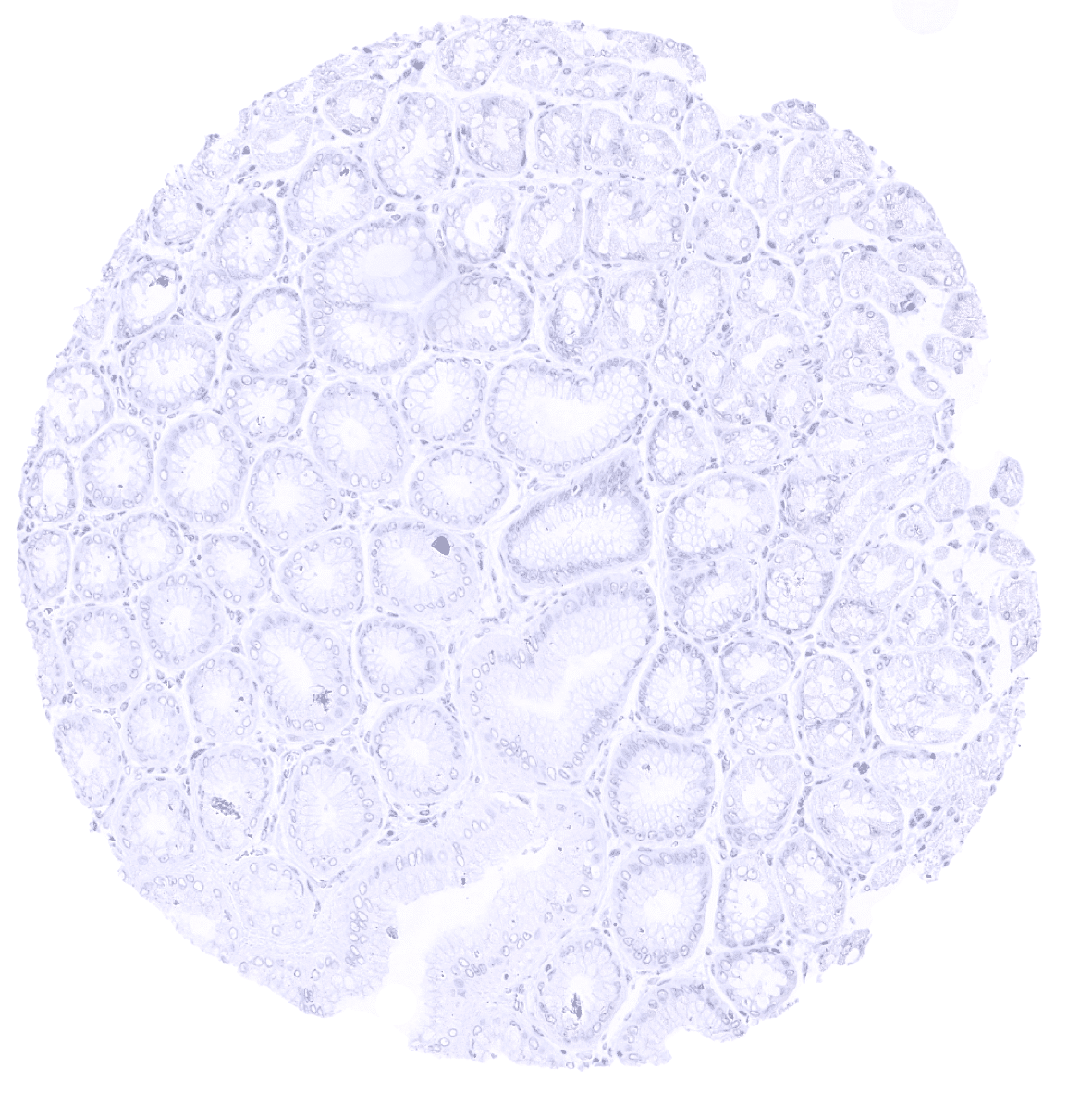
Colon descendens, mucosa

Colon descendens, mucosa
Colon descendens, muscular wall

Duodenum, Brunner gland - A weak to moderate GP2 staining occurs in Brunner glands of the duodenum. The staining is cytoplasmic but shows a distinct apical predominance.
Duodenum, Brunner gland - A weak to moderate GP2 staining occurs in Brunner glands of the duodenum. The staining is cytoplasmic but shows a distinct apical predominance.
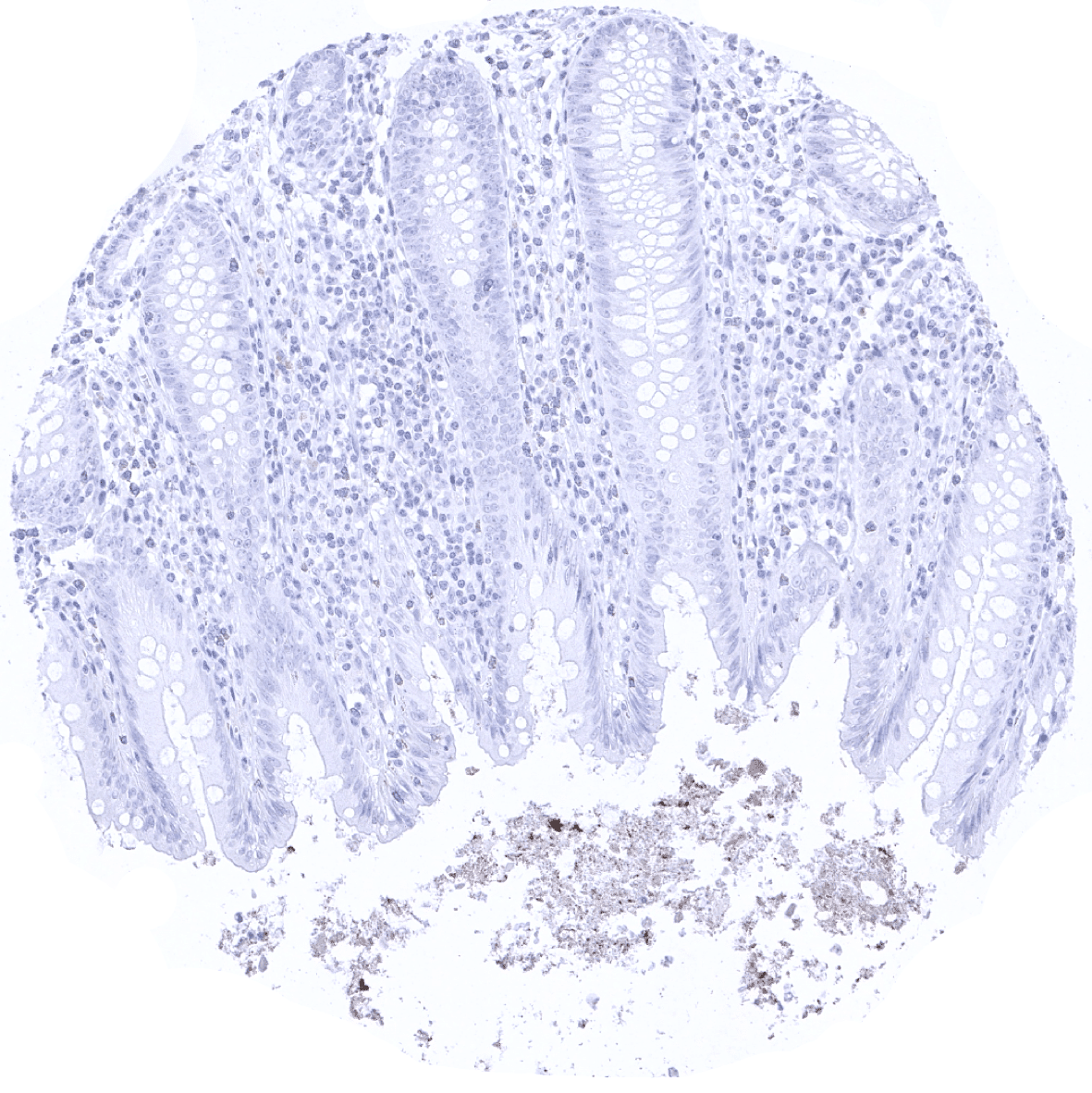
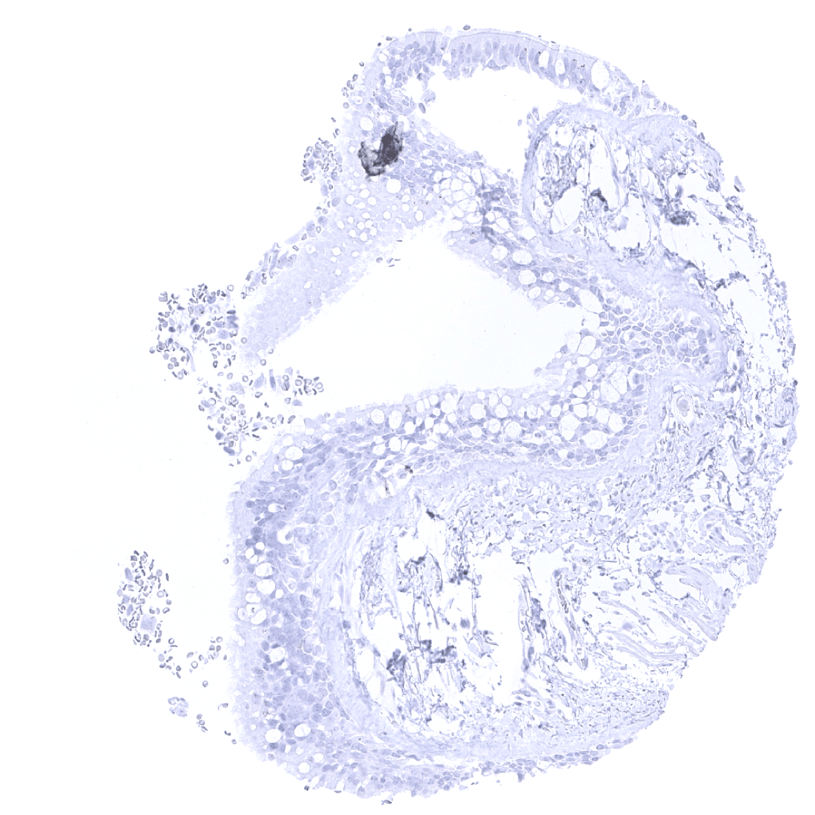
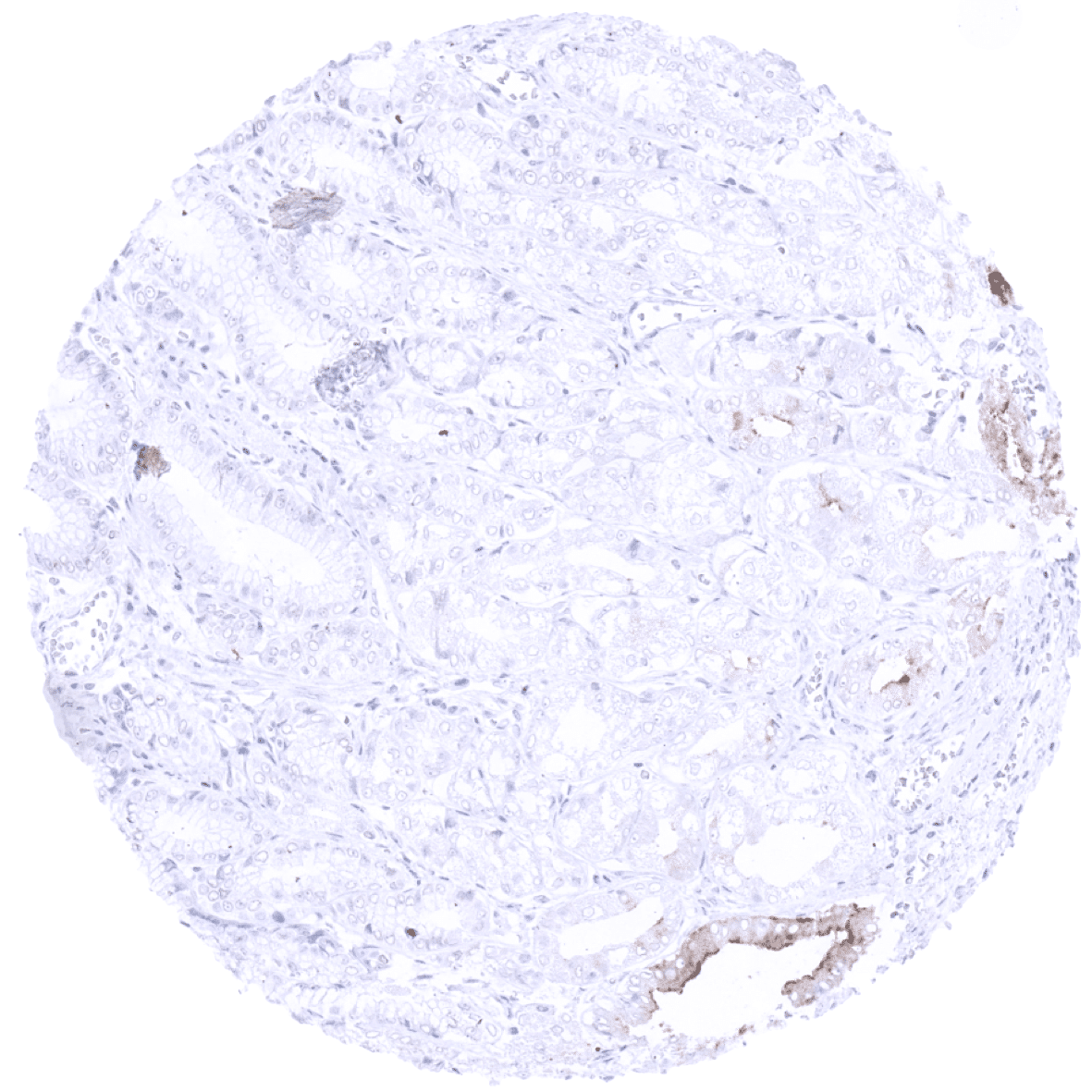
Duodenum, mucosa
Epididymis
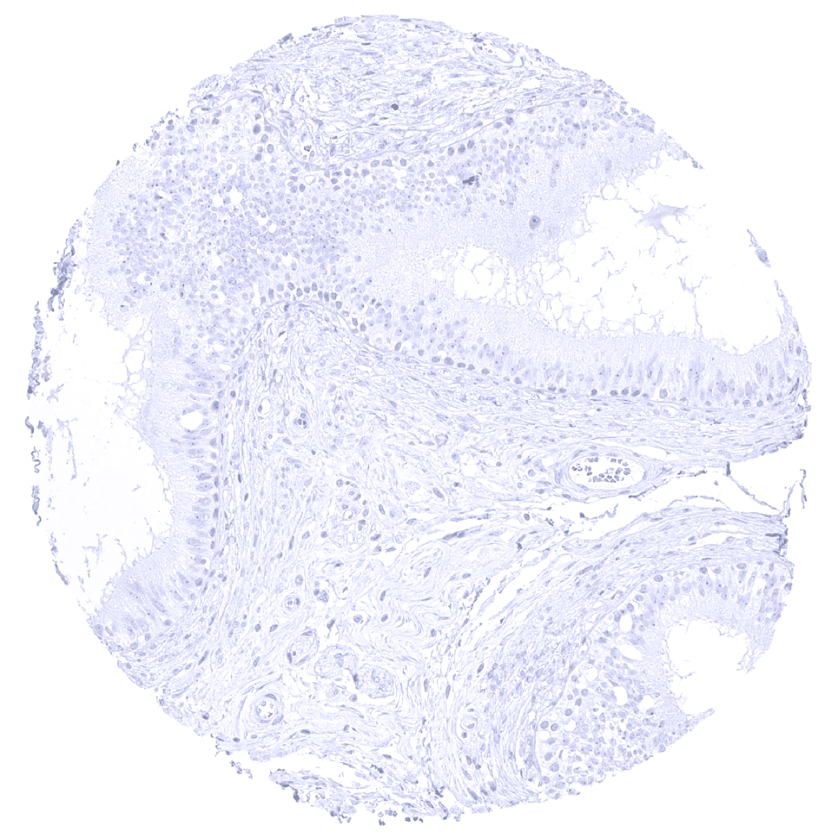
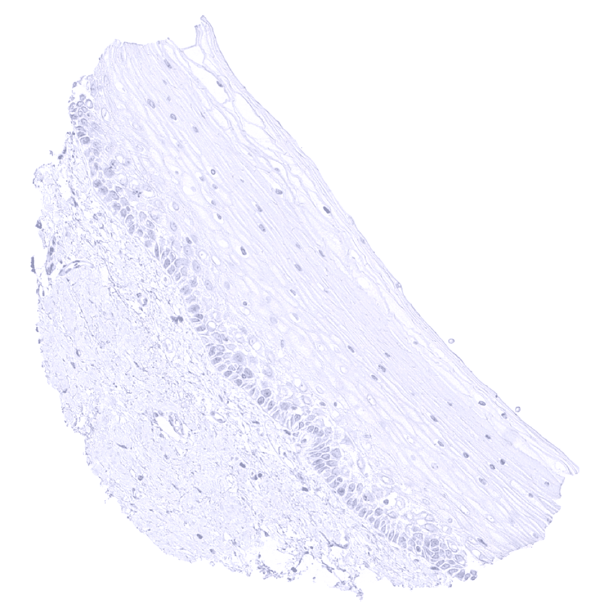
Esophagus, squamous epithelium

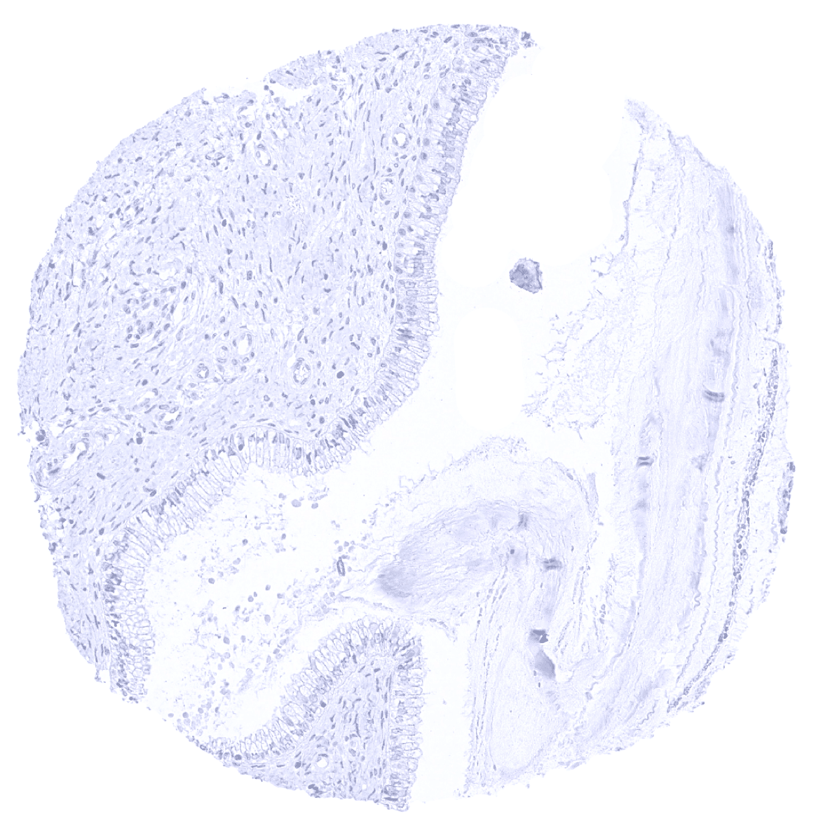
Fallopian tube
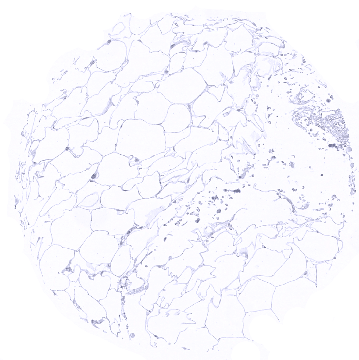
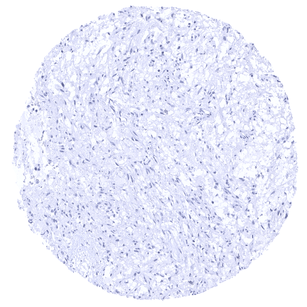
Fat

Gallbladder, epithelium
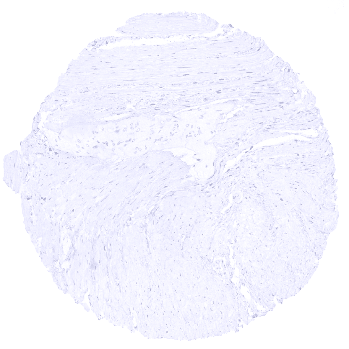
Heart muscle

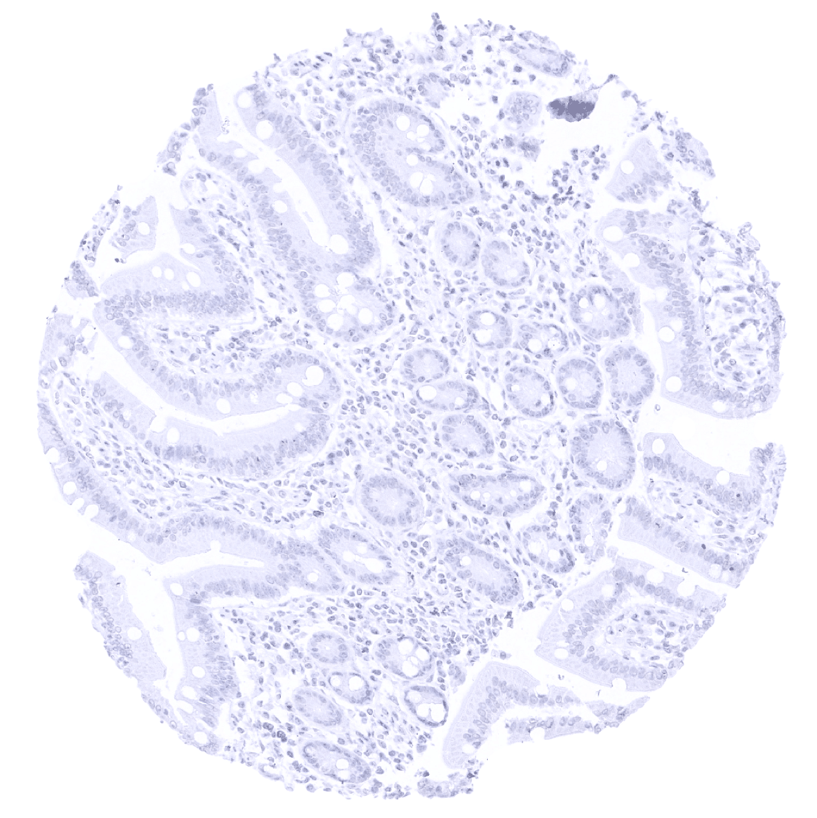
Ileum, mucosa
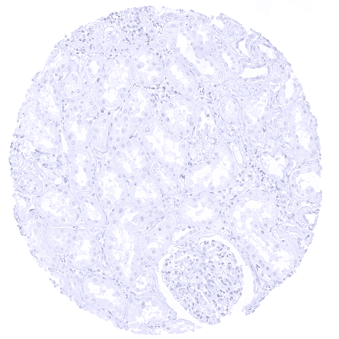
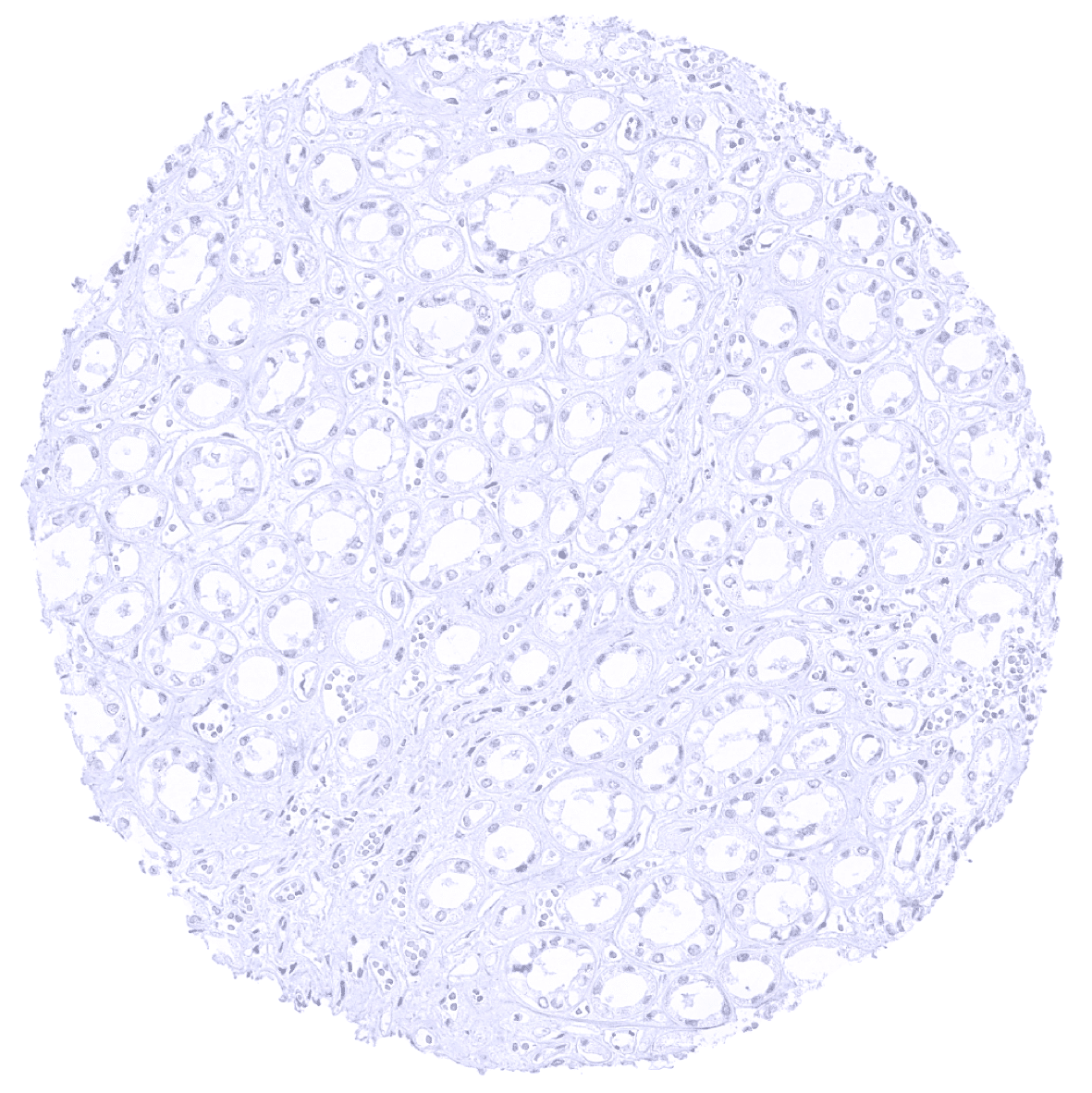
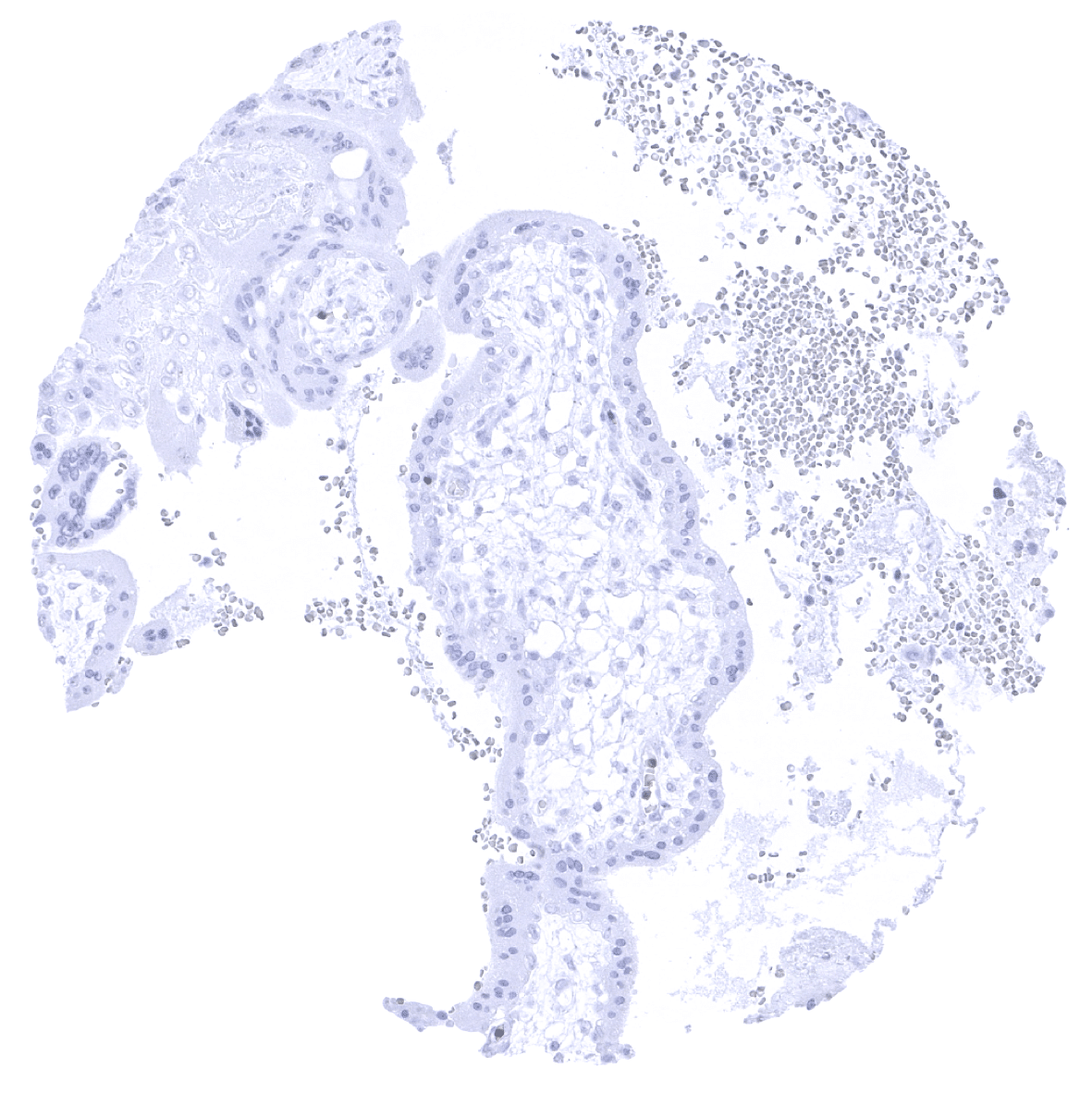
Kidney, cortex
Kidney, medulla
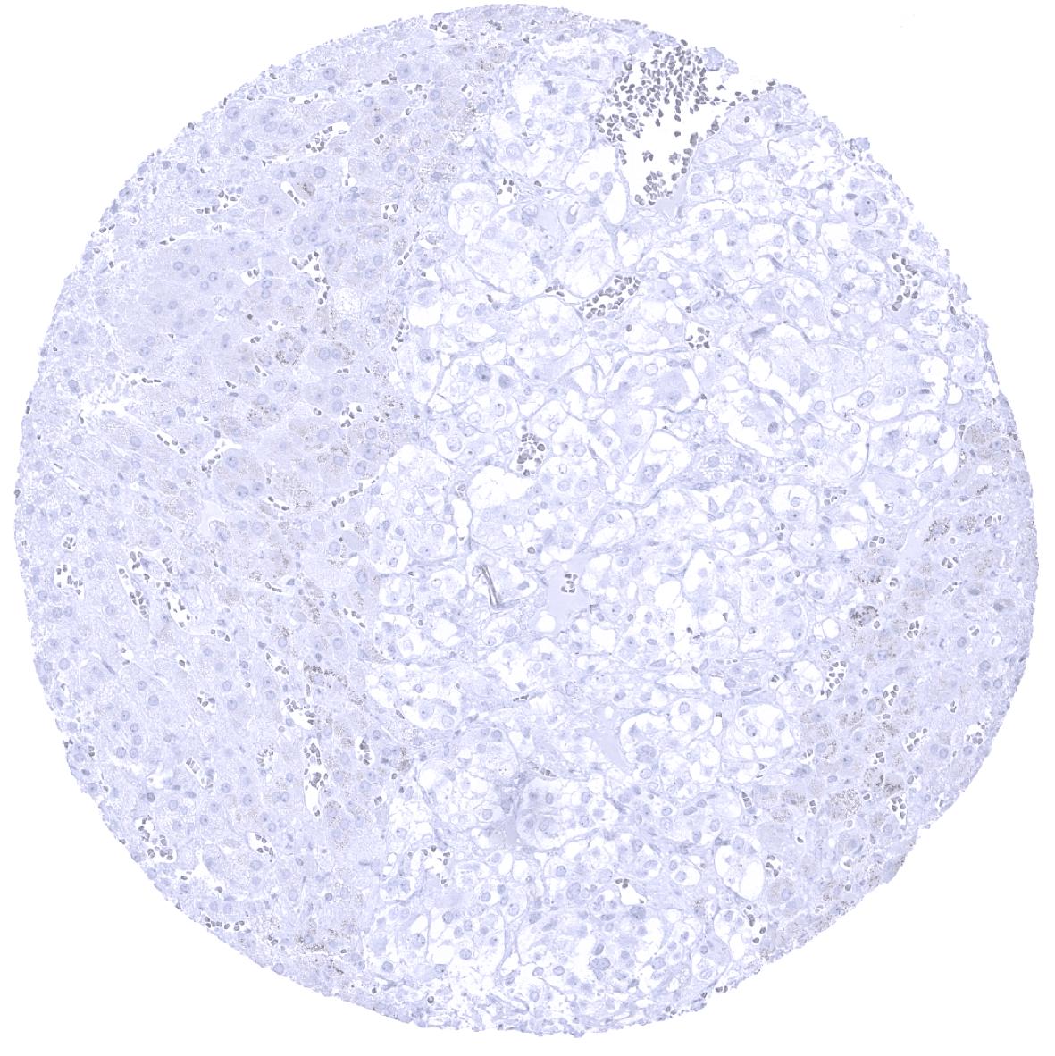
Liver
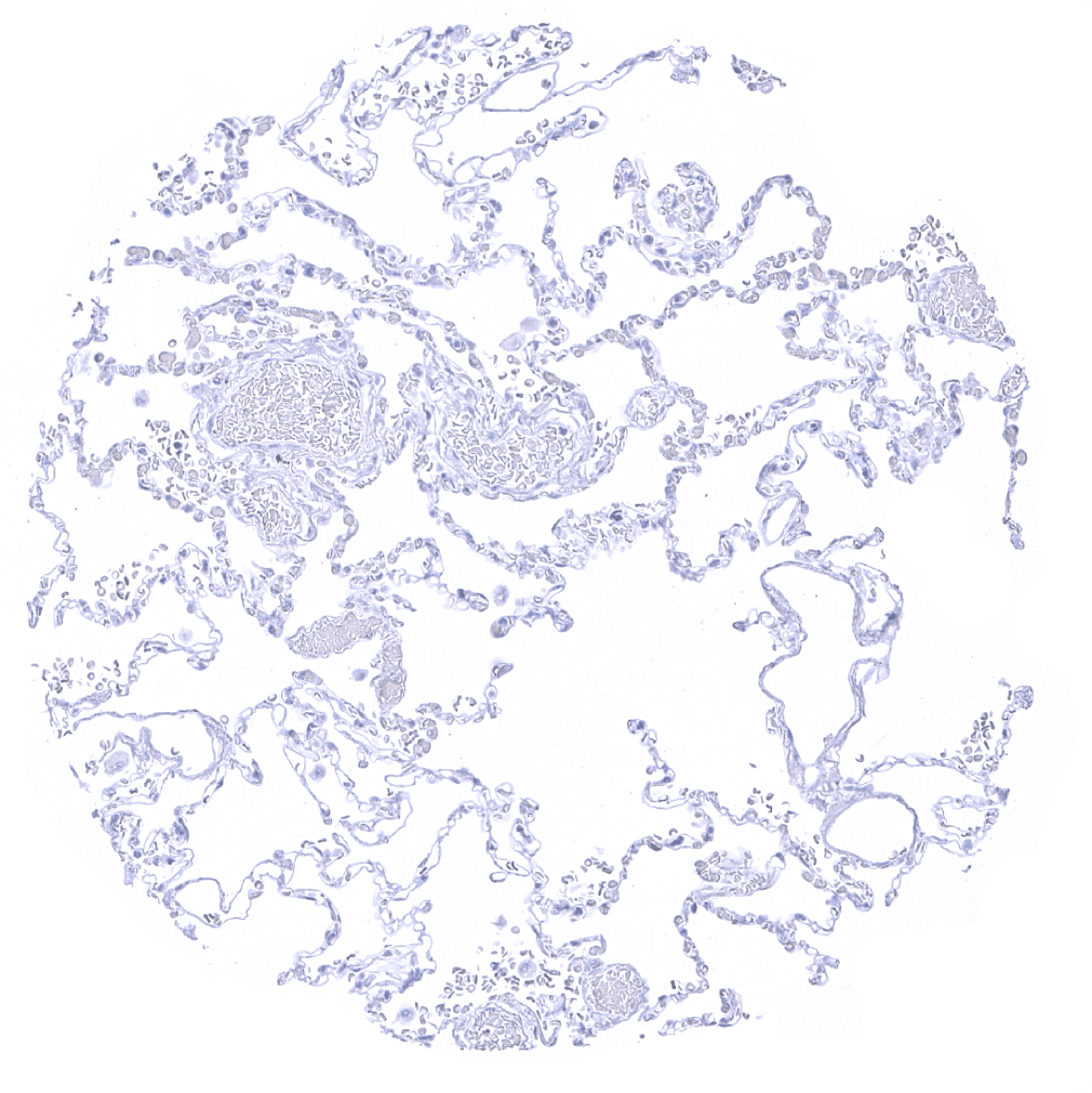
Lung
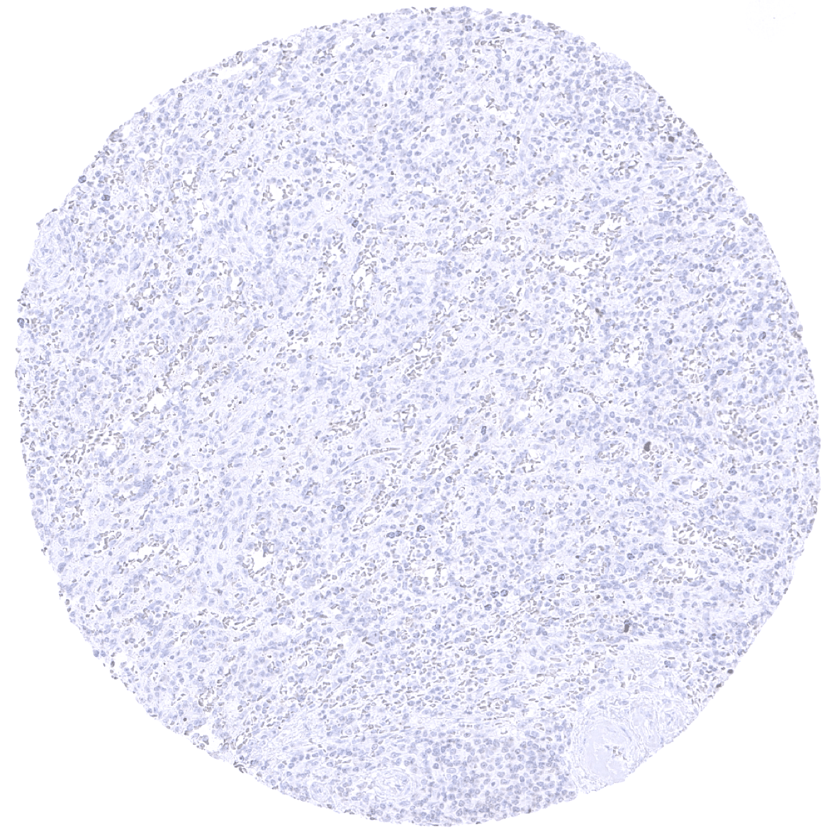
Lymph node
Ovary, stroma
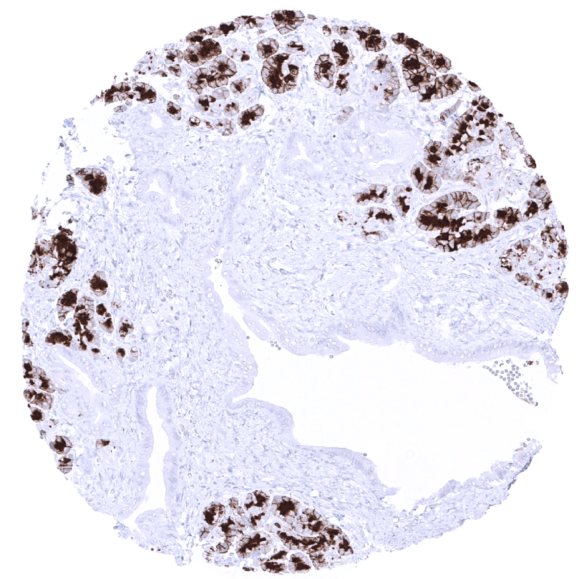
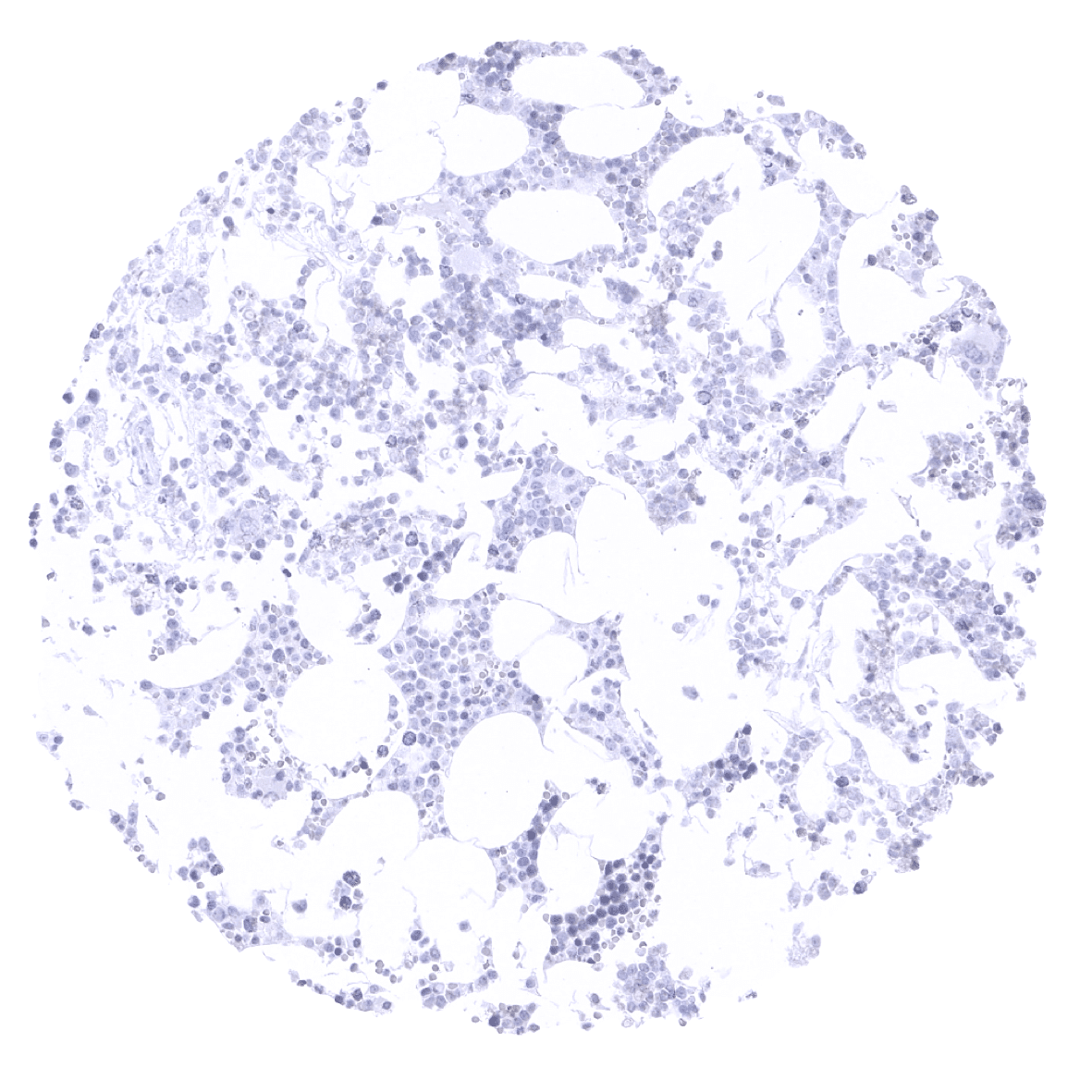
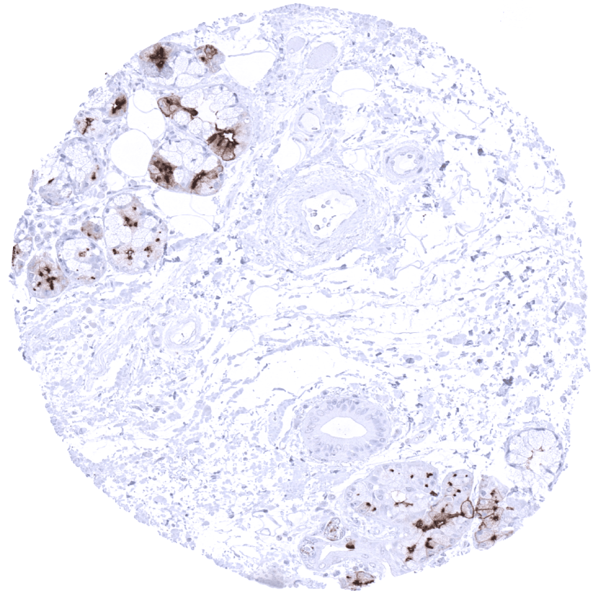
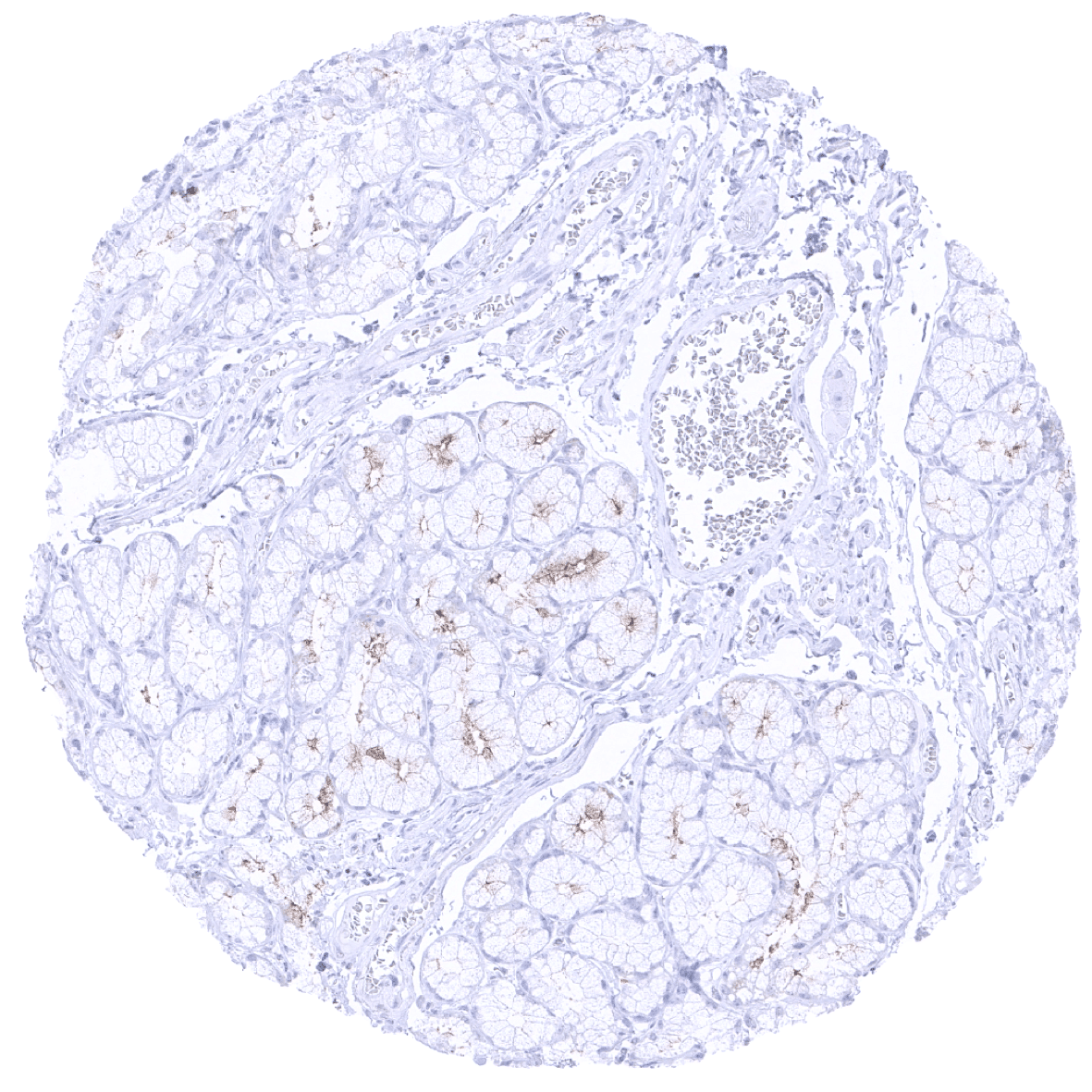
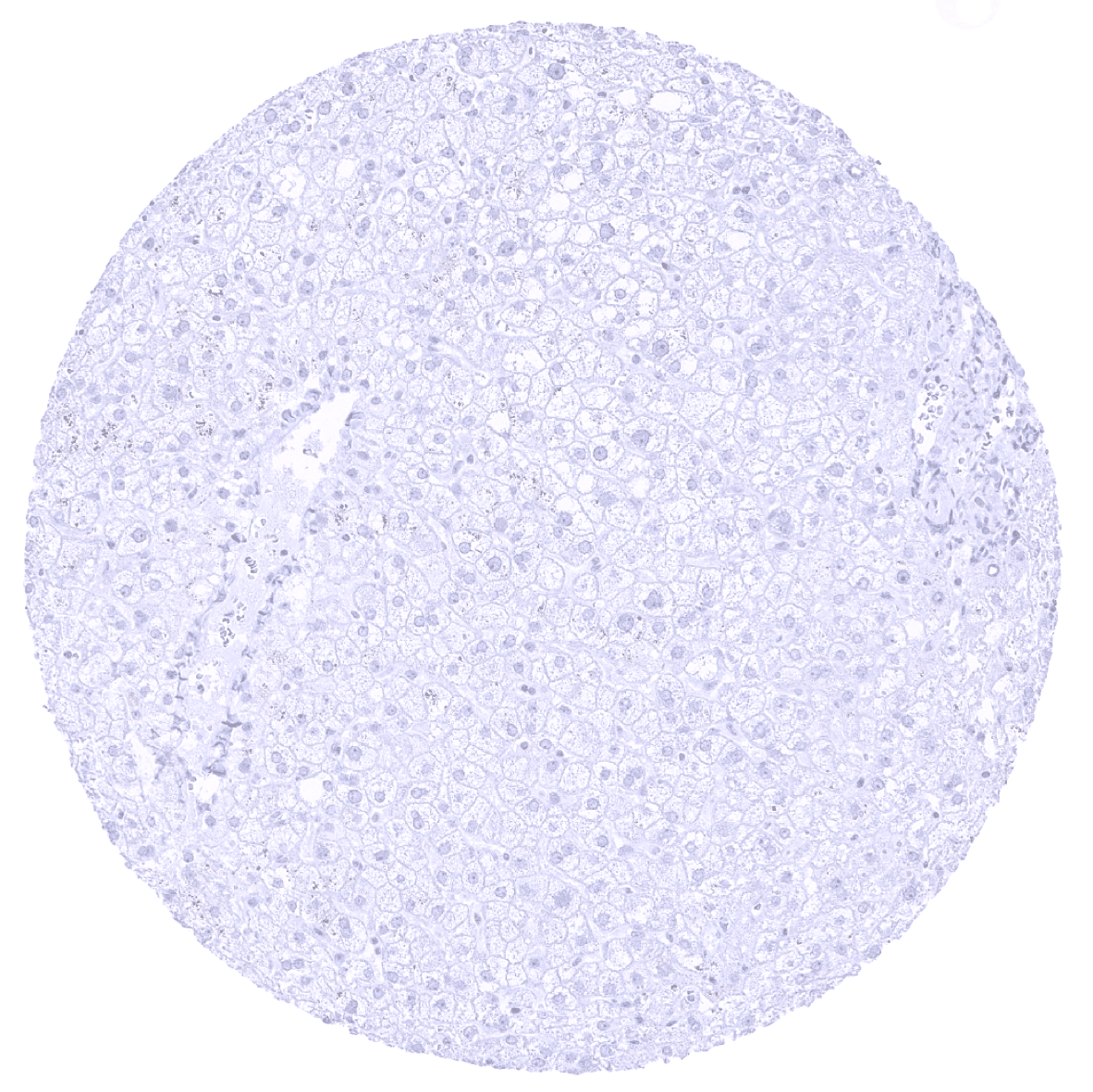
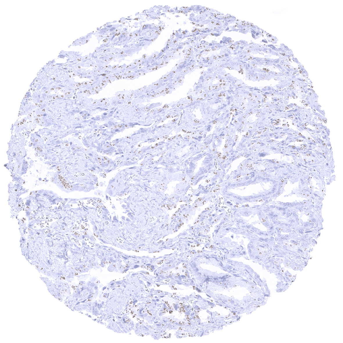
Pancreas - A strong GP2 staining is seen in acinar cells of the pancreas. GP2 staining is lacking in pancreatic ducts.

Pancreas - A strong GP2 staining is seen in acinar cells of the pancreas. The staining is cytoplasmic and membranous and shows a distinct apical predominance.

Pancreas - A strong GP2 staining is seen in acinar cells of the pancreas. The staining is cytoplasmic and membranous and shows a distinct apical predominance.
Parathyroid gland

Parotid gland
Pituitary gland, anterior lobe
Pituitary gland, posterior lobe

Placenta (amnion and chorion)
Placenta, first trimenon
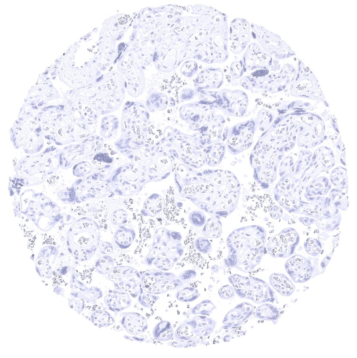
Placenta, mature

Prostate

Rectum, mucosa
Seminal vesicle
Sinus paranasales

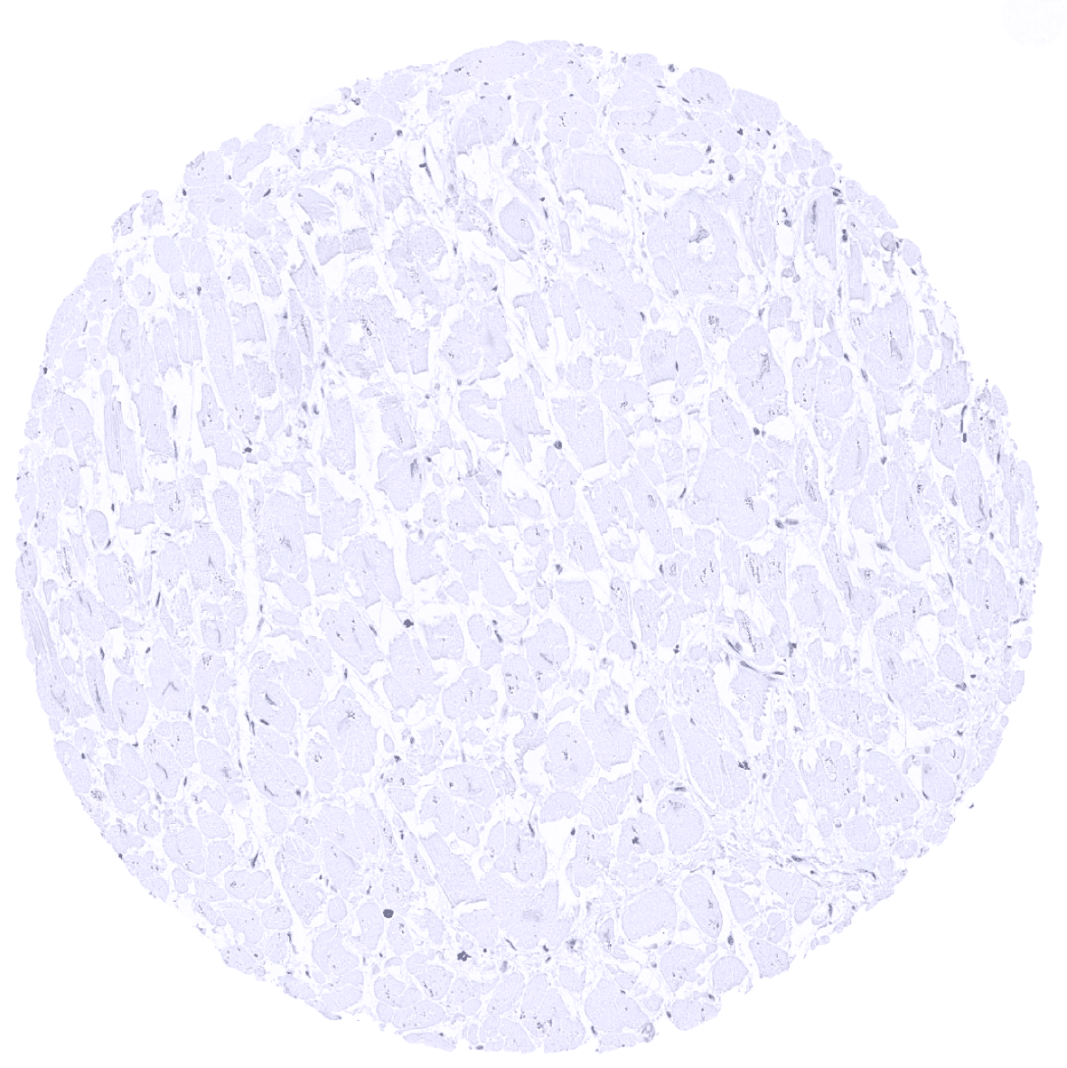
Skeletal muscle
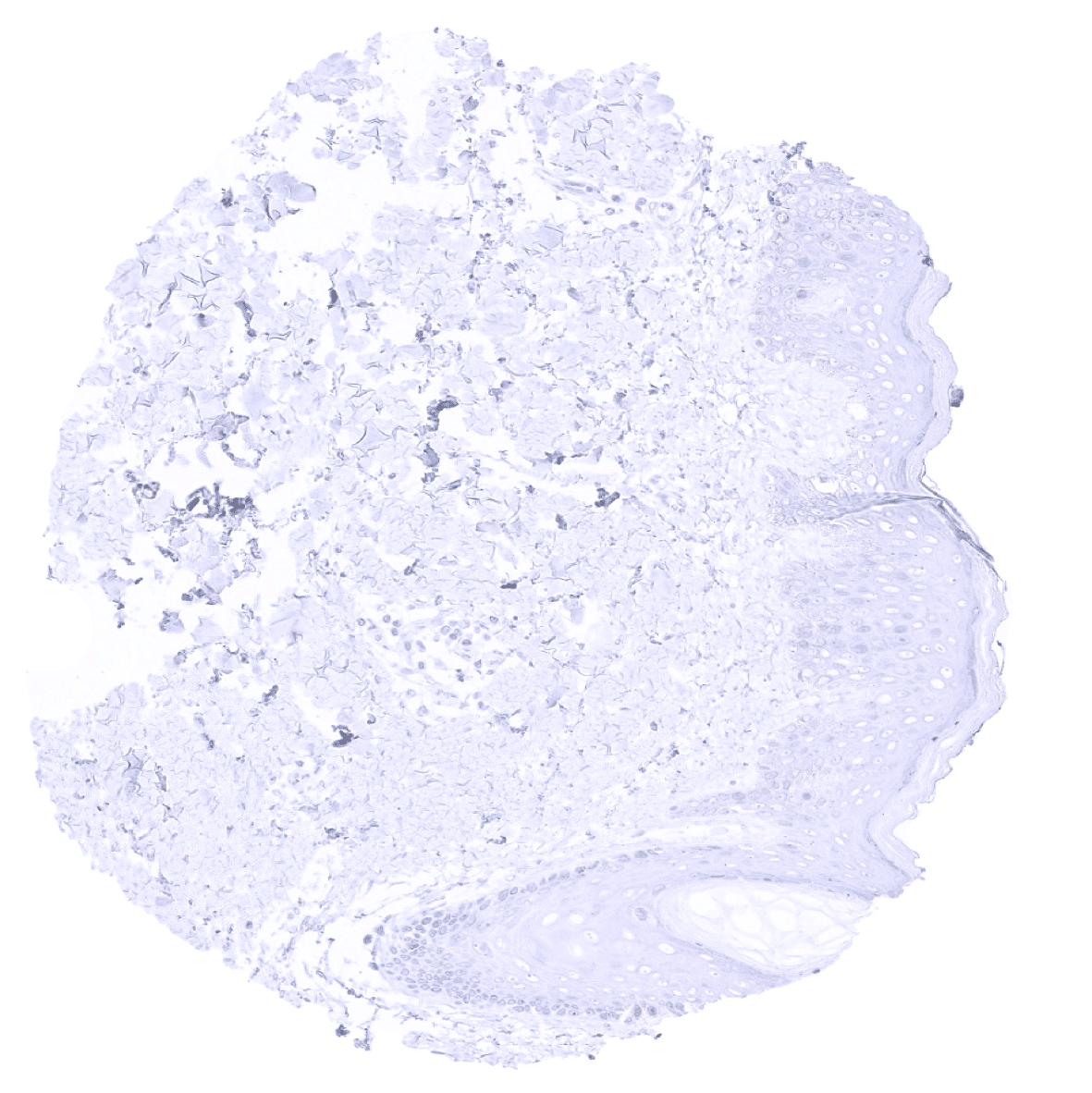
Skin
Spleen
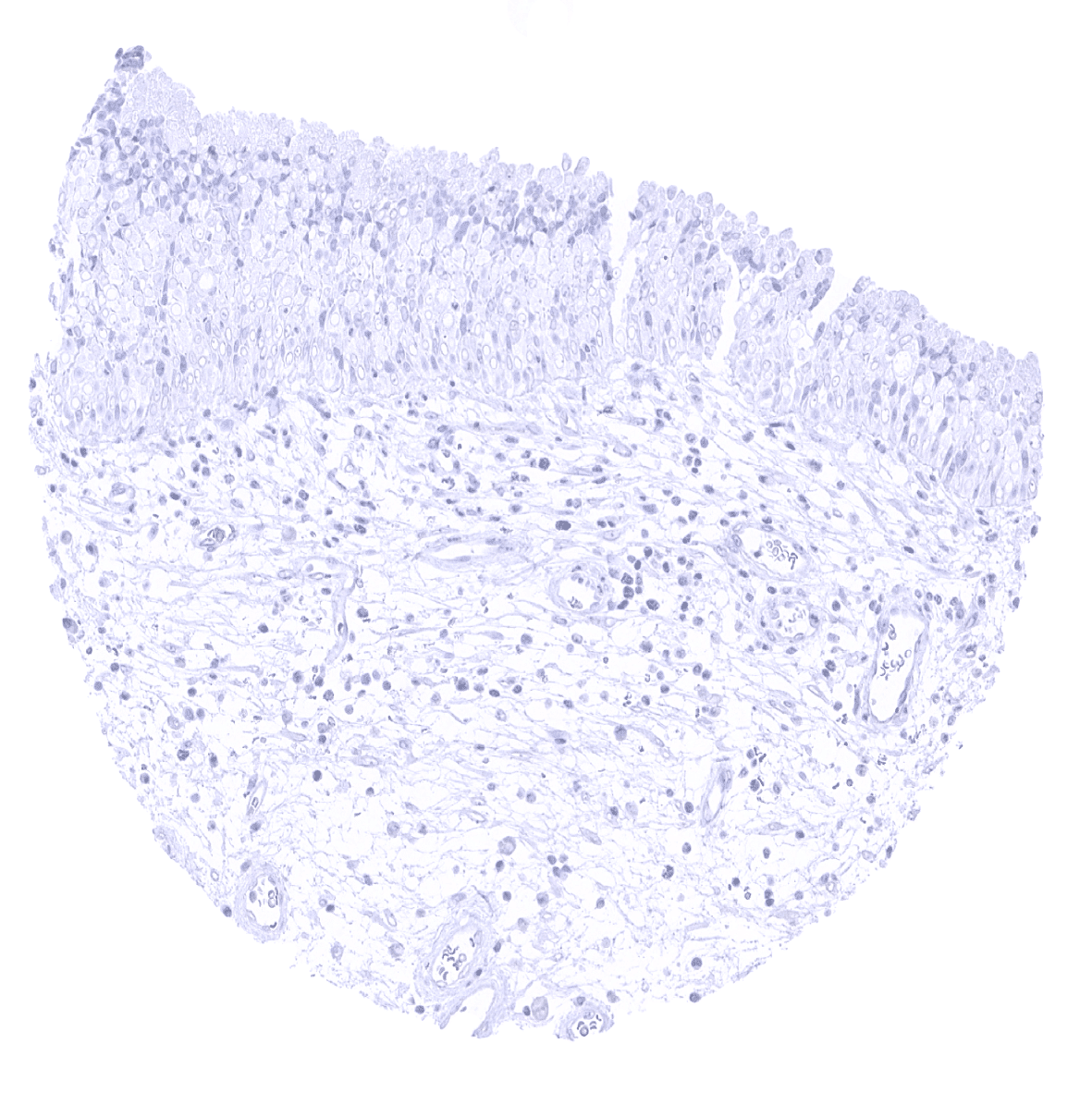
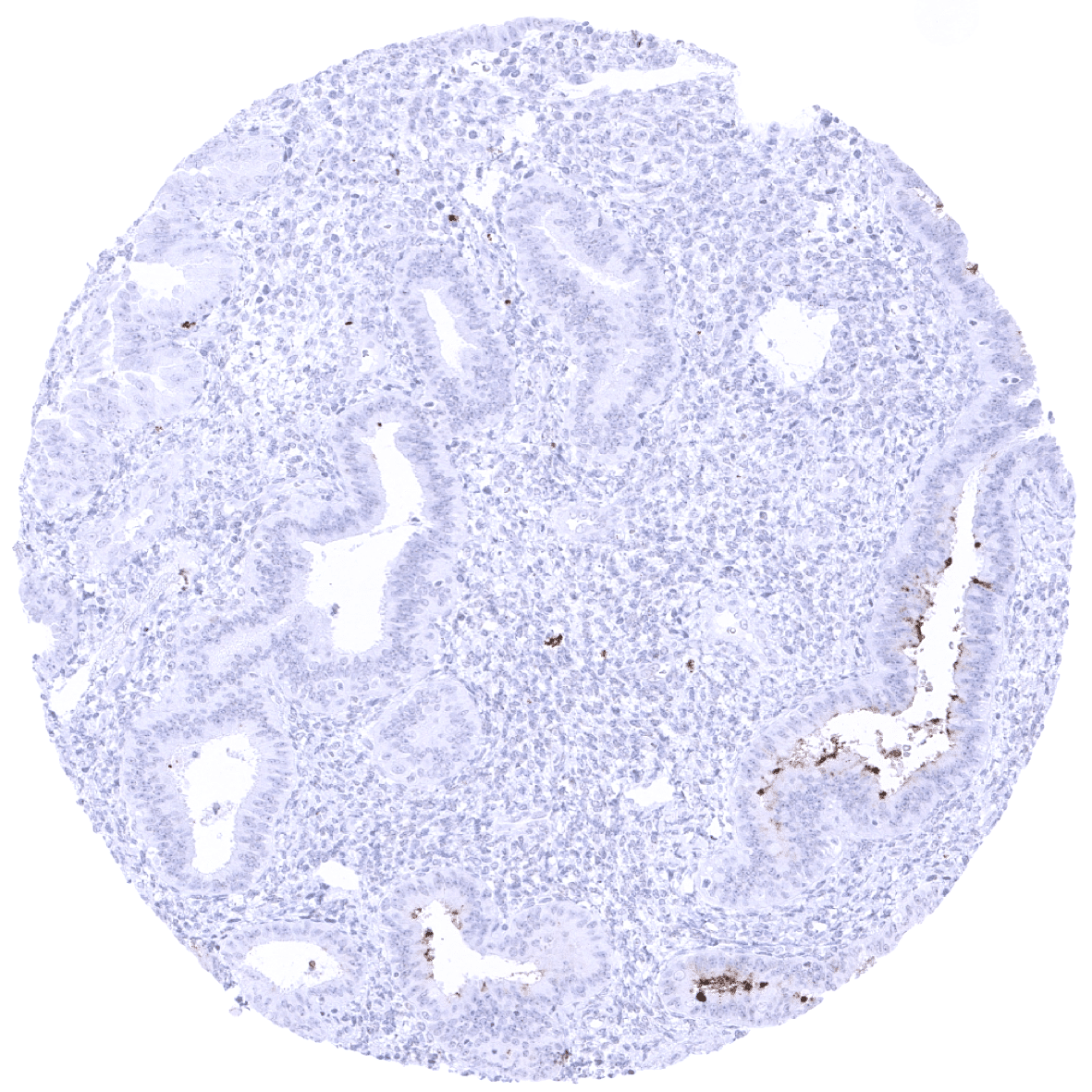
Stomach, antrum
Stomach, antrum - A weak to moderate GP2 staining can occur in some gland of the stomach antrum. The staining is cytoplasmic and shows a distinct predominance at the apical membrane.

Testis
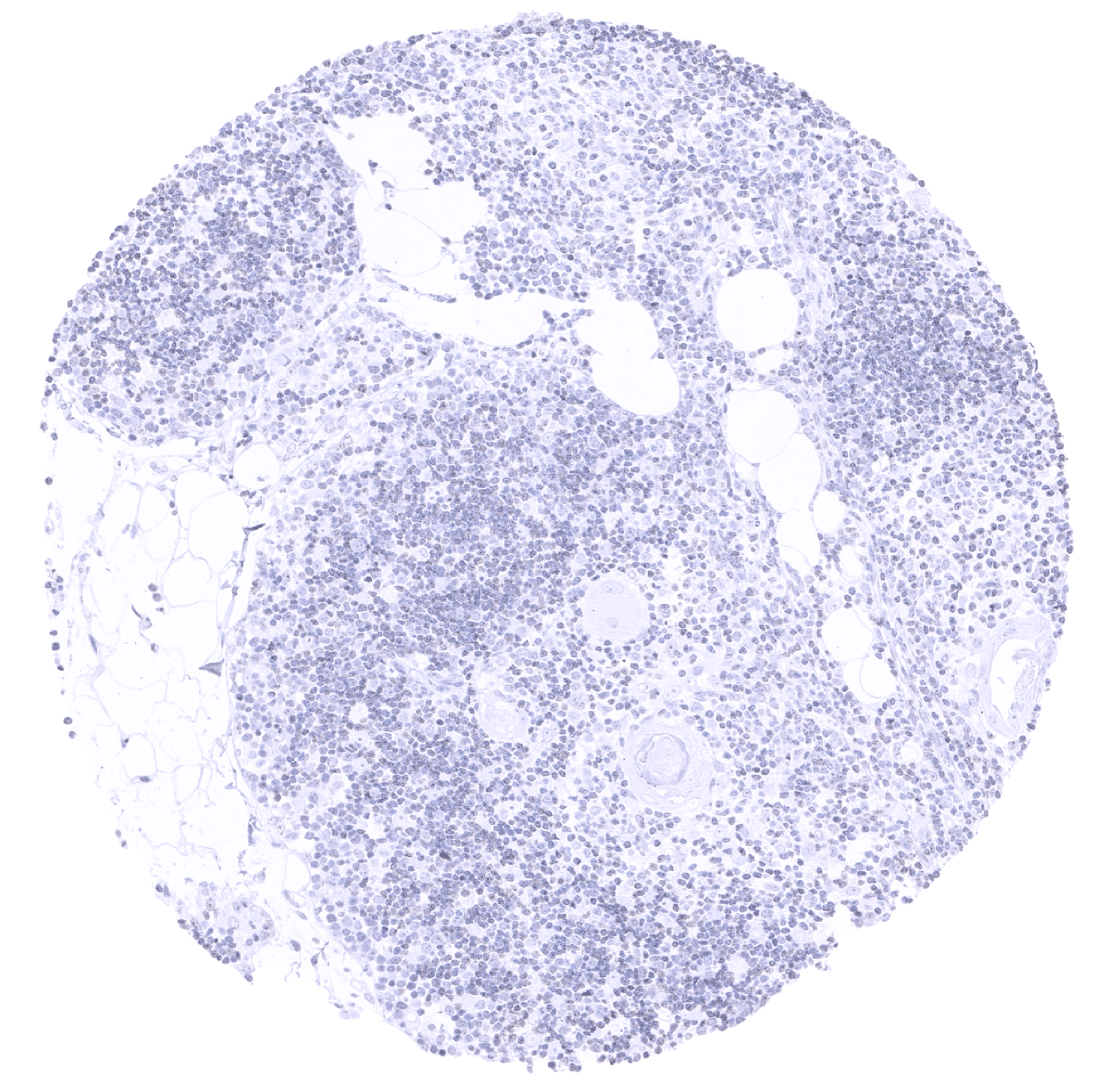
Thymus
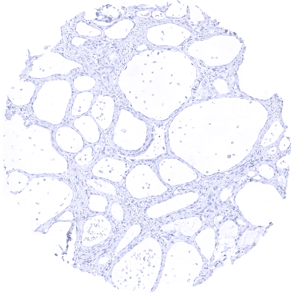
Thyroid gland
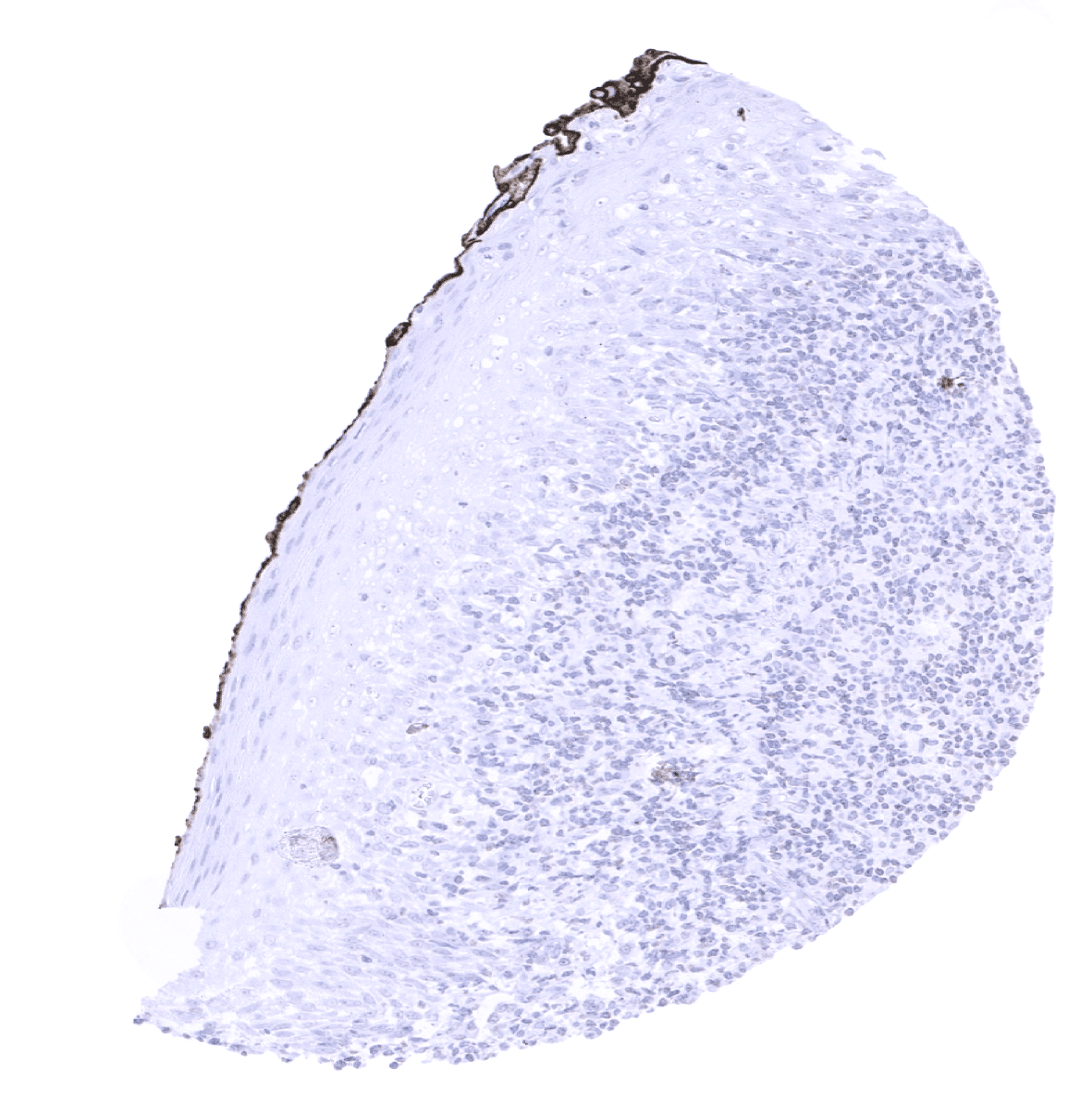
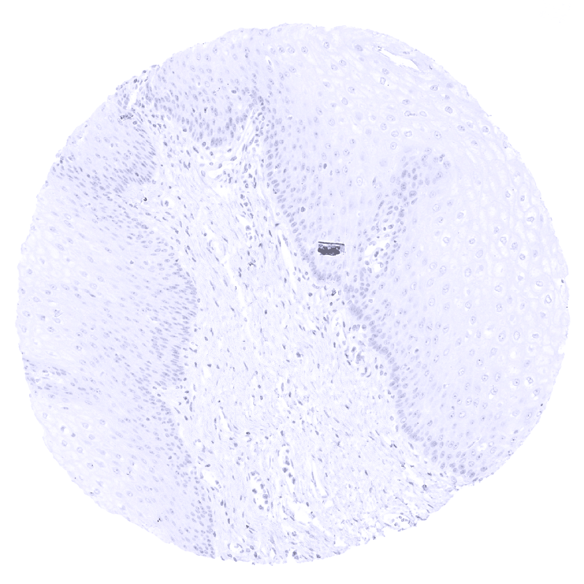
Tonsil, surface epithelium: The black staining at the surface represents an "inking artifact".

Tonsil
Urinary bladder, muscular wall
Urinary bladder, urothelium
Uterus, ectocervix
Uterus, endocervix
Uterus, endometrium (pregnancy)

Uterus, endometrium (proliferation)
Uterus, endometrium (secretion) - A weak to moderate GP2 staining can occur in few endometrial glands. The staining is predominantly located at the apical membrane.

Uterus, endometrium (secretion)

Uterus, myometrium