

295,00 € – 995,00 €
Product details
Synonyms = Chymotrypsin like elastase family member 3B (CELA3B); ELA3B; Elastase IIIB; Protease E
Antibody type = Recombinant Mouse monoclonal / IgG
Clone = MSVA-410M
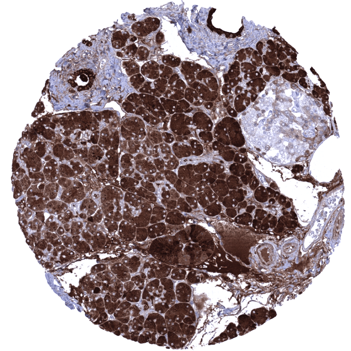
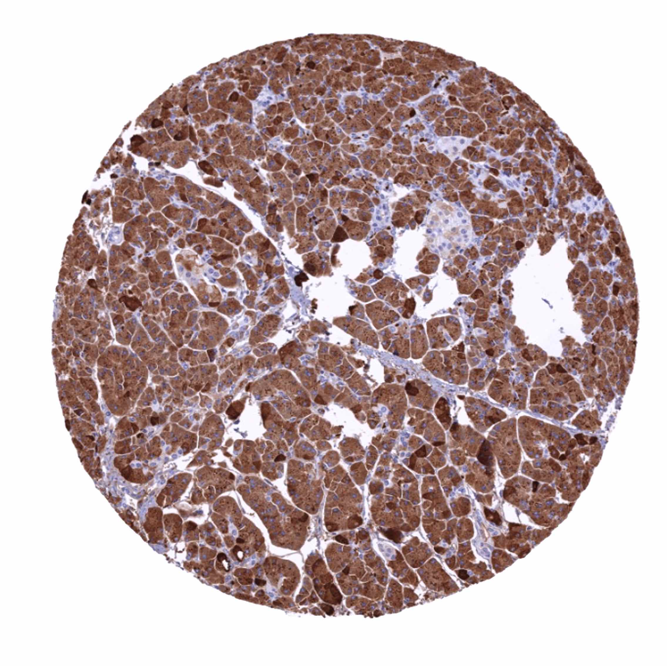
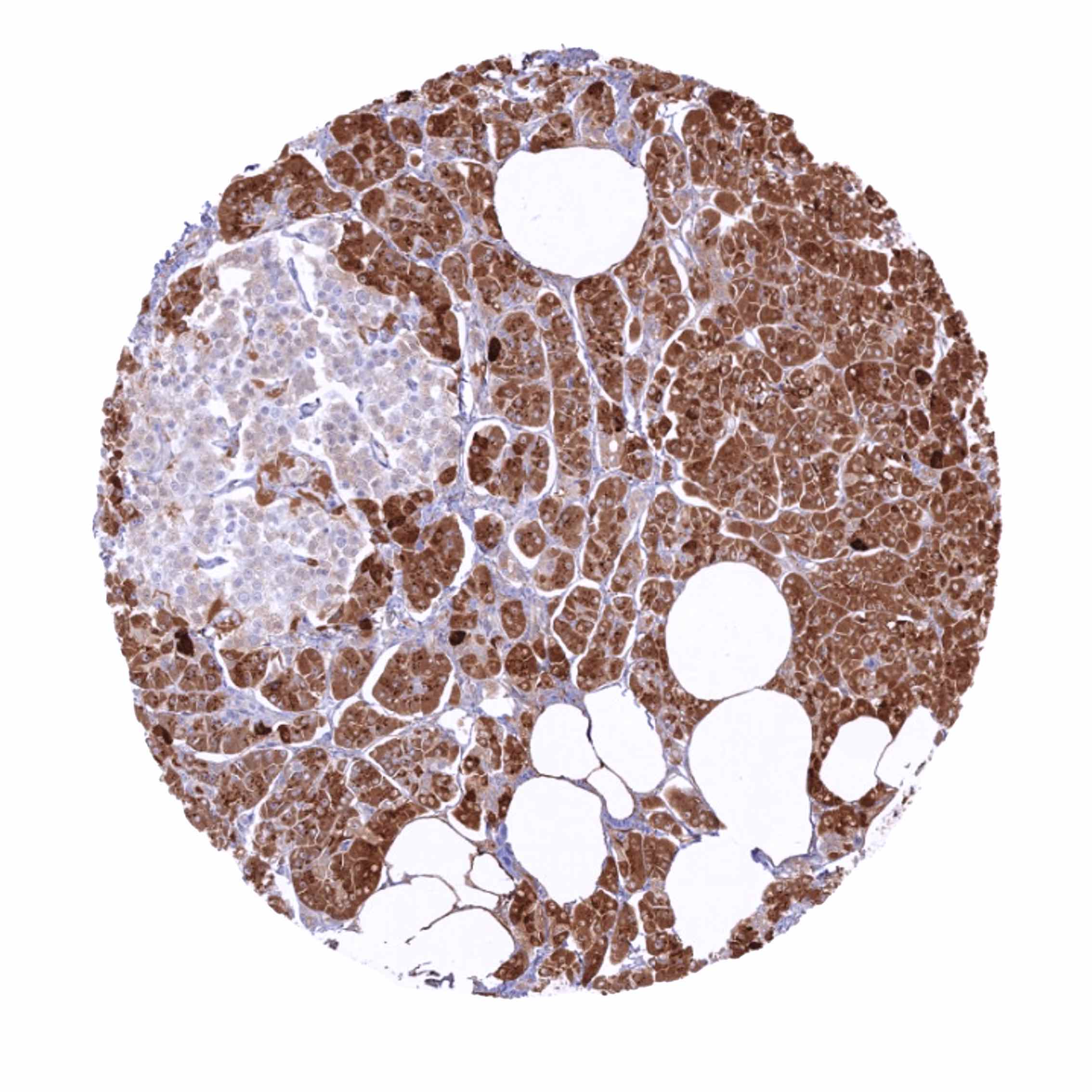
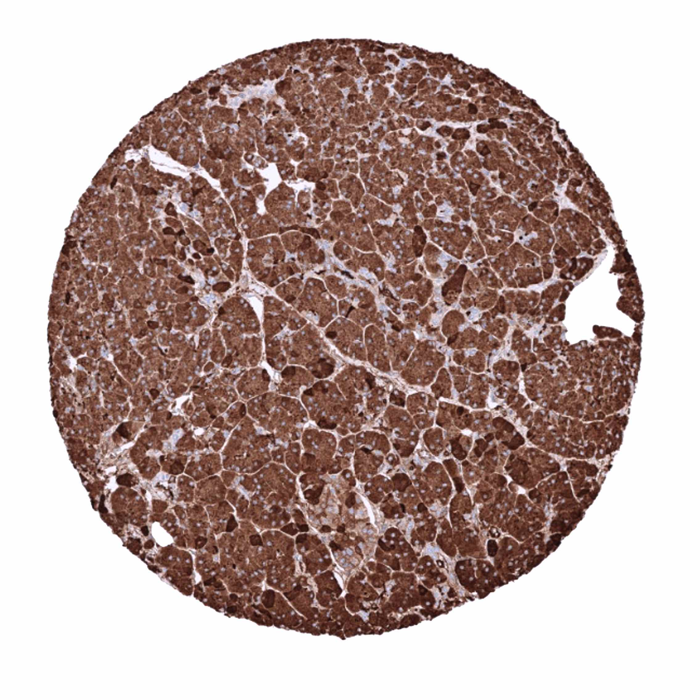
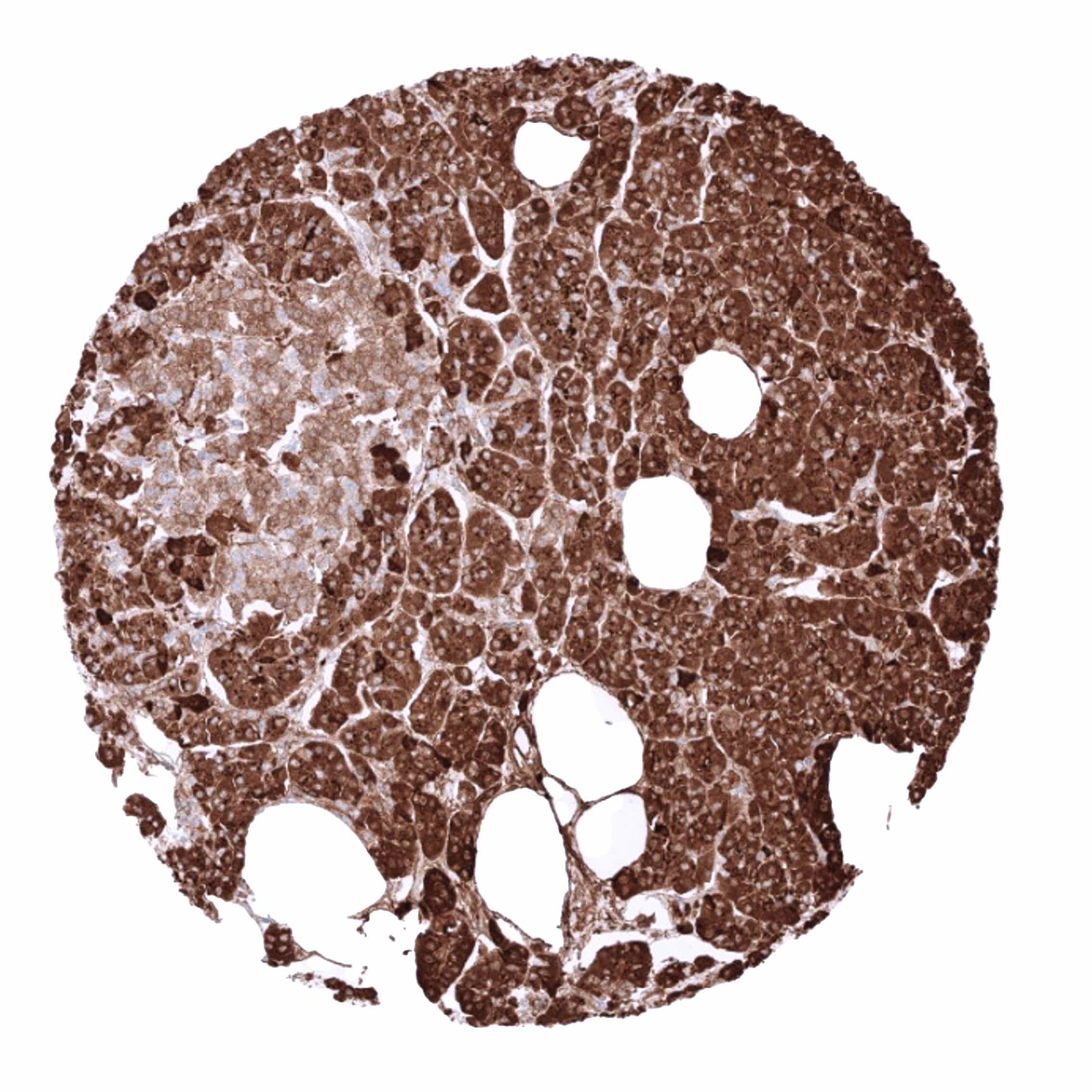
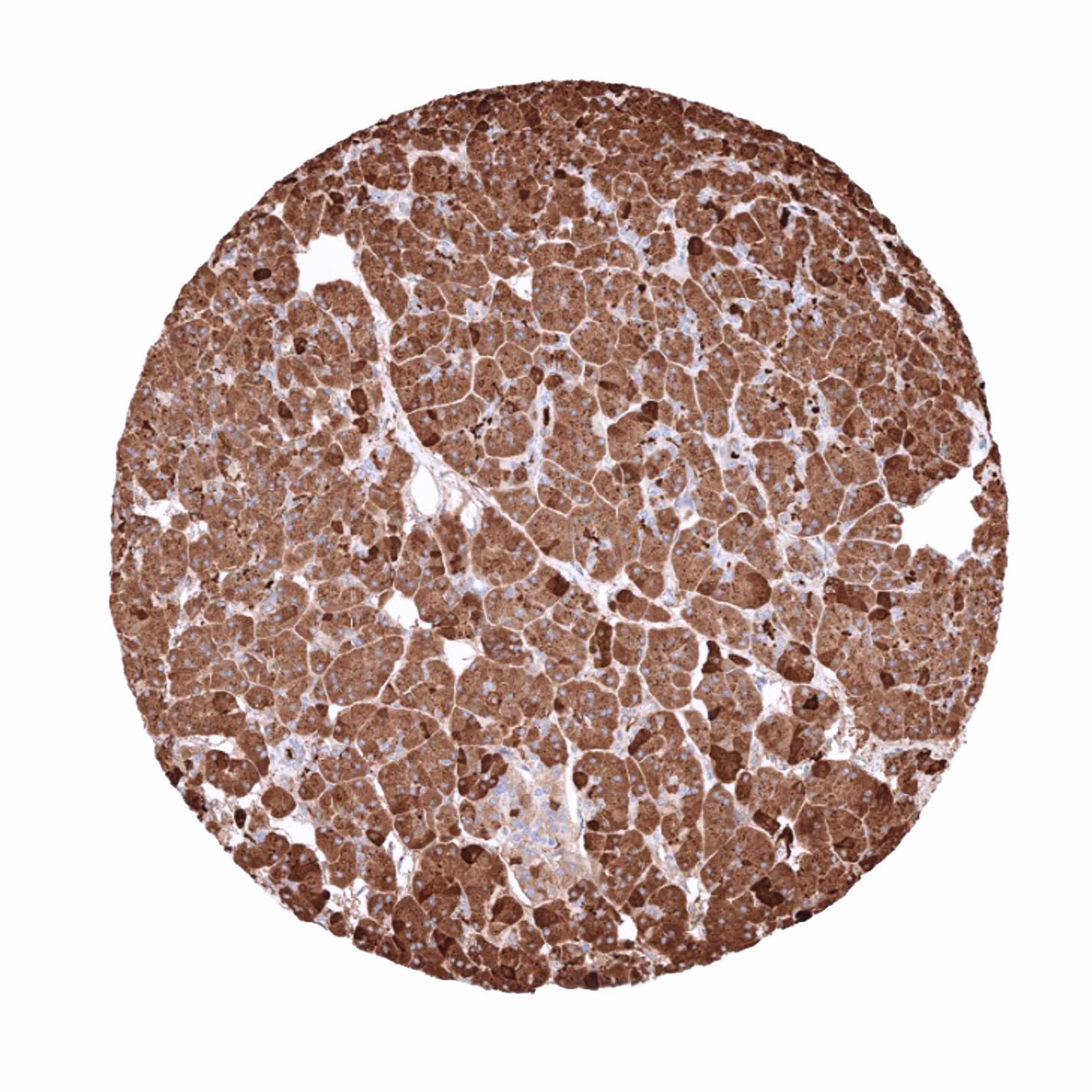
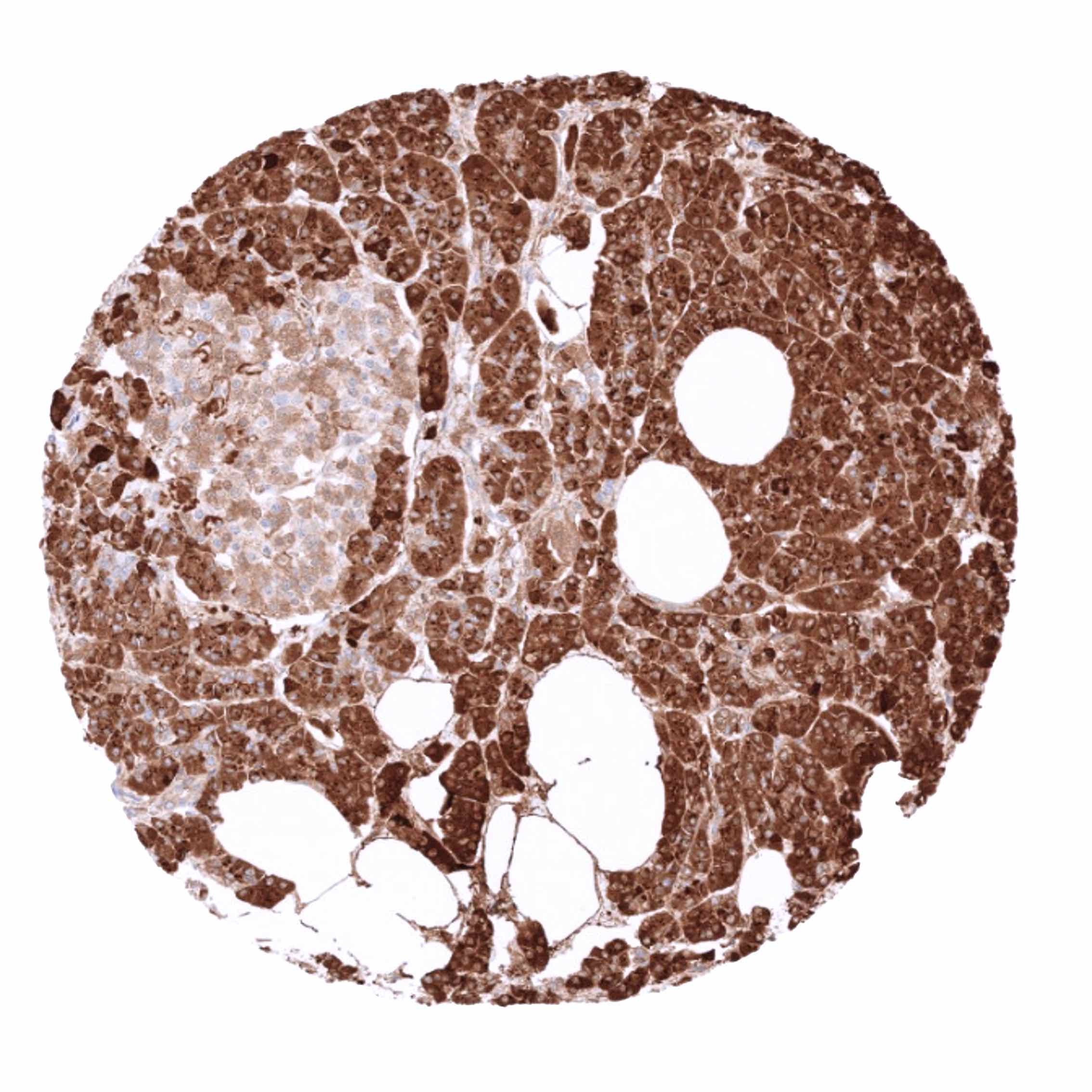
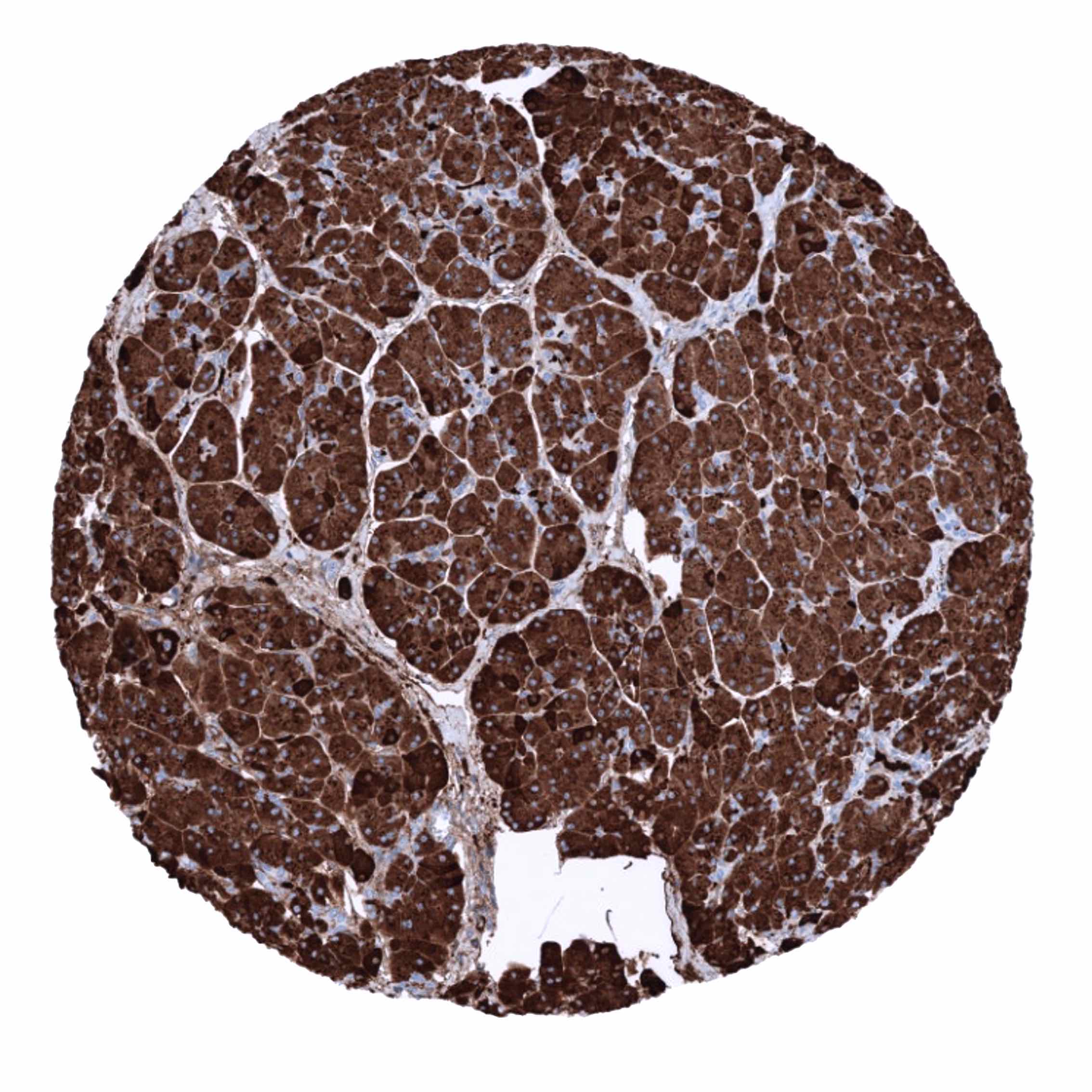
Positive control = Pancreas: Acinar cells should show a strong CELA3B immunostaining with apical predominance.
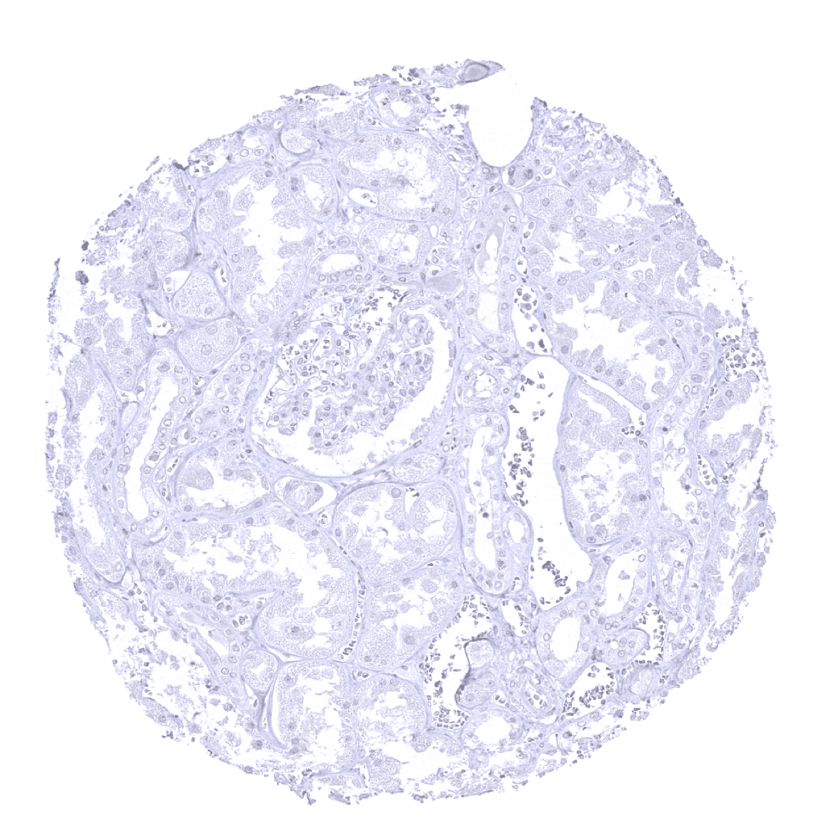
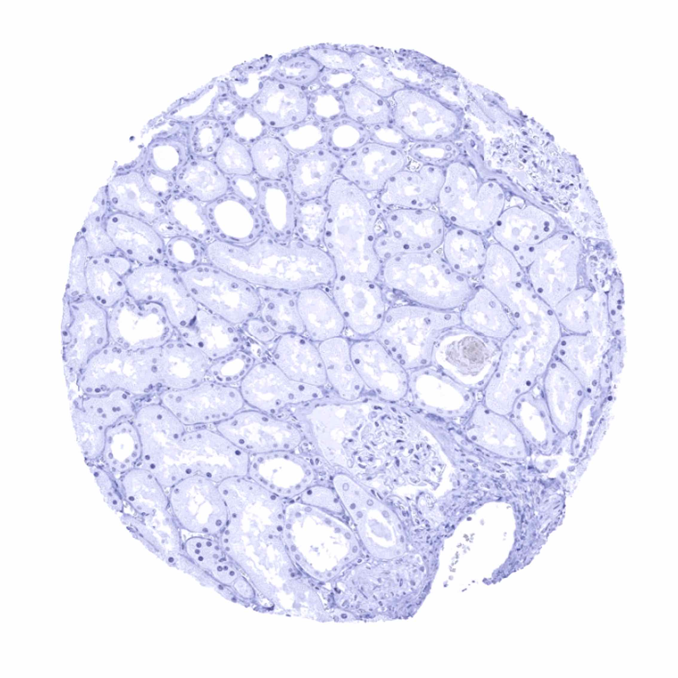
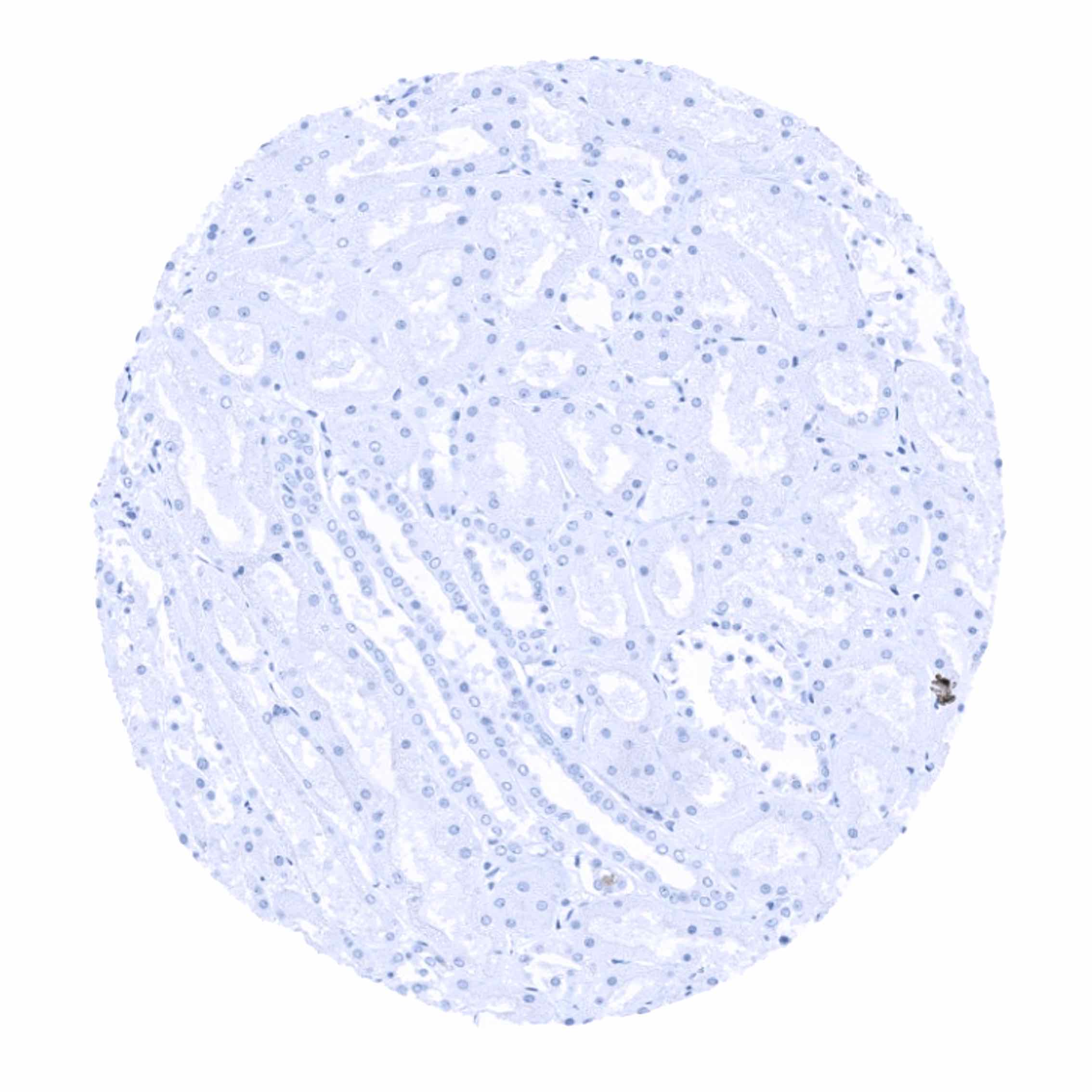
Negative control = Kidney: CELA3B staining should be absent in all epithelial cells.
Cellular localization = Cytoplasmic
Reactivity = Human
Application = Immunohistochemistry
Dilution = 1:100 – 1:200
Intended Use = Research Use Only
Relevance of Antibody
CELA3B is expressed on acinus cells of the pancreas.
Biology Behind
Chymotrypsin-like elastase family member 3B is a protease with a molecular weight of 29 kDa. The protein is encoded by the CELA3B gene located at human chromosome 1p36.12. Elastases are a subfamily of serine proteases that cleave elastin but also many other proteins. CELA3B and its related elastases (elastase 1, 2, 2A, and 3A) are secreted from the pancreas and support digestion of nutrients in the intestine. CELA3B has also been connected to cholesterol metabolism. Both CELA3A and CELA3B are used as analytes to estimate pancreatic function in feces samples.
Staining Pattern in Normal Tissues
CELA3B staining pattern in Normal Tissues with antibody MSVA-410M (images are shown in our “Normal Tissue Gallery”)
| Brain | Cerebrum | Negative. |
| Cerebellum | Negative. | |
| Endocrine Tissues | Thyroid | Negative. |
| Parathyroid | Negative. | |
| Adrenal gland | Negative. | |
| Pituitary gland | Negative. | |
| Respiratory system | Respiratory epithelium | Negative. |
| Lung | Negative. | |
| Gastrointestinal Tract | Salivary glands | Negative. |
| Esophagus | Negative. | |
| Stomach | Negative. | |
| Duodenum | Strong CELA3B staining of apical membranes of surface epithelial cells (in most samples). | |
| Small intestine | Strong CELA3B staining of apical membranes of surface epithelial cells (in most samples). | |
| Appendix | Weak CELA3B staining of apical membranes of surface epithelial cells (in few samples). | |
| Colon | Weak to moderate CELA3B staining of apical membranes of surface epithelial cells (in some samples). | |
| Rectum | Weak to moderate CELA3B staining of apical membranes of surface epithelial cells (in some samples). | |
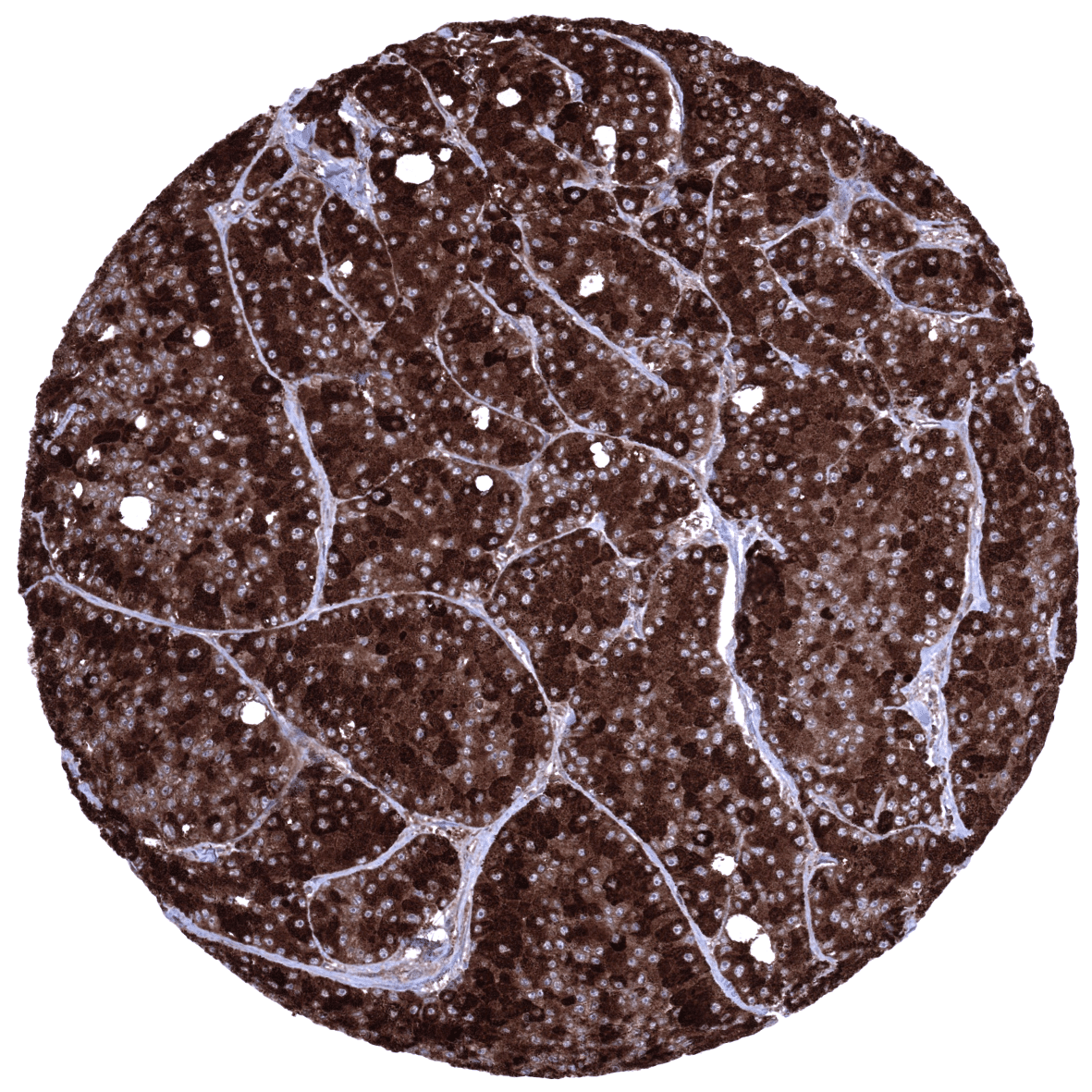
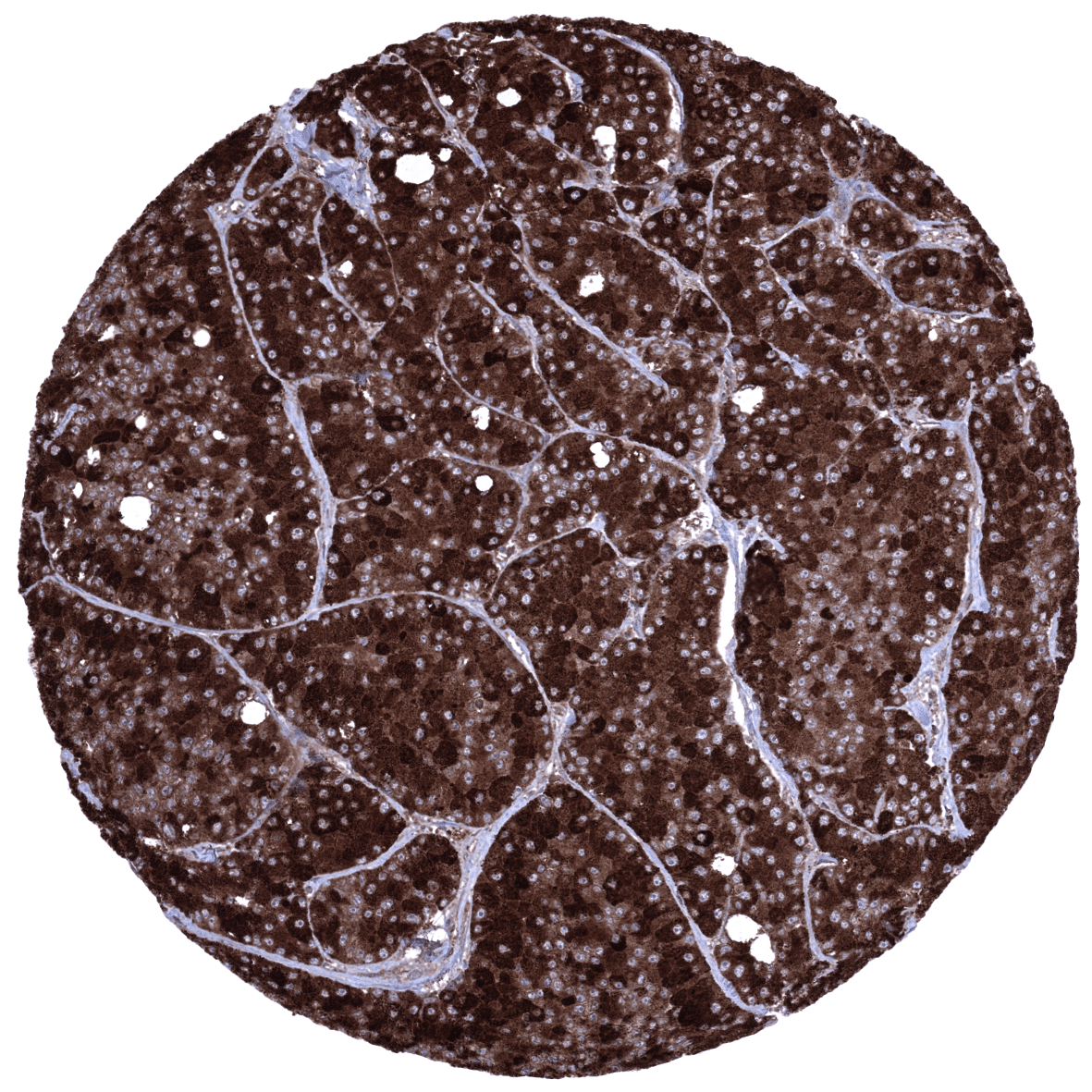
| Liver | Negative. | |
| Gallbladder | Negative. | |
| Pancreas | Strong cytoplasmic CELA3B staining of all acinar cells and a fraction of ductal cells. | |
| Genitourinary | Kidney | Negative. |
| Urothelium | Negative. | |
| Male genital | Prostate | Negative. |
| Seminal vesicles | Negative. | |
| Testis | Negative. | |
| Epididymis | Negative. | |
| Female genital | Breast | Negative. |
| Uterus, myometrium | Negative. | |
| Uterus, ectocervix | Negative. | |
| Uterus endocervix | Negative. | |
| Uterus, endometrium | Negative. | |
| Fallopian Tube | Negative. | |
| Ovary | Negative. | |
| Placenta early | Negative. | |
| Placenta mature | Negative. | |
| Amnion | Negative. | |
| Chorion | Negative. | |
| Skin | Epidermis | Negative. |
| Sebaceous glands | Negative. | |
| Muscle/connective tissue | Heart muscle | Negative. |
| Skeletal muscle | Negative. | |
| Smooth muscle | Negative. | |
| Vessel walls | Negative. | |
| Fat | Negative. | |
| Stroma | Negative. | |
| Endothelium | Negative. | |
| Bone marrow/ lymphoid tissue | Bone marrow | Negative. |
| Lymph node | Negative. | |
| Spleen | Negative. | |
| Thymus | Negative. | |
| Tonsil | Negative. | |
| Remarks |
These findings are largely comparable to the RNA and protein data described in the Human Protein Atlas (Tissue expression CELA3B) even though a CELA3B RNA expression in the surface epithelium of the small intestine and the colorectum is not described in the protein atlas. These structures constitute only small subsets of the total amount of cells in their respective organs and may have been underrepresented in RNA analyses. Images provided in the protein atlas suggest, however, that apical membrane staining was also observed in the colon and the small intestine by using the antibody HPA045650.
Positive control = Pancreas: Acinar cells should show a strong CELA3B immunostaining with apical predominance.
Negative control = Kidney: CELA3B staining should be absent in epithelial cells.

Staining Pattern in Relevant Tumor Types
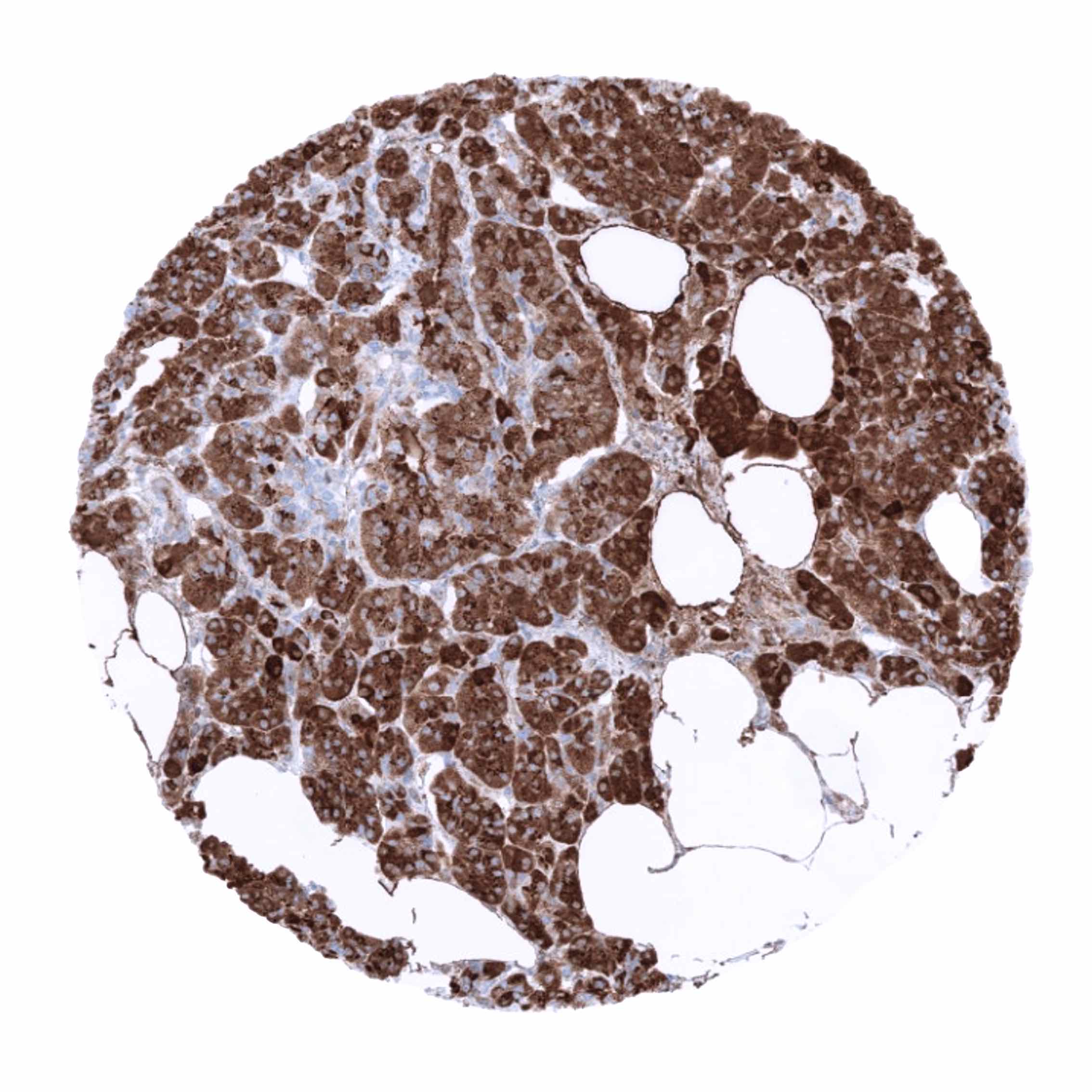
CELA3B is expressed in the majority of acinar cell carcinomas of the pancreas. A lower level of CELA3B expression may rarely be seen in several other tumor entities.
The TCGA findings on CELA3B RNA expression in different tumor categories have been summarized in the Human Protein Atlas.


Compatibility of Antibodies
CELA3B (MSVA-410M) publication summary
Relevant publication: Uhlig et al. “CELA3B immunostaining is a highly specific marker for acinar cell carcinoma of the pancreas”. Published in PLoS One. 2023 Jun 28;18(6):e0287528. PMID: 37379306
A total of 13,223 tumors were analyzed from 132 different tumor categories by using the following protocol: Heat-induced antigen retrieval for 5 minutes in an autoclave at 121°C in pH7,8 Target Retrieval Solution buffer. MSVA-601M at a dilution of 1:150 at 37°C for 60 minutes. Visualization of bound antibody was by the EnVision Kit (Dako, Agilent). This protocol was also used for all stainings depicted in our tumor and normal tissue galleries.
A positive CELA3B immunostaining was seen in all 12 of 16 analyzed pancreatic acinar cell carcinomas and in only 5 further tumors (from 3 different tumor entities) out of 12263 tumors from 131 other tumor categories. The distribution of positive staining results is shown in an “organ-systematic” figure and in a “ranking order” below (images based on data from Uhlig et al.).
Authors conclusions on diagnostic utility with respect to the distinction of benign versus malignant (Uhlig et al.):
- not applicable.
Authors conclusions on diagnostic utility with respect to the distinction of different tumor entities (Uhlig et al.):
- CELA3B is a specific marker for acinar cell carcinoma of the pancreas.
Authors conclusions on prognostic/predictive role of CPA1 expression.
- not applicable.
Data from the publication: Uhlig et al. “CELA3B immunostaining is a highly specific marker for acinar cell carcinoma of the pancreas”. Published in PLoS One. 2023 Jun 28;18(6):e0287528. PMID: 37379306
Summarized in own graphics:
Figure1. CELA3B staining in tumors (“organ-specific ; according to Uhlig et al.”) with antibody MSVA-410M

Figure 2. CELA3B staining in tumors (“ranking-order ; according to Uhlig et al.”) by positivity with antibody MSVA-410M

Protocol Recommendations
IHC users have different preferences on how the stains should look like. Some prefer high staining intensity of the target stain and even accept some background. Others favor absolute specificity and lighter target stains. Factors that invariably lead to more intense staining include higher concentration of the antibody and visualization tools, longer incubation time, higher temperature during incubation, higher temperature and longer duration of the heat induced epitope retrieval (slide pretreatment). The impact of the pH during slide pretreatment has variable effects and depends on the antibody and the target protein.
All images and data shown here are obtained by the manual protocol described below. Other protocols resulting in equivalent staining are described as well.
Manual protocol
Freshly cut sections should be used (less than 10 days between cutting and staining). Heat-induced antigen retrieval for 5 minutes in an autoclave at 121°C in pH 7,8 Target Retrieval Solution buffer. Apply MSVA-410M at a dilution of 1:150 at 37°C for 60 minutes. Visualization of bound antibody by the EnVision Kit (Dako, Agilent) according to the manufacturer’s directions.
Agilent / Dako – Autostainer Link 48
Pretreatment in PT-Link for 30 minutes at 95°C (pH high); FLEX peroxidase blocking for 5 minutes (room temperature), MSVA-410M 1:150 for 20 minutes (room temperature), FLEX+ mouse/rabbit (LINKER) for 15 minutes (room temperature), horseradish peroxidase (HRP) for 20 minutes (room temperature), FLEX DAB+Sub-Chromo for 10 minutes (room temperature), FLEX hematoxylin for 5 minutes (room temperature).

These images reflect stainings by the protocol described above. It is of note that a comparable staining result can also be obtained by different protocols. In general, a longer pretreatment, a longer incubation time of the primary antibody, a higher antibody concentration, and a longer incubation time of FLEX+LINKER result in stronger staining, potentially at the cost of more background staining. Modifications of the protocol with a strengthening effect on staining intensity in combination with changes of other parameters that result in lower staining intensity can result in a comparable result as shown above.
Leica – BOND RX
Dewax at 72°C for 30 seconds; Pretreatment in Bond Epitope Retrieval Solution (ER2 – EDTA pH9) for 20 minutes at 100°C; Peroxidase blocking for 5 minutes (room temperature), MSVA-410M 1:150 for 15 minutes (room temperature), Post primary (rabbit anti mouse) for 8 minutes (room temperature), Polymer (goat anti rabbit) for 8 minutes (room temperature), mixed DAB refine for 10 minutes (room temperature), hematoxylin for 5 minutes (room temperature).

These images reflect stainings by the protocol described above. It is of note that a comparable staining result can also be obtained by different protocols. In general, a longer pretreatment, a longer incubation time of the primary antibody, a higher antibody concentration, a higher temperature during incubation, and a longer incubation time of Post primary and or the Polymer result in stronger staining, potentially at the cost of more background staining. Modifications of the protocol with a strengthening effect on staining intensity in combination with changes of other parameters that result in lower staining intensity can result in a comparable result as shown above.
Roche – Ventana Discovery ULTRA
Pretreatment for 64 minutes at 100°C (pH 8,4); CM peroxidase blocking for 12 minutes (room temperature), MSVA-410M 1:150 for 20 minutes at 36°C, secondary antibody (anti-mouse HQ) for 12 minutes at 36°C, anti-HQ HRP for 12 minutes at room temperature, DAB at room temperature, hematoxylin II at room temperature for 8 minutes, bluing reagent at room temperature for 4 minutes.
These images depict staining results obtained by the protocol described above. It is of note, that the Ventana machines generally require higher antibody concentrations than other commonly used autostainers because the antibodies are automatically diluted during the procedure. Various other protocols can result in an identical result as shown above. A longer pretreatment, a longer incubation time of the primary antibody, a higher antibody concentration, a higher temperature during incubation, and a longer incubation time of secondary antibody and or the anti-HQ HRP result in stronger staining, potentially at the cost of more background staining.
Potential Research Applications
- The diagnostic utility of CELA3B immunohistochemistry for the distinction of pancreatic acinar cell carcinomas from other pancreatic and extra-pancreatic neoplasms should be investigated.
- The function of CELA3B is largely unknown and needs to be investigated.
- The prevalence of CELA3b expression is unknown for most tumor entities
Evidence for Antibody Specificity in IHC
There are two ways, how the specificity of antibodies can be documented for immunohistochemistry on formalin fixed tissues. These are: 1. comparison with a second independent method for target expression measurement across a large number of different tissue types (orthogonal strategy), and 2. Comparison with one or several independent antibodies for the same target and showing that all positive staining results are also seen with other antibodies for the same target (independent antibody strategy).
Orthogonal validation: For the antibody MSVA-410M specificity is documented by the strong concordance of the immunostaining with RNA expression data derived from the Human Protein Atlas (HPA) RNA-seq tissue dataset, the FANTOM5 project, and the Genotype-Tissue Expression (GTEx) project which are all summarized in the Human Protein Atlas (Tissue expression CELA3B) Immunostaining by using MSVA-410M was predominantly detected in the pancreas which was the only organ with documented RNA expression. In addition, a positive immunostaining was also found in the surface epithelium of the small intestine and the colorectum. These structures constitute small subsets of the total amount of cells in these organs and may have been largely underrepresented in RNA analyses.
Comparison of antibodies: True expression of CELA3B in surface epithelium of the small intestine and the colorectum is suggested by the finding of an identical staining pattern by the unrelated antibody HPA045650 shown in the human protein atlas. Images depicted there also show a weak apical membranous staining in the colon and the small intestine
Moreover, no staining was seen in tissues notorious for non-specific IHC background such as kidney, colonic mucosa, and epidermis.
