
395,00 € – 1.495,00 €
Product details
Synonyms = GATA3; GATA binding protein-3; GATA-binding factor 3; GATA3; HDR; HDRS; Transacting T-cell-specific transcription factor GATA-3
Antibody type = Mouse monoclonal / IgG
Clone = MSVA-450M
Positive control
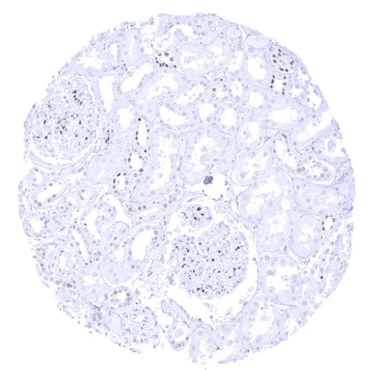
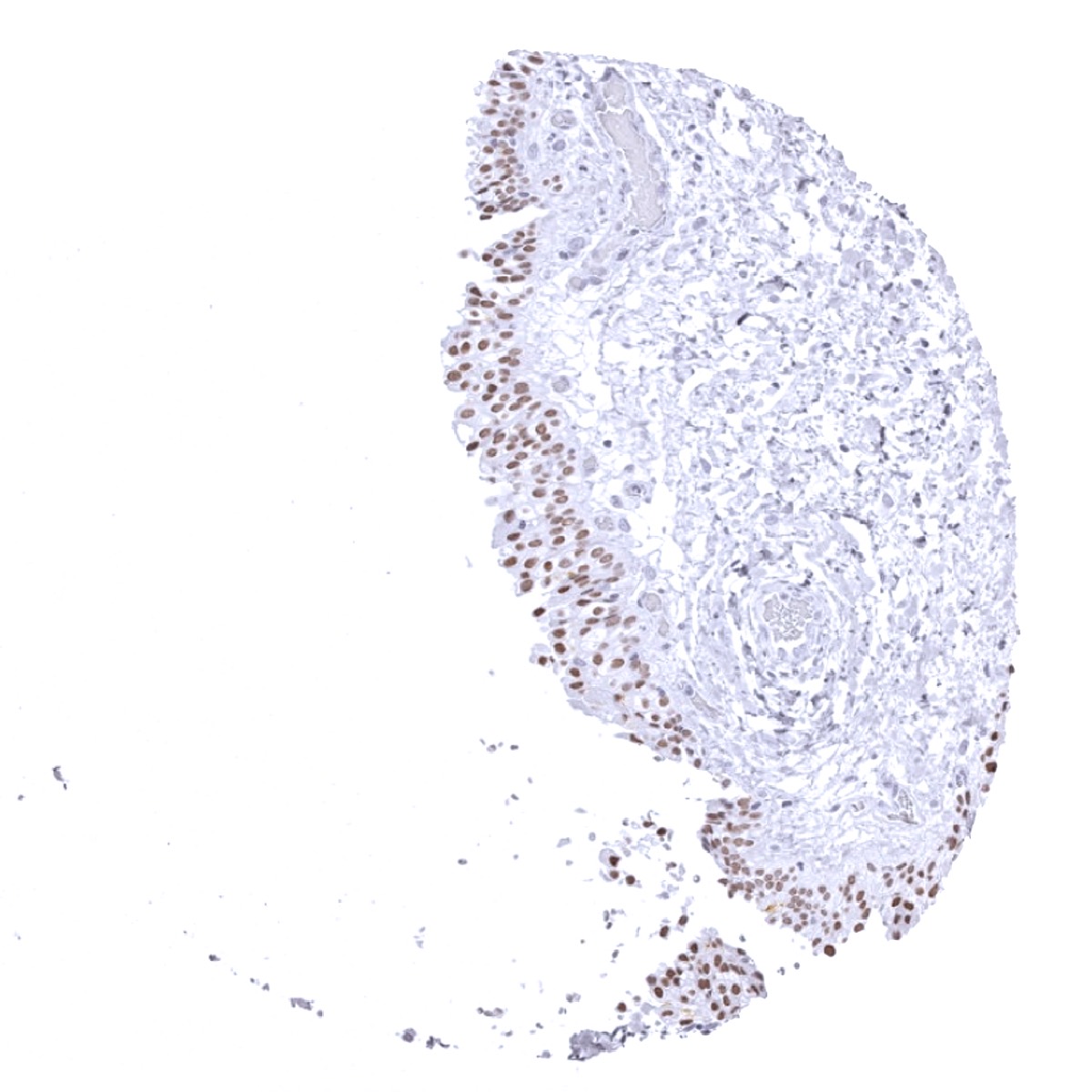
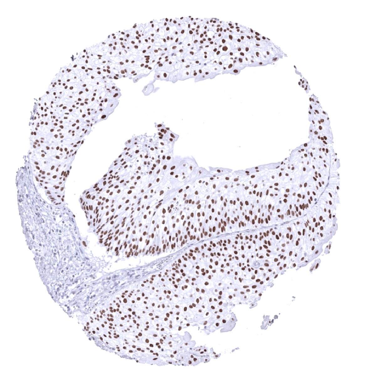
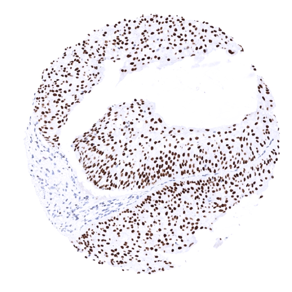
-Kidney: A moderate to strong nuclear staining reaction should be seen in a fraction of collecting duct cells and podocytes in glomeruli.
-Tonsil: The vast majority of T helper cells (Th2) in the T-zones should show a weak to moderate nuclear staining.
Negative control
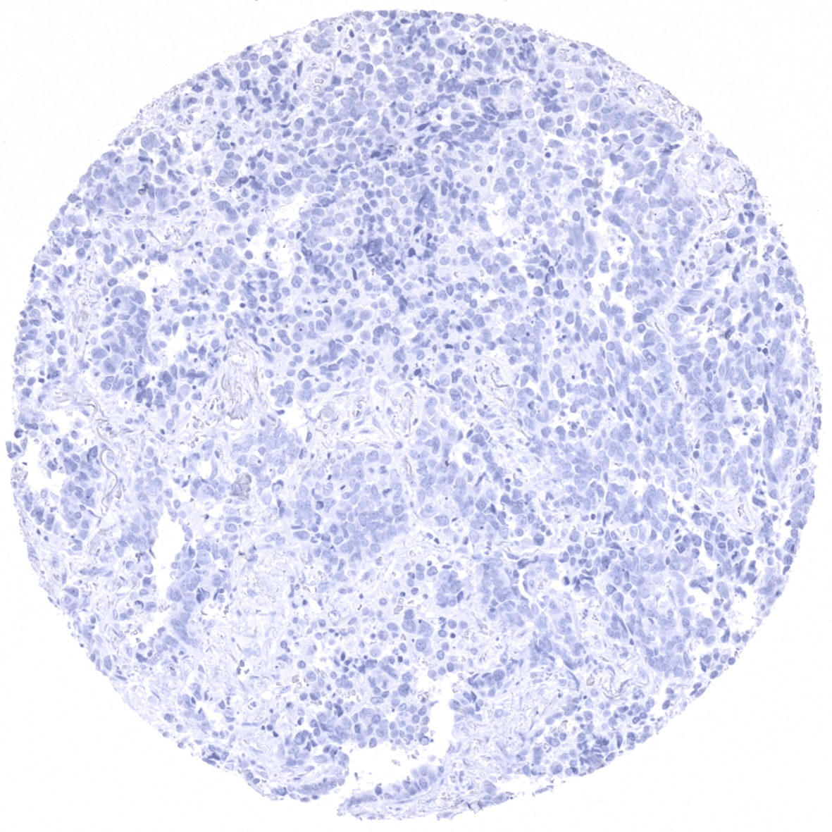
-Kidney: Staining should be absent in proximal and distal tubuli as well as in blood vessels.
-Tonsil: Staining should be absent in B-cells.
Cellular localization = Nuclear
Reactivity = Human
Application = Immunohistochemistry
Dilution = 1:50 – 1:100
Intended Use = Research Use Only
Relevance of Antibody
GATA3 is expressed in breast epithelium, urothelial cells, and a subset of lymphocytes.
Biology Behind
GATA3 gene at 10p14 consists of 8 exons and codes for GATA3, variant 1, and GATA3, variant 2, proteins of 48 KDa that which differ in one amino acid and may have identical functions. GATA3 is characterized by two conserved zinc finger domains, which bind DNA at the nucleotide sequence GATA. Like other proteins of the GATA family (GATA1-6), GATA3 is a transcription factor that regulates various cellular programs during embryogenesis and differentiation in several organ systems, including parathyroid gland, kidney, breast epithelium, urothelium, and the nervous system. A key role has been identified in T cell development, where GATA3 is the downstream of Notch in differentiation of T-helper cells and important for allergic and humoral immune responses. A role of GATA3 has also been demonstrated for tumor development and progression. GATA3 is frequently found mutated in breast cancer, and downregulation of the protein has been linked to poor prognosis of the disease.
Staining Pattern in Normal Tissues
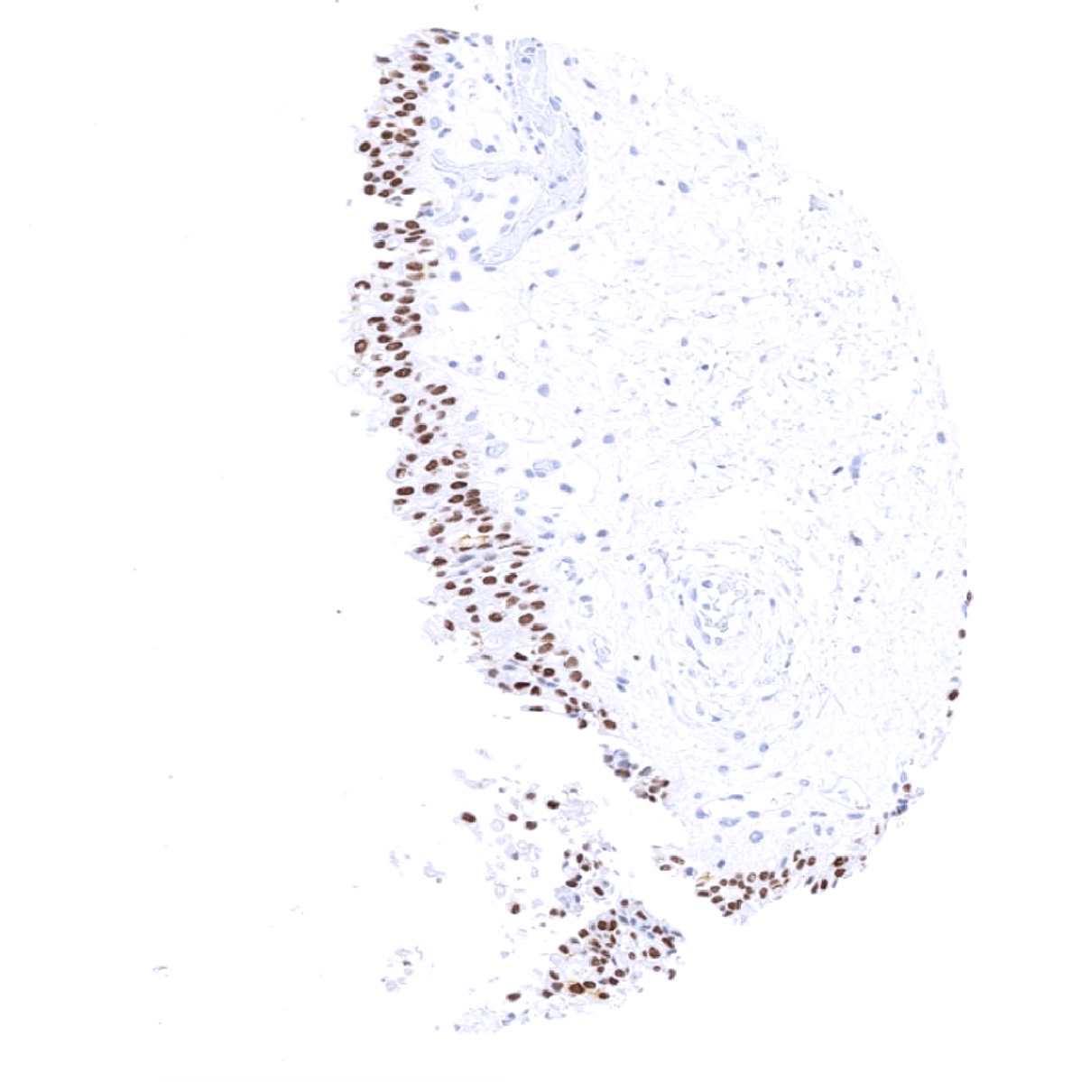
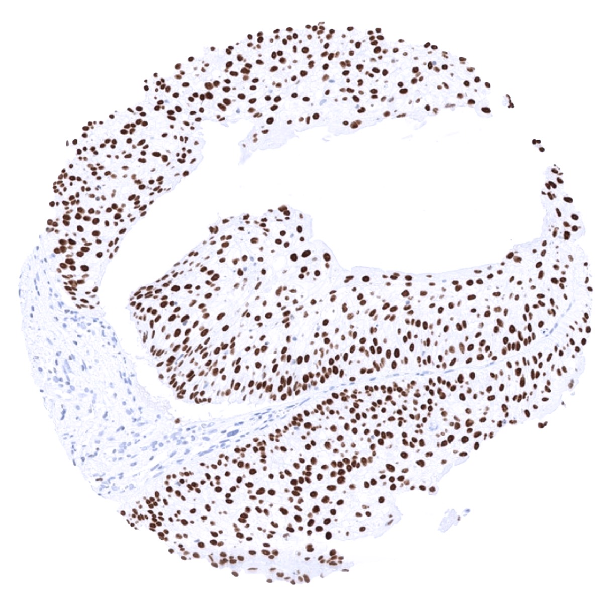
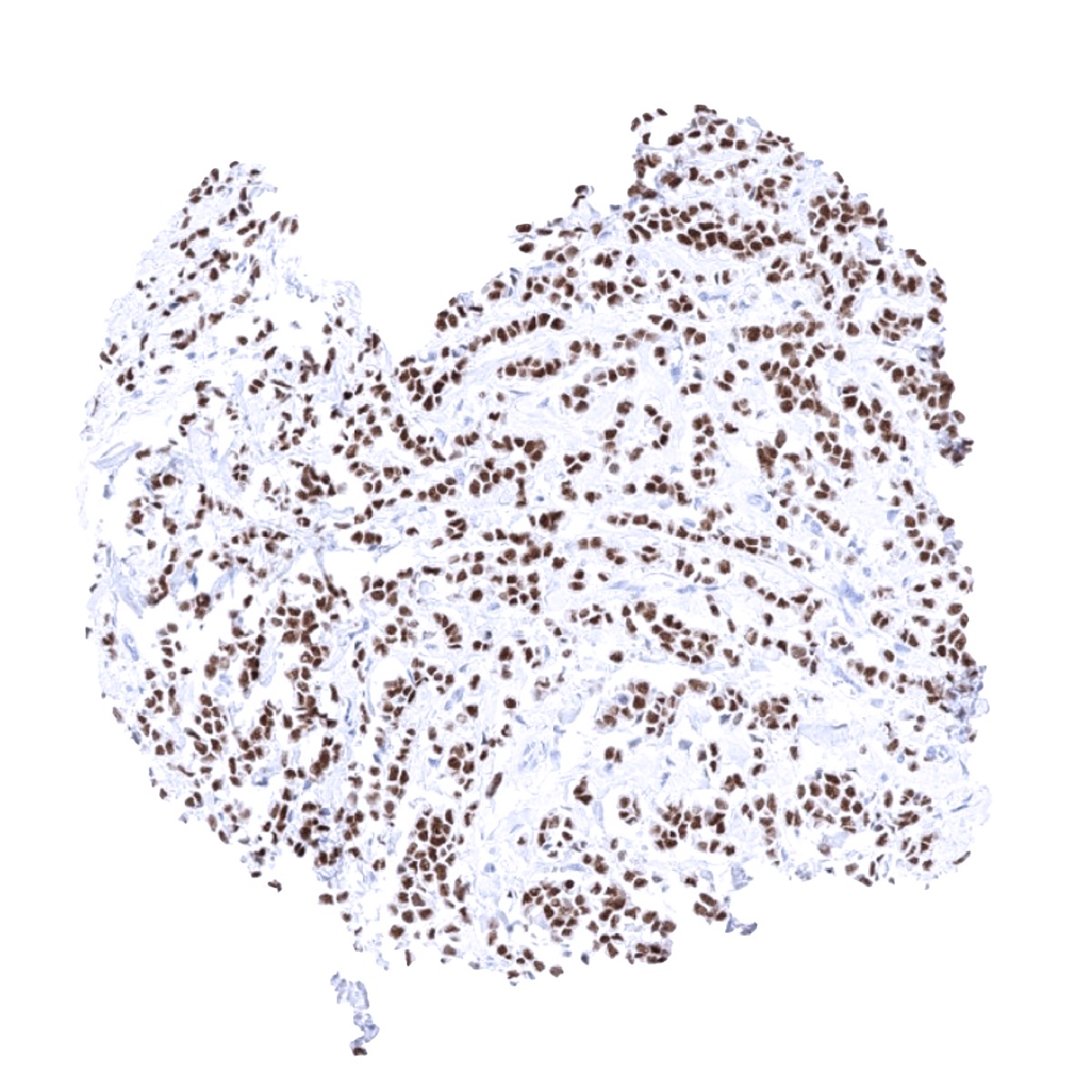
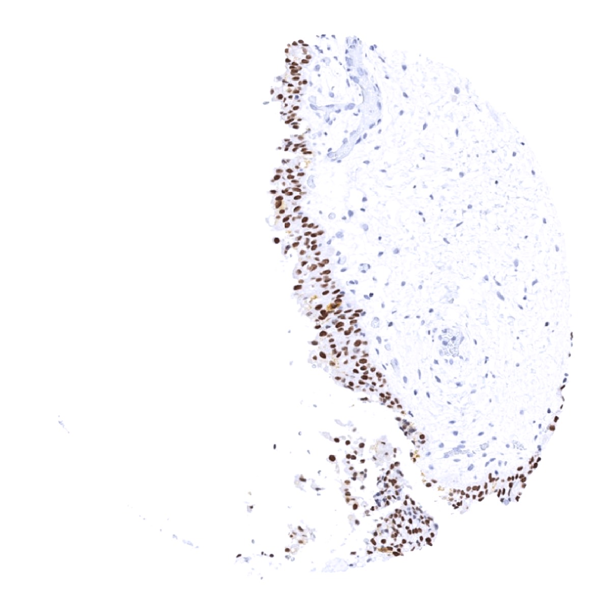
GATA3 staining pattern in Normal Tissues with antibody MSVA-450M (images are shown in our “Normal Tissue Gallery”)
| Brain | Cerebrum | Negative. |
| Cerebellum | Negative. | |
| Endocrine Tissues | Thyroid | Negative. |
| Parathyroid | Strong GATA3 positivity in all epithelial cells. | |
| Adrenal gland | Negative. | |
| Pituitary gland | Negative. | |
| Respiratory system | Respiratory epithelium | A weak cytoplasmic GATA3 staining can occur in goblet cells of respiratory epithelium (probably non-specific staining). |
| Lung | Negative. | |
| Gastrointestinal Tract | Salivary glands | Weak GATA3 staining of glandular cells (mostly mucinous). |
| Esophagus | Negative. | |
| Stomach | A weak cytoplasmic GATA3 staining can occur in gastric glands (probably non-specific staining). | |
| Duodenum | Negative. | |
| Small intestine | Negative. | |
| Appendix | Negative. | |
| Colon | Negative. | |
| Rectum | Negative. | |
| Liver | Negative. | |
| Gallbladder | Negative. | |
| Pancreas | Negative. | |
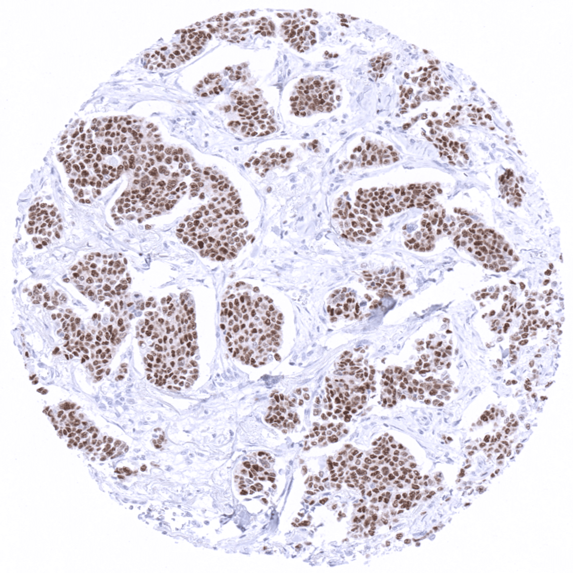
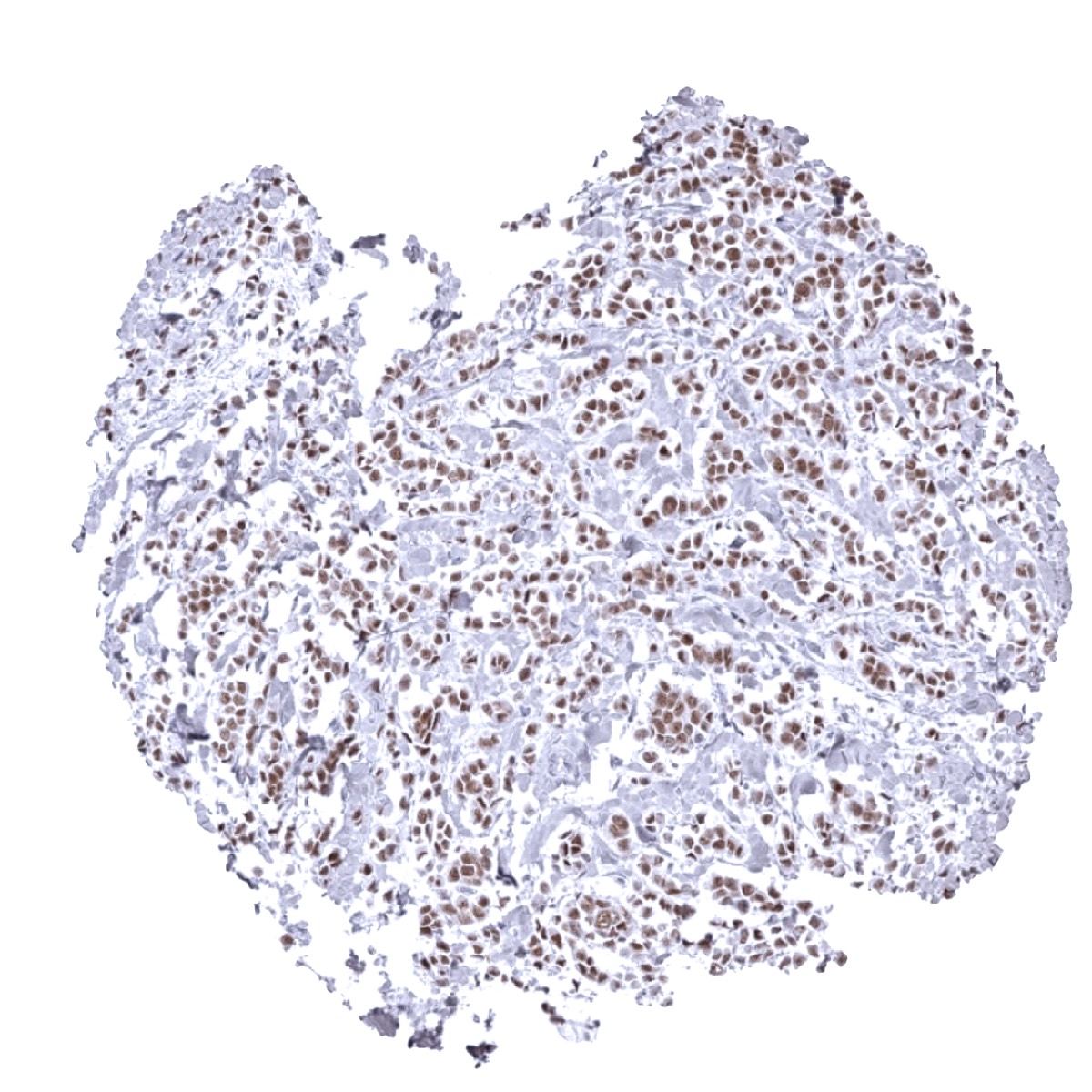
| Genitourinary | Kidney | Strong GATA3 positivity collecting ducts, moderate staining in glomerular podocytes. |
| Urothelium | Strong GATA3 positivity in all urothelial cells. | |
| Male genital | Prostate | Weak GATA3 positivity in some basal cells (not in all samples). |
| Seminal vesicles | Moderate to strong GATA3 positivity of a epithelial cells. | |
| Testis | Negative. | |
| Epididymis | Weak to moderate staining of tall columnar cells and basal cells of the corpus. | |
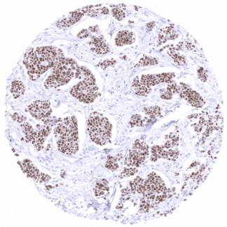
| Female genital | Breast | Moderate to strong GATA3 positivity of a fraction of luminal cells. |
| Uterus, myometrium | Negative. | |
| Uterus, ectocervix | Negative. | |
| Uterus endocervix | Negative. | |
| Uterus, endometrium | Negative. | |
| Fallopian Tube | Negative. | |
| Ovary | Negative. | |
| Placenta early | Moderate to strong of GATA3 positivity of trophoblast cells. | |
| Placenta mature | Weak to moderate GATA3 positivity of trophoblast cells. | |
| Amnion | Weak GATA3 positivity in amnion cells. | |
| Chorion | Strong GATA3 positivity in chorion cells. | |
| Skin | Epidermis | Variable (weak to strong) GATA3 positivity in squamous epithelial cells. |
| Sebaceous glands | Weak to moderate GATA3 positivity in epithelial cells including hair follicles. | |
| Muscle/connective tissue | Heart muscle | Negative. |
| Skeletal muscle | Negative. | |
| Smooth muscle | Negative. | |
| Vessel walls | Negative. | |
| Fat | Negative. | |
| Stroma | Negative. | |
| Endothelium | Negative. | |
| Bone marrow/ lymphoid tissue | Bone marrow | Negative. |
| Lymph node | Weak to moderate GATA3 positivity of a fraction of lymphocytes. | |
| Spleen | Negative. | |
| Thymus | Moderate GATA3 positivity of a significant fraction of lymphocytes. | |
| Tonsil | Weak to moderate GATA3 positivity of a fraction of lymphocytes. | |
| Remarks | GATA3 immunostaining is always nuclear. |
The findings obtained by MSVA-450M almost perfectly match the RNA data summarized in the Human Protein Atlas (Tissue expression GATA3) and identify the cell types responsible for RNA expression. The only protein atlas findings which could not be corroborated was GATA3 expression in the vagina. The validation experiments performed with MSVA-450M had suggested that GATA3 expression occurs in all keratinized squamous epithelia but not in non-keratinized squamous epithelia.
Suggested positive tissue control:
-Kidney: A moderate to strong nuclear staining reaction should be seen in a fraction of collecting duct cells and podocytes in glomeruli.
-Tonsil: The vast majority of T helper cells (Th2) in the T-zones should show a weak to moderate nuclear staining.
Suggested negative tissue control:
-Kidney: Staining should be absent in proximal and distal tubuli as well as in blood vessels.
-Tonsil: Staining should be absent in B-cells.

Staining Pattern in Relevant Tumor Types
Detectable expression of GATA3 is primarily seen in breast cancer and in urothelial carcinomas. Other tumors for which GATA3 expression has been reported include renal carcinoma, lung cancer and testicular tumors. At low frequency, GATA3 expression can occur in additional tumor entities.
The TCGA findings on GATA3 RNA expression in different tumor categories have been summarized in the Human Protein Atlas.
Compatibility of Antibodies
GATA3 (MSVA-450M) publication summary
Relevant publication: Reiswich et al.GATA3 expression in human tumors: A tissue microarray study on 16’557 tumors. Published in Pathobiology 2023 Jan 17:1-14. Epub ahead of print. PMID: 36649695.
A total of 13’093 tumors were analyzed from 131 different tumor categories by using the following protocol: Heat-induced antigen retrieval for 5 minutes in an autoclave at 121°C in pH 7,8 Target Retrieval Solution buffer. MSVA-450M at a dilution of 1:50 at 37°C for 60 minutes. Visualization of bound antibody by the EnVision Kit (Dako, Agilent). This protocol was also used for all stainings depicted in our tumor and normal tissue galleries.
Overall, 70 (53%) of 131 tumor categories showed detectable GATA3 expression in at least one case and 24 (18%) tumor categories included at least one case with strong GATA3 positivity. The highest positivity rates and the highest levels of expression was found in various categories of breast and urinary bladder neoplasms as well as in basal cell carcinoma of the skin. At lower frequency – and often at lower intensity – GATA3 staining could also be seen in various categories of salivary gland tumors, neuroendocrine tumors as well as in squamous cell carcinomas or tumors containing squamous cell elements such as endometiroid carcinomas. The distribution of positive staining results is shown in “organ-systematic” and in “ranking order” figures below (images based on data from Reiswich et al.).
Authors conclusions on diagnostic utility of GATA3 IHC with respect to the distinction of benign versus malignant (Reiswich et al.):
- Not applicable.
Authors conclusions on diagnostic utility of GATA3 IHC with respect to the distinction of different tumor entities (Reiswich et al.):
- Distinction of metastatic urothelial and breast carcinomas (often GATA3 pos) from other metastatic carcinomas (mostly GATA3 negative).
- Distinction of urothelial carcinoma (mostly GATA3 positive) from prostatic carcinoma (mostly GATA3 neg) in tumor masses at the base of the bladder.
- Distinction of metastatic lobular carcinoma of the breast (often GATA3 positive) from gastric signet ring cell carcinoma (GATA3 negative)
It is of note, however, that a weak or even moderate to strong GATA3 immunostaining can occasionally occur in various further clinically important tumor entities including neuroendocrine cancer, adenocarcinoma of the lung, malignant mesothelioma, germ cell tumor, renal cell carcinoma, prostate cancer, and pancreatic adenocarcinoma.
Authors conclusions on the prognostic role of GATA3 immunostaining results (Reiswich et al.):
- Reduced GATA3 expression is linked to unfavorable tumor phenotype, HER2 positivity, ER/PR negativity, and poor prognosis in breast cancer.
Data from the publication: Reiswich et al.GATA3 expression in human tumors: A tissue microarray study on 16’557 tumors. Published in Pathobiology 2023 Jan 17:1-14. Epub ahead of print. PMID: 36649695. Summarized in own graphics.
Figure 1. GATA3 staining in cancer (“organ-systematic”; according to Reiswich et al.)

Figure 2. GATA3 staining in cancer (“ranking list”; according to Reiswich et al.)

Protocol Recommendations
IHC users have different preferences on how the stains should look like. Some prefer high staining intensity of the target stain and even accept some background. Others favor absolute specificity and lighter target stains. Factors that invariably lead to more intense staining include higher concentration of the antibody and visualization tools, longer incubation time, higher temperature during incubation, higher temperature and longer duration of the heat induced epitope retrieval (slide pretreatment). The impact of the pH during slide pretreatment has variable effects and depends on the antibody and the target protein.
All images and data shown here and in our image galleries are obtained by the manual protocol described below. Other protocols resulting in equivalent staining are described as well.
Manual protocol
Freshly cut sections should be used (less than 10 days between cutting and staining). Heat-induced antigen retrieval for 5 minutes in an autoclave at 121°C in pH 7,8 Target Retrieval Solution buffer. Apply MSVA-450M at a dilution of 1:80 at 37°C for 60 minutes. Visualization of bound antibody by the EnVision Kit (Dako, Agilent) according to the manufacturer’s directions.
Agilent / Dako – Autostainer Link 48
Pretreatment in PT-Link for 30 minutes at 95°C (pH high); FLEX peroxidase blocking for 5 minutes (room temperature), MSVA-450M 1:80 for 20 minutes (room temperature), FLEX+ mouse/rabbit (LINKER) for 15 minutes (room temperature), horseradish peroxidase (HRP) for 20 minutes (room temperature), FLEX DAB+Sub-Chromo for 10 minutes (room temperature), FLEX hematoxylin for 5 minutes (room temperature).
These images reflect stainings by the protocol described above. It is of note that a comparable staining result can also be obtained by different protocols. In general, a longer pretreatment, a longer incubation time of the primary antibody, a higher antibody concentration, and a longer incubation time of FLEX+LINKER result in stronger staining, potentially at the cost of more background staining. Modifications of the protocol with a strengthening effect on staining intensity in combination with changes of other parameters that result in lower staining intensity can result in a comparable result as shown above.
Leica – BOND RX
Dewax at 72°C for 30 seconds; Pretreatment in Bond Epitope Retrieval Solution (ER2 – EDTA pH9) for 20 minutes at 100°C; Peroxidase blocking for 5 minutes (room temperature), MSVA-450M 1:80 for 15 minutes (room temperature), Post primary (rabbit anti mouse) for 8 minutes (room temperature), Polymer (goat anti rabbit) for 8 minutes (room temperature), mixed DAB refine for 10 minutes (room temperature), hematoxylin for 5 minutes (room temperature).

These images reflect stainings by the protocol described above. It is of note that a comparable staining result can also be obtained by different protocols. In general, a longer pretreatment, a longer incubation time of the primary antibody, a higher antibody concentration, a higher temperature during incubation, and a longer incubation time of Post primary and or the Polymer result in stronger staining, potentially at the cost of more background staining. Modifications of the protocol with a strengthening effect on staining intensity in combination with changes of other parameters that result in lower staining intensity can result in a comparable result as shown above.
Potential pitfalls
GATA3 is used as a marker for urinary bladder cancer. However, squamous cell carcinoma of the cervix uteri can also express GATA3 at low levels. This may cause misinterpretation as “urothelial carcinoma” in case of a cervical carcinoma infiltrating the urinary bladder.
Potential Research Applications
- Because of partly controversial data, the diagnostic utility of GATA3 IHC should be investigated in a large cohort of tumors from different entities.
- The prognostic role of GATA3 expression in tumor types that are positive in only a fraction of cases is unclear.
- GATA3 is one of the three genes mutated in >10% of breast cancers (Cancer Genome Atlas) and plays a role in estrogen and androgen receptor signaling. Further studies are needed to elucidate the role, if any, of GATA3 in the development of breast cancer.
- GATA3 delineates important subgroups of inflammatory cells of which the exact biological and clinical role is not fully understood. GATA3 is thus an important component of antibody panels for use in multicolor immunofluorescence analyses.