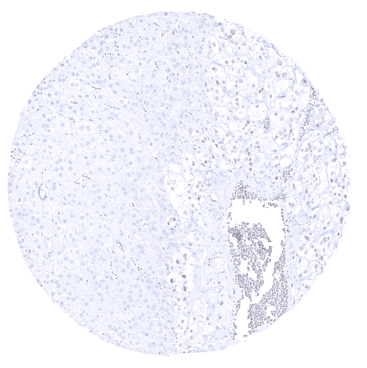
Adrenal gland

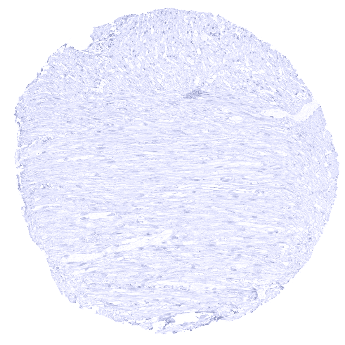
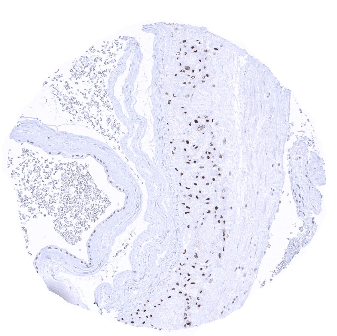
Aorta, media
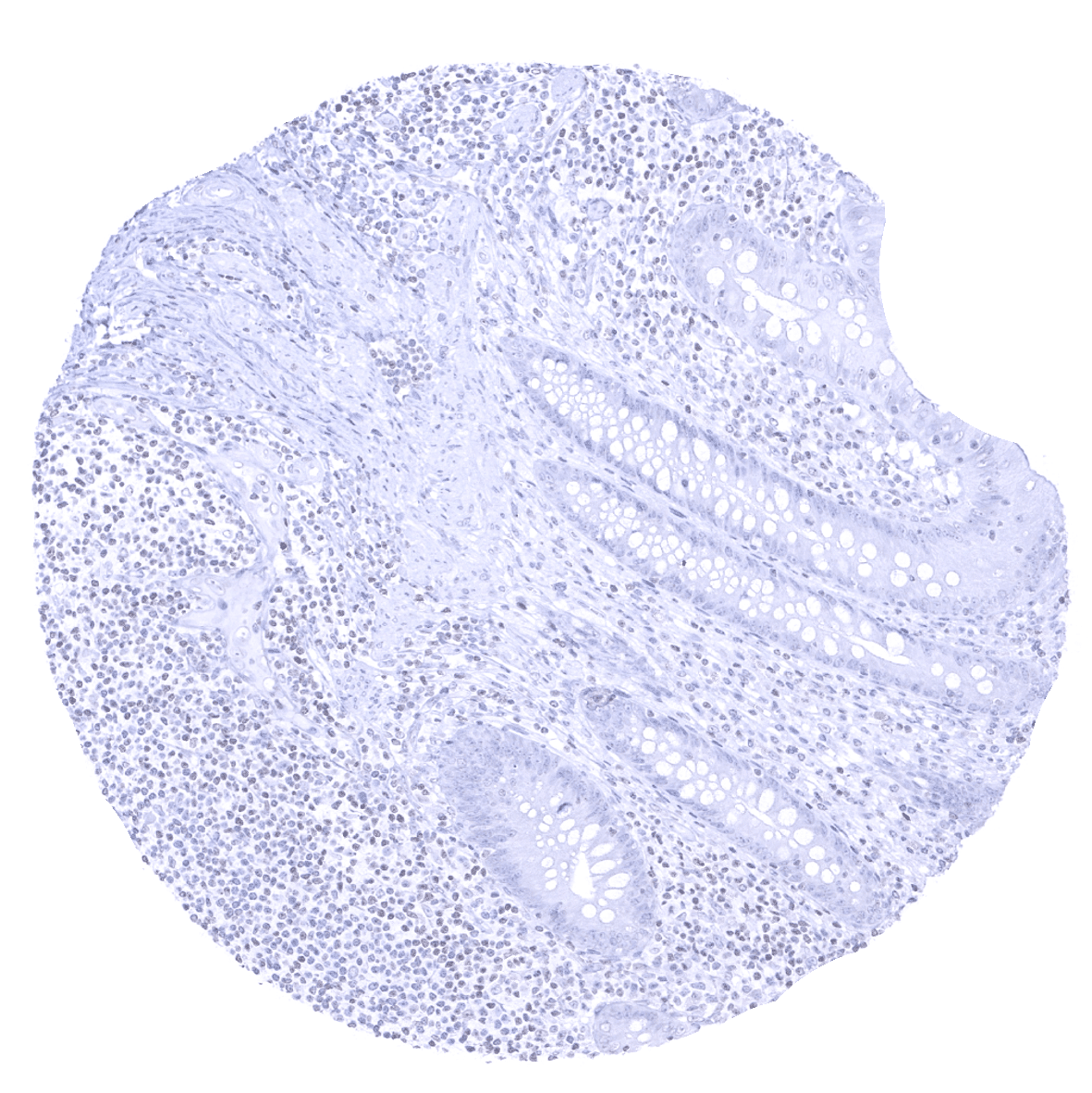
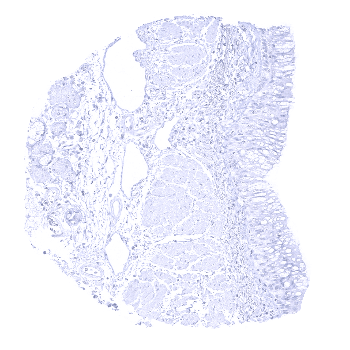
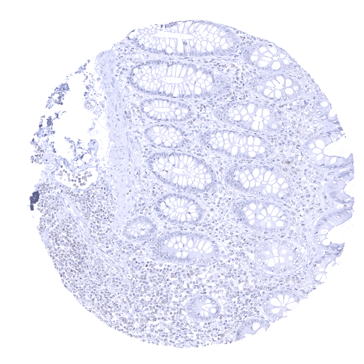
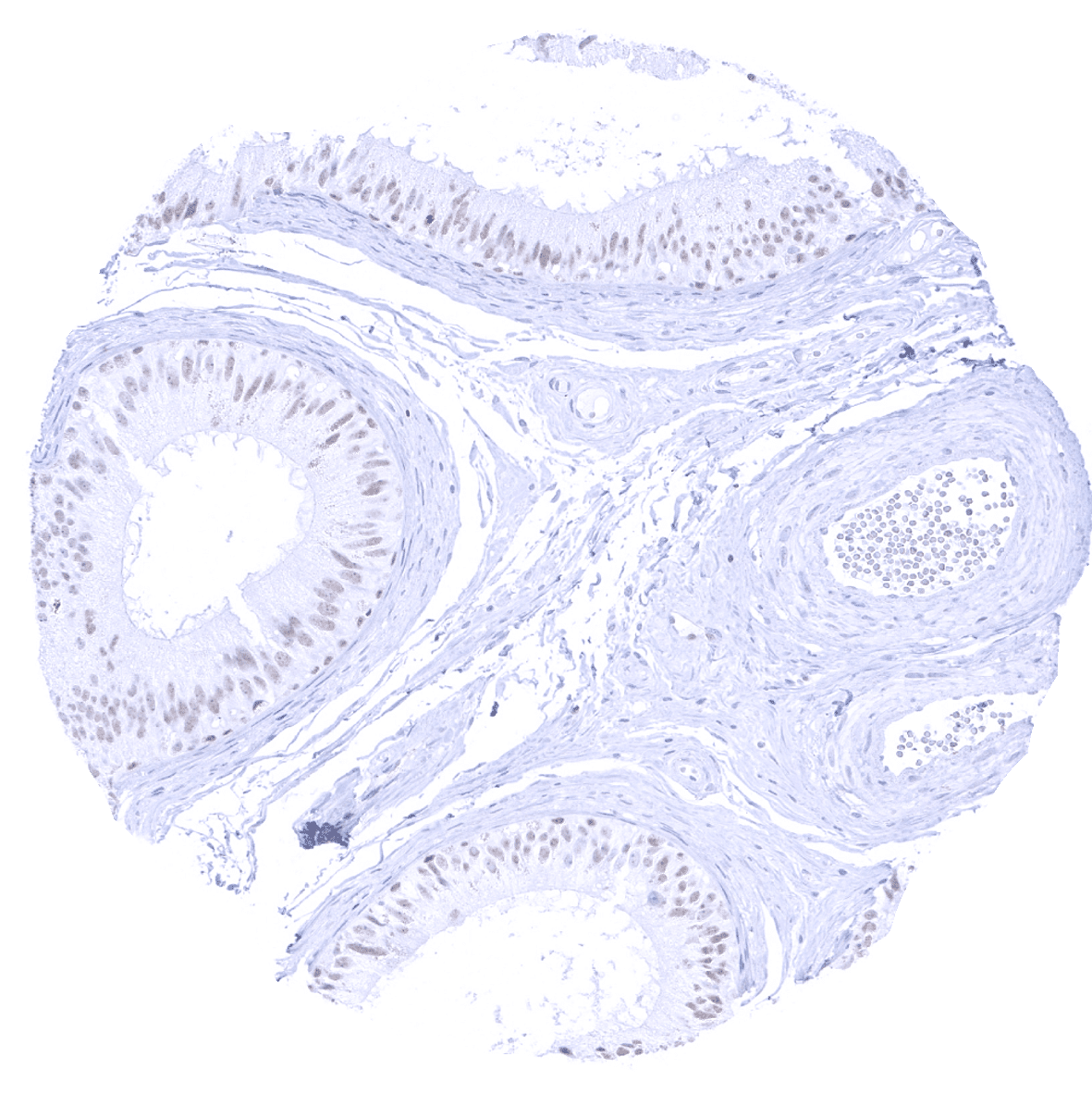
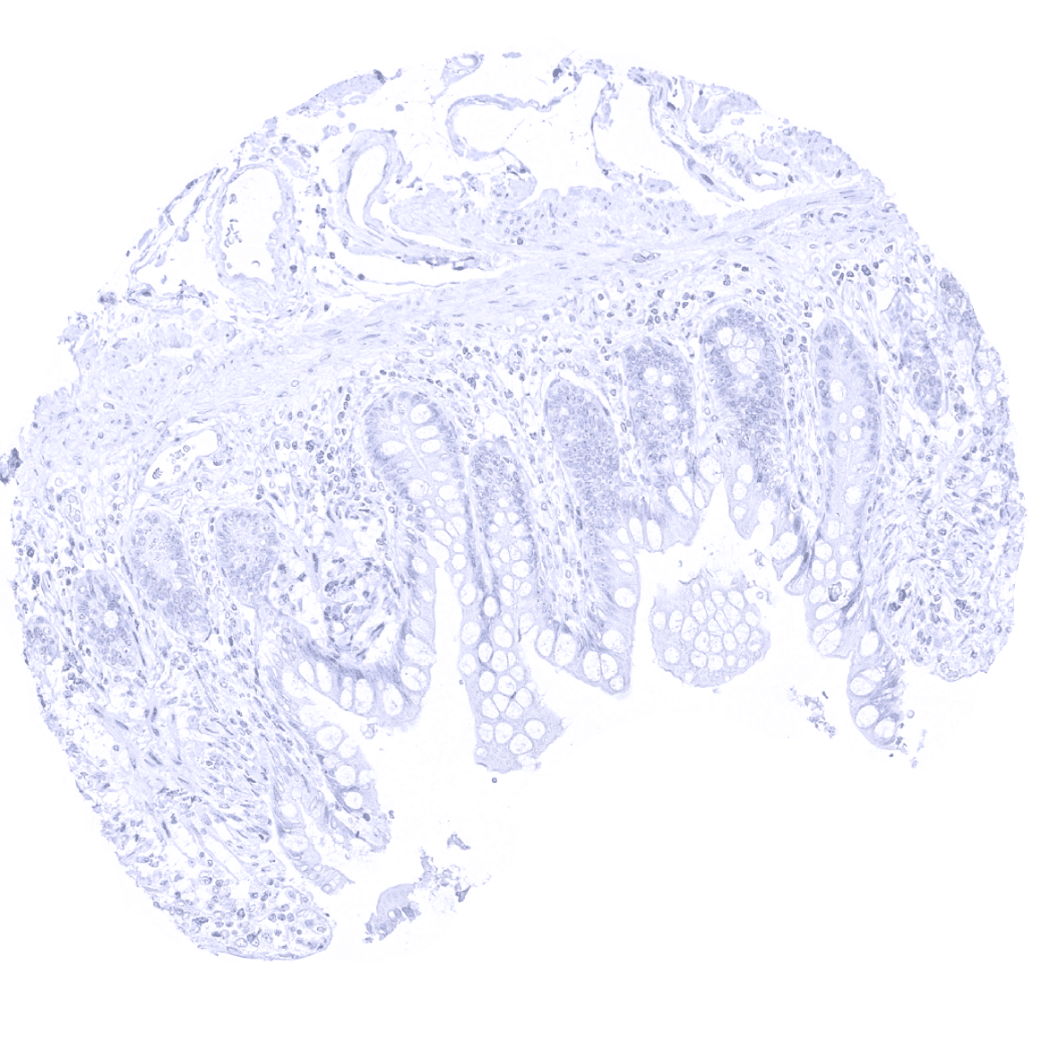
Appendix, mucosa
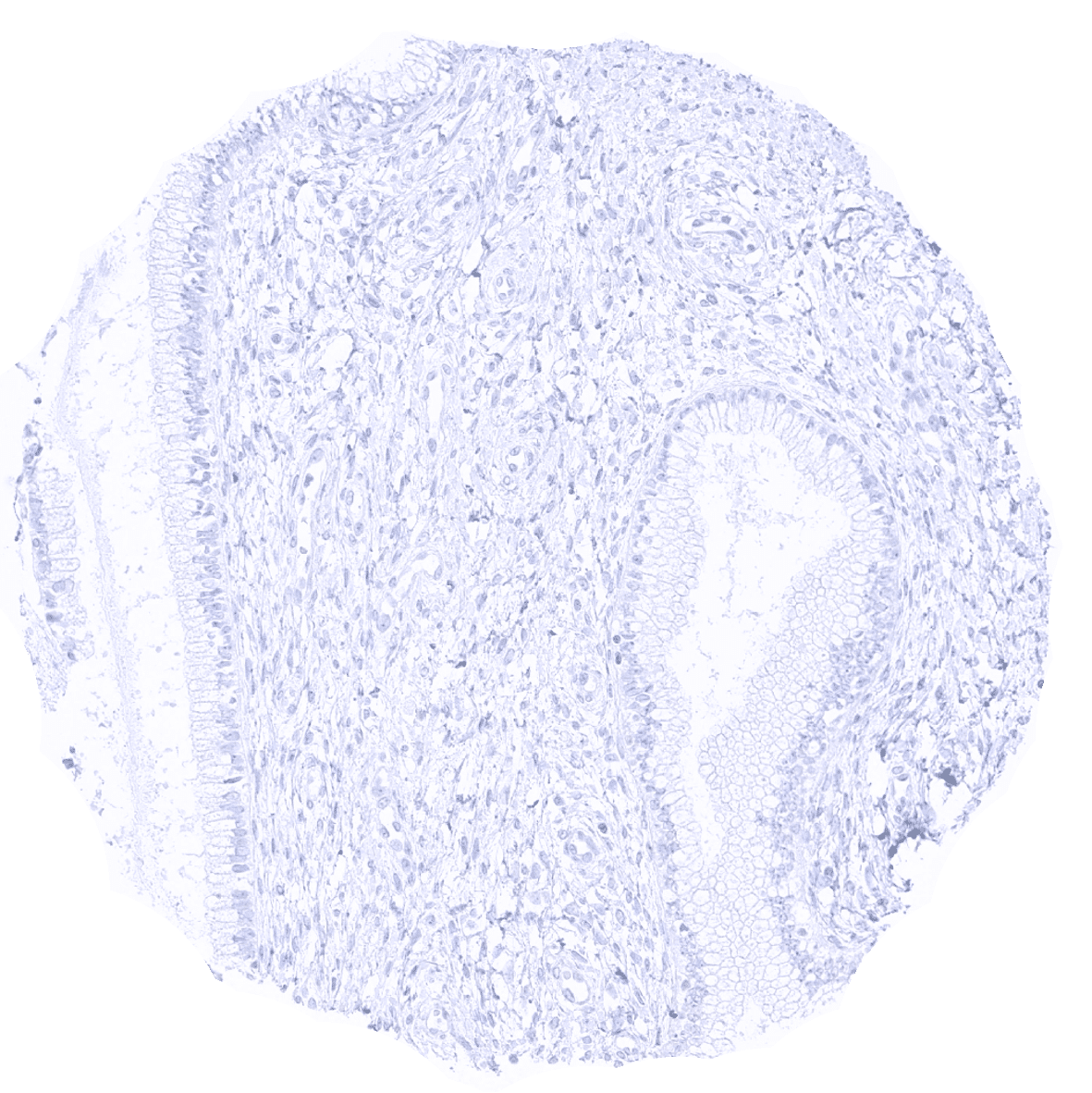
Appendix, muscular wall
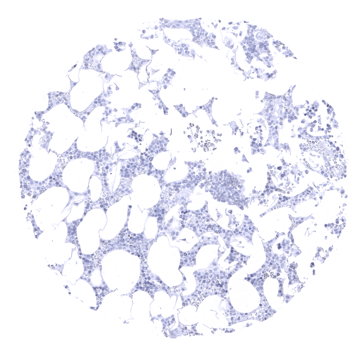
Bone marrow
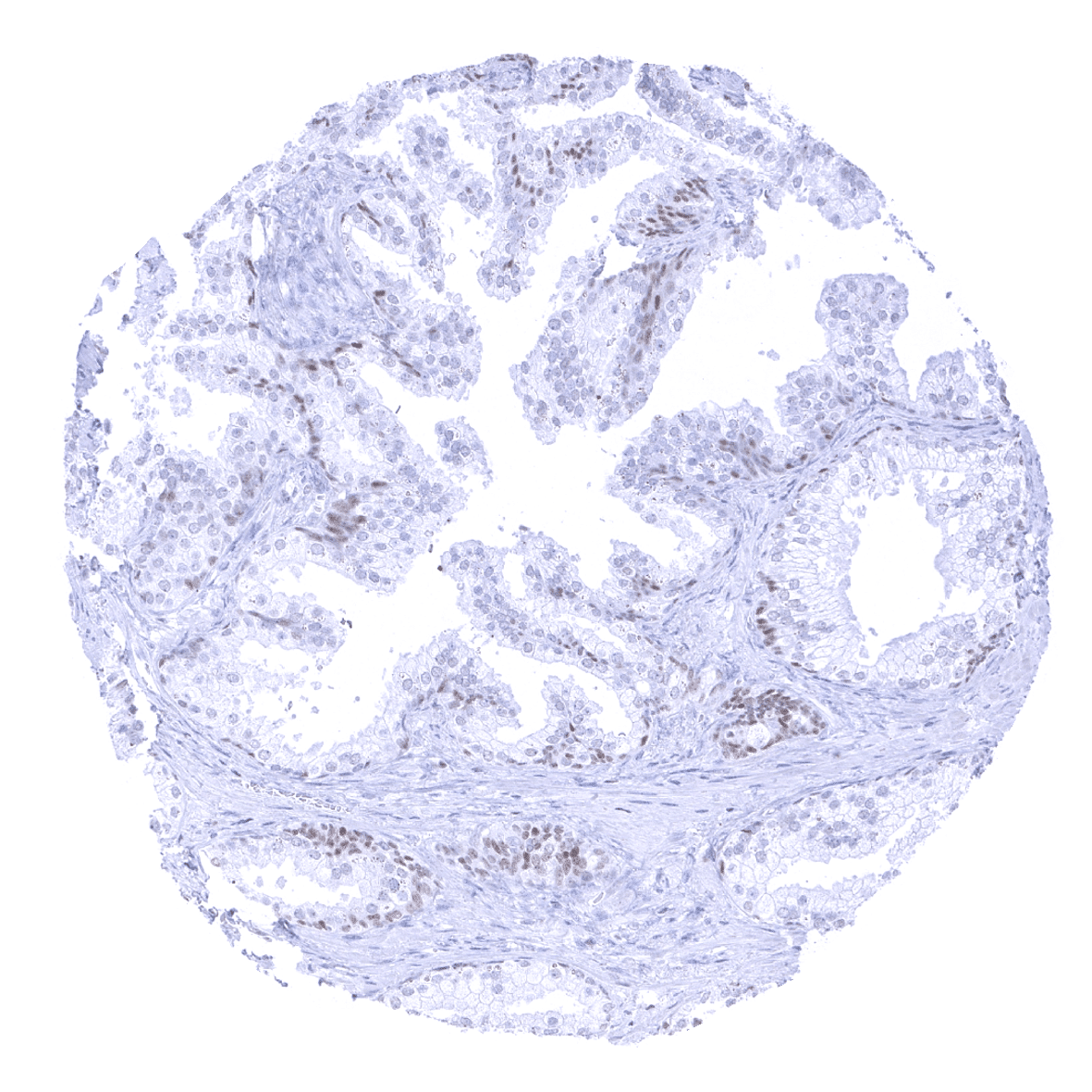
Breast
Bronchus, mucosa

Cerebellum (molecular, Purkinje and, granule cell layers; white matter)
Cerebellum (granule cell layer; white matter)
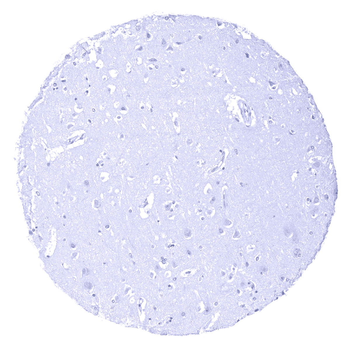
Cerebrum, grey matter
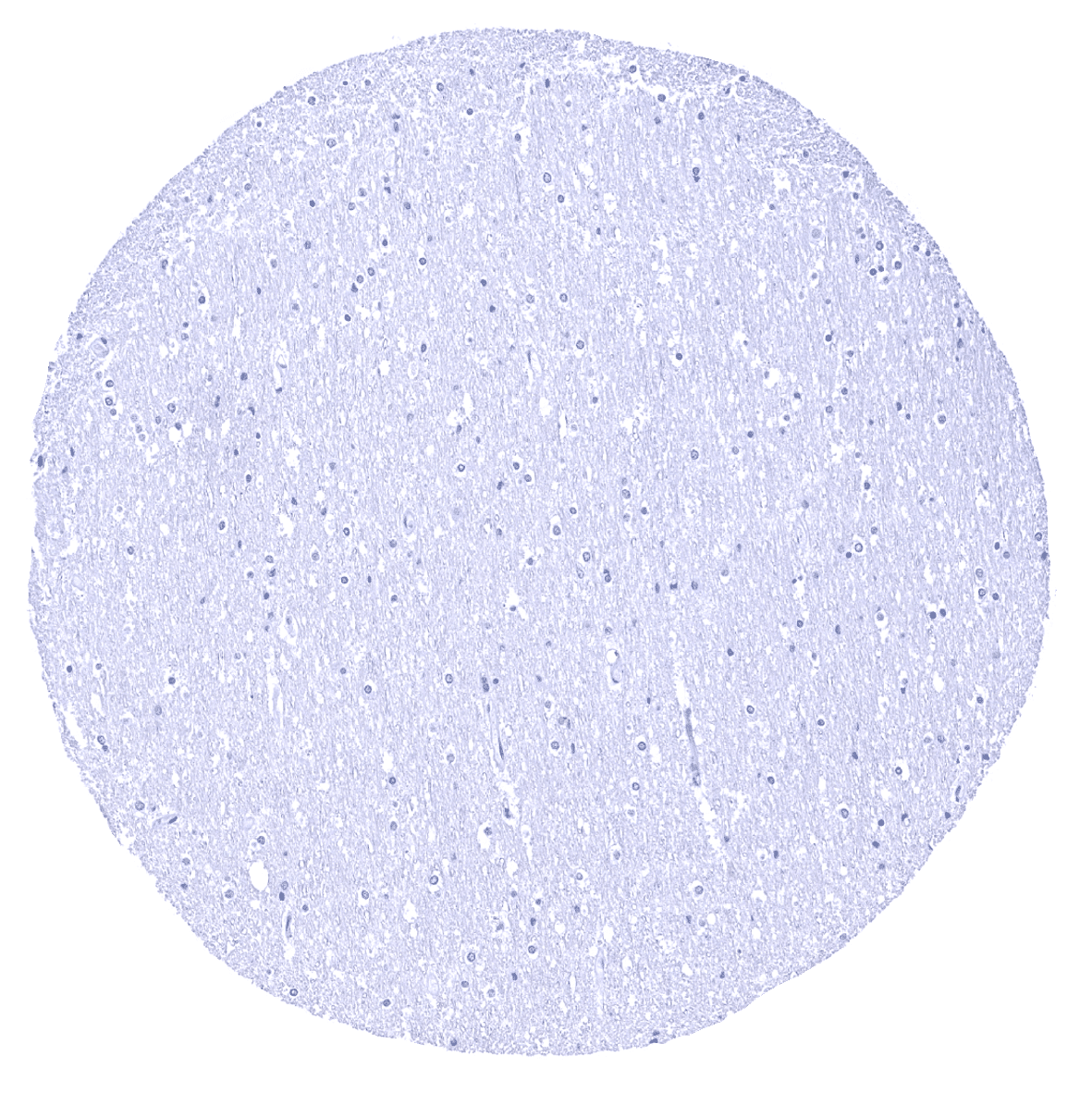
Cerebrum, white matter
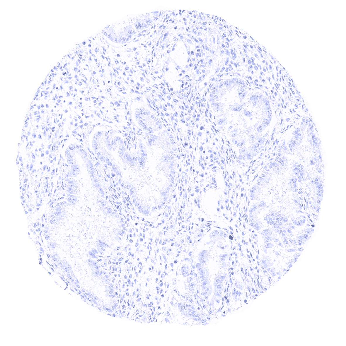
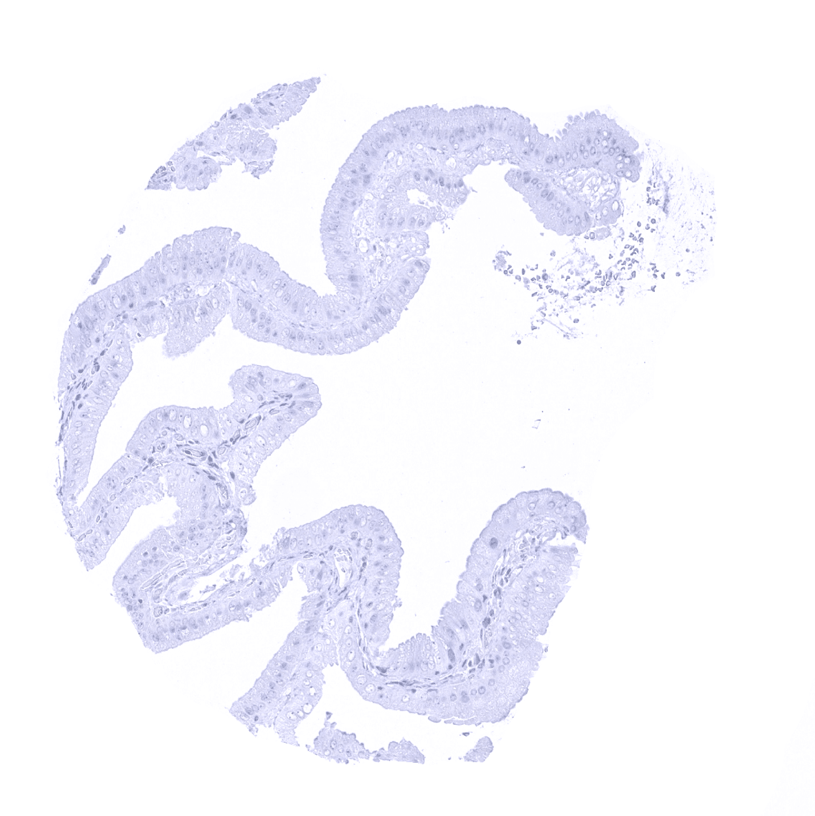
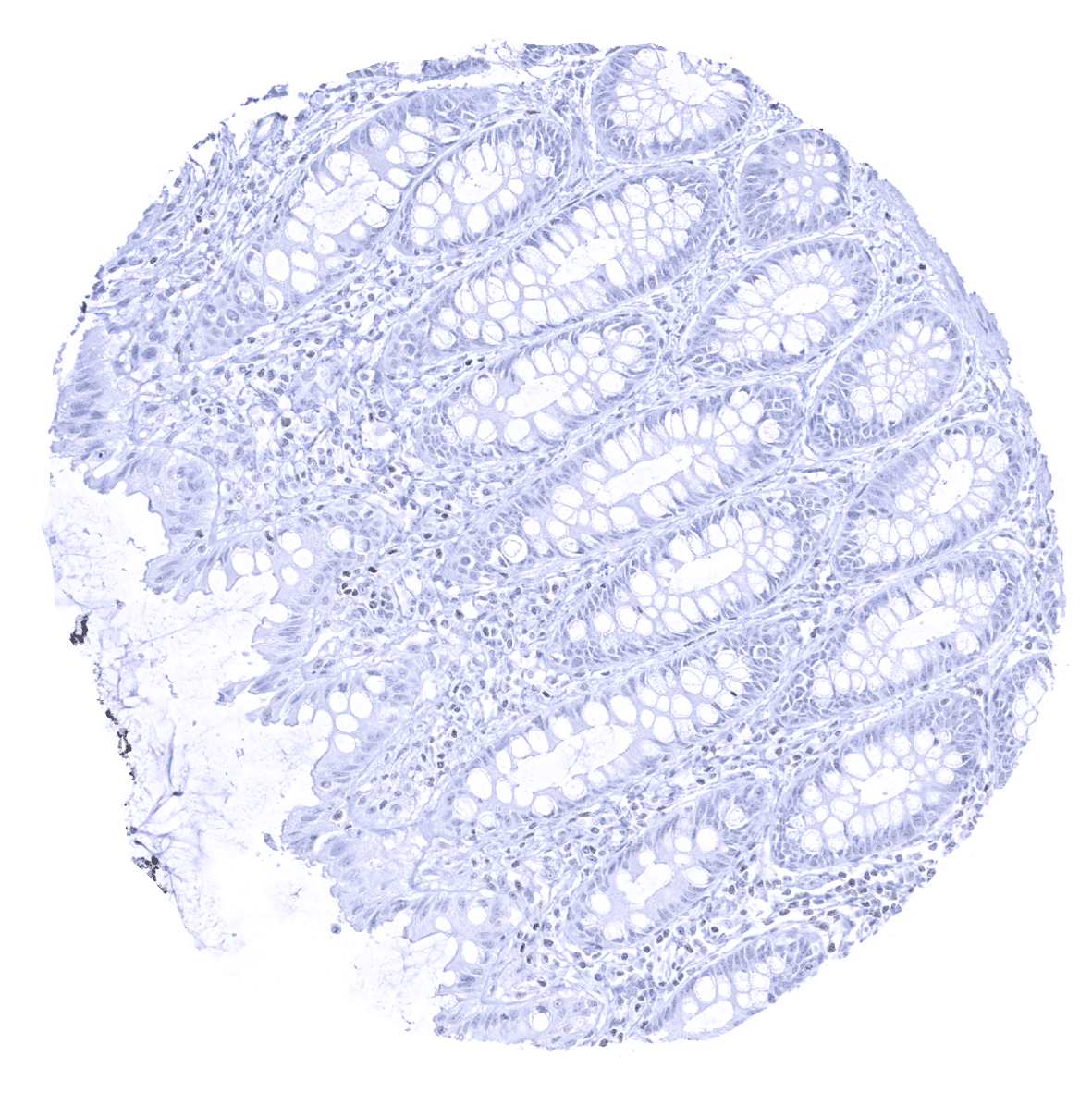
Colon descendens, mucosa

Colon descendens, muscular wall
Duodenum, Brunner gland
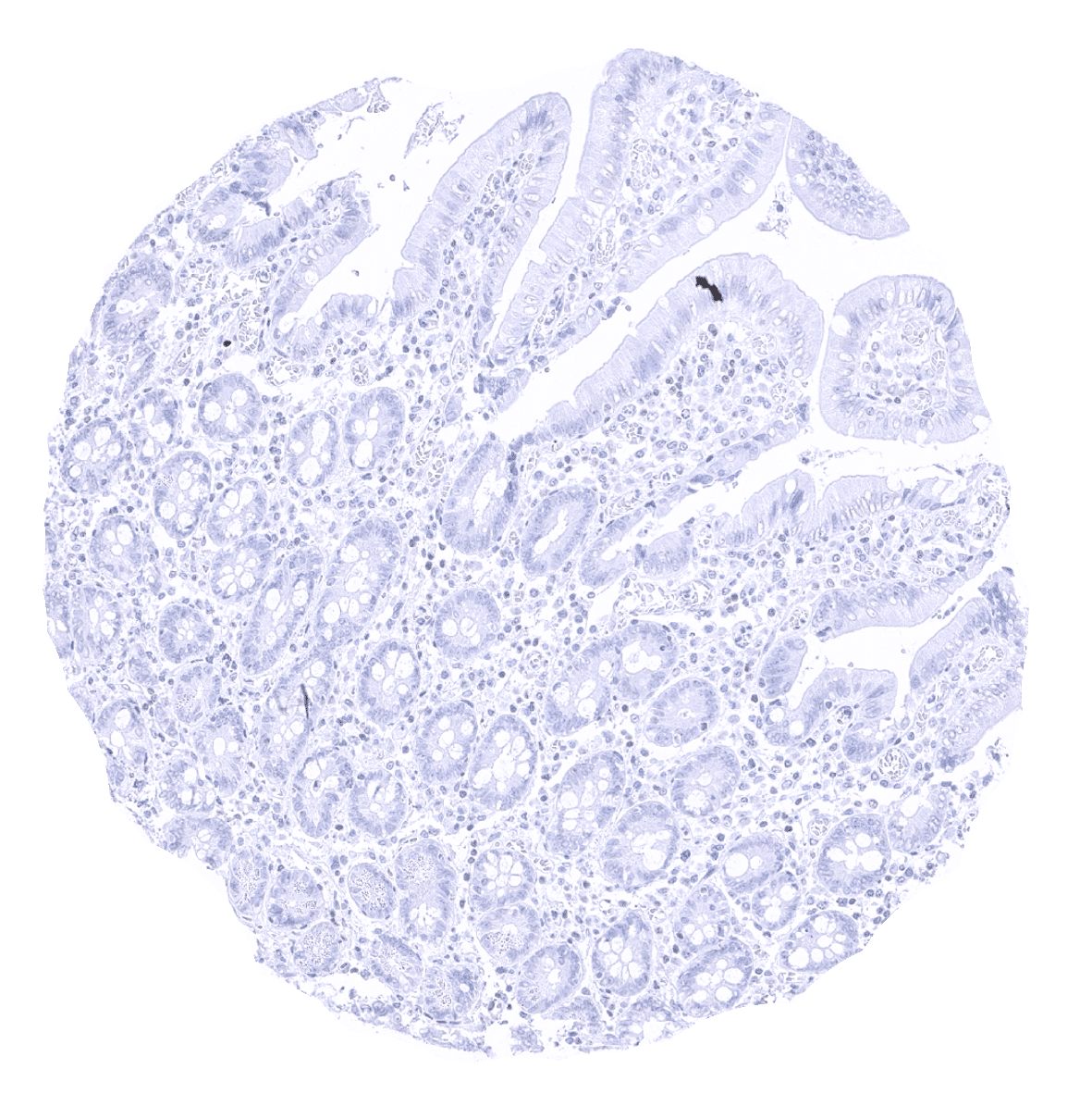
Duodenum, mucosa
Endocervix
Endometrium, proliferation
Endometrium, secretion
Epididymis - A weak to moderate GATA3 immunostaining is present in a fraction of tall columnar cells.
Esophagus, squamous epithelium - Non-keratinizing squamous epithelia lack GATA3 immunostaining.
Fallopian tube, mucosa
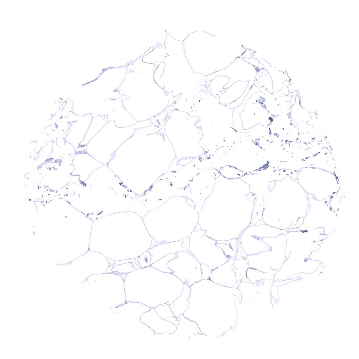
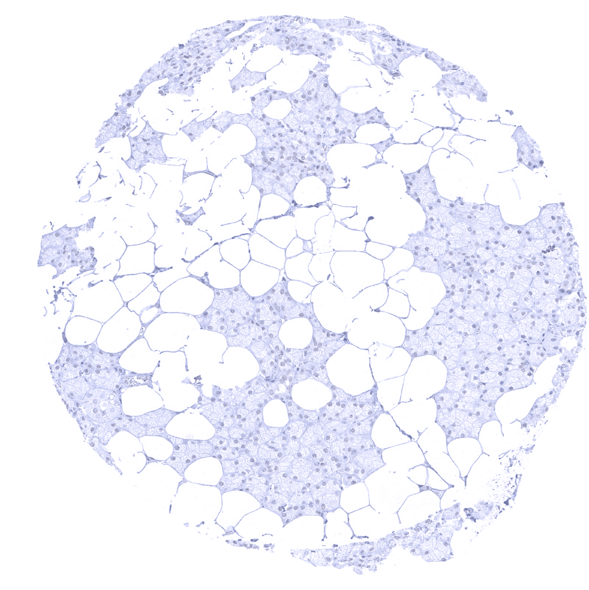
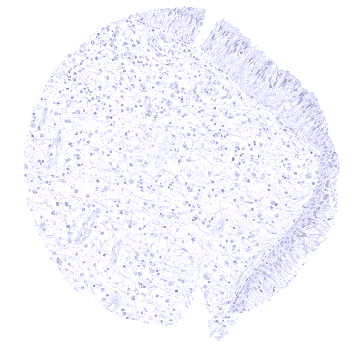
Fat
Gallbladder, epithelium
Heart
Ileum, mucosa
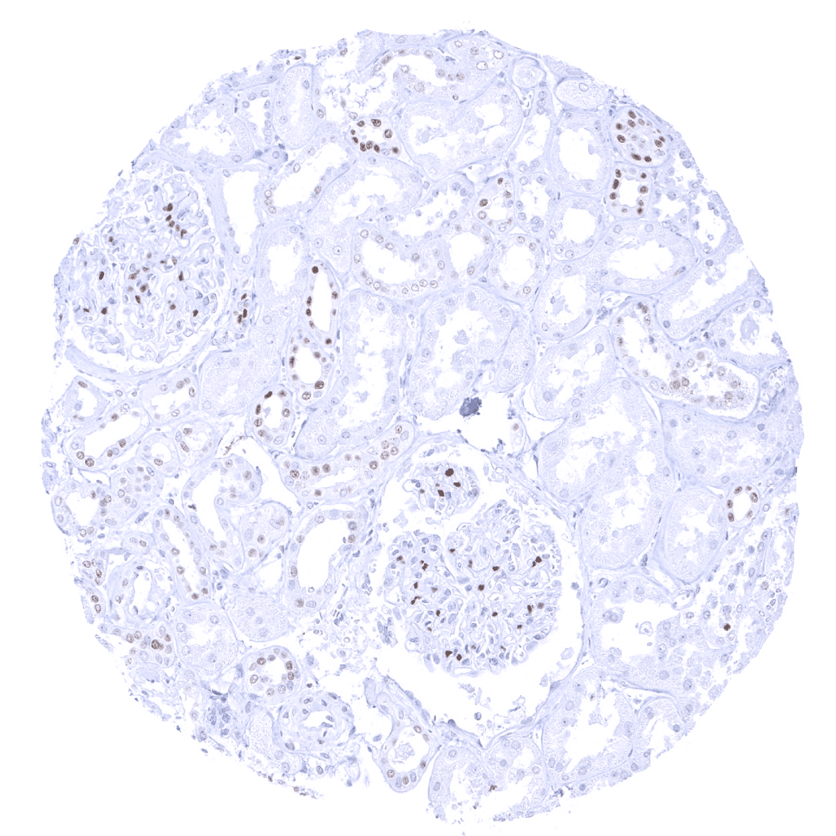
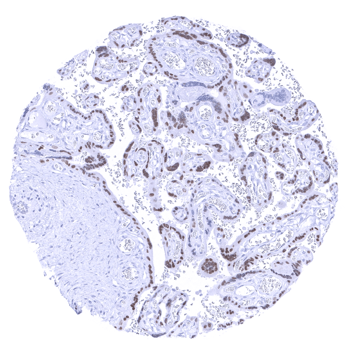
Kidney, cortex - A moderate to strong GATA3 positivity occurs in podocytes and in collecting ducts.
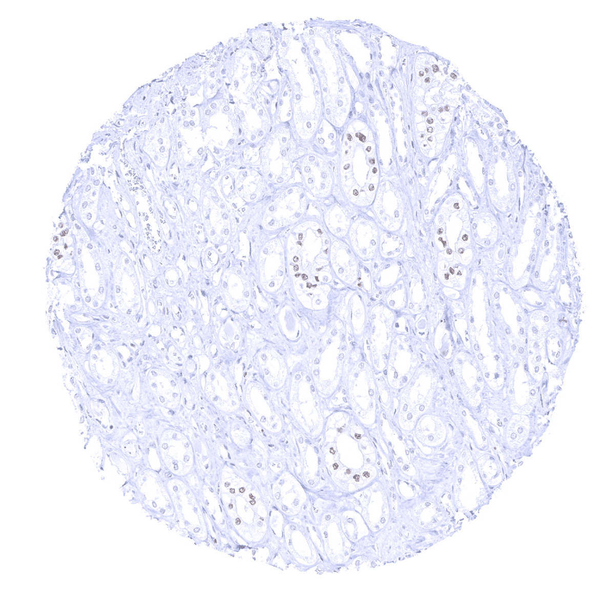
Kidney, medulla - A moderate to strong GATA3 positivity is seen in collecting ducts.
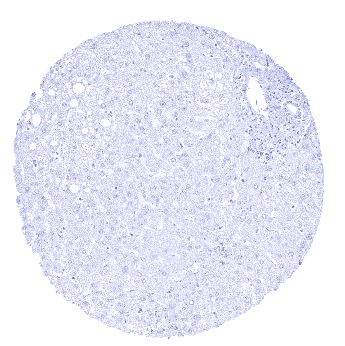
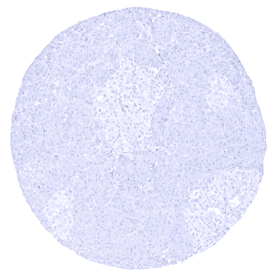
Liver
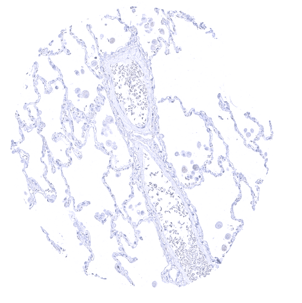
Lung
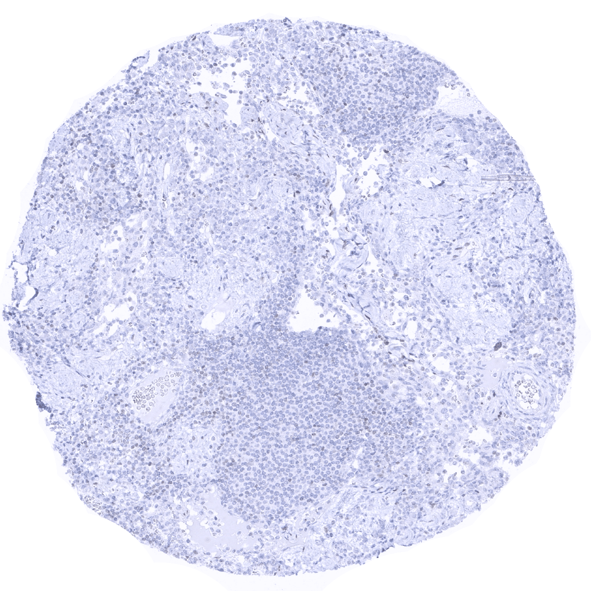
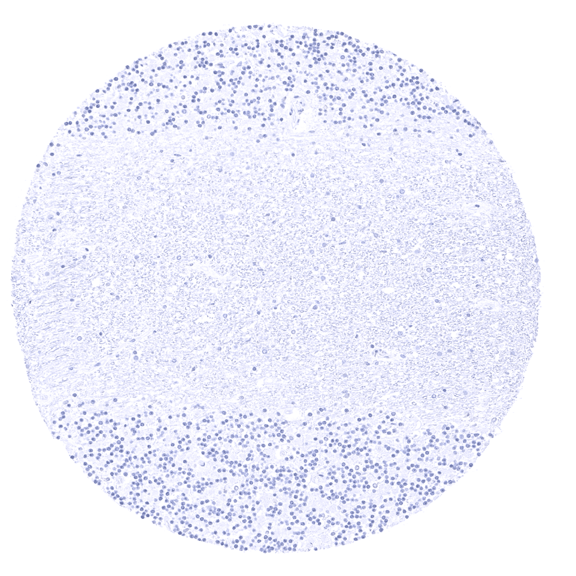
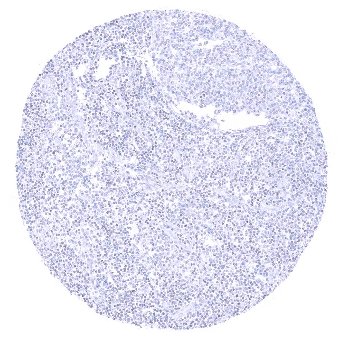
Lymph node
Ectocervix - Non-keratinizing squamous epithelia lack GATA3 immunostaining.
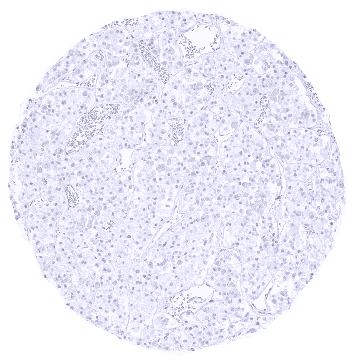
Pancreas
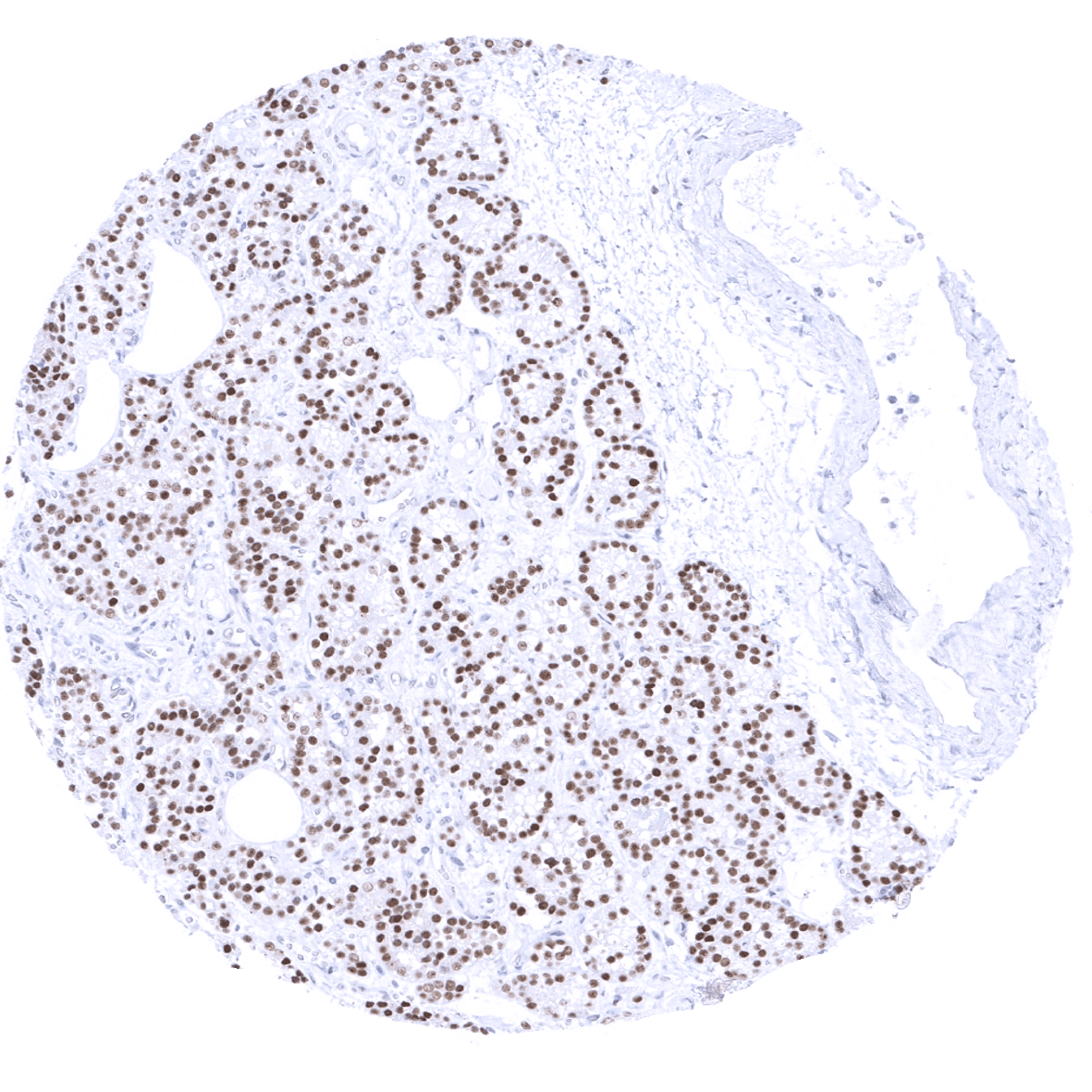
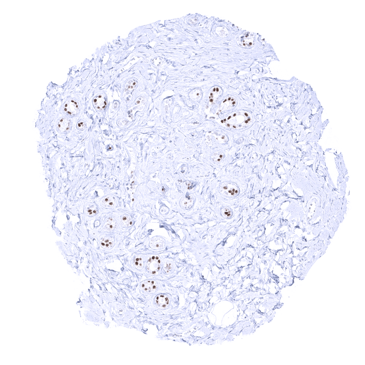
Parathyroid - Glandular cells show a strong GATA3 positivity.
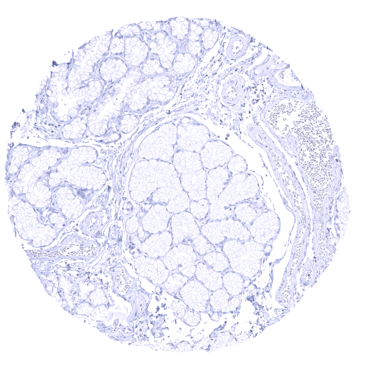
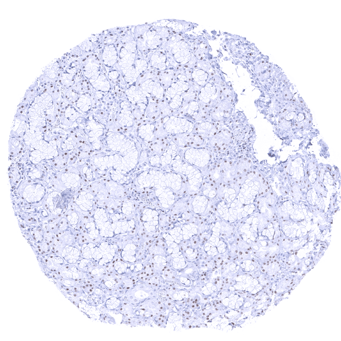
Parotid gland
Pituitary gland, anterior lobe
Pituitary gland, posterior lobe

Pregnant uterus (decidua)
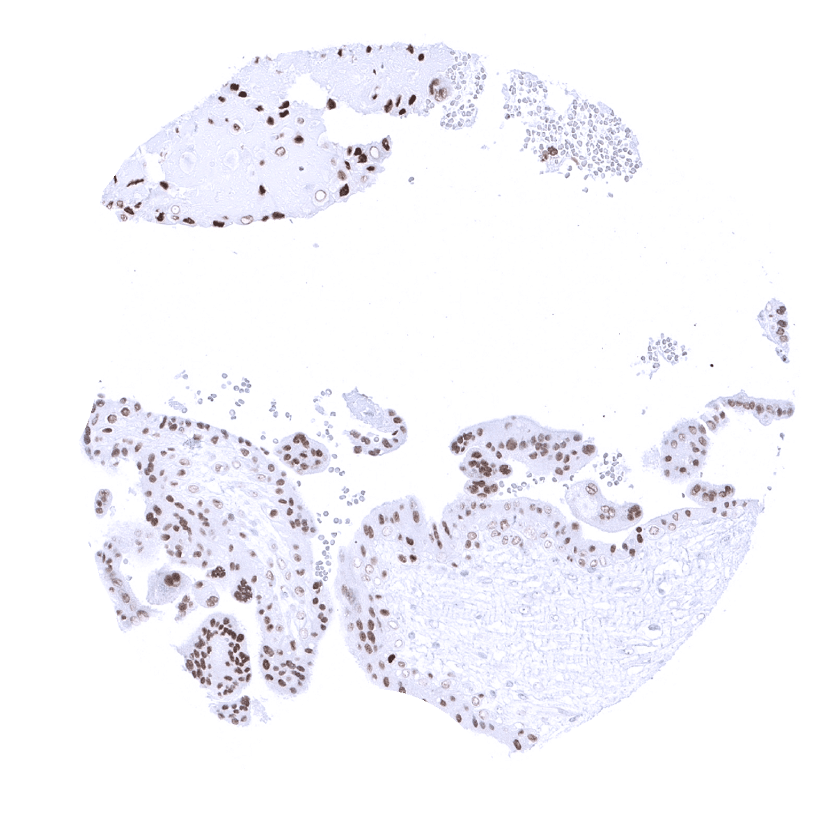
Placenta, early - A strong GATA3 positivity is seen trophoblast cells. Staining is strongest in the first trimenon.
Placenta, mature - GATA3 positivity is seen trophoblast cells. Staining intensity decreases during maturation.
Placenta (amnion and chorion) - A strong GATA3 immunostaining is seen chorion cells. Amnion cells stain weakly.
Prostate - In the prostate, a weak to moderate GATA3 staining occurs in basal cells.
Rectum, mucosa
Seminal vesicle - Seminal vesicle epithelium exhibits a moderate to strong GATA3 positivity.
Sinus paranasales
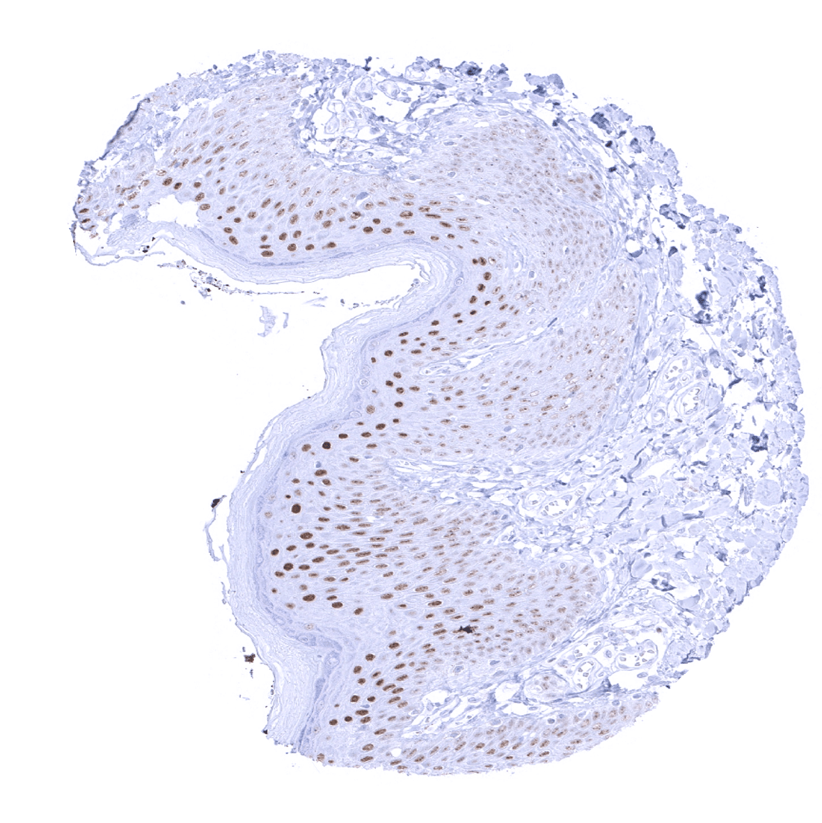
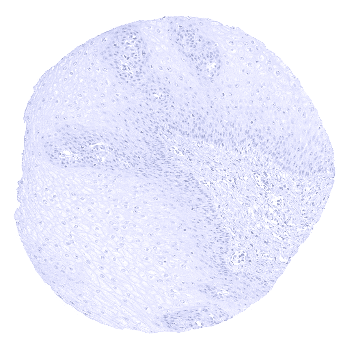
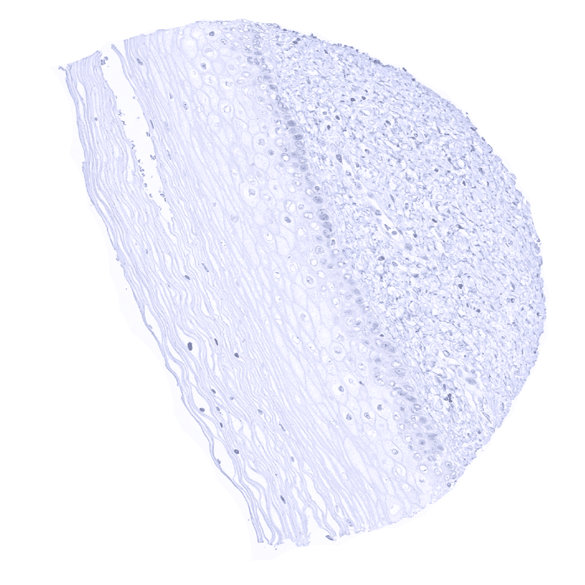
Skin - A moderate to strong GATA3 immunostaining is seen in the squamous epithelium of the skin. Strongest intensity occurs in the upper layers.
Skin, hairfollicel and sebaceous glands - A moderate to strong nuclear GATA3 immunostaining is seen in sebaceous glands.
Spleen

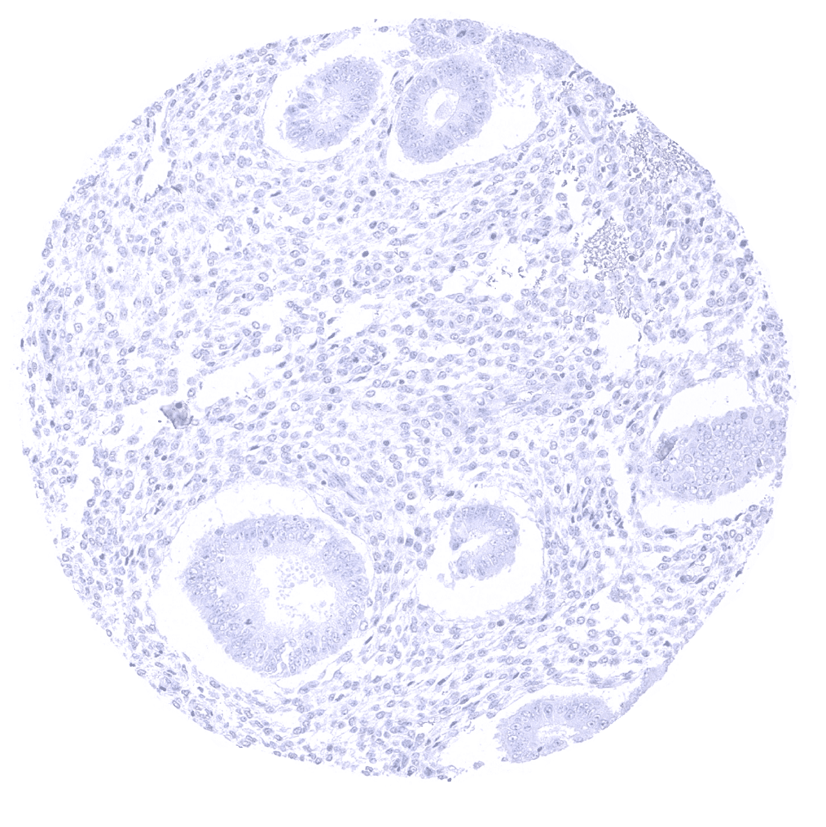
Stomach, antrum
Stomach, corpus
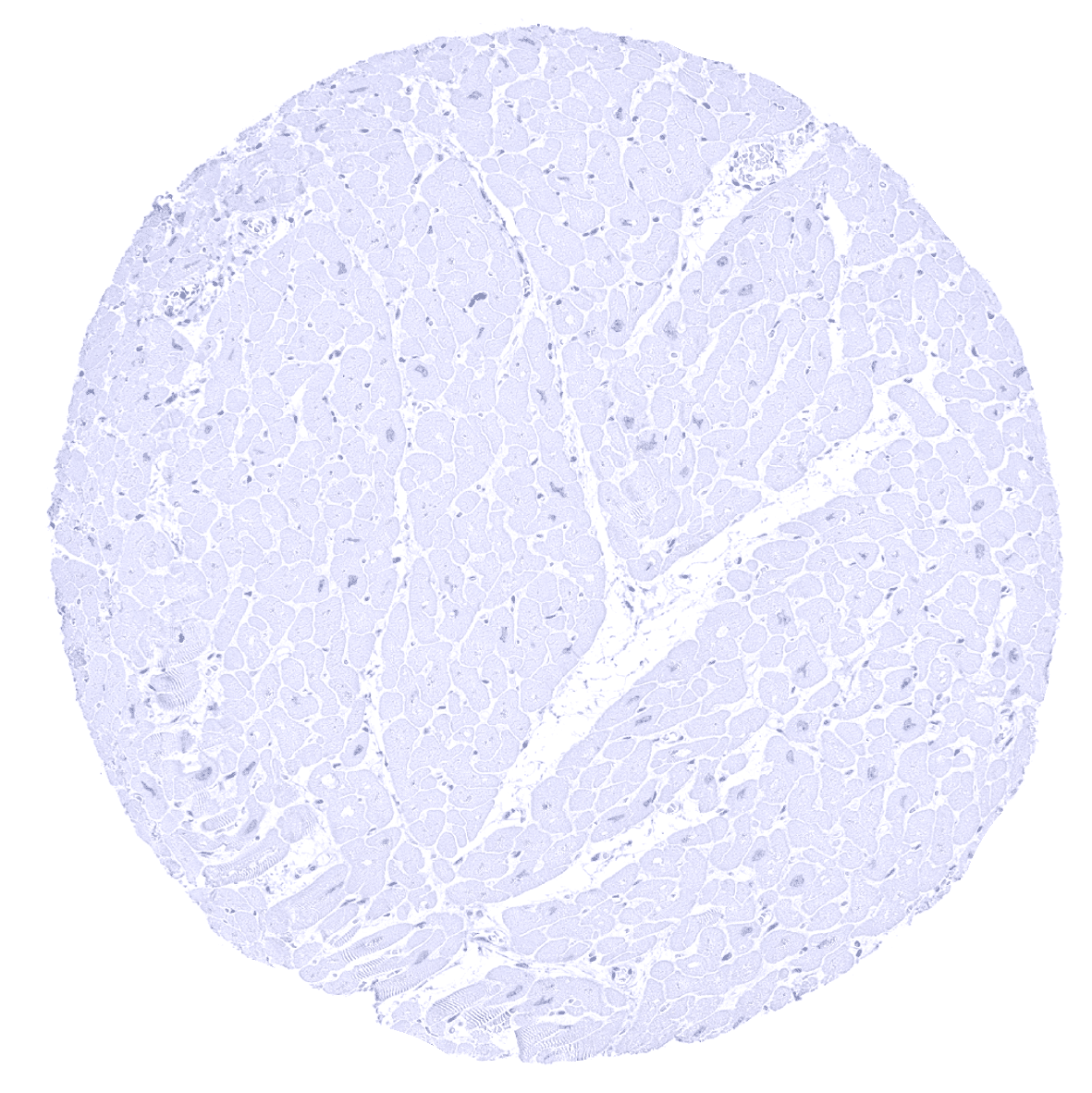
Striated muscle
Sublingual gland - A weak GATA3 positivity occurs in glandular cells of salivary glands.
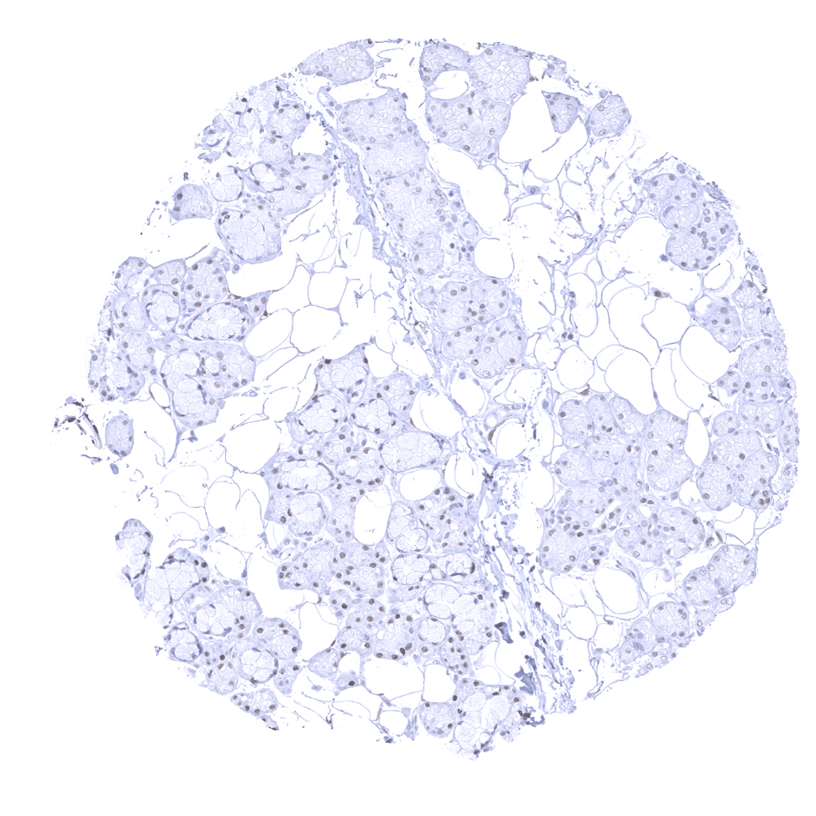
Submandibular gland - A weak GATA3 positivity occurs in glandular cells of salivary glands.
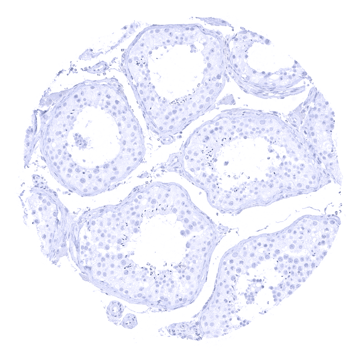
Testis

Thymus - GATA3 immunostaining is seen in a fraction of lymphocytes, especially in the thymus.
Thyroid gland

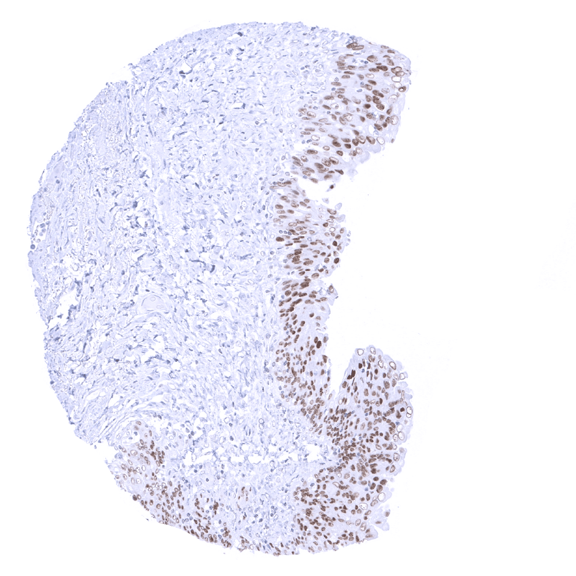
Tonsil, surface epithelium
Tonsil

Urinary bladder, muscular wall

Ovary, stroma
Urinary bladder, urothelium - A strong GATA3 positivity occurs in the urothelium.
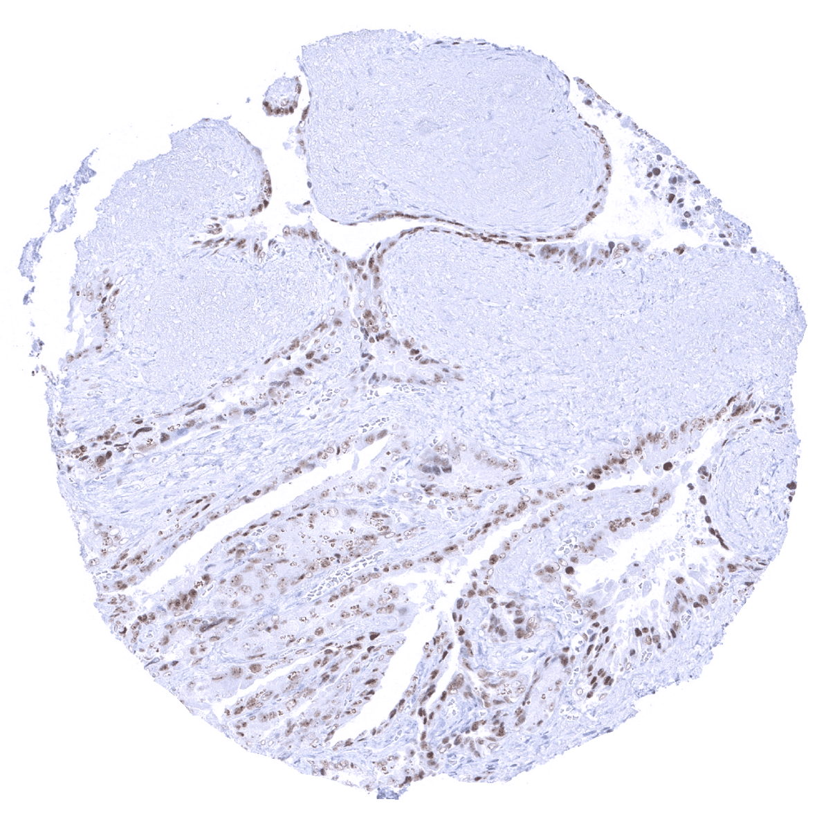
Uterus, myometrium