295,00 € – 895,00 €
Product details
Synonyms = 130kDa pemphigus vulgaris antigen (PVA); Balding (Bal); Cadherin family member 6 (CDHF6); Desmoglein-3 (DSG3)
Antibody type = Mouse monoclonal / IgG
Clone = MSVA-543M
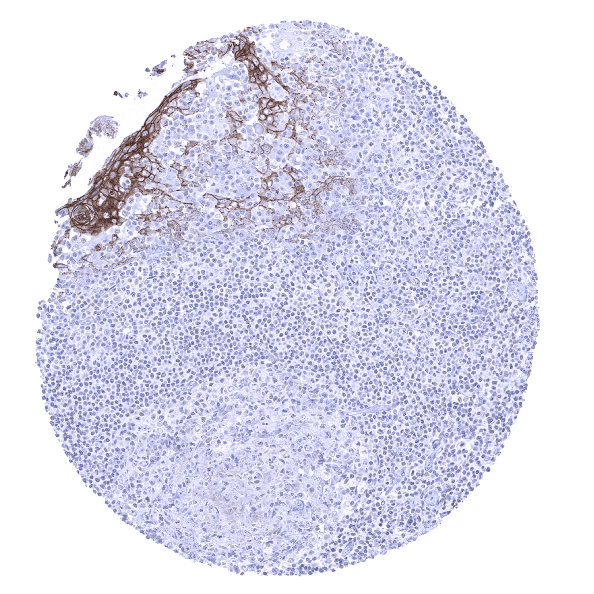
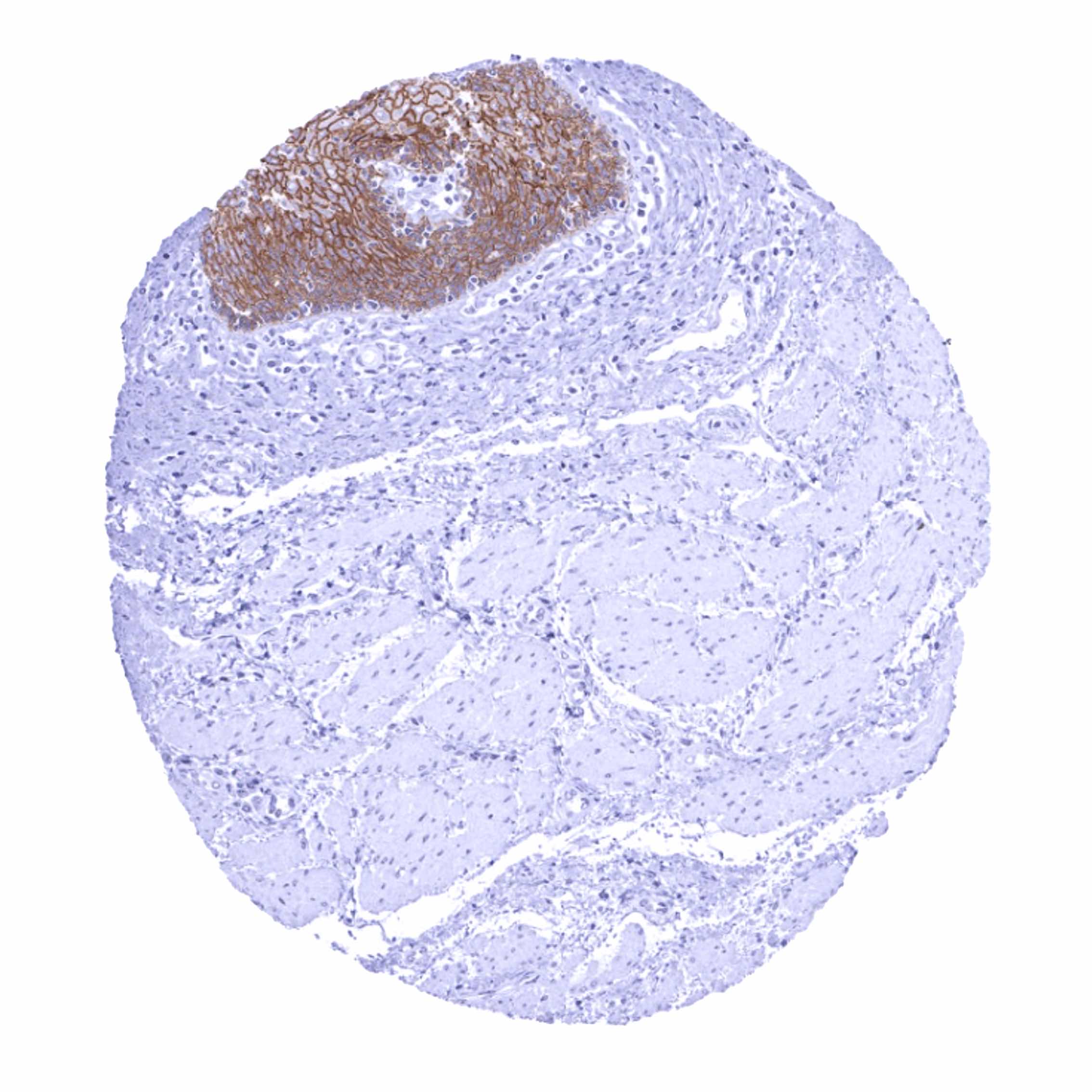
Positive control = Tonsil: A moderate to strong desmoglein 3 immunostaining should be seen in a fraction of squamous epithelial cells.
Negative control = Tonsil: Desmoglein 3 immunostaining should be absent in all non-squamous epithelial cell types.
Cellular localization = Cell Surface.
Reactivity = Human
Application = Immunohistochemistry
Dilution = 1:75 – 1:150
Intended Use = Research Use Only
Relevance of Antibody
Desmoglein 3 is expressed in squamous epithelium and cells undergoing squamous differentiation.
Biology Behind
Desmoglein-3 (Dsg3) is a 130 kDa calcium-binding transmembrane glycoprotein which is coded by the DSG3 gene at chromosome 18q12.1. Dsg3, a cadherin superfamily member, is an adhesion protein in desmosomes. In addition to its significant role in cell-cell adhesion, Dsg3 acts as a regulator of pathways governing proliferation, differentiation, and migration of keratinocytes. Because of its suppression of the function of p53, Dsg3 is considered a keratinocyte anti-stress protein. Dsg3 is down-regulated in pemphigus vulgaris where Dsg3 serves as a major antigen (PVA) for autoantibodies, causing disruption of cell–cell cohesion and pemphigus acantholysis in Dsg3-expressing tissues. Dsg3 has also been suggested to play a role in the progression and metastasis of cancer where it was found upregulated.
Staining Pattern in Normal Tissues
Desmoglein 3 staining pattern in Normal Tissues with antibody MSVA-543M (images are shown in our “Normal Tissue Gallery”)
| Brain | Cerebrum | Negative. |
| Cerebellum | Negative. | |
| Endocrine Tissues | Thyroid | Negative. |
| Parathyroid | Negative. | |
| Adrenal gland | Negative. | |
| Pituitary gland | Negative. | |
| Respiratory system | Respiratory epithelium | Weak to moderate Dsg3 staining of a subset of respiratory epithelial cells. |
| Lung | Negative. | |
| Gastrointestinal Tract | Salivary glands | Weak to moderate Dsg3 staining in some basal cell layers of small excretory ducts. |
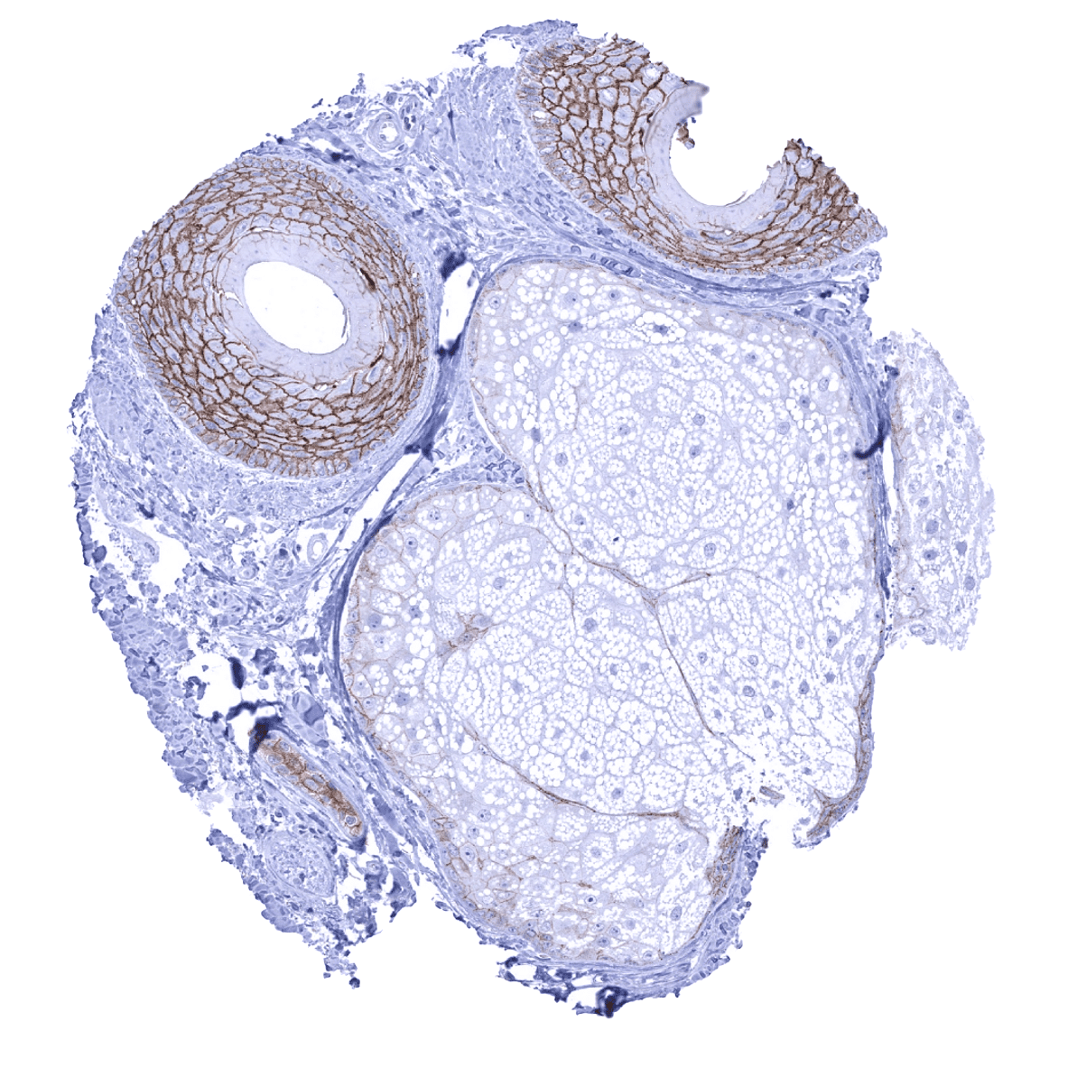
| Esophagus | Moderate to strong membranous Dsg3 staining of squamous epithelial cells. The staining intensity decreases towards the surface of the epithelium. | |
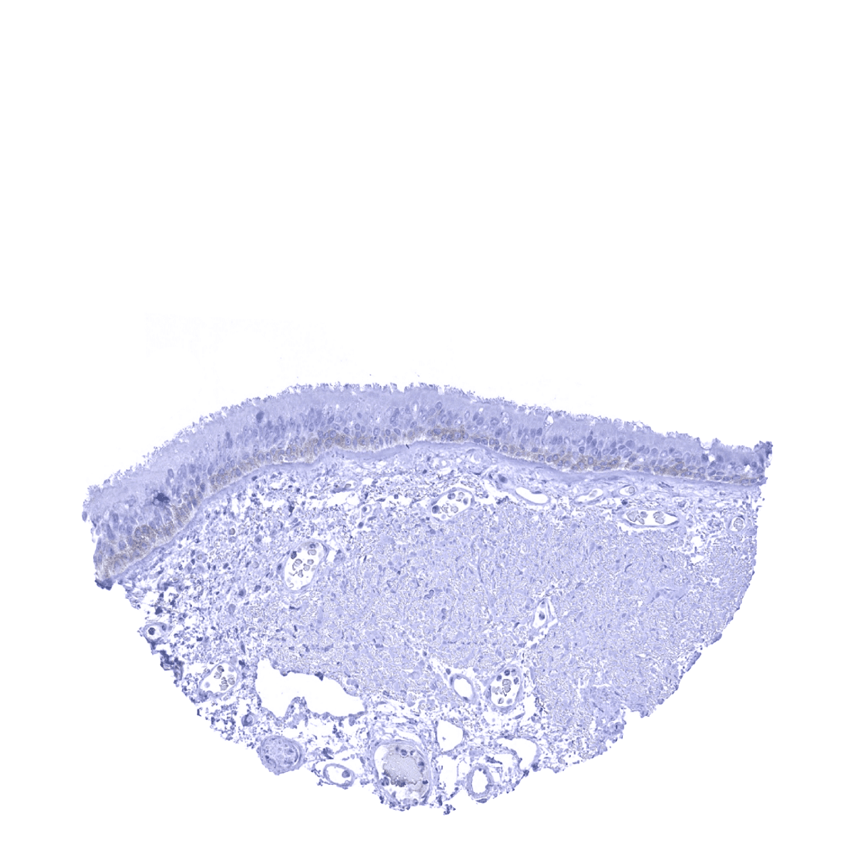
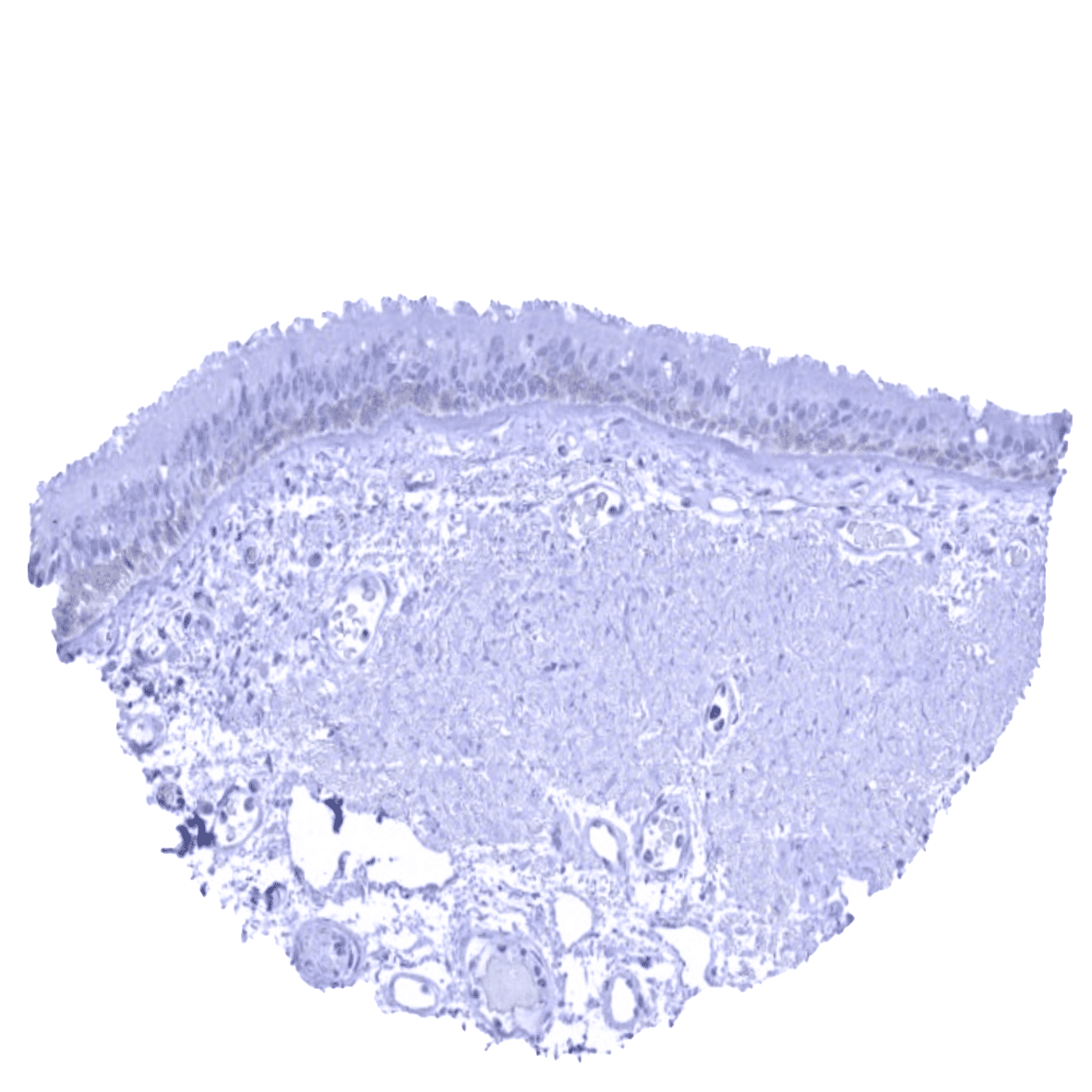
| Stomach | Negative. | |
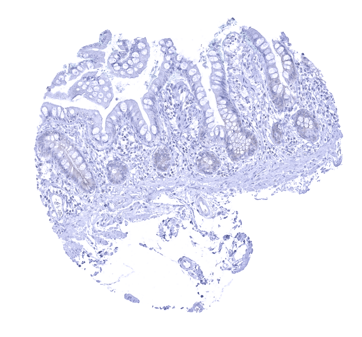
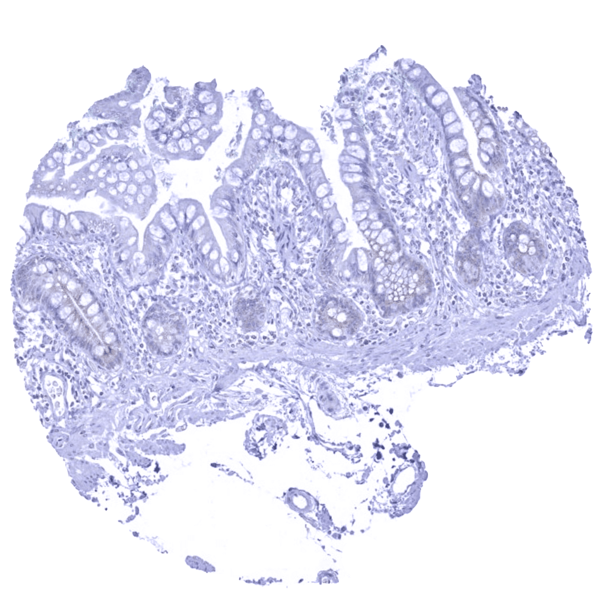
| Duodenum | Weak to moderate apical membranous staining of a subset crypt epithelial cells. | |
| Small intestine | Weak to moderate apical membranous staining of a subset crypt epithelial cells. | |
| Appendix | Negative. | |
| Colon | Negative. | |
| Rectum | Negative. | |
| Liver | Negative. | |
| Gallbladder | Negative. | |
| Pancreas | Negative. | |
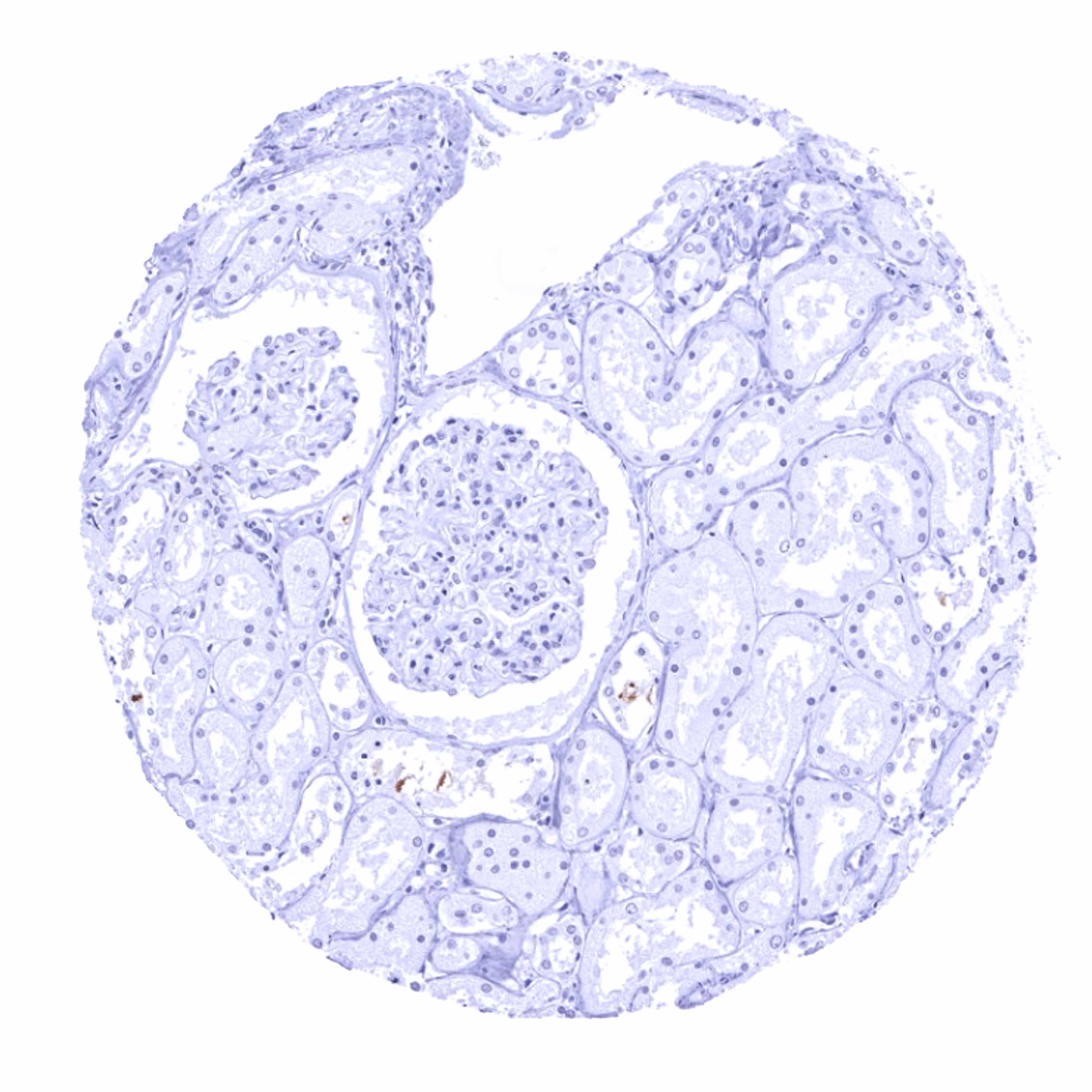
| Genitourinary | Kidney | Negative. |
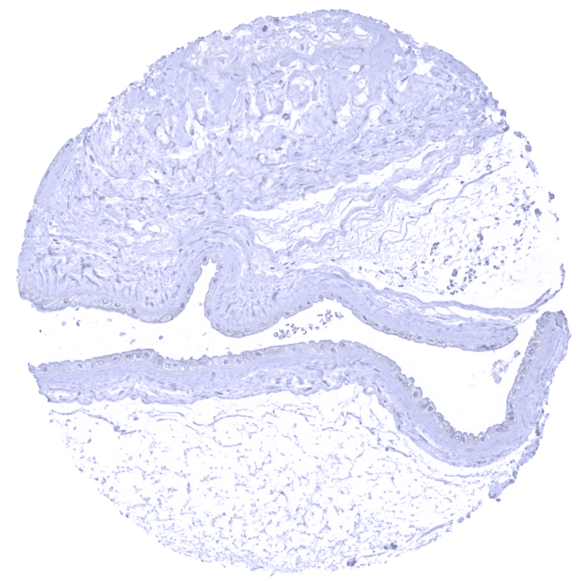
| Urothelium | Usually negative. Some Dsg3 staining of basal cell layers was observed in case of inflammation. | |
| Male genital | Prostate | Negative. |
| Seminal vesicles | Negative. | |
| Testis | Negative. | |
| Epididymis | Negative. | |
| Female genital | Breast | Negative. |
| Uterus, myometrium | Negative. | |
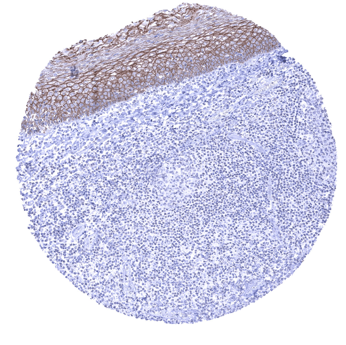
| Uterus, ectocervix | Moderate to strong membranous Dsg3 staining of squamous epithelial cells. The staining intensity decreases towards the surface of the epithelium. | |
| Uterus endocervix | Negative. | |
| Uterus, endometrium | Negative. | |
| Fallopian Tube | Negative. | |
| Ovary | Negative. | |
| Placenta early | Negative. | |
| Placenta mature | Negative. | |
| Amnion | Low level Dsg3 expression may occur in amnion cells. | |
| Chorion | Negative. | |
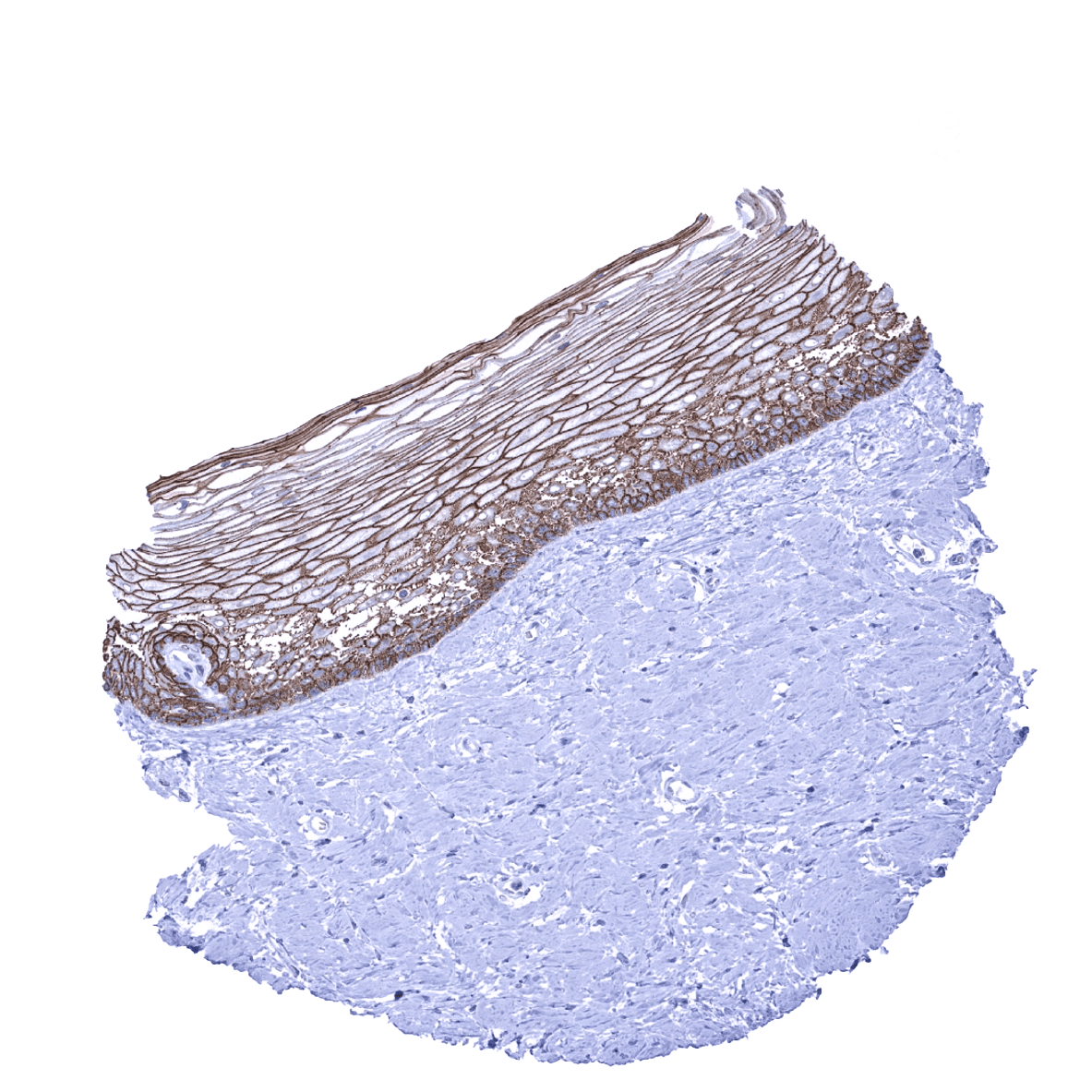
| Skin | Epidermis | Moderate to strong membranous Dsg3 staining of squamous epithelial cells. The staining intensity markedly decreases towards the surface of the epithelium. The top 50% of the epithelium is often Dsg3 negative. |
| Sebaceous glands | Moderate to strong membranous Dsg3 staining. | |
| Muscle/connective tissue | Heart muscle | Negative. |
| Skeletal muscle | Negative. | |
| Smooth muscle | Negative. | |
| Vessel walls | Negative. | |
| Fat | Negative. | |
| Stroma | Negative. | |
| Endothelium | Negative. | |
| Bone marrow/lymphoid tissue | Bone marrow | Negative. |
| Lymph node | Negative. | |
| Spleen | Negative. | |
| Thymus | Weak to moderate membranous Dsg3 staining of corpuscles of Hassall’s and (weaker) also of other thymic epithelial cells. | |
| Tonsil | Moderate to strong membranous Dsg3 staining of squamous epithelial cells. Only a fraction of cells are stained in the crypts. | |
| Remarks |
These findings are largely consistent with the RNA and protein data described in the Human Protein Atlas (Tissue expression Desmoglein 3)
Positive control: Tonsil: A moderate to strong desmoglein 3 immunostaining should be seen in a fraction of squamous epithelial cells.
Negative control: Tonsil: Desmoglein 3 immunostaining should be absent in all non-squamous epithelial cell types.
Staining Pattern in Relevant Tumor Types
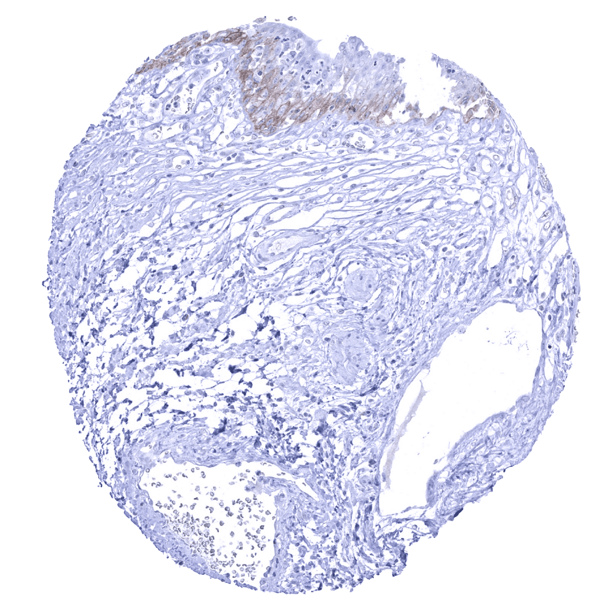
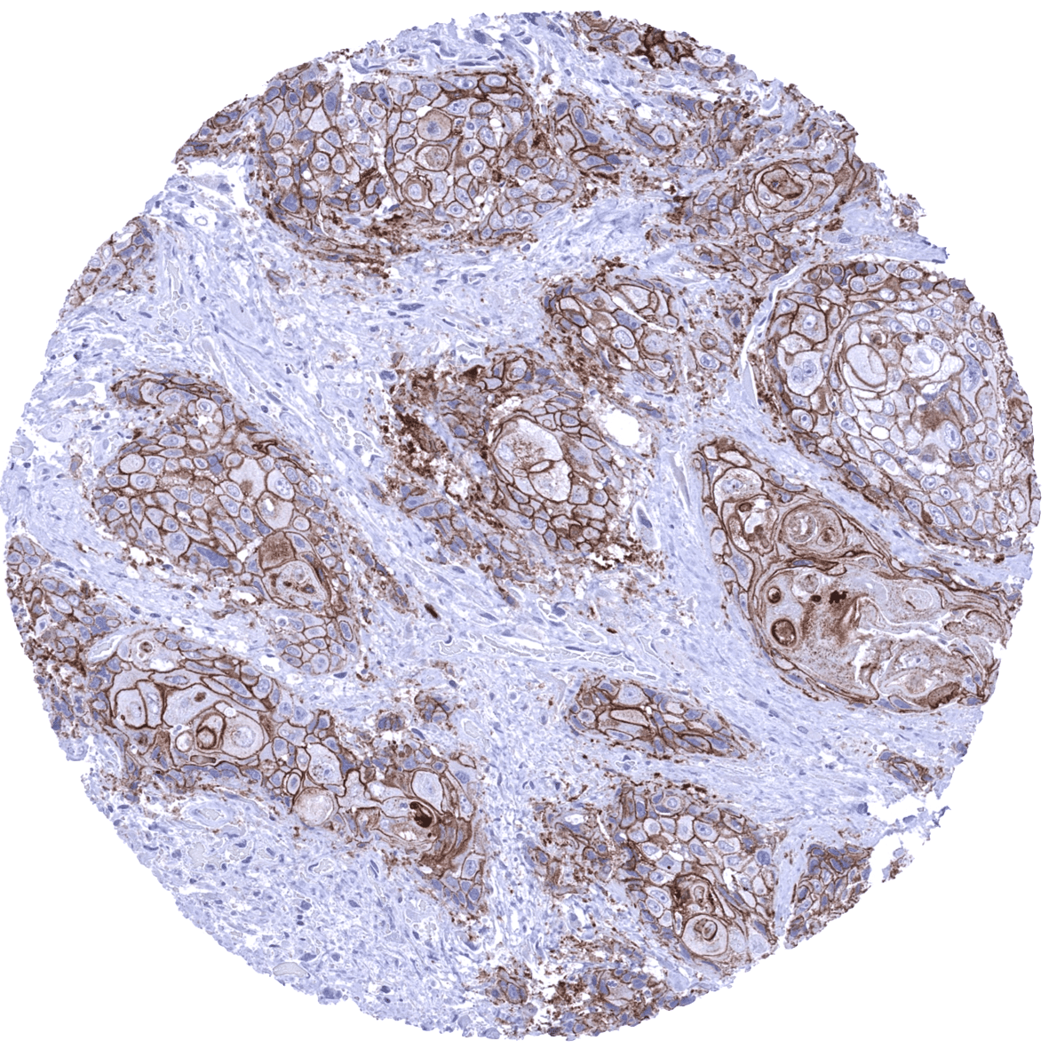
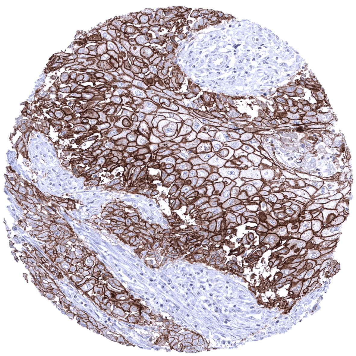
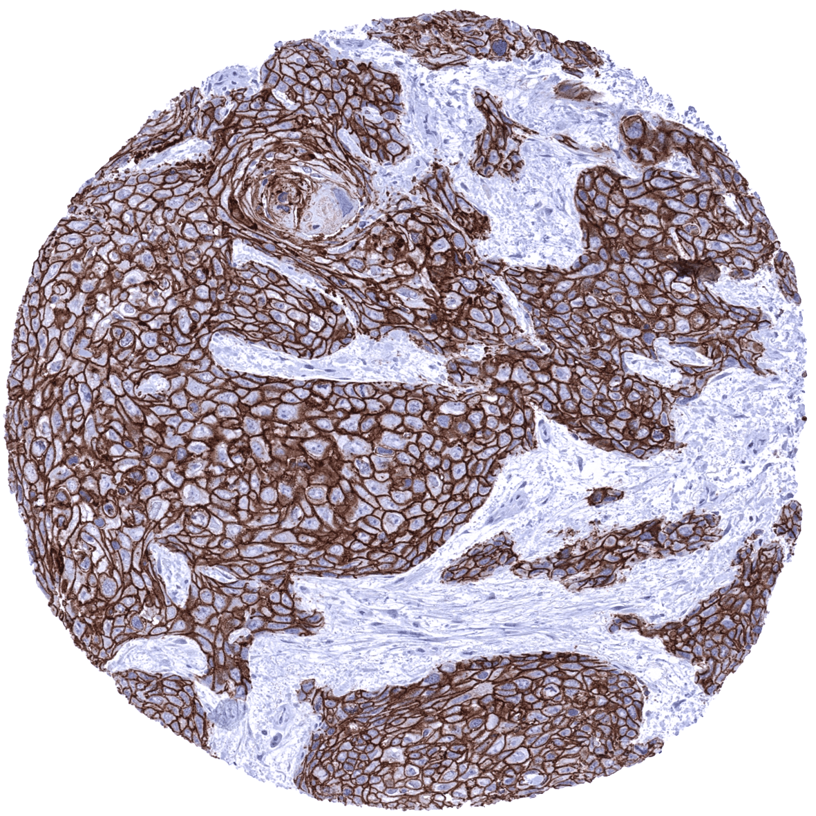
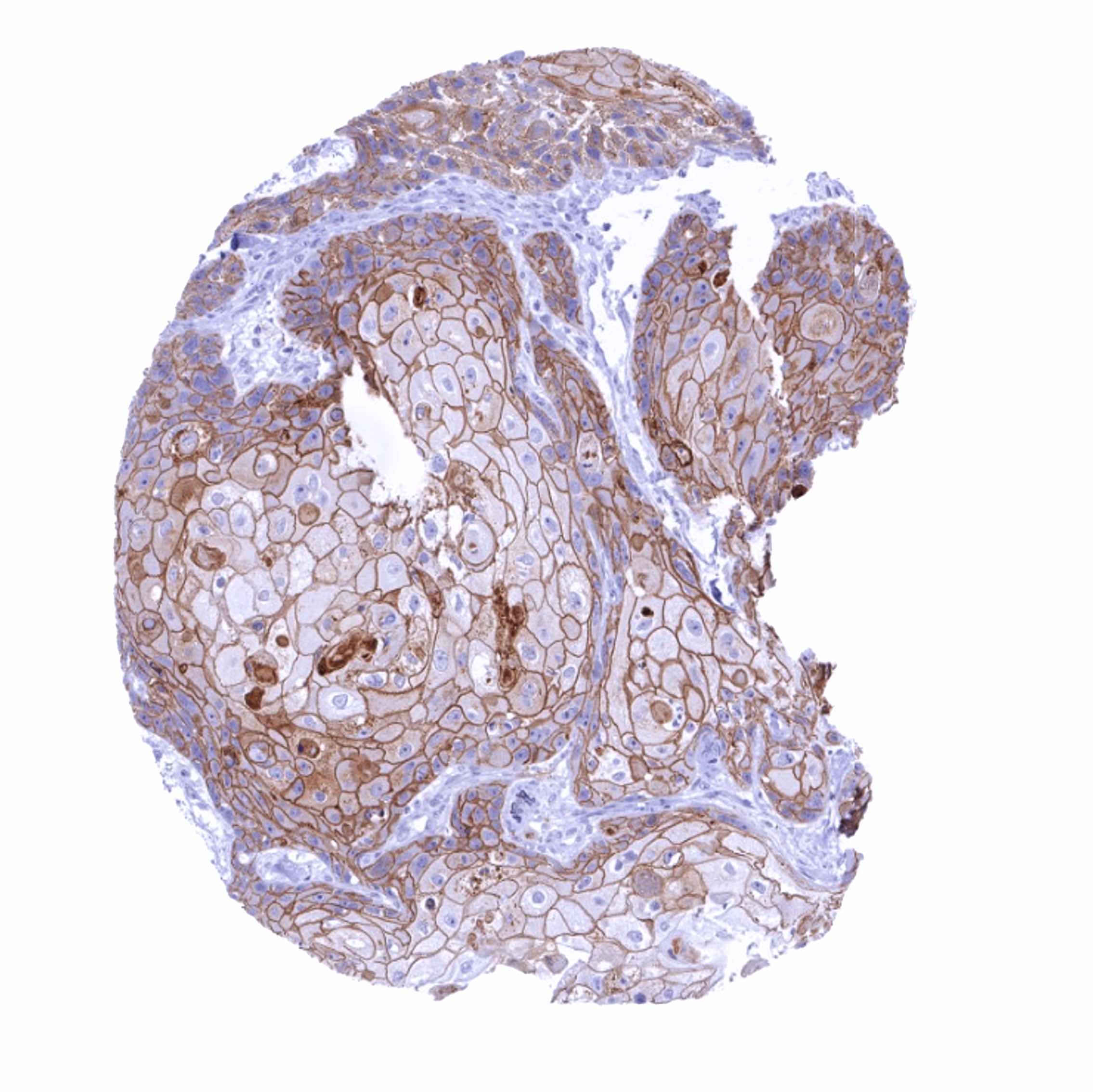
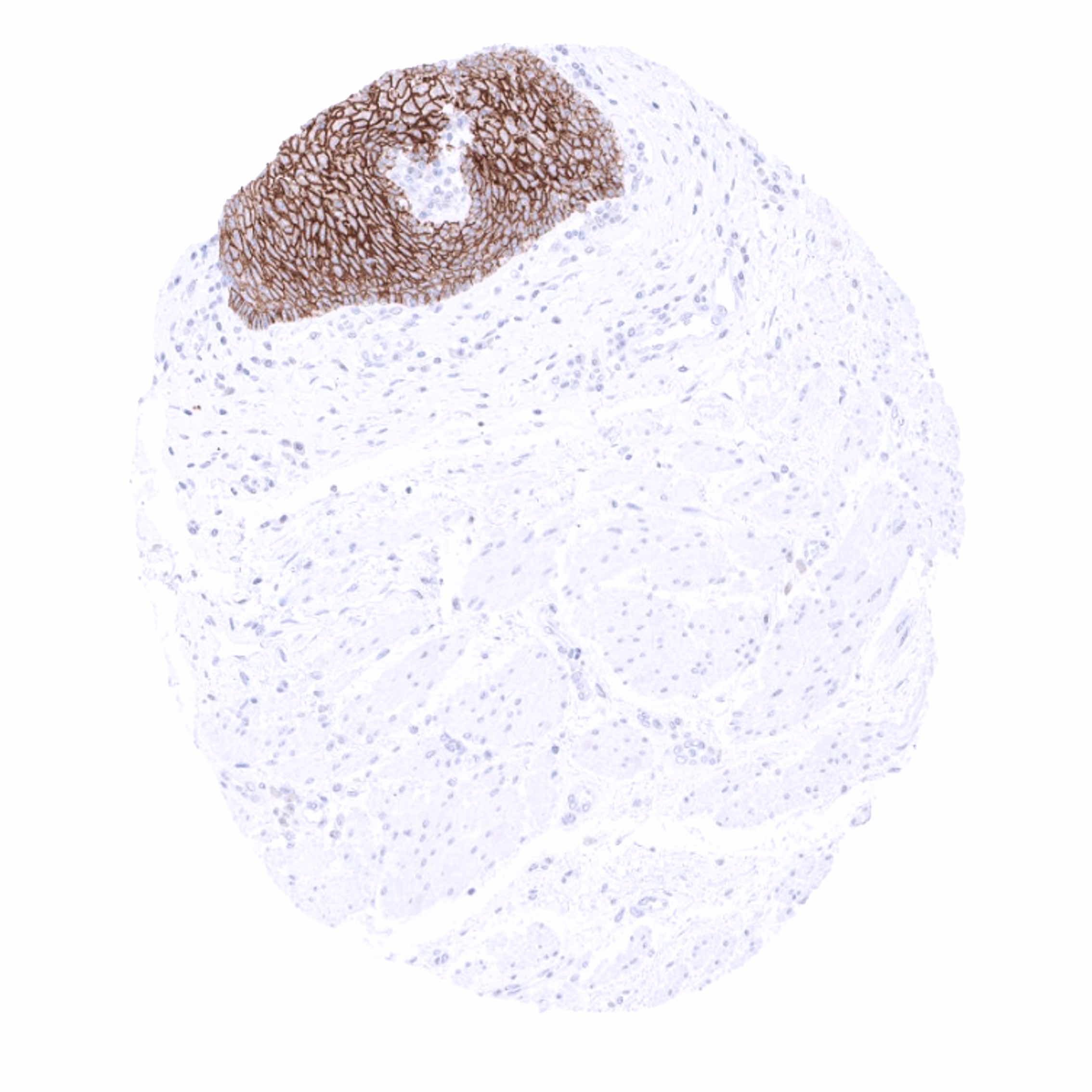
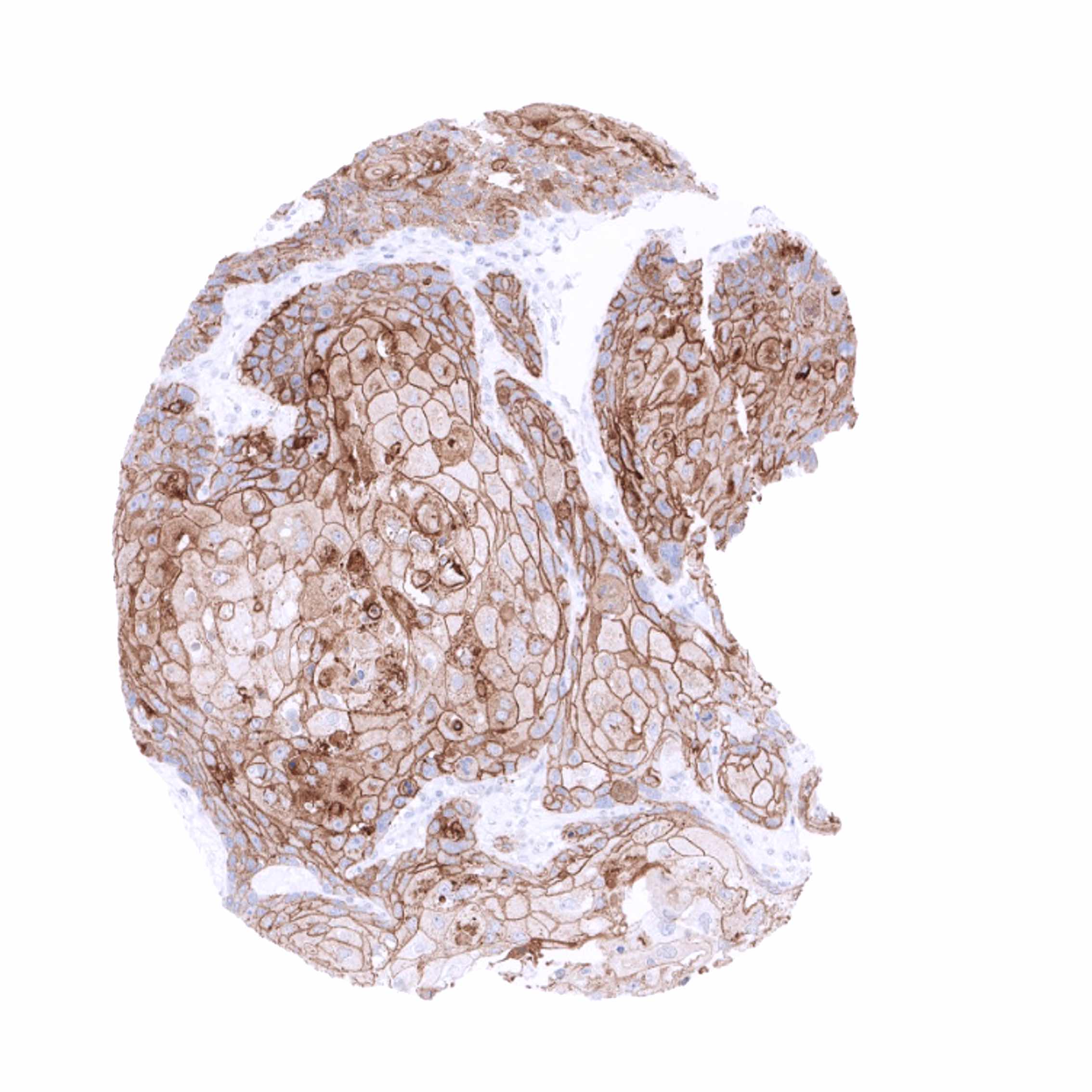
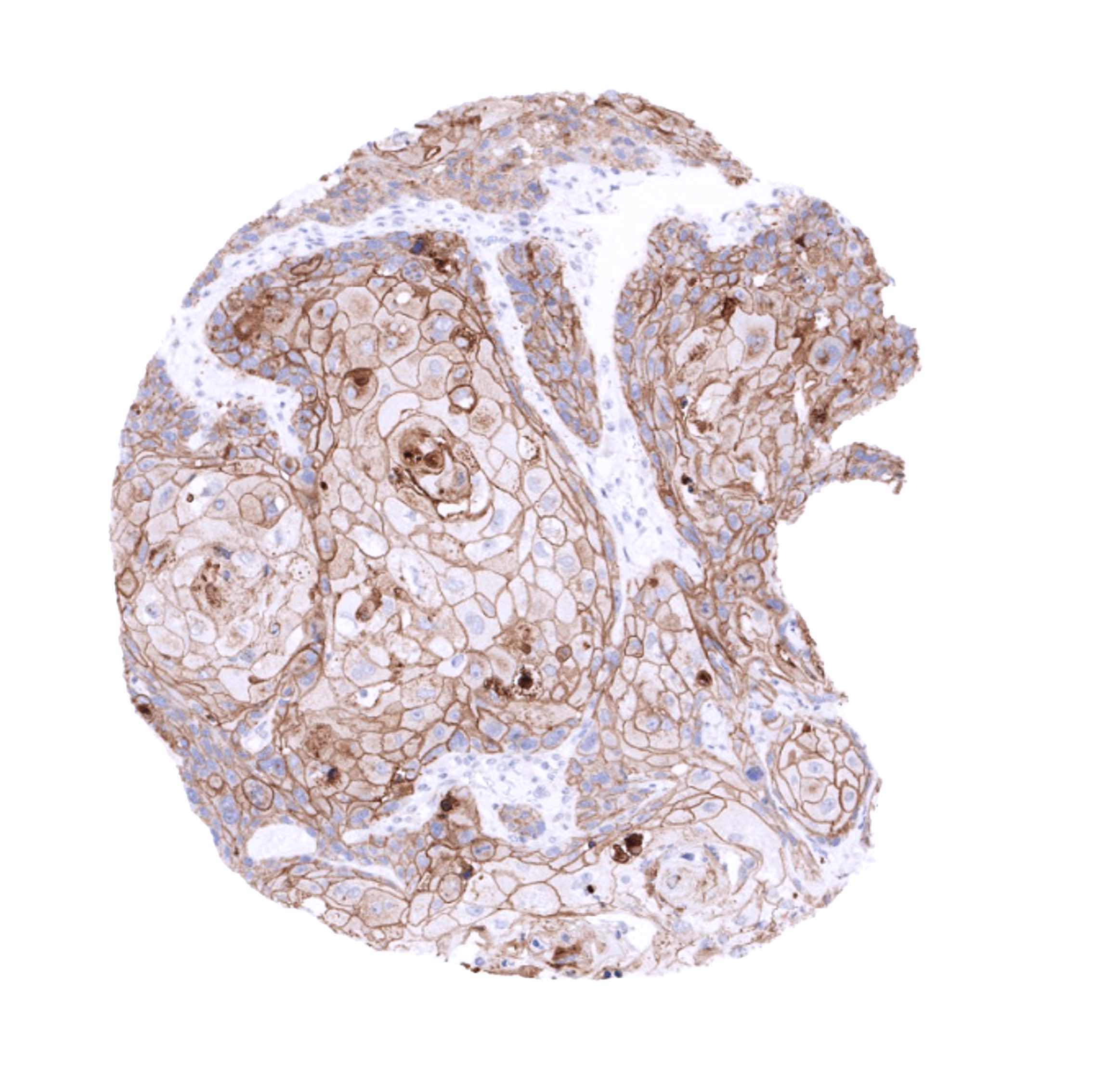
A positive desmoglein 3 immunostaining is usually seen in squamous cell carcinomas irrespective of their site of origin and in tumor areas exhibiting squamous differentiation. Dsg3 immunostaining can also be seen in various other tumor entities including urothelial cancer, gastric cancer, pancreatic adenocarcinoma, and endometrioid carcinoma. It for example occurs in areas of squamous differentiation.
The TCGA findings on Desmoglein 3 RNA expression in different tumor categories have been summarized in the Human Protein Atlas.
Compatibility of Antibodies
Desmoglein 3 (MSVA-543M) publication summary
Relevant publication: Viehweger et al. “Desmoglein 3 (Dsg3) expression in cancer: A tissue microarray study on 15,869 tumors” Published in Pathology – Research and Practice 2022 Dec;240:154200. PMID: 36375372.
A total of 14’094 tumors from 137 different tumor categories were successfully analyzed by using the following protocol: Heat-induced antigen retrieval for 5 minutes in an autoclave at 121°C in pH 7,8 Target Retrieval Solution buffer. MSVA-543M at a dilution of 1:150 at 37°C for 60 minutes. Visualization of bound antibody by the EnVision Kit (Dako, Agilent). This protocol was also used for all stainings depicted in our tumor and normal tissue galleries.
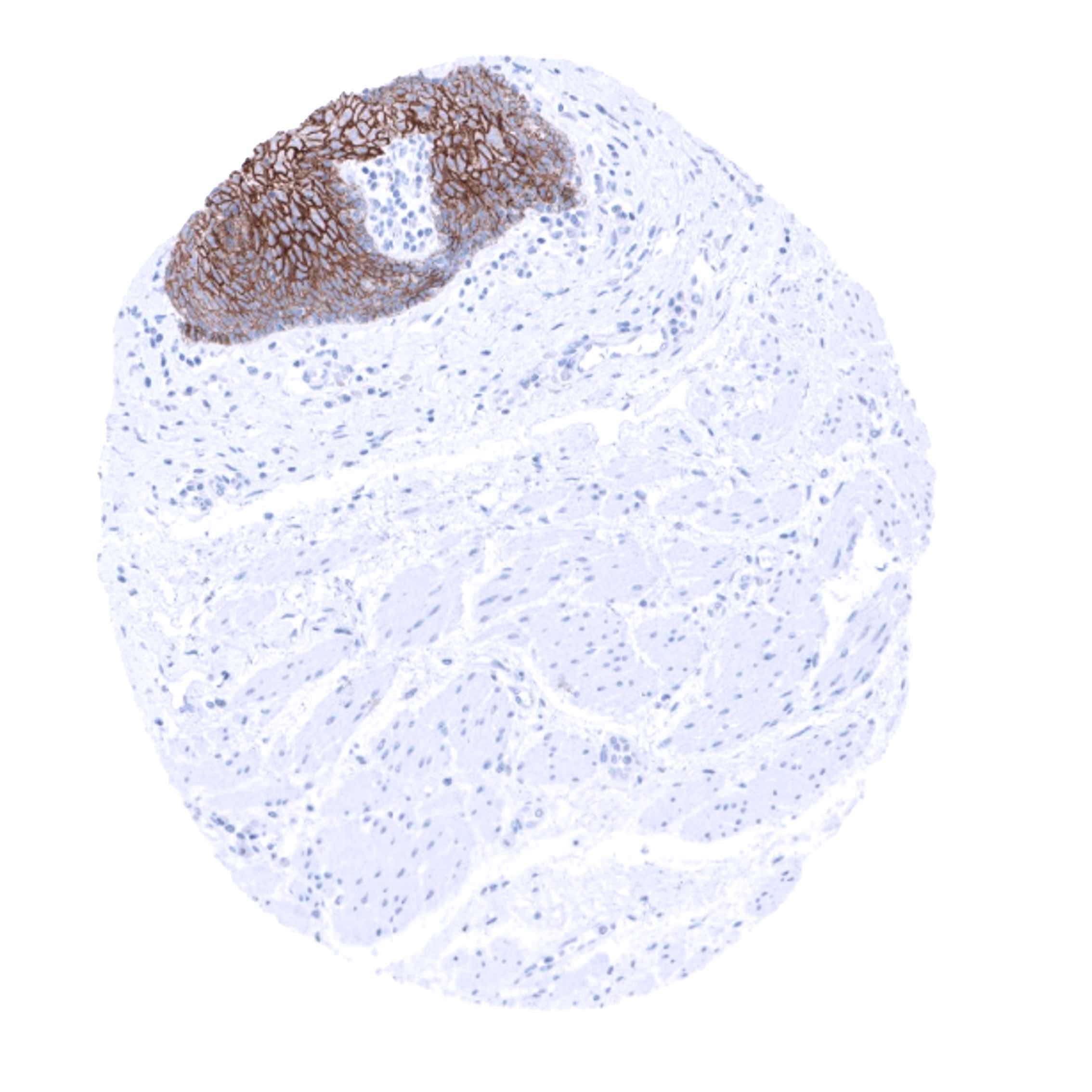
Overall, 47 (34,3%) of 137 tumor categories showed detectable Dsg3 expression in at least one case and 15 (10,9%) tumor categories included at least one case with strong Dsg3 positivity. The highest rate of Dsg3 staining occurred in squamous cell carcinomas from various sites of origin (positive in 71.2–97.3 %), basal cell carcinomas of the skin (41.9 %), various benign tumors, mainly derived from salivary glands (8.0–38.9%) as well as in urothelial tumors (2.1–20.7 %). A Dsg3 positivity in less than 10% of cases occurred in 23 additional tumor categories. Dsg3 staining was almost always weak and rarely moderate in tumors that were not derived from squamous epithelium. In many cases, focal Dsg3 staining appeared in areas of aberrant squamous differentiation, but Dsg3 expression could also be seen in tumors without features of squamous differentiation. The distribution of positive staining results is shown in an “organ-systematic” (Figure 1) and in a “ranking order” figure (Figure 2) below (images based on data from Viehweger et al.). Data on associations with histopathological and clinical parameters of tumor aggressiveness in several cancer types are also summarized below (Figure 3; based on data described by Viehweger et al.).
Authors conclusions on diagnostic utility of Dsg3 IHC with respect to the distinction of different tumor entities (Viehweger et al.):
- Dsg3 is a marker for squamous cell differentiation. This may be diagnostically used for the distinction of squamous cell carcinomas of the lung and the identification of (focal) squamous differentiation in urothelial neoplasms as well as in other cancers.
Authors conclusions on the prognostic role of Dsg3 immunostaining results (Viehweger et al.):
- Detectable Dsg3 expression was linked to tumor invasiveness in urothelial cancer (p<0,0001).
- Dsg3 positivity was associated with advanced pT stage (p=0,0102), nodal metastasis (p=0,0162), L1 (p=0,0151), V1 (p=0,0189), and MSI (p=0,0023) in colorectal cancer.
Figure 1. Desmoglein 3 staining in cancer (“organ-systematic”; according to Viehweger et al.)
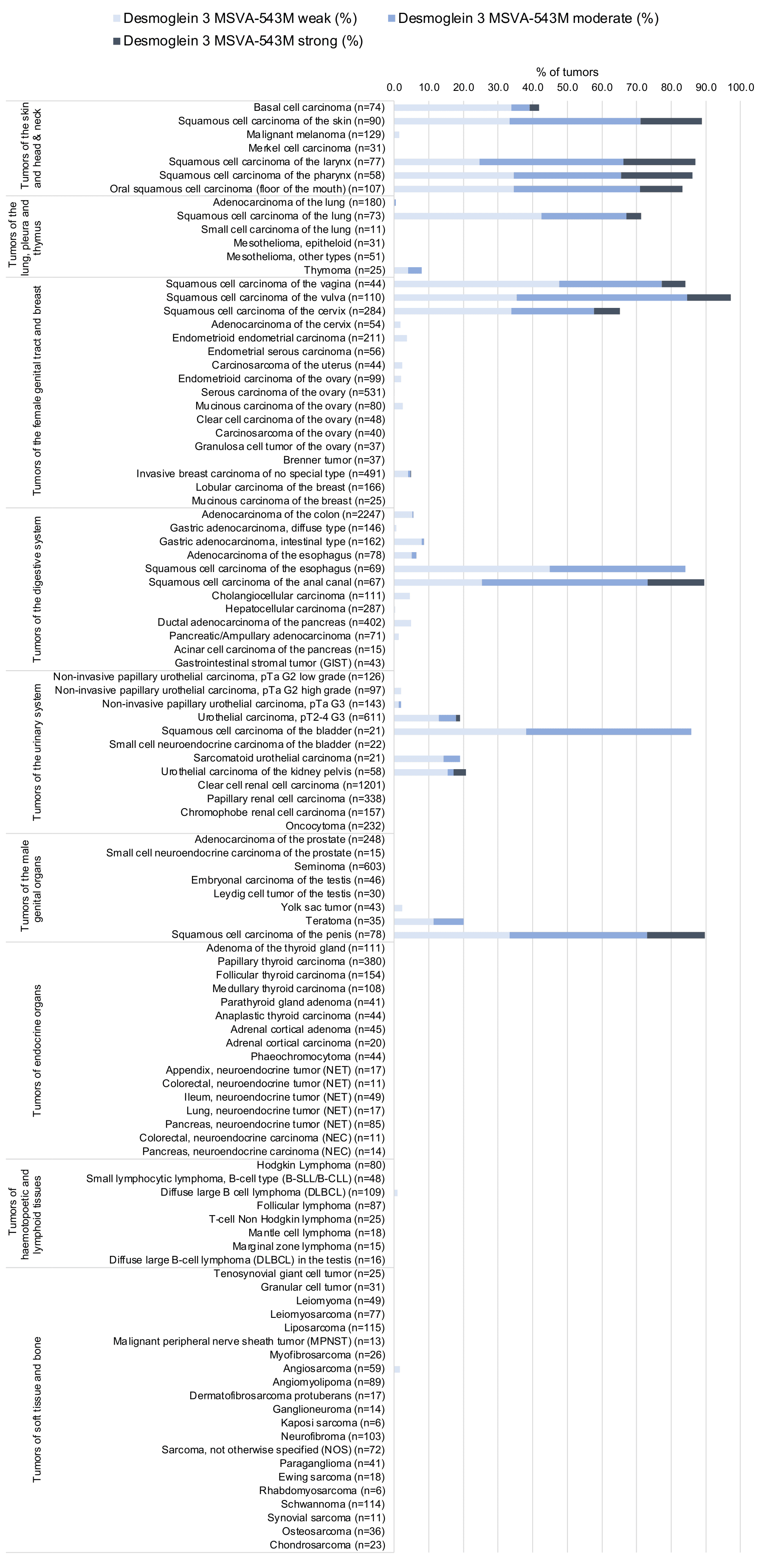
Figure 2. Desmoglein 3 staining in cancer (“ranking list”; according to Viehweger et al.)

Protocol Recommendations
IHC users have different preferences on how the stains should look like. Some prefer high staining intensity of the target stain and even accept some background. Others favor absolute specificity and lighter target stains. Factors that invariably lead to more intense staining include higher concentration of the antibody and visualization tools, longer incubation time, higher temperature during incubation, higher temperature and longer duration of the heat induced epitope retrieval (slide pretreatment). The impact of the pH during slide pretreatment has variable effects and depends on the antibody and the target protein.
All images and data shown here and in our image galleries are obtained by the manual protocol described below. Other protocols resulting in equivalent staining are described as well.
Manual protocol
Freshly cut sections should be used (less than 10 days between cutting and staining). Heat-induced antigen retrieval for 5 minutes in an autoclave at 121°C in pH 7,8 Target Retrieval Solution buffer. Apply MSVA-543M at a dilution of 1:100 at 37°C for 60 minutes. Visualization of bound antibody by the EnVision Kit (Dako, Agilent) according to the manufacturer’s directions.
Agilent / Dako – Autostainer Link 48
Pretreatment in PT-Link for 30 minutes at 95°C (pH high); FLEX peroxidase blocking for 5 minutes (room temperature), MSVA-543M 1:100 for 20 minutes (room temperature), FLEX+ mouse/rabbit (LINKER) for 15 minutes (room temperature), horseradish peroxidase (HRP) for 20 minutes (room temperature), FLEX DAB+Sub-Chromo for 10 minutes (room temperature), FLEX hematoxylin for 5 minutes (room temperature).

These images reflect stainings by the protocol described above. It is of note that a comparable staining result can also be obtained by different protocols. In general, a longer pretreatment, a longer incubation time of the primary antibody, a higher antibody concentration, and a longer incubation time of FLEX+LINKER result in stronger staining, potentially at the cost of more background staining. Modifications of the protocol with a strengthening effect on staining intensity in combination with changes of other parameters that result in lower staining intensity can result in a comparable result as shown above.
Leica – BOND RX
Dewax at 72°C for 30 seconds; Pretreatment in Bond Epitope Retrieval Solution (ER2 – EDTA pH9) for 20 minutes at 100°C; Peroxidase blocking for 5 minutes (room temperature), MSVA-543M 1:100 for 15 minutes (room temperature), Post primary (rabbit anti mouse) for 8 minutes (room temperature), Polymer (goat anti rabbit) for 8 minutes (room temperature), mixed DAB refine for 10 minutes (room temperature), hematoxylin for 5 minutes (room temperature).


These images reflect stainings by the protocol described above. It is of note that a comparable staining result can also be obtained by different protocols. In general, a longer pretreatment, a longer incubation time of the primary antibody, a higher antibody concentration, a higher temperature during incubation, and a longer incubation time of Post primary and or the Polymer result in stronger staining, potentially at the cost of more background staining. Modifications of the protocol with a strengthening effect on staining intensity in combination with changes of other parameters that result in lower staining intensity can result in a comparable result as shown above.
Roche – Ventana Discovery ULTRA
Pretreatment for 64 minutes at 100°C (pH 8,4); CM peroxidase blocking for 12 minutes (room temperature), MSVA-543M 1:100 for 20 minutes at 36°C, secondary antibody (anti-mouse HQ) for 12 minutes at 36°C, anti-HQ HRP for 12 minutes at room temperature, DAB at room temperature, hematoxylin II at room temperature for 8 minutes, bluing reagent at room temperature for 4 minutes.


These images depict staining results obtained by the protocol described above. It is of note, that the Ventana machines generally require higher antibody concentrations than other commonly used autostainers because the antibodies are automatically diluted during the procedure. Various other protocols can result in an identical result as shown above. A longer pretreatment, a longer incubation time of the primary antibody, a higher antibody concentration, a higher temperature during incubation, and a longer incubation time of secondary antibody and or the anti-HQ HRP result in stronger staining, potentially at the cost of more background staining.
Potential Research Applications
- The diagnostic utility of desmoglein 3 IHC should be investigated in a large cohort of tumors from different entities.
- Desmoglein 3 has been suggested as a prognostic parameter in several different cancer types. This subject deserves further investigation.
Evidence for Antibody Specificity in IHC
There are two ways how the specificity of antibodies can be documented for immunohistochemistry on formalin fixed tissues. These are: 1. comparison with a second independent method for target expression measurement across a large number of different tissue types (orthogonal strategy), and 2. Comparison with one or several independent antibodies for the same target and showing that all positive staining results are also seen with other antibodies for the same target (independent antibody strategy).
Orthogonal validation: Data derived from three independent RNA screening studies, including the Human Protein Atlas (HPA) RNA-seq tissue dataset, the FANTOM5 project, and the Genotype-Tissue Expression (GTEx) project, which are all summarized in the Human Protein Atlas (Tissue expression Desmoglein 3) suggested that desmoglein 3 expression only occurs in tissues that are covered by squamous epithelium. This fits very well with protein expression data obtained by MSVA-543M. In addition, a weak to moderate desmoglein 3 immunostaining was seen in amnion cells of the placenta and (only in some samples) in basal or suprabasal urothelial cells, basal cells of respiratory epithelium and crypt base cells of the duodenum and the ileum. Given the paucity of these cell types in their respective organs, it is very likely, that desmoglein 3 expression can be missed in RNA analyses. Because desmoglein 3 and desmoglein 1 are usually co-expressed, true expression of desmoglein 3 in amnion cells of the placenta is indirectly suggested by the observation of desmoglein 1 expression in amnion cells (data shown in MSVA-544M Desmoglein 1).
Comparison of antibodies: True expression of desmoglein 3 in amnion cells of the placenta and (only in some samples) in basal or suprabasal urothelial cells, basal cells of respiratory epithelium and crypt base cells of the duodenum and the ileum was further suggested by comparable findings obtained by a second independent antibody (MSVA-C. Dsg3) tested in comparison with MSVA-543M (images are shown below).