
295,00 € – 995,00 €
Product details
Synonyms = K1B; KRT1B; Keratin, type II cytoskeletal 1b; K77; CK-1B; Keratin 1B; Keratin-77; Cytokeratin-1B; Type-II Keratin Kb39
Antibody type = Recombinant Rabbit monoclonal / IgG
Clone = MSVA-000R
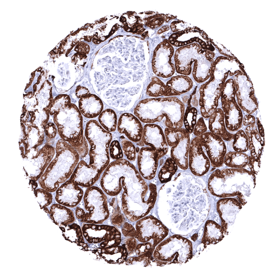
Positive control = Liver: A strong staining of all bile duct epithelial cells and an at least moderate, predominantly membranous staining of most hepatocytes should be seen.
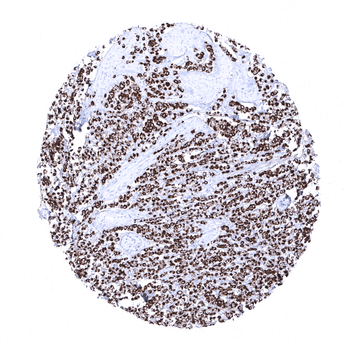
Negative control = Tonsil: All lymphocytes must not stain. However, interstitial reticulum cells with dendritic/reticular pattern show a weak to moderate fibrillar staining.
Cellular localization = Cytoplasmic
Reactivity = Human
Application = Immunohistochemistry
Dilution = 1:100 – 1:200
Intended Use = Research Use Only
Relevance of Antibody
Pan-cytokeratin (CKpan) is an antibody cocktail to be used for staining as many as possible cells of epithelial origin.
Biology Behind
“Pan-cytokeratin” (CKpan) is an antibody cocktail that contains most known cytokeratins and thus binds to almost all human epithelial and mesothelial cells. CKpan cocktails are broadly used in diagnostic immunohistochemistry for securing the epithelial nature of cells with equivocal morphology and visualization of cancer cells that are difficult to see in routine stains in pathology.
Staining Pattern in Normal Tissues
“Pan Cytokeratin” staining pattern in Normal Tissues with antibody MSVA-000R (images are shown in our “Normal Tissue Gallery”)
| Brain | Cerebrum | Negative. |
| Cerebellum | Negative. | |
| Endocrine Tissues | Thyroid | Strong CKpan positivity of all epithelial cells. |
| Parathyroid | Strong CKpan positivity of all epithelial cells. | |
| Adrenal gland | Weak to moderate staining of only a fraction of cells which are typically grouped together. | |
| Pituitary gland | Strong CKpan positivity of all epithelial cells of the adenohypophysis. | |
| Respiratory system | Respiratory epithelium | Strong CKpan positivity of all epithelial cells. |
| Lung | “Only” moderate to strong staining in pneumocytes. | |
| Gastrointestinal Tract | Salivary glands | Strong CKpan positivity of all epithelial cells. |
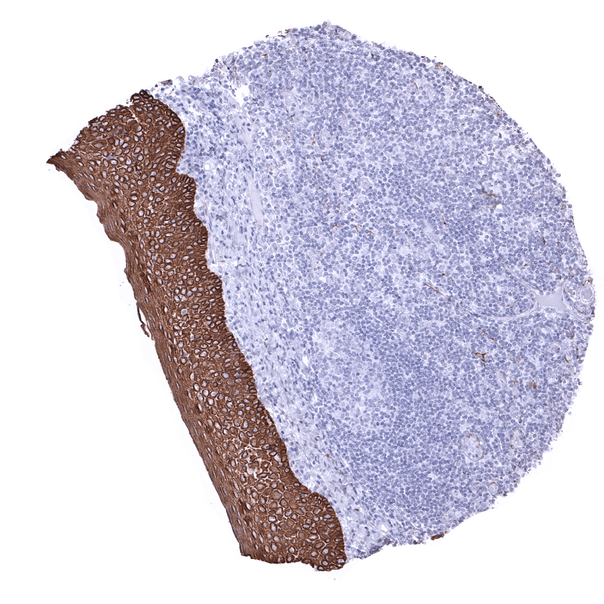
| Esophagus | Strong CKpan positivity of all epithelial cells. | |
| Stomach | Strong CKpan positivity of all epithelial cells. | |
| Duodenum | Strong CKpan positivity of all epithelial cells. | |
| Small intestine | Strong CKpan positivity of all epithelial cells. | |
| Appendix | Strong CKpan positivity of all epithelial cells. | |
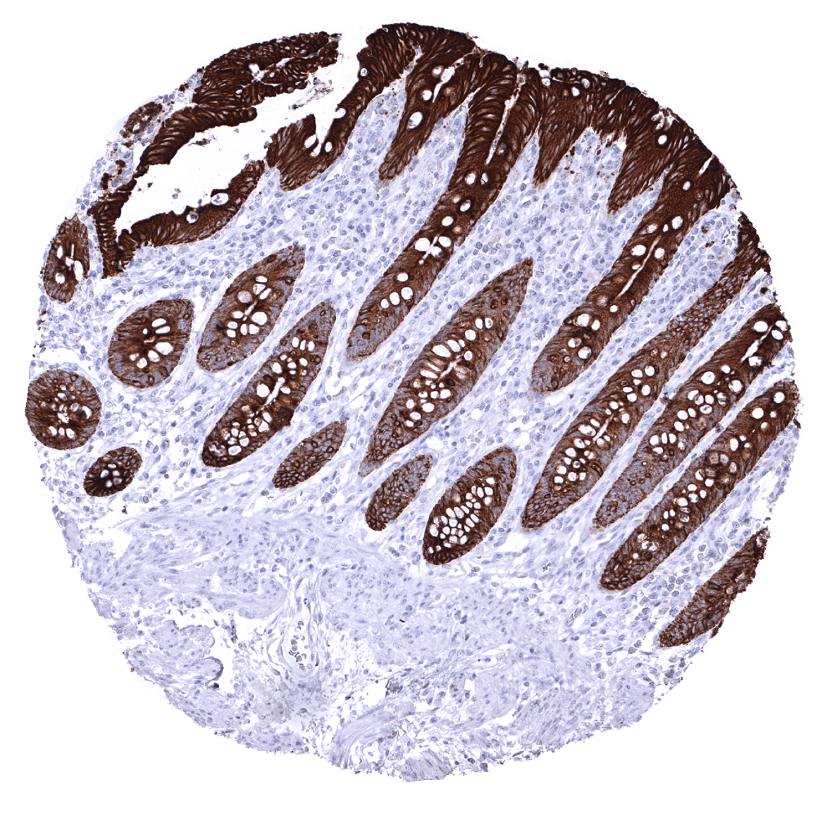
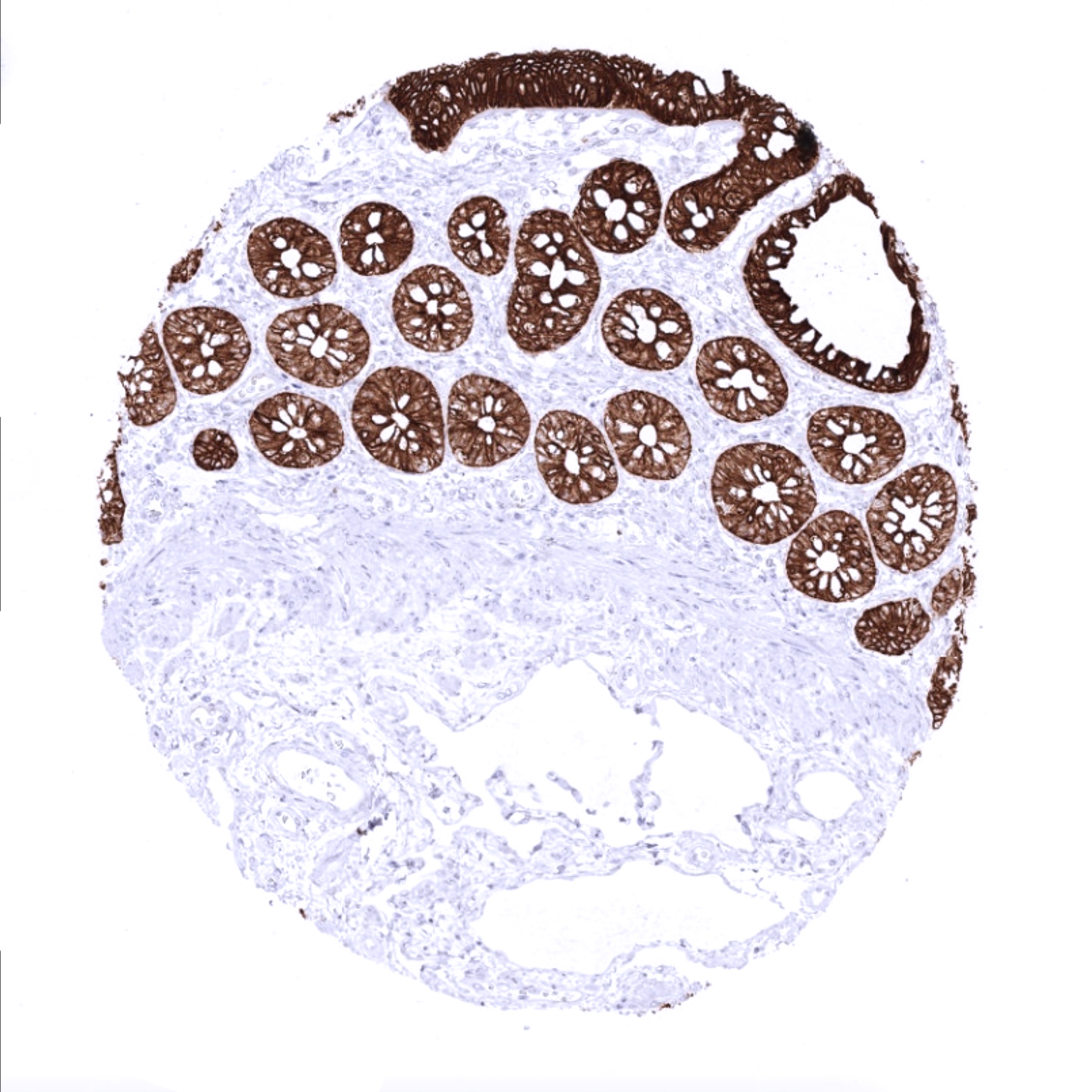
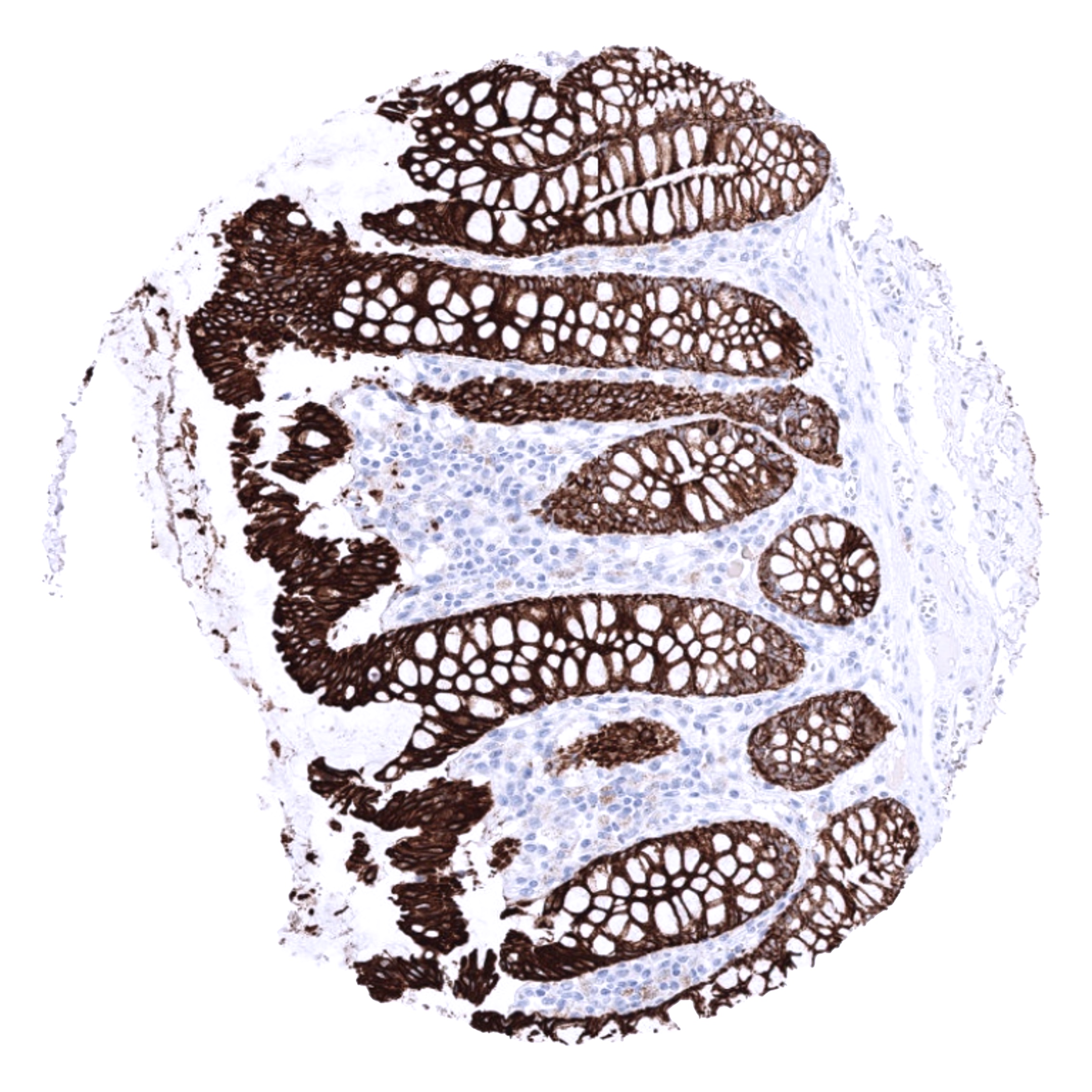
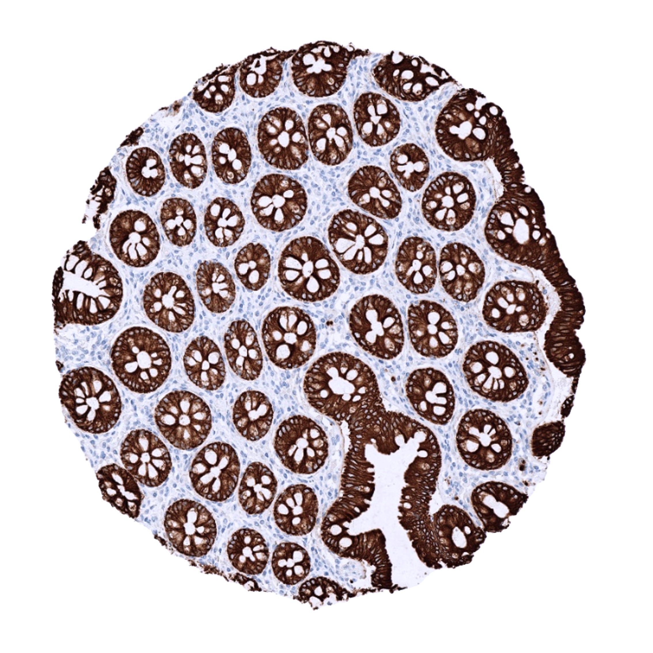
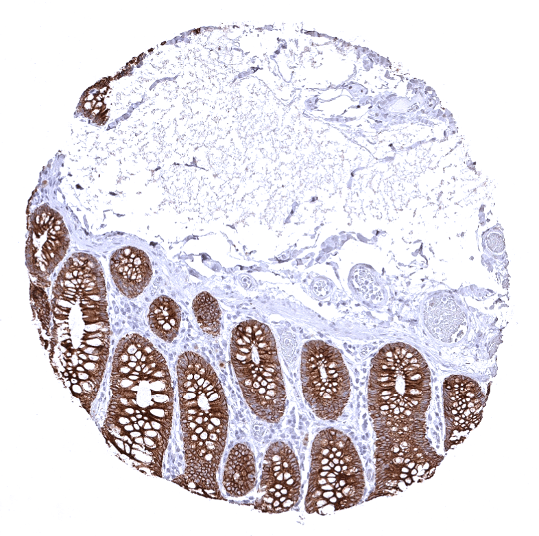
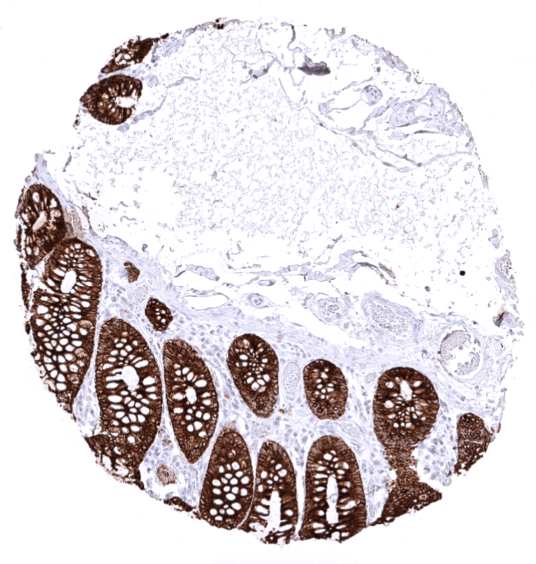
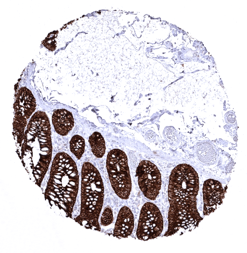
| Colon | Strong CKpan positivity of all epithelial cells. | |
| Rectum | Strong CKpan positivity of all epithelial cells. | |
| Liver | Variable, weak to strong staining of hepatocytes. Strong CKpan positivity of intrahepatic bile ducts. | |
| Gallbladder | Strong CKpan positivity of all epithelial cells. | |
| Pancreas | “Only” moderate to strong staining in islet cells. | |
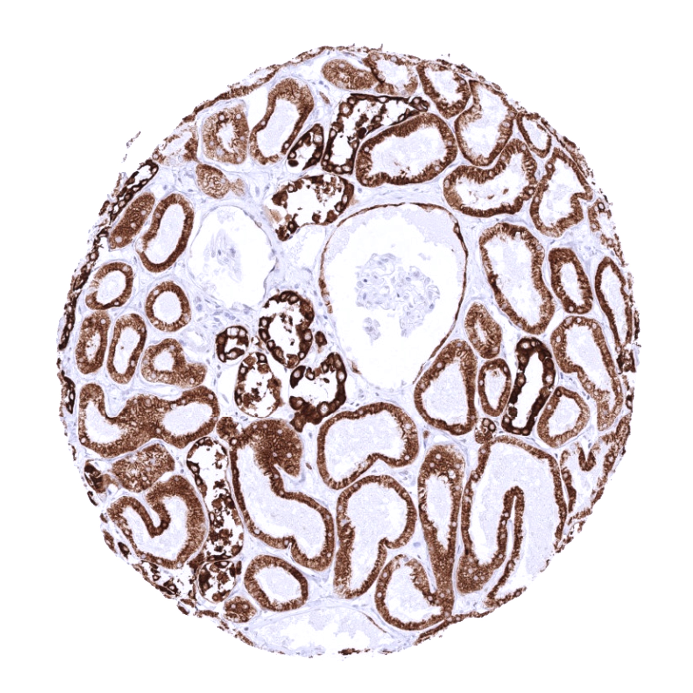
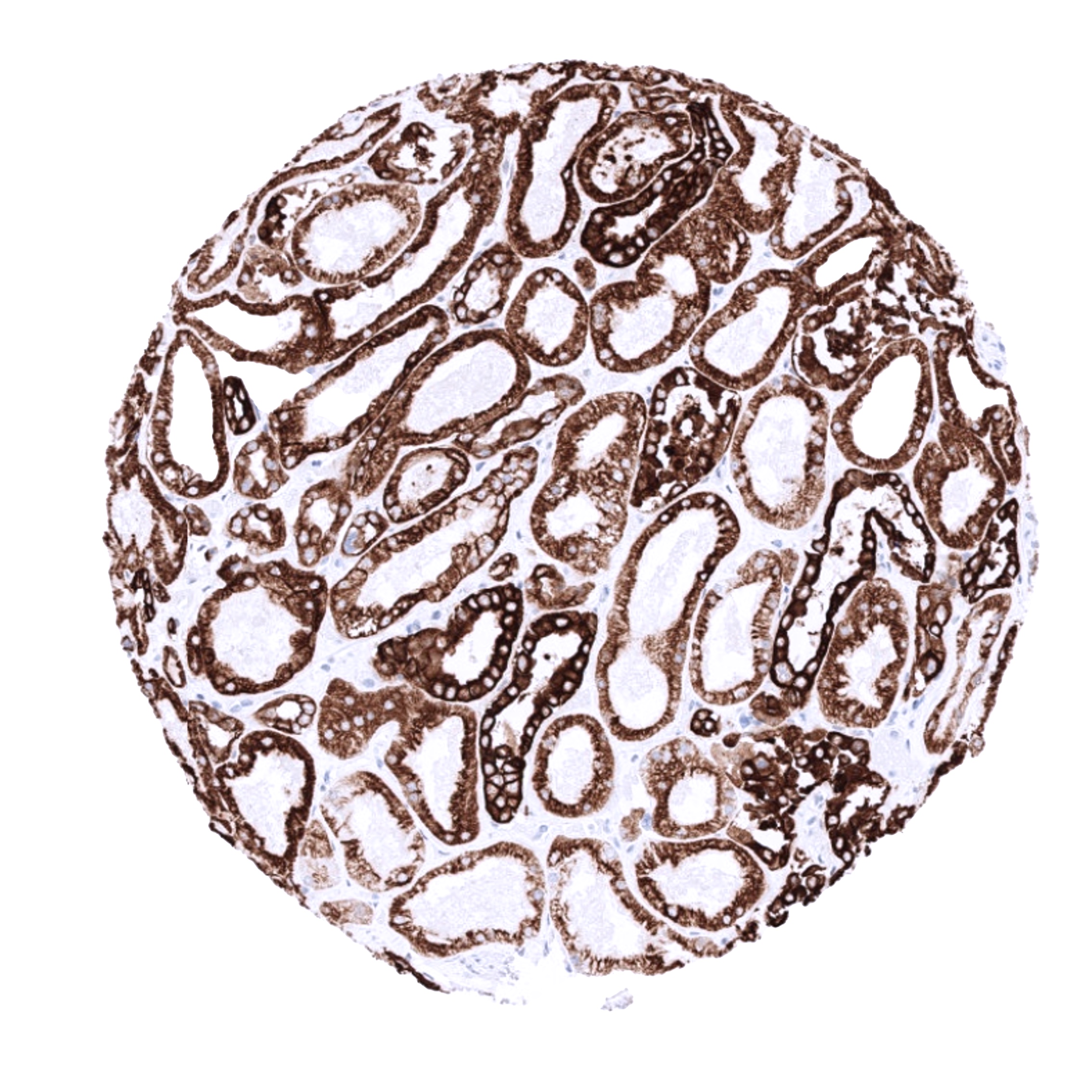
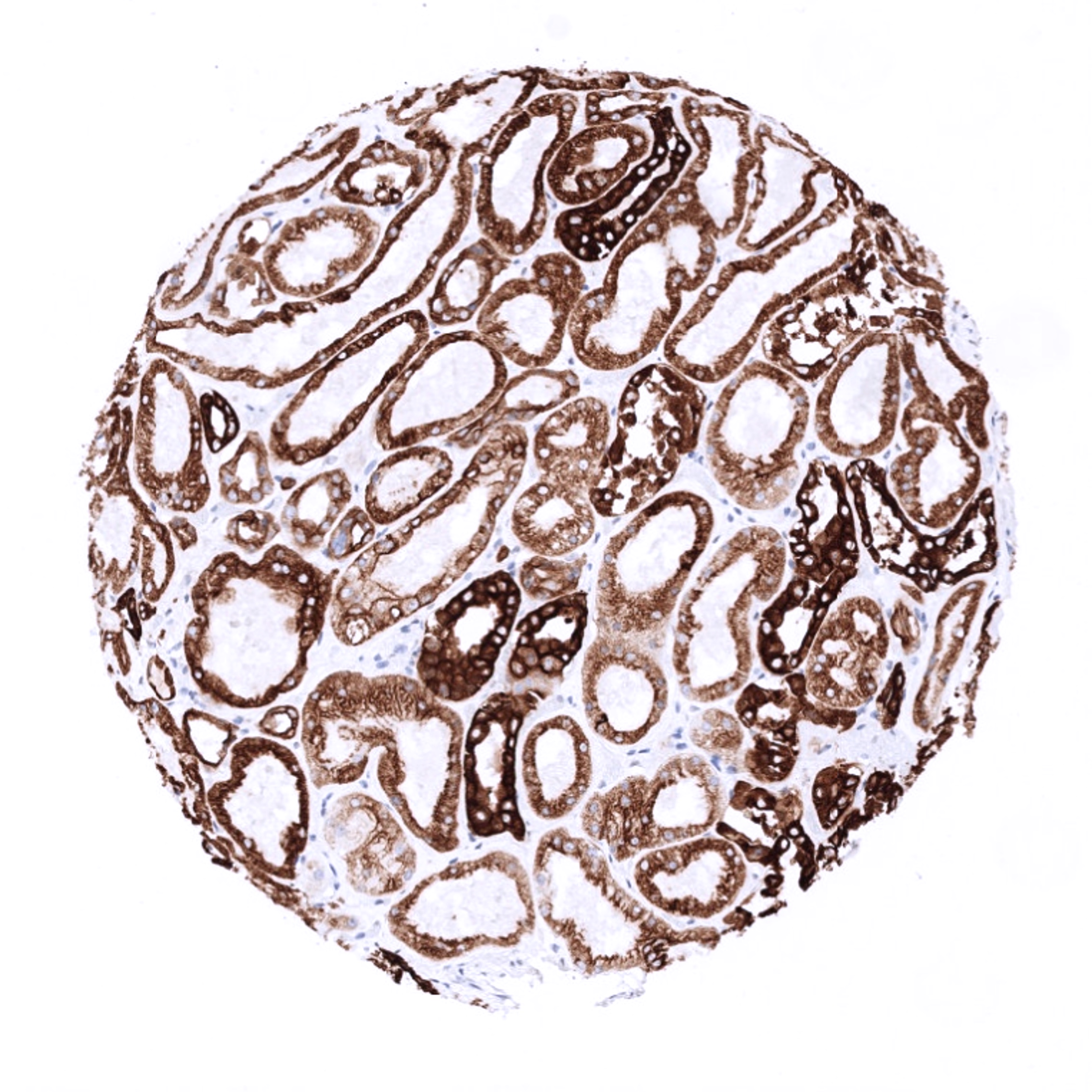
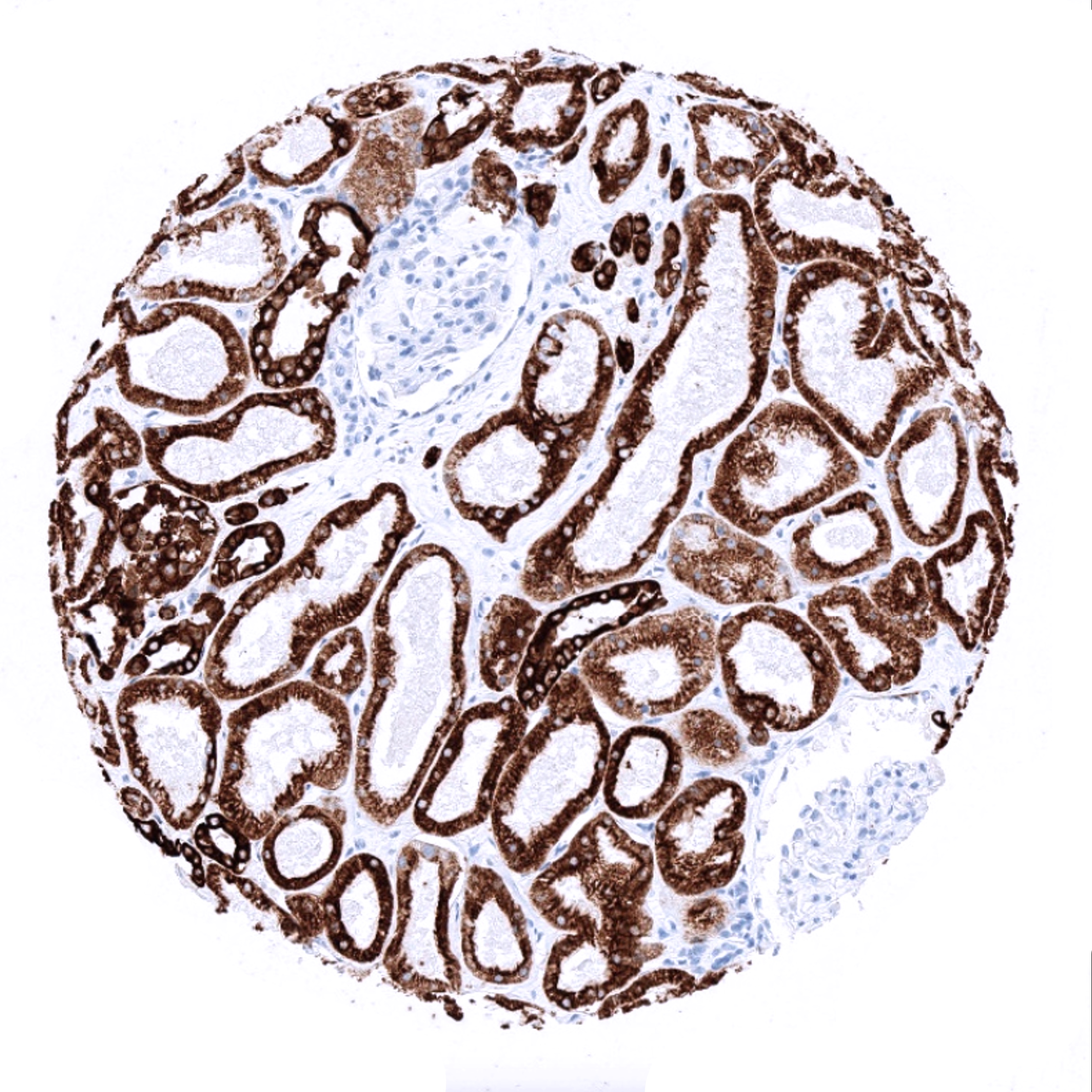
| Genitourinary | Kidney | Strong CKpan positivity of all epithelial cells. |
| Urothelium | Strong CKpan positivity of all epithelial cells. | |
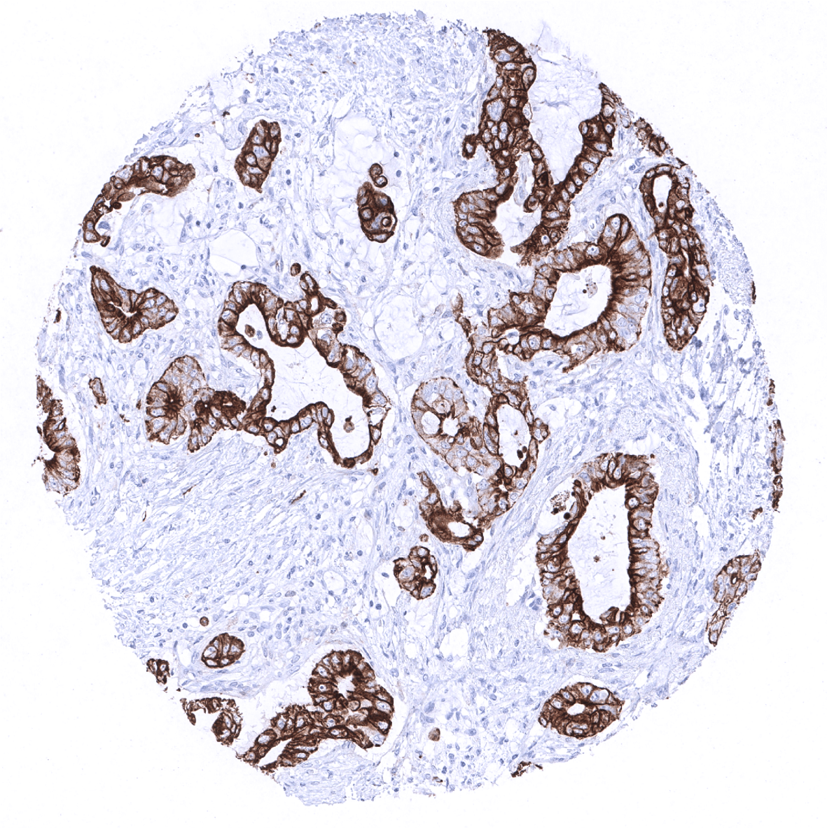
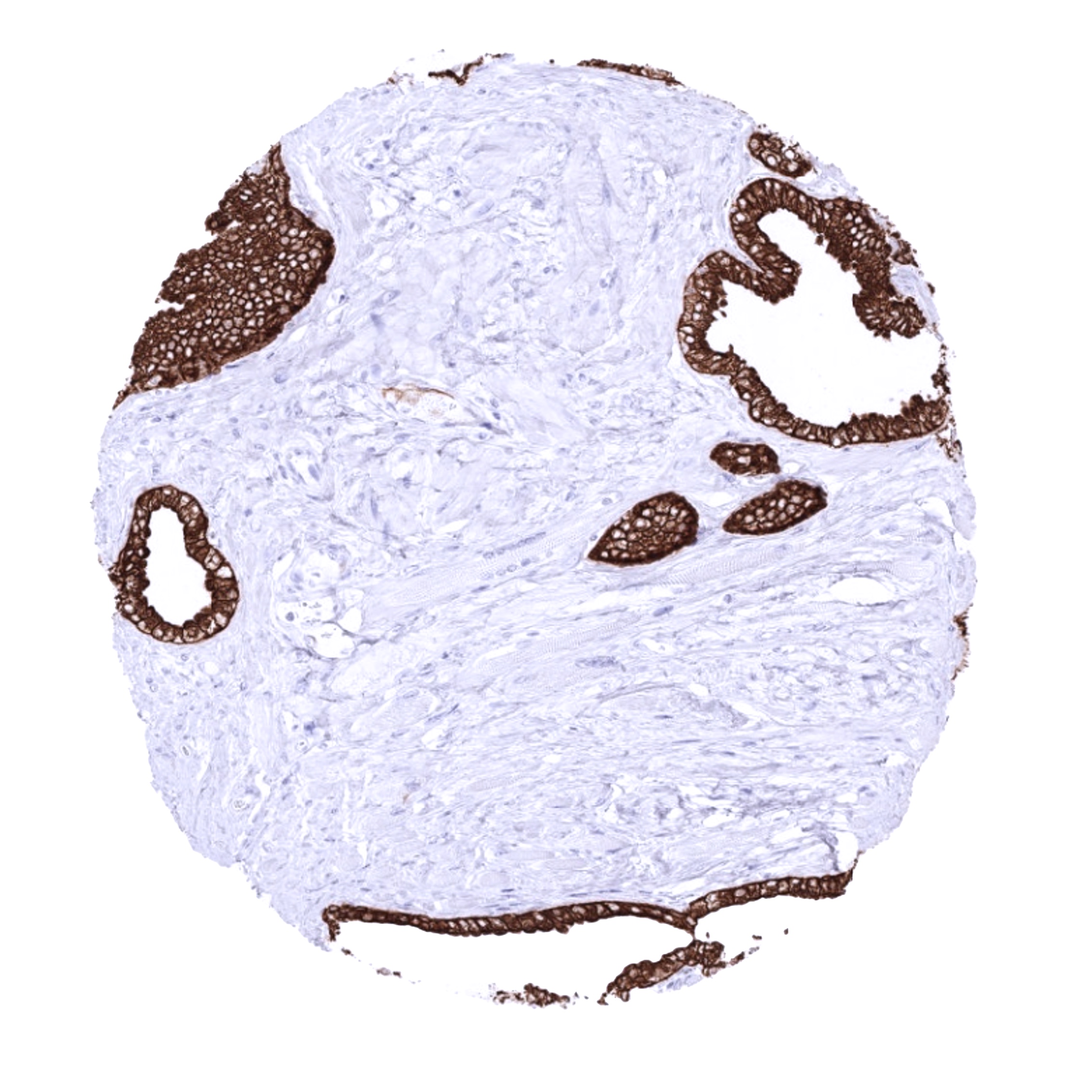
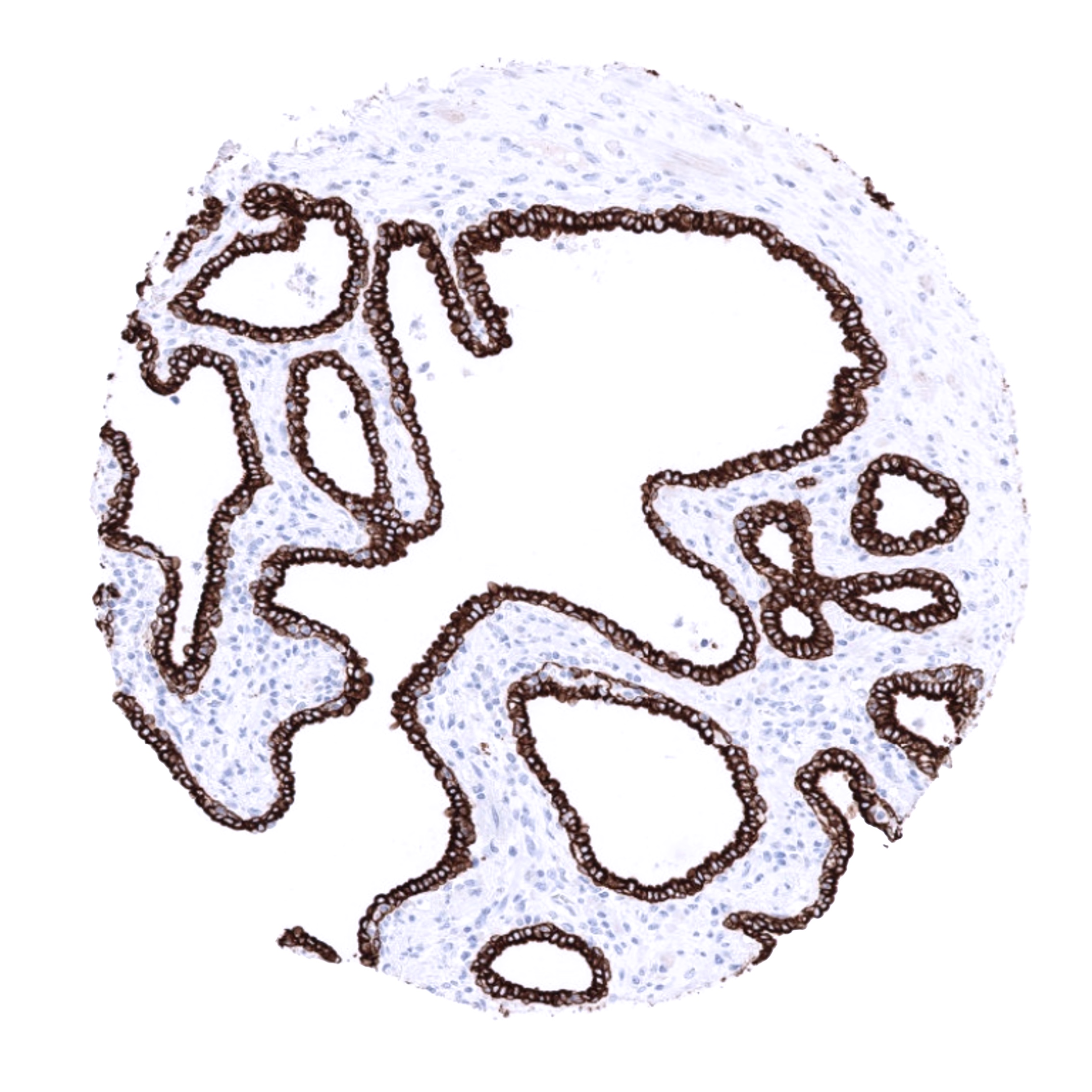
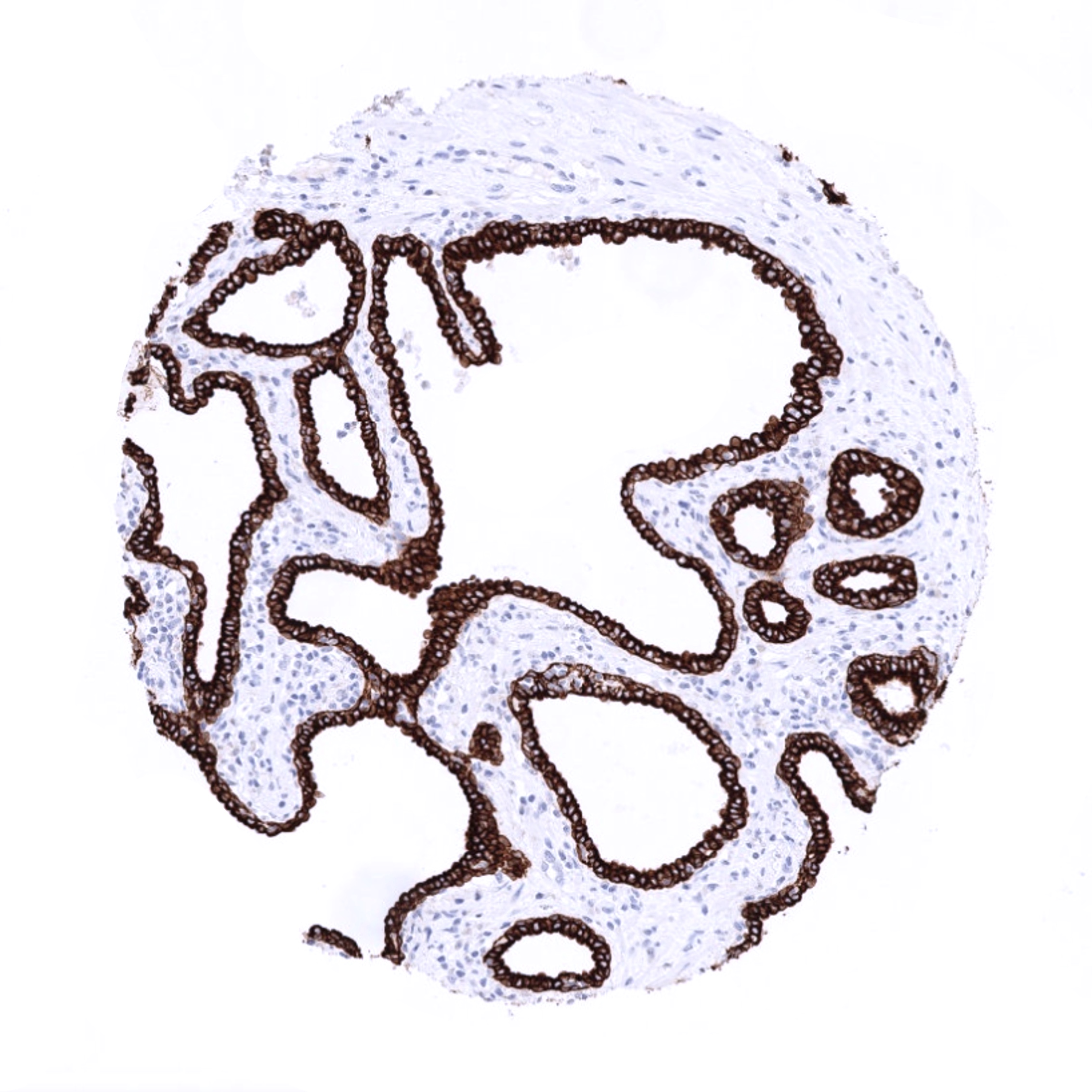
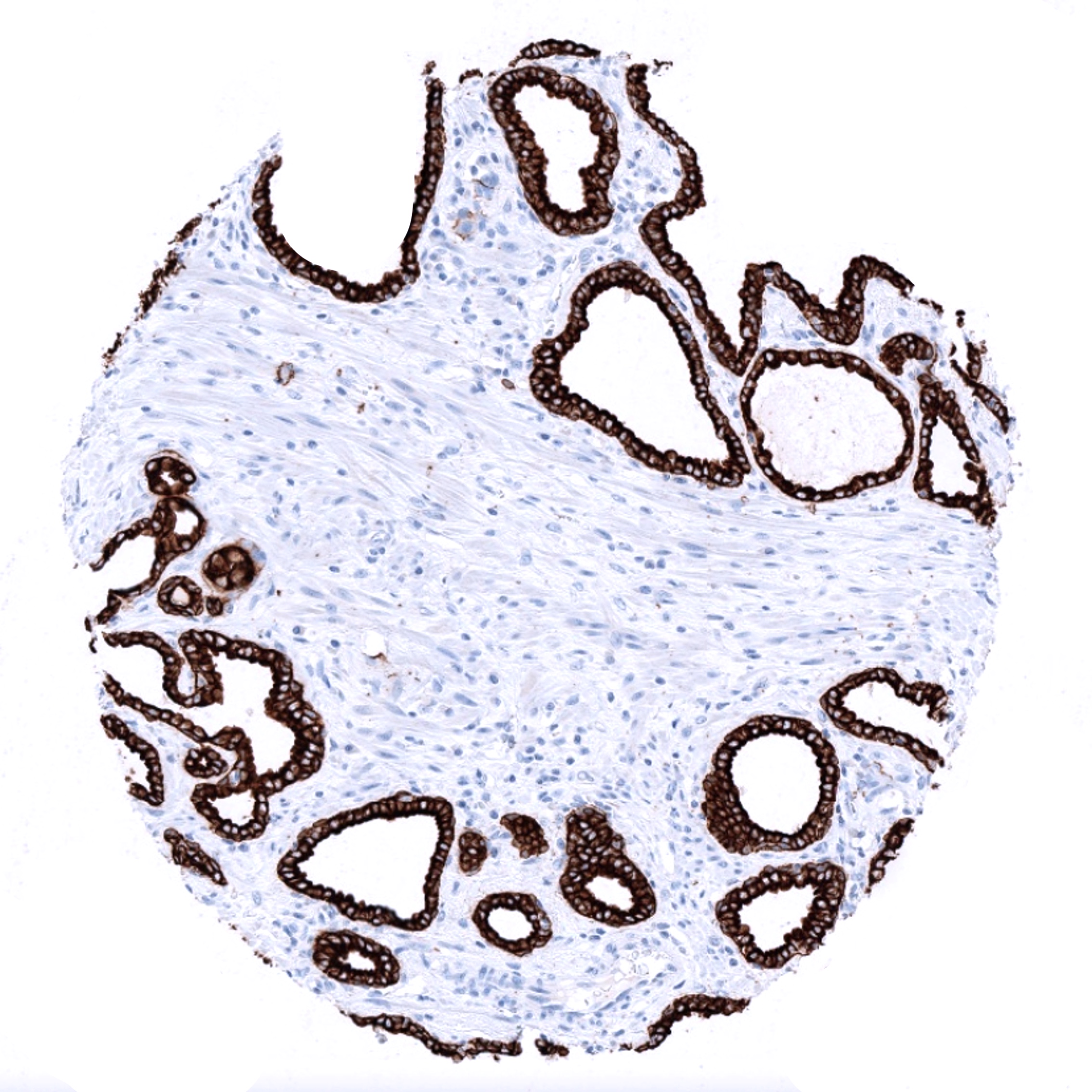
| Male genital | Prostate | Strong CKpan positivity of all epithelial cells. |
| Seminal vesicles | Strong CKpan positivity of all epithelial cells. | |
| Testis | Negative. | |
| Epididymis | Strong CKpan positivity of all epithelial cells. | |
| Female genital | Breast | Strong CKpan positivity of all epithelial cells. |
| Uterus, myometrium | CKpan positivity of some bundles of muscular cells. | |
| Uterus, ectocervix | Strong CKpan positivity of all epithelial cells. | |
| Uterus endocervix | Strong CKpan positivity of all epithelial cells. | |
| Uterus, endometrium | Strong CKpan positivity of all epithelial cells. | |
| Fallopian Tube | Strong CKpan positivity of all epithelial cells. | |
| Ovary | “Only” moderate to strong CKpan staining in corpus luteum cells. | |
| Placenta early | Strong CKpan positivity of all epithelial cells. | |
| Placenta mature | Strong CKpan positivity of all epithelial cells. | |
| Amnion | Strong CKpan positivity of all epithelial cells. | |
| Chorion | Strong CKpan positivity of all epithelial cells. | |
| Skin | Epidermis | Strong CKpan positivity of all epithelial cells. |
| Sebaceous glands | Strong CKpan positivity of all epithelial cells. | |
| Muscle/connective tissue | Heart muscle | Negative. |
| Skeletal muscle | Negative. | |
| Smooth muscle | Negative. | |
| Vessel walls | Negative. | |
| Fat | Negative. | |
| Stroma | CKpan positive myoepithelial cells can occur in all tissues, especially in case of tissue damage. | |
| Endothelium | Negative. | |
| Bone marrow/ lymphoid tissue | Bone marrow | Negative. |
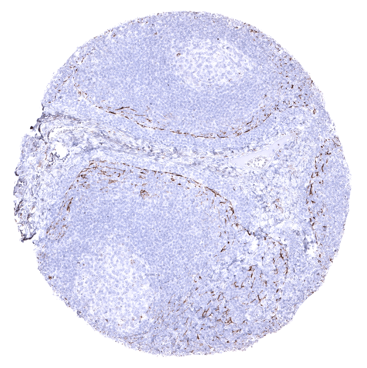
| Lymph node | Delicate fibrillar CKpan staining in the interfollicular area caused by fibroblastic reticulum cells. | |
| Spleen | Delicate fibrillar CKpan staining in the interfollicular area caused by fibroblastic reticulum cells. | |
| Thymus | Strong CKpan positivity of all epithelial cells. | |
| Tonsil | Delicate fibrillar CKpan staining in the interfollicular area caused by fibroblastic reticulum cells. | |
| Remarks |
These findings are largely comparable to the sum of RNA and protein data described on various cytokeratins in the Human Protein Atlas
Suggested positive tissue control: Liver: A strong staining of all bile duct epithelial cells and an at least moderate, predominantly membranous staining of most hepatocytes should be seen.
Suggested negative tissue control: Tonsil: All lymphocytes must not stain. However, interstitial reticulum cells with dendritic/reticular pattern show a weak to moderate fibrillar staining.
Staining Pattern in Relevant Tumor Types
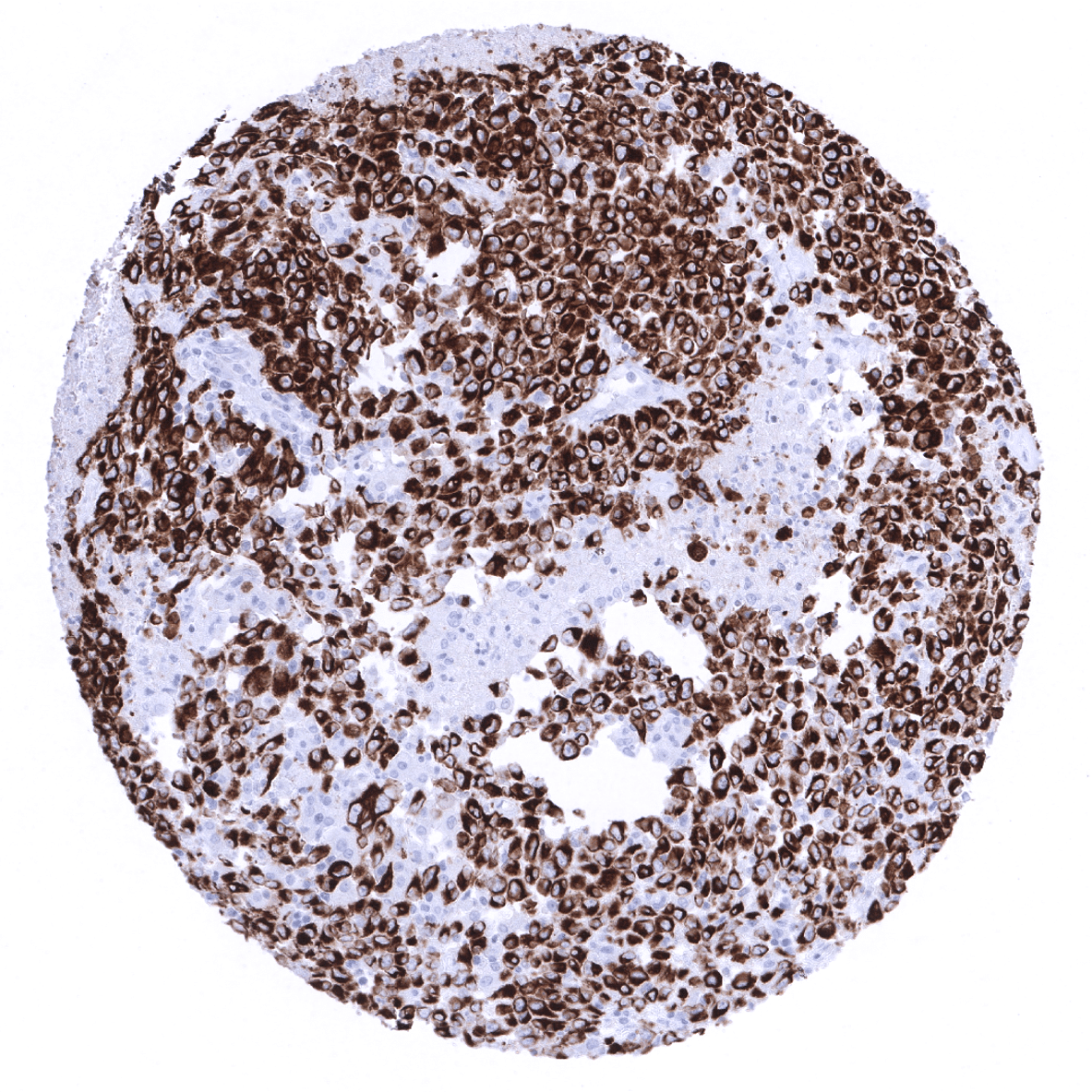
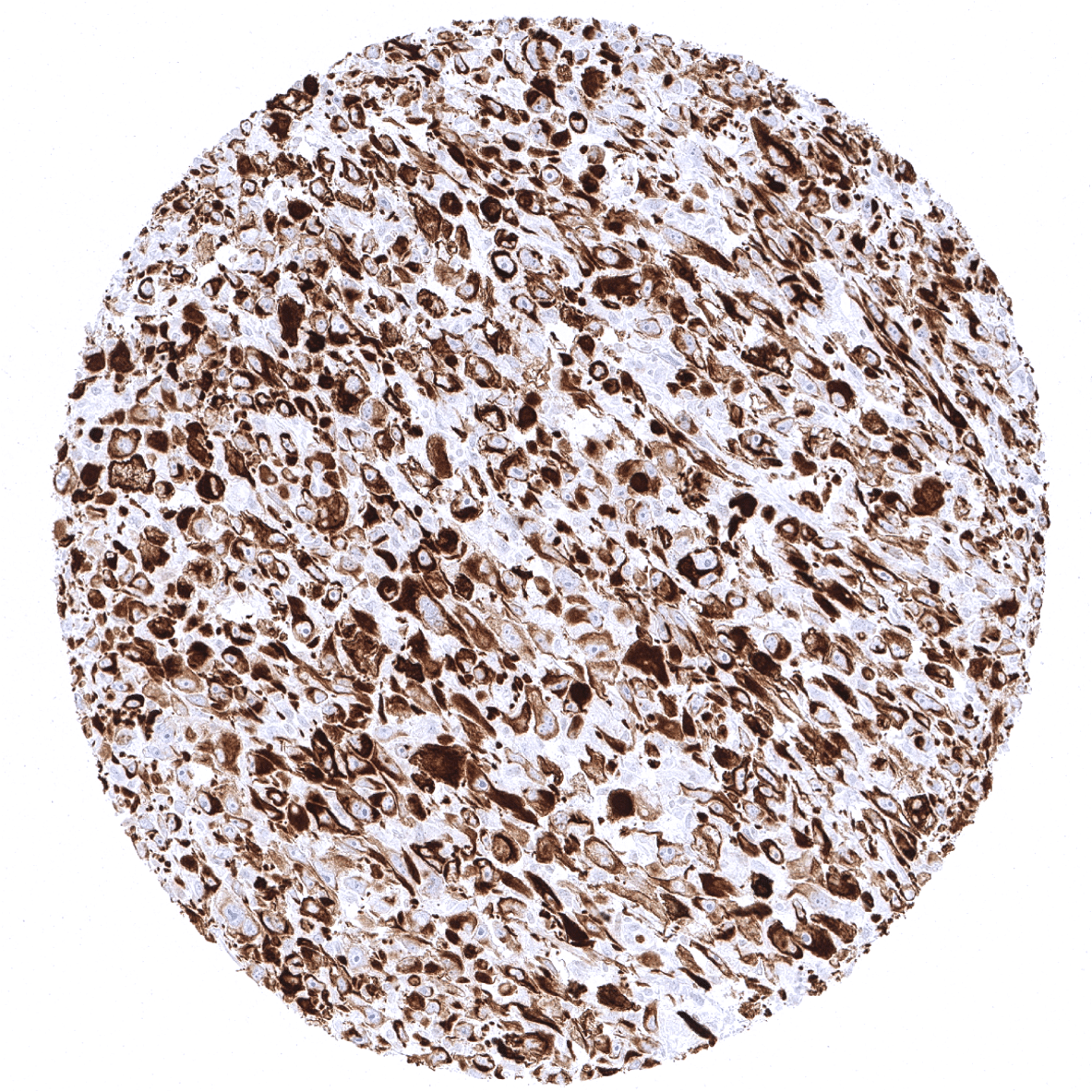
CKpan staining is observed in the vast majority of epithelial cancers. However, CKpan staining can be absent in a fraction of epithelial tumors. For most epithelial tumor types the rate of CKpan negativity is low (0,5-5%). Particularly high rates of CKpan negativity are seen in renal cell carcinoma, hepatocellular carcinoma, and adrenal cortical neoplasms. Although CKpan staining is normally absent in non-epithelial tumors, a small fraction of sarcomas do express cytokeratins.
Compatibility of Antibodies
Cytokeratin, Pan (MSVA-000R) publication summary
Relevant publication: Menz et al.: “Pan-keratin Immunostaining in Human Tumors: A Tissue Microarray Study of 15,940 Tumors.” Published in International Journal of Surgical Pathology August 9th Epub ahead of print. PMID: 35946088.
A total of 13’501 tumors from 121 different tumor categories were successfully analyzed for cytokeratin (pan) expression by using the following protocol: Heat-induced antigen retrieval for 5 minutes in an autoclave at 121°C in pH 9 Target Retrieval Solution buffer. MSVA-000R at a dilution of 1:150 at 37°C for 60 minutes. Visualization of bound antibody by the EnVision Kit (Dako, Agilent). This protocol was also used for all CKpan immunostainings depicted in our tumor and normal tissue galleries.
At least one case with a positive CKpan immunostaining was seen 101 of 121 (84%) different tumor categories. Among 75 epithelial tumor entities, the CKpan positivity rate was 100% in 50 (67%) and 98-99.9% in 12 categories (16%). These tumors included almost all adenocarcinomas, squamous cell and urothelial carcinomas. A total of 15 tumor entities showed CKpan positivity in 0.9%-25%, most of these were of mesenchymal origin and often showed a weaker staining than epithelial neoplasms. All 20 (17%) tumor entities that were always CKpan negative were of mesenchymal or hemato-lymphatic origin. The distribution of positive staining results is shown in an “organ-systematic” (Figure 1) and in a “ranking order” figure (Figure 2) below (images based on data from Menz et al). Data on associations with histopathological and clinical parameters of tumor aggressiveness in several cancer types are also summarized below (Figure 3; based on data described by Menz et al).
Authors conclusions on diagnostic utility with respect to the distinction of different tumor entities (Menz et al):
- -Among carcinomas, low level or even absent CKpan staining is particularly common in renal cell carcinomas, adrenocortical, neuroendocrine and endocrine tumors, as well as in very poorly differentiated cancers, such as small cell, sarcomatoid or anaplastic carcinomas.
Authors conclusions on prognostic/predictive role of CK7 expression (Menz et al.):
- Reduced/absent CKpan staining is associated with high UICC stage (p<0.0001), high Thoenes grade (p<0.05), high Fuhrman grade (p<0.005), advanced tumor stage (p< 0.0001) and nodal metastasis (p<0.05) in clear cell renal cell carcinoma.
- Reduced/absent CKpan staining is linked to advanced pT stage (p<0.001) in papillary renal cell carcinoma.
- Reduced/absent CKpan staining is related to advanced stage (p<0.005), high grade (p=0.0005) as well as loss of ER and PR expression (p<0.0001 each) in invasive breast carcinoma of no special type (NST).
Data from the publication: “Pan-keratin Immunostaining in Human Tumors: A Tissue Microarray Study of 15,940 Tumors.” Published by Menz et al. in International Journal of Surgical Pathology August 9th Epub ahead of print. PMID: 35946088.
Figure 1. CKpan staining in cancer (“organ-systematic”; according to Menz et al.)

Figure 2. CKpan staining in cancer (“ranking list”; according to Menz et al.)

Figure 3. Clinico-pathological associations described by Menz et al. (p-value)

Protocol Recommendations
IHC users have different preferences on how the stains should look like. Some prefer high staining intensity of the target stain and even accept some background. Others favor absolute specificity and lighter target stains. Factors that invariably lead to more intense staining include higher concentration of the antibody and visualization tools, longer incubation time, higher temperature during incubation, higher temperature and longer duration of the heat induced epitope retrieval (slide pretreatment). The impact of the pH during slide pretreatment has variable effects and depends on the antibody and the target protein.
All images and data shown here and in our image galleries are obtained by the manual protocol described below. Protocols resulting in equivalent staining by using an automated immunostainer are described as well.
Manual protocol
Freshly cut sections should be used (less than 10 days between cutting and staining). Heat-induced antigen retrieval for 5 minutes in an autoclave at 121°C in pH 9 Target Retrieval Solution buffer. Apply MSVA-000R at a dilution of 1:150 at 37°C for 60 minutes. Visualization of bound antibody by the EnVision Kit (Dako, Agilent) according to the manufacturer’s directions.
Agilent / Dako – Autostainer Link 48
Pretreatment in PT-Link for 30 minutes at 95°C (pH high); FLEX peroxidase blocking for 5 minutes (room temperature), MSVA-000R 1:150 for 20 minutes (room temperature), FLEX+ mouse/rabbit (LINKER) for 15 minutes (room temperature), horseradish peroxidase (HRP) for 20 minutes (room temperature), FLEX DAB+Sub-Chromo for 10 minutes (room temperature), FLEX hematoxylin for 5 minutes (room temperature).
These images reflect stainings by the protocol described above. It is of note that a comparable staining result can also be obtained by different protocols. In general, a longer pretreatment, a longer incubation time of the primary antibody, a higher antibody concentration, and a longer incubation time of FLEX+LINKER result in stronger staining, potentially at the cost of more background staining. Modifications of the protocol with a strengthening effect on staining intensity in combination with changes of other parameters that result in lower staining intensity can result in a comparable result as shown above.
Leica – BOND RX
Dewax at 72°C for 30 seconds; Pretreatment in Bond Epitope Retrieval Solution (ER2 – EDTA pH9) for 20 minutes at 100°C; Peroxidase blocking for 5 minutes (room temperature), MSVA-000R 1:150 for 30 minutes (room temperature), Post primary (rabbit anti mouse) for 8 minutes (room temperature), Polymer (goat anti rabbit) for 8 minutes (room temperature), mixed DAB refine for 10 minutes (room temperature), hematoxylin for 5 minutes (room temperature).

These images reflect stainings by the protocol described above. It is of note that a comparable staining result can also be obtained by different protocols. In general, a longer pretreatment, a longer incubation time of the primary antibody, a higher antibody concentration, a higher temperature during incubation, and a longer incubation time of Post primary and or the Polymer result in stronger staining, potentially at the cost of more background staining. Modifications of the protocol with a strengthening effect on staining intensity in combination with changes of other parameters that result in lower staining intensity can result in a comparable result as shown above.
Roche – Ventana Discovery ULTRA
Pretreatment for 64 minutes at 100°C (pH 8,4); CM peroxidase blocking for 12 minutes (room temperature), MSVA-000R 1:50 for 40 minutes at 36°C, secondary antibody (anti-rabbit HQ) for 12 minutes at 36°C, anti-HQ HRP for 12 minutes at room temperature, DAB at room temperature, hematoxylin II at room temperature for 8 minutes, bluing reagent at room temperature for 4 minutes.
These images depict staining results obtained by the protocol described above. It is of note, that the Ventana machines generally require higher antibody concentrations than other commonly used autostainers because the antibodies are automatically diluted during the procedure. Various other protocols can result in an identical result as shown above. A longer pretreatment, a longer incubation time of the primary antibody, a higher antibody concentration, a higher temperature during incubation, and a longer incubation time of secondary antibody and or the anti-HQ HRP result in stronger staining, potentially at the cost of more background staining.
Impact of pH
MSVA-000R results in strongest staining if pH9,0 is used for slide pretreatment. pH7,8 is acceptable but lower pH results in a significant reduction of sensitivity.
Potential pitfalls
- Normal epithelial cells entrapped within tumor tissue can result in false positive interpretation of cytokeratin IHC.
- Pan cytokeratin staining regularly occurs in myofibroblasts/myoepithelial cells. These are for example often seen in the bladder wall after transurethral resection and should not be mistaken for residual tumor.
Potential Research Applications
- In suitable tissues CKpan can be used for the distinction of cancerous from non-cancerous cells and can thus be employed in various experimental multicolor immunofluorescence approaches (For example: automated assessment of the Ki67 labeling index in cancer cells)
Evidence for Antibody Specificity in IHC
Specificity of MSVA-000R is documented by strong staining in all epithelial cell types that are well documented to express cytokeratins and absence of staining in all tissues known to not express cytokeratins
