
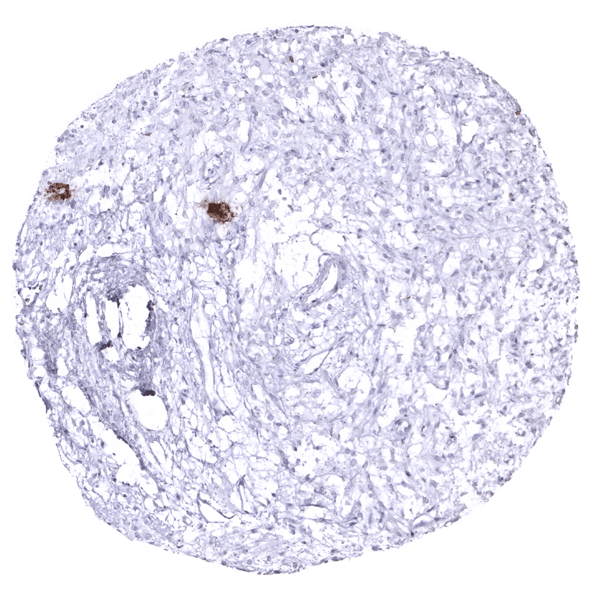
Adrenal gland - In the adrenal gland, only a fraction of cortical cells, typically arranged in groups, fascicles or sheets, show weak to moderate Ckpan immunostaining.
Adrenal gland - In the adrenal gland, only a fraction of cortical cells, typically arranged in groups, fascicles or sheets, show weak to moderate Ckpan immunostaining.
Aorta, media
Appendix, mucosa
Appendix, muscular wall

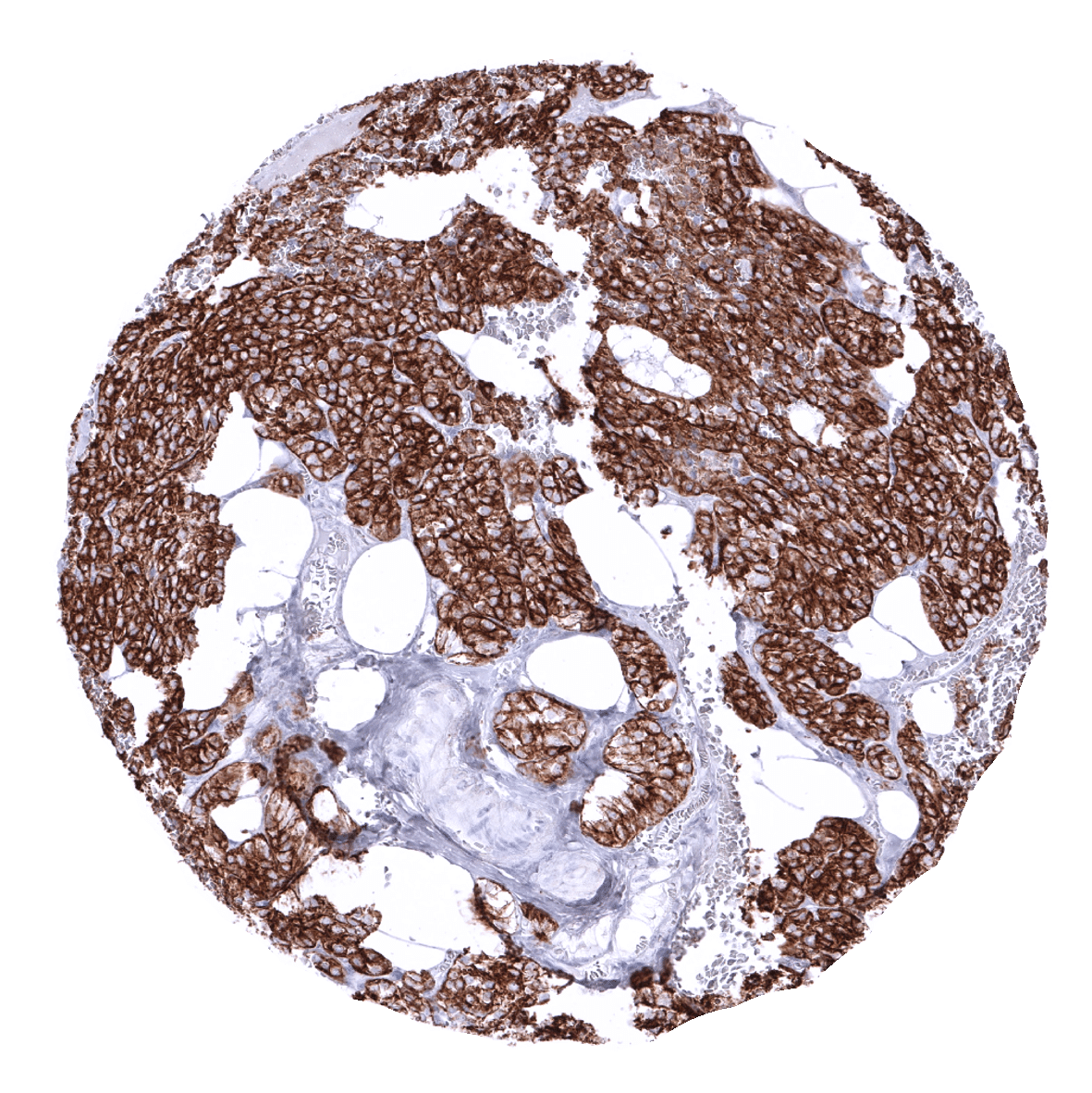
Breast - In the breast, the intensity of Ckpan immunostaining is markedly weaker in basal cells than in luminal cells.
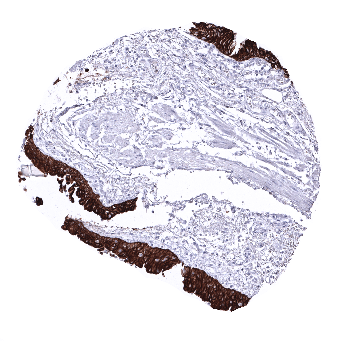
Bronchus, mucosa
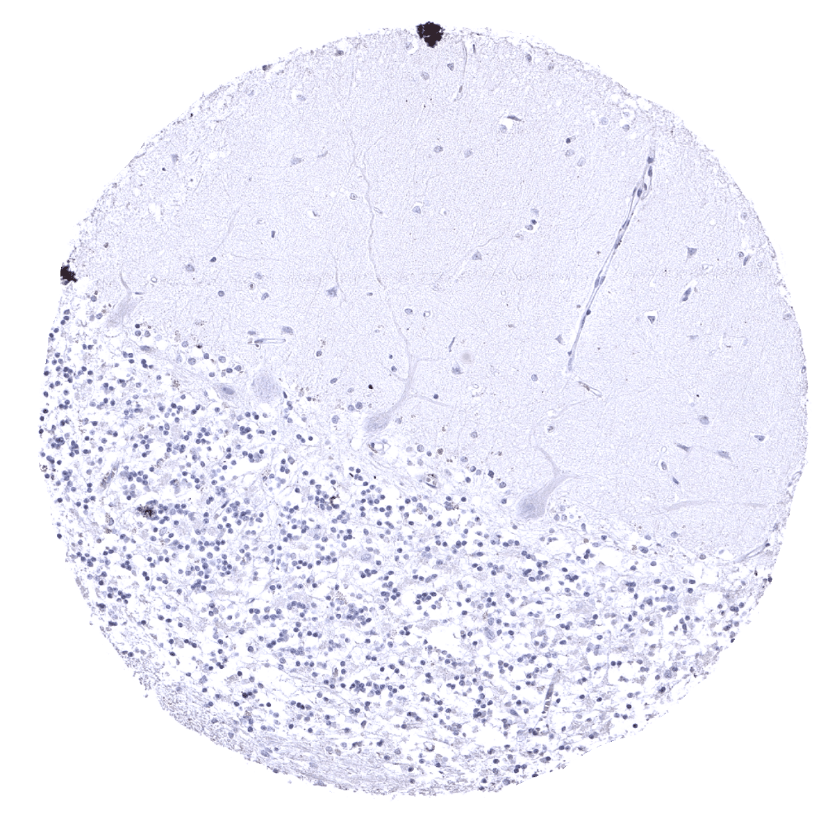
Cerebellum (molecular layer, Purkinje cell layer, granule cell layer)
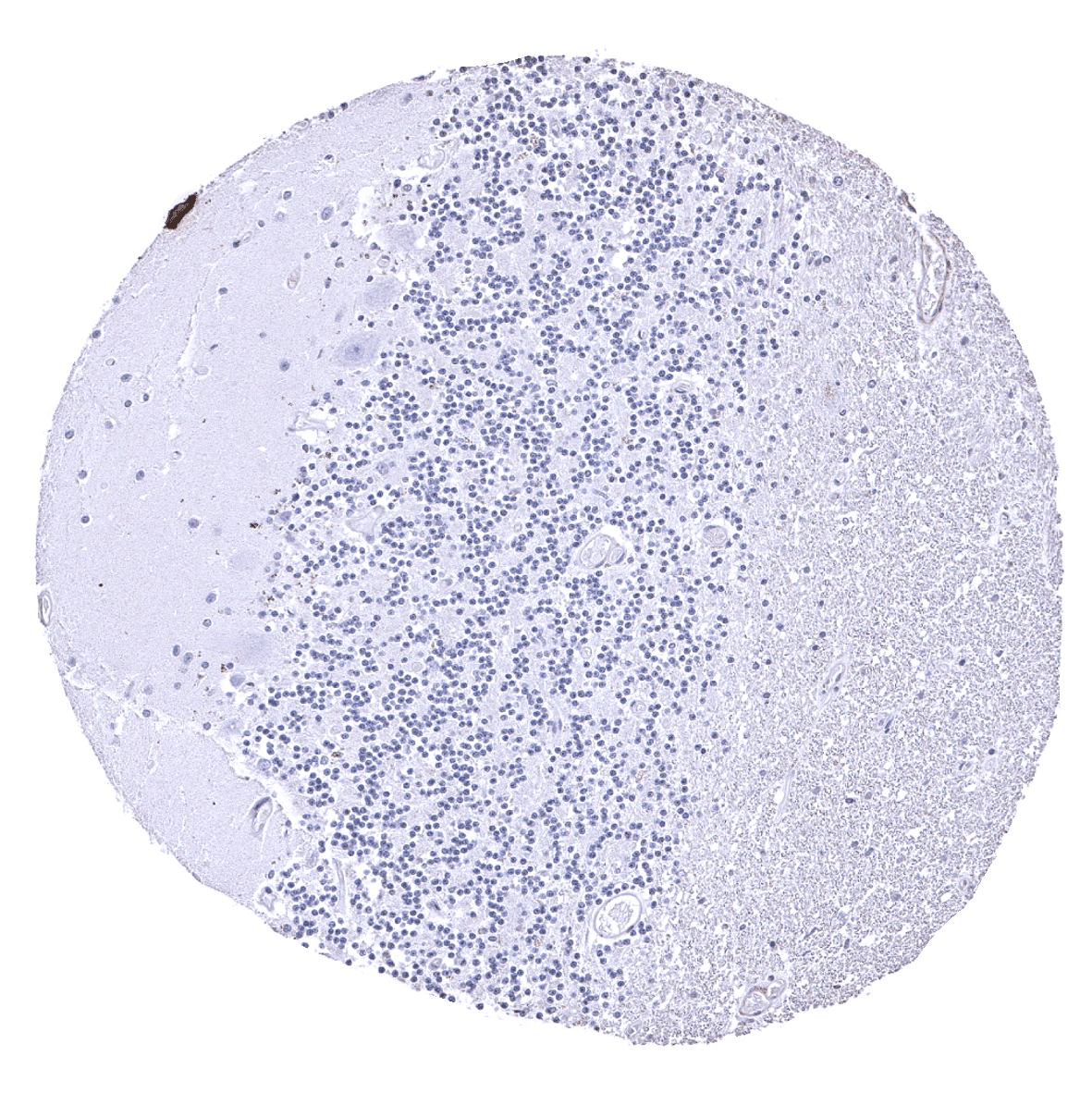
Cerebellum (molecular layer, Purkinje cell layer, granule cell layer, white matter)
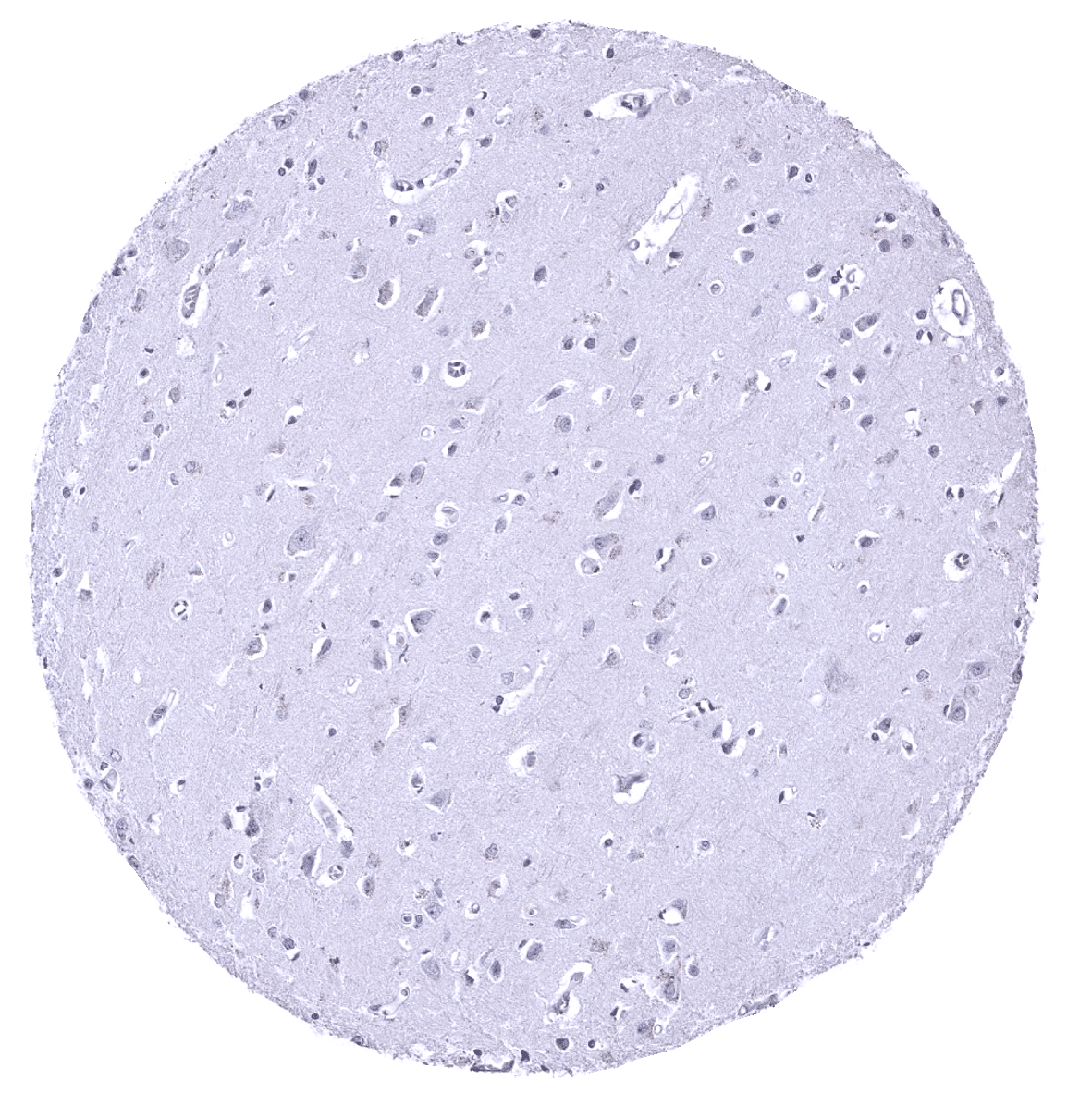
Cerebrum, grey matter

Cerebrum, white matter
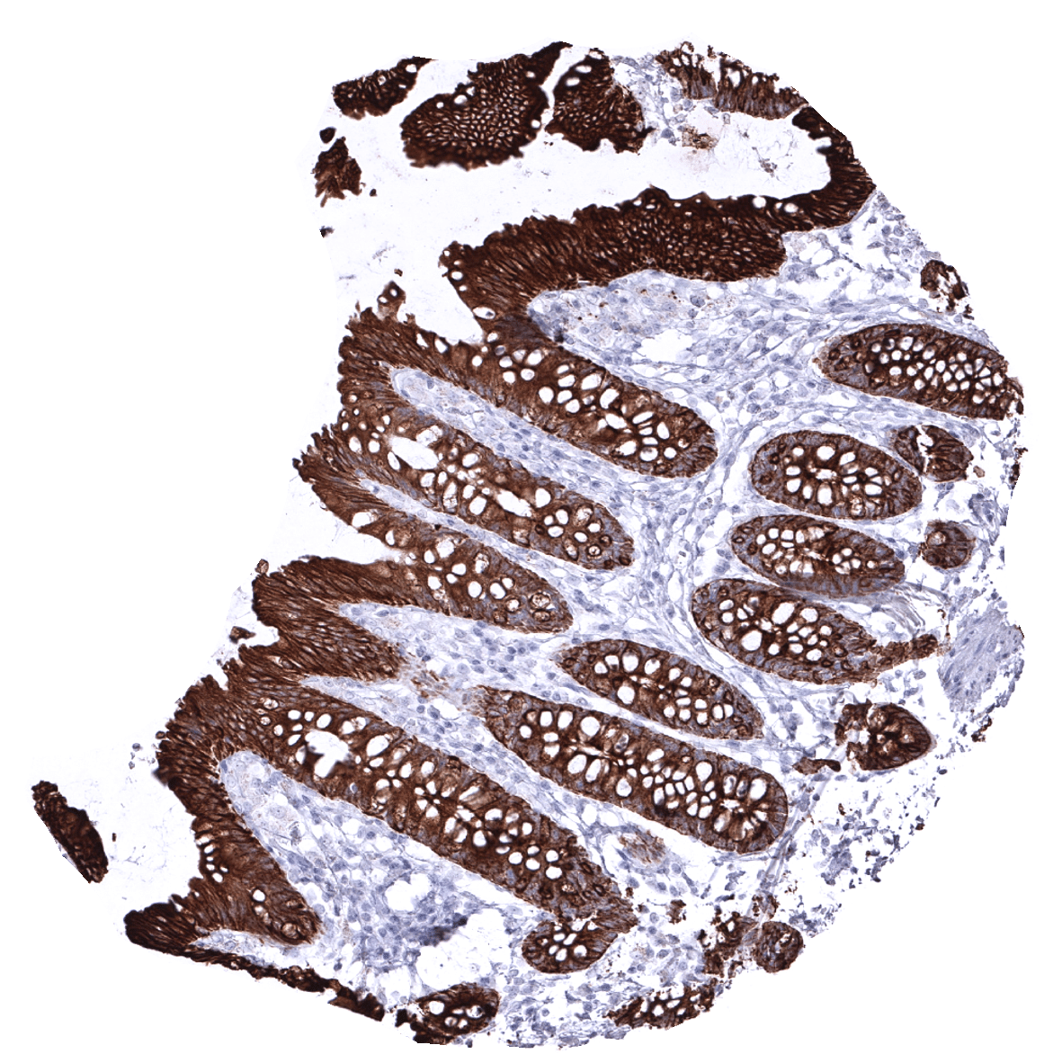
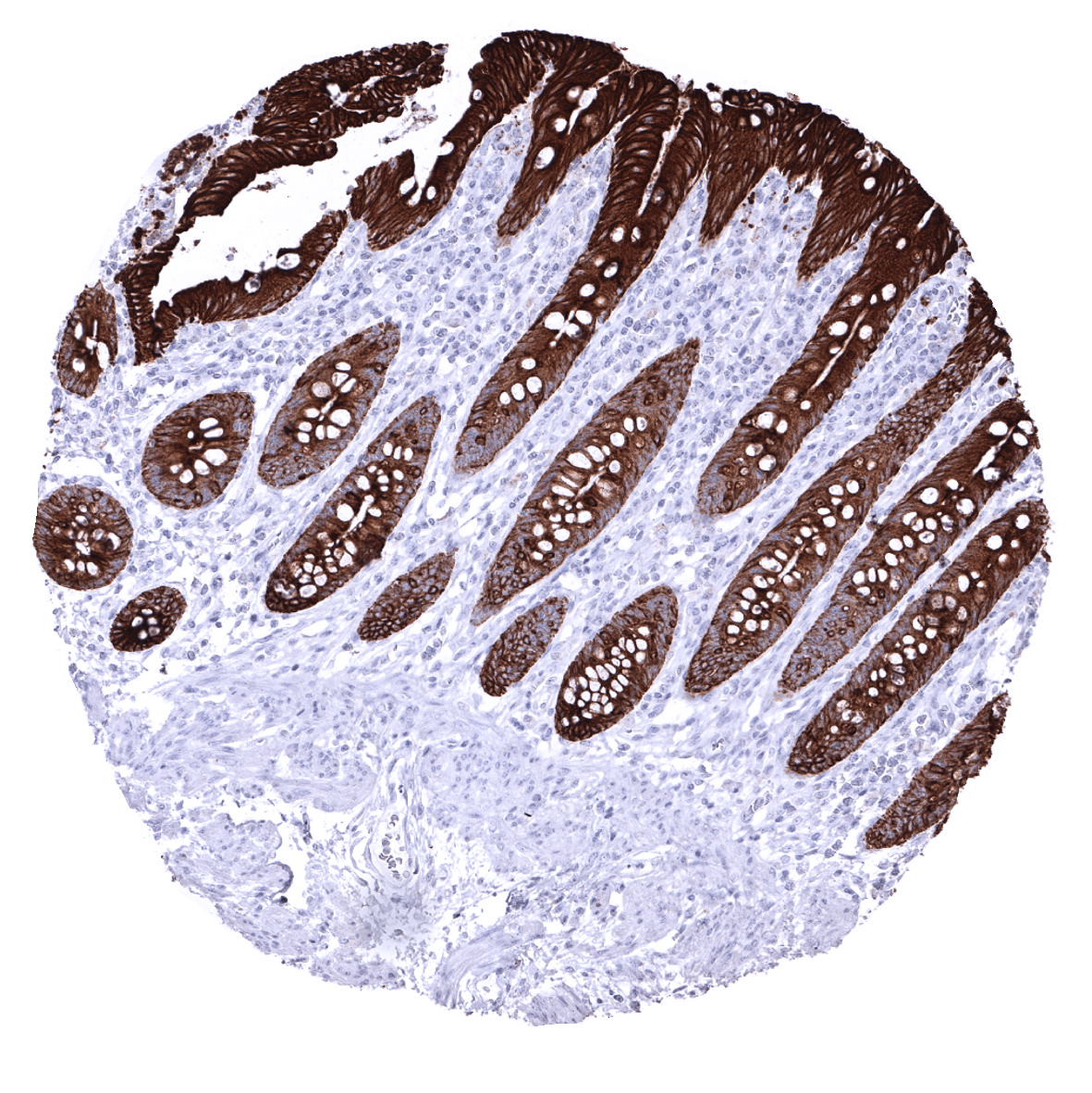
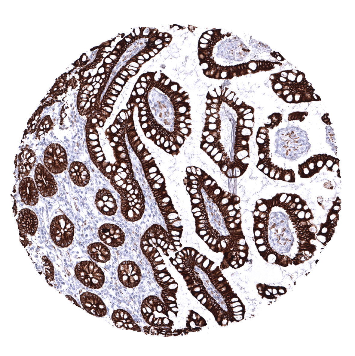
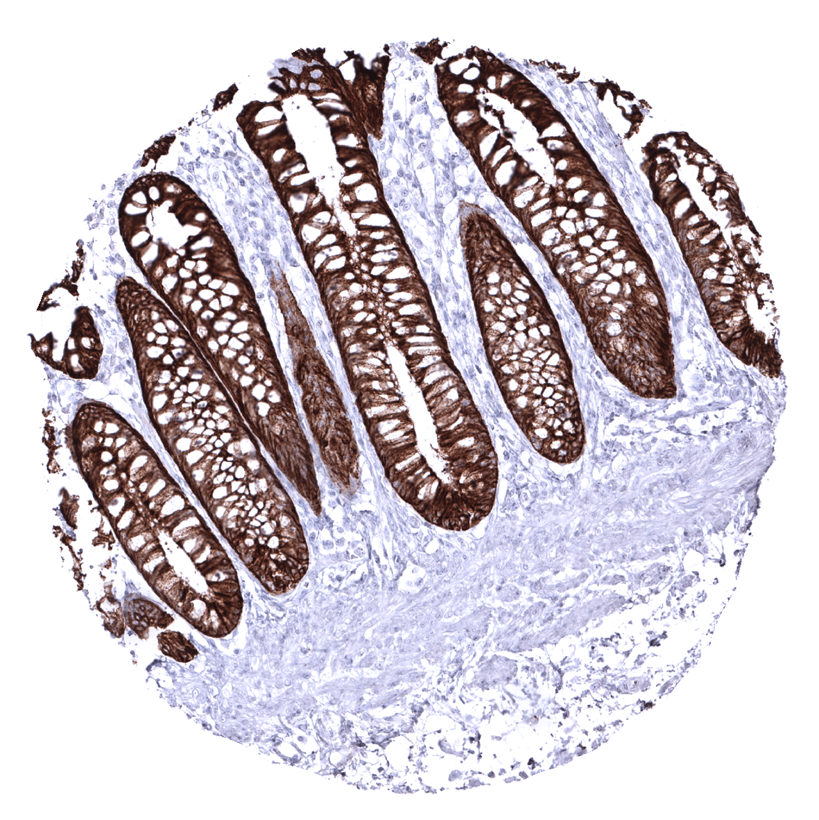
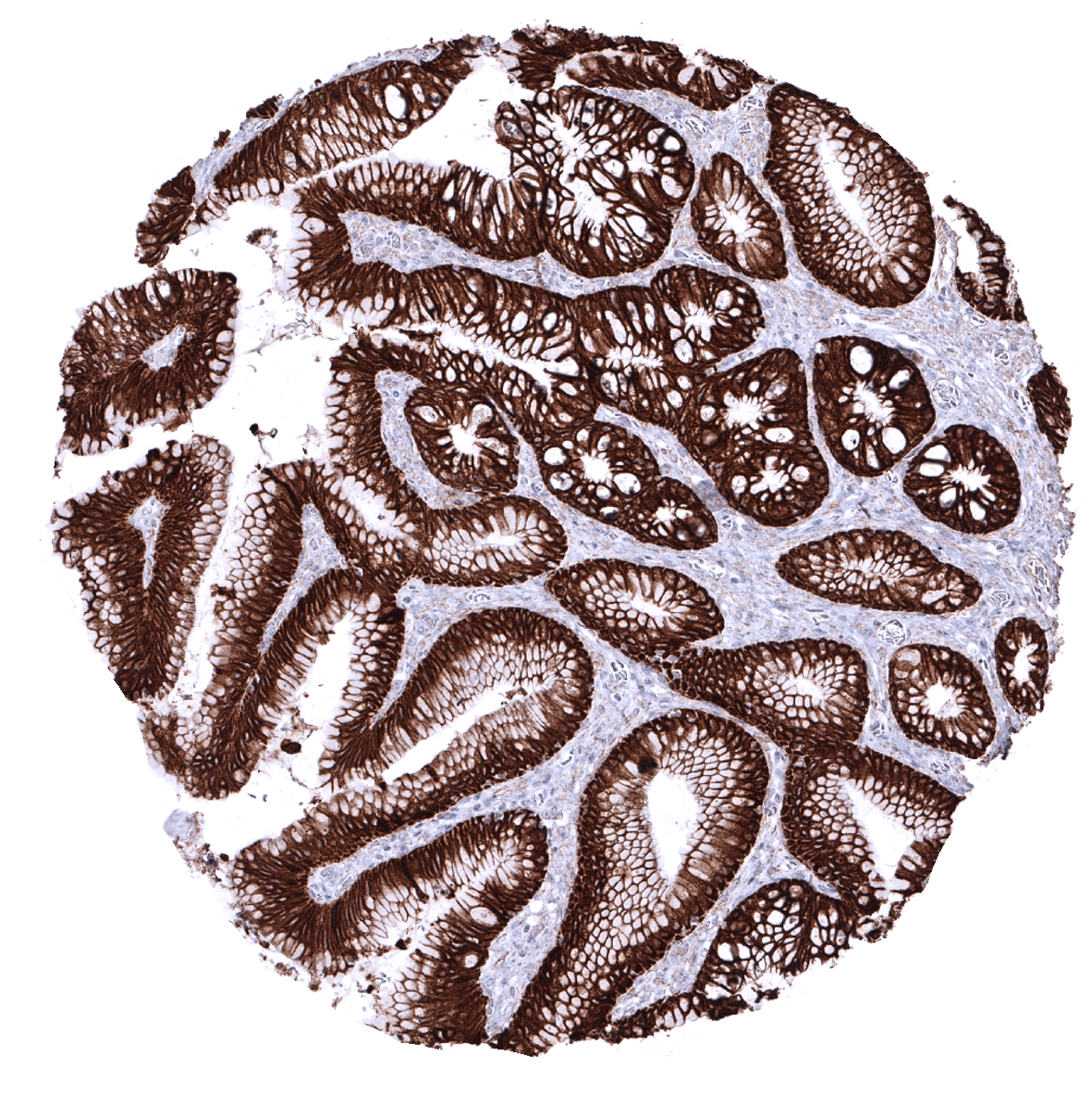
Colon descendens, mucosa
Colon descendens, muscular wall

Duodenum, Brunner gland
Duodenum, mucosa

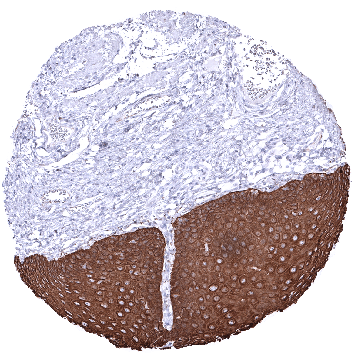
Ectocervix
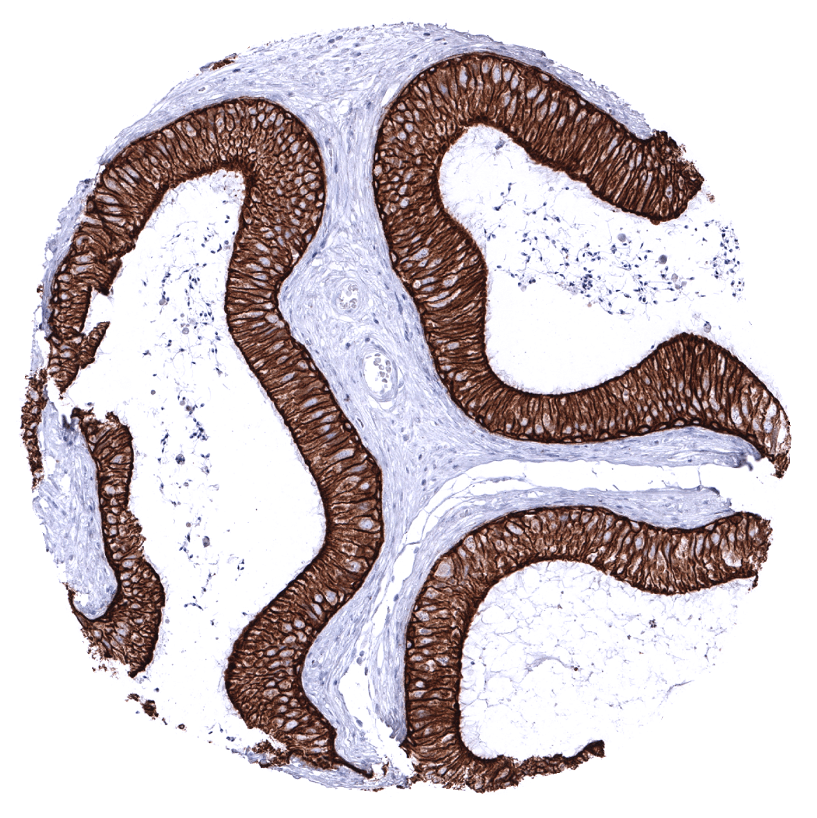
Endocervix
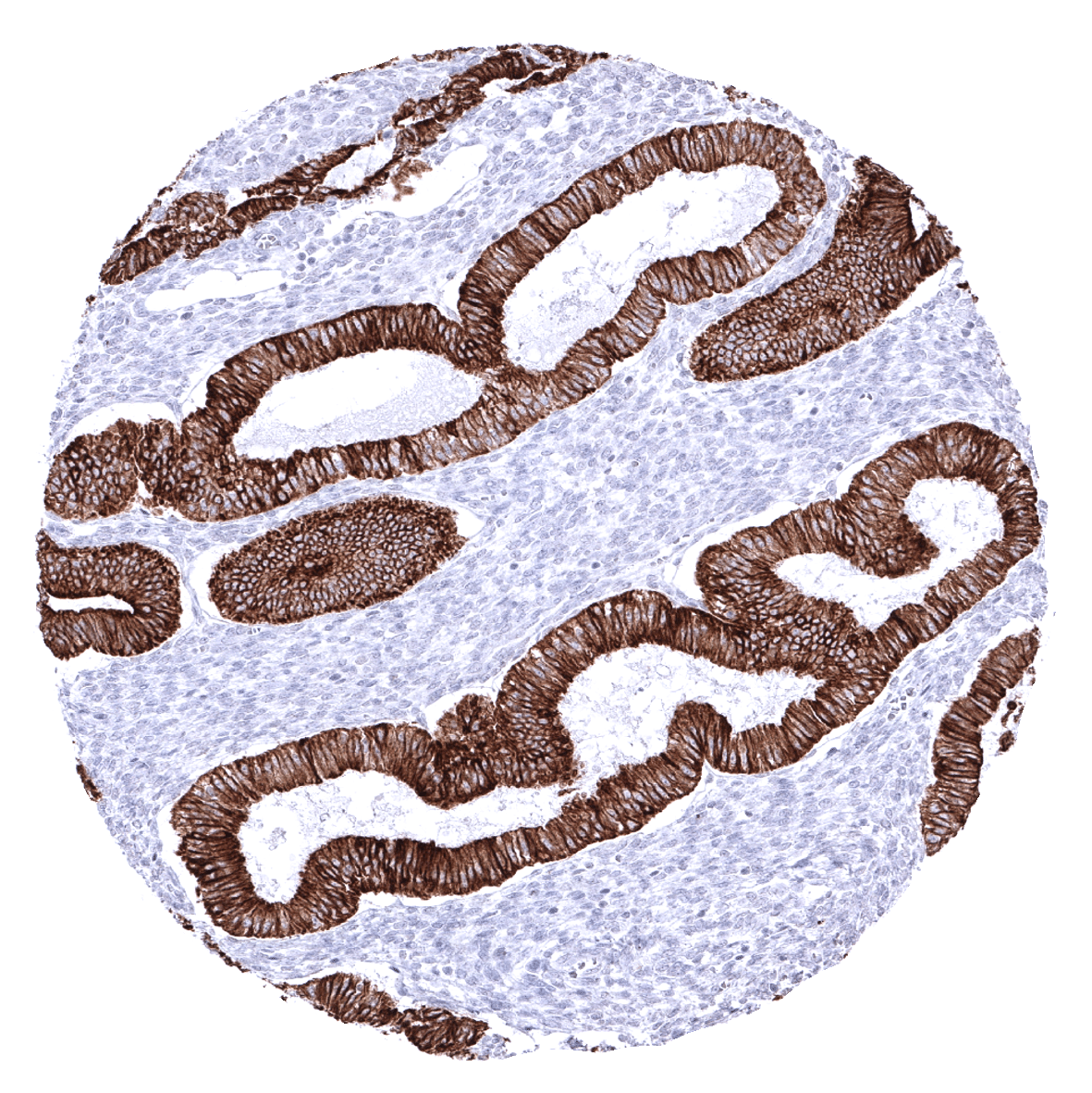
Endometrium, proliferation
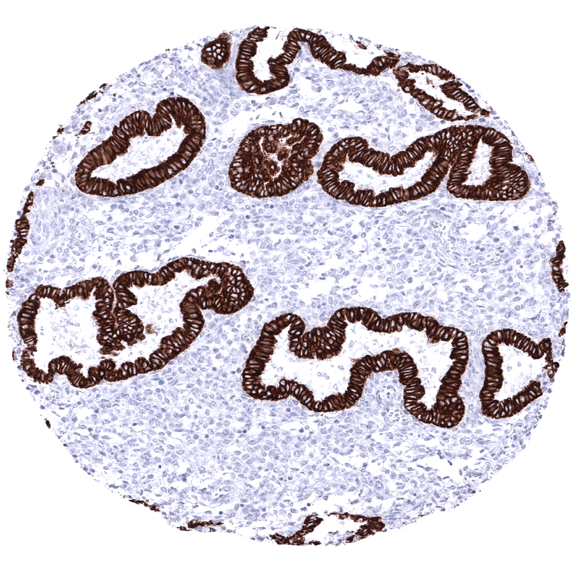
Endometrium, secretion
Epididymis
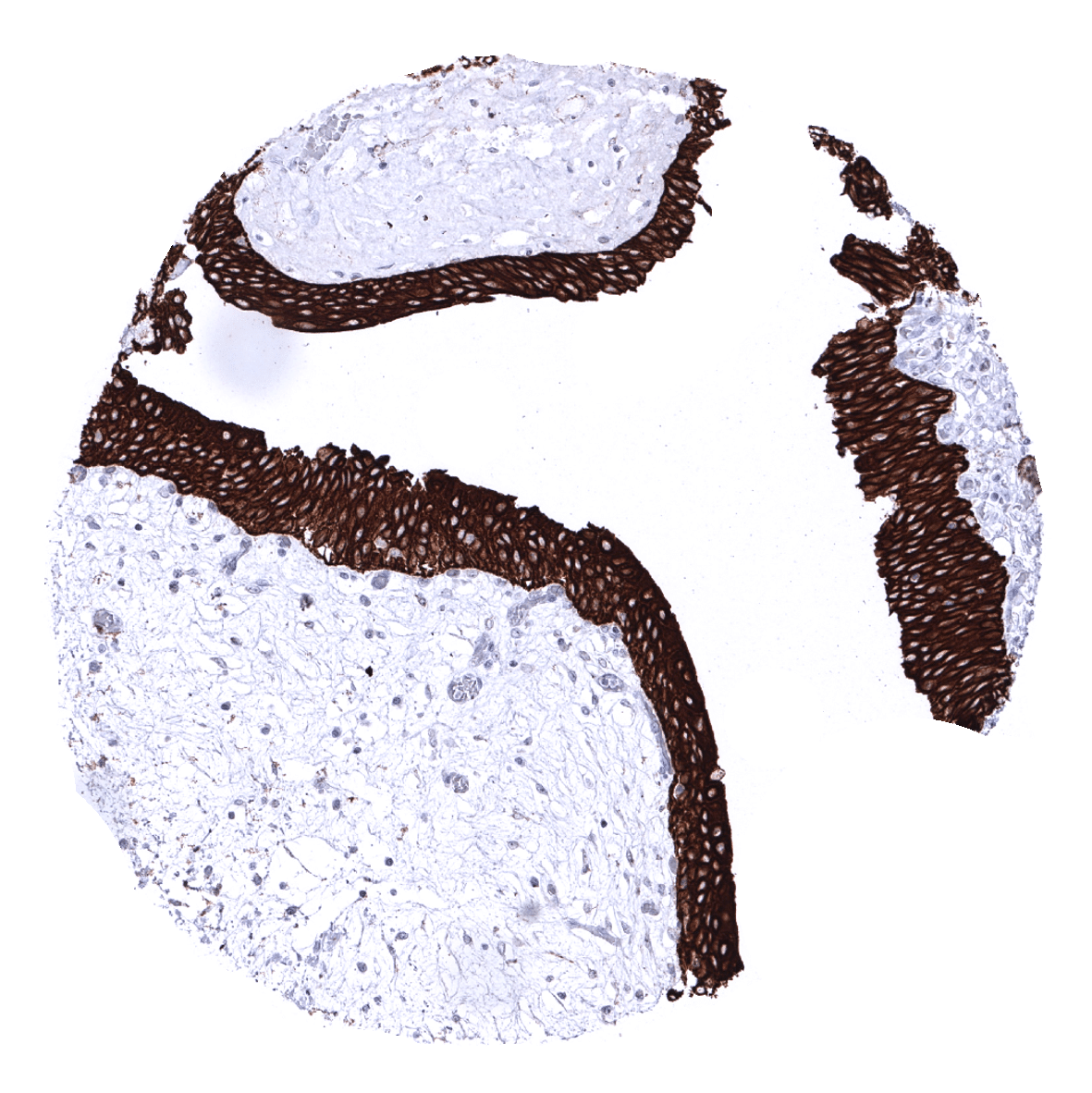
Esophagus, squamous epithelium

Fat
Gallbladder, epithelium
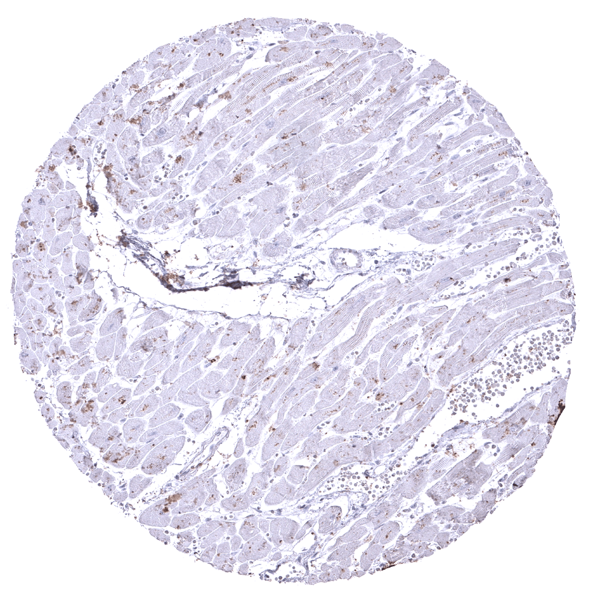
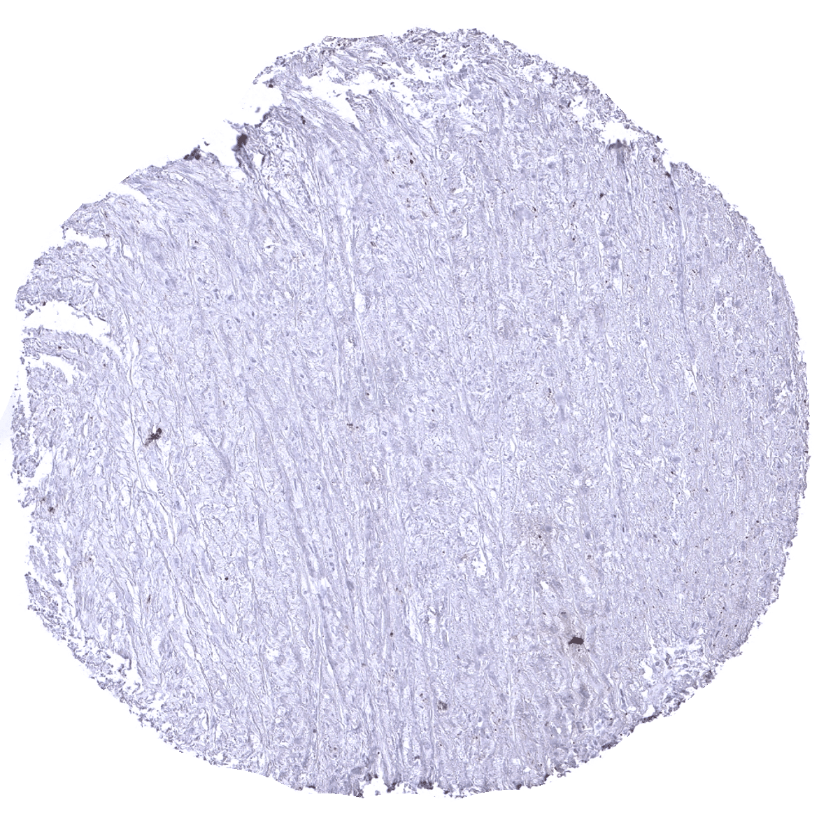
Heart - Ckpan negative heart muscle containing lipofuscine pigment.
Ileum, mucosa

Kidney, cortex
Kidney, medulla
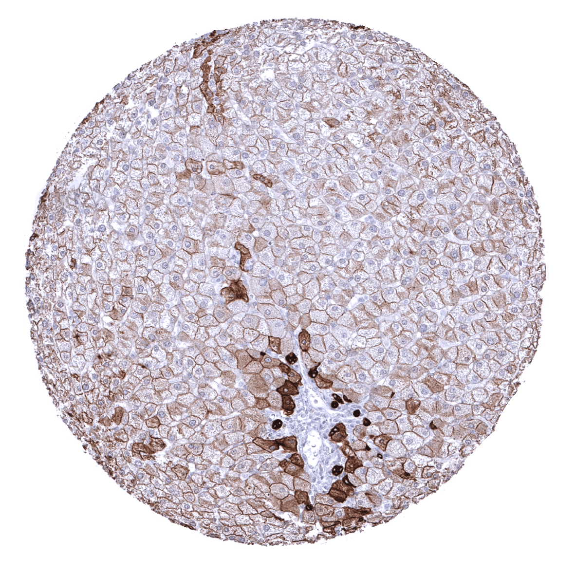
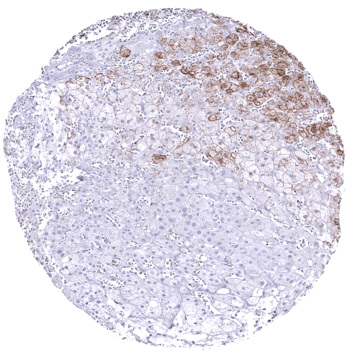
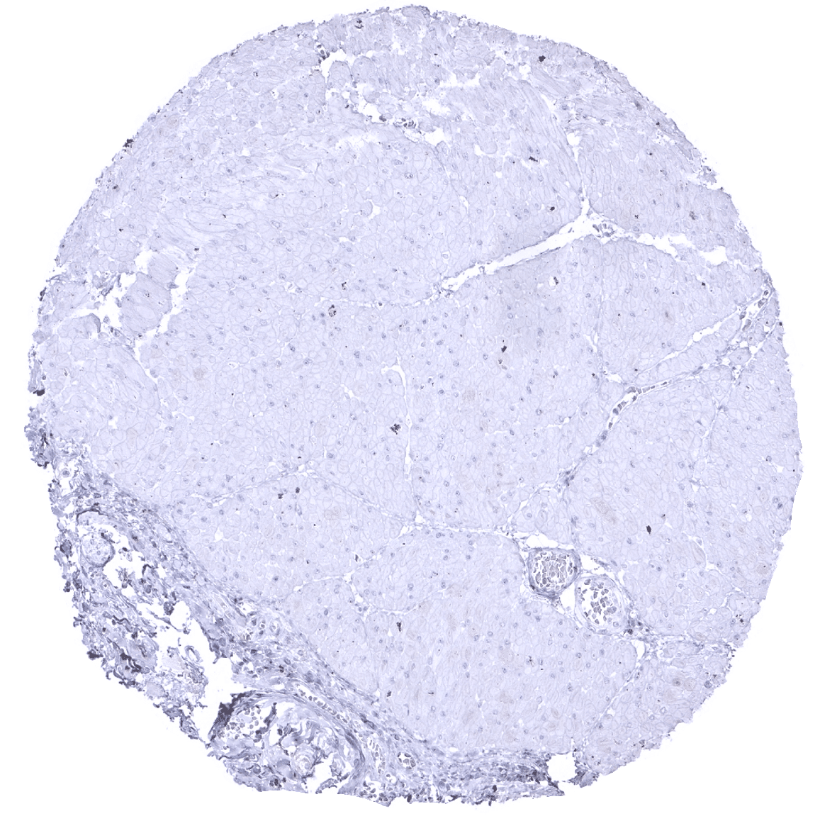
Liver - In the liver, a strong cytoplasmic staining of all bile ductal epithelial cells and an at least moderate, predominantly membranous immunostaining of hepatocytes is seen.
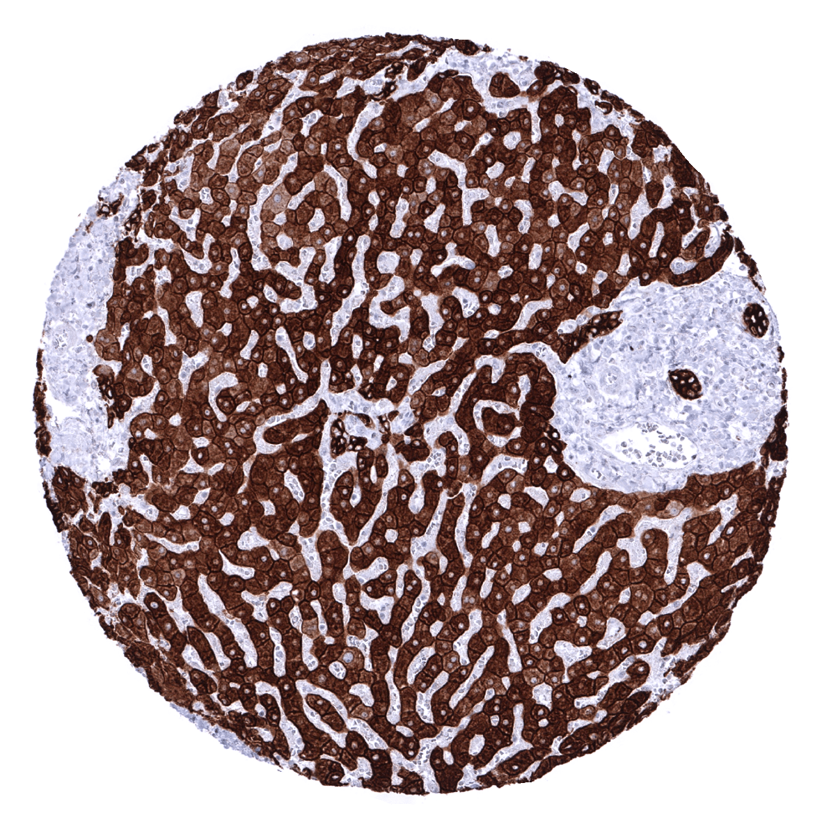
Liver - In the liver, a strong cytoplasmic staining of all bile ductal epithelial cells and an at least moderate, predominantly membranous immunostaining of hepatocytes is seen.
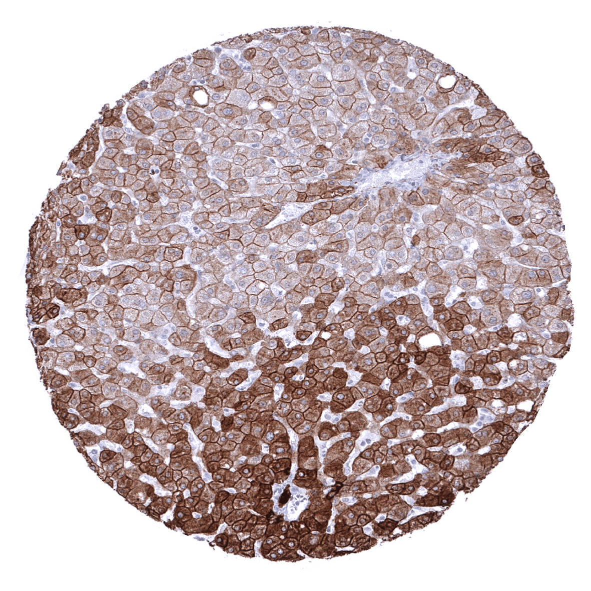
Liver - The Ckpan immunostaining pattern of hepatocytes can show a zonal variability.

Lung
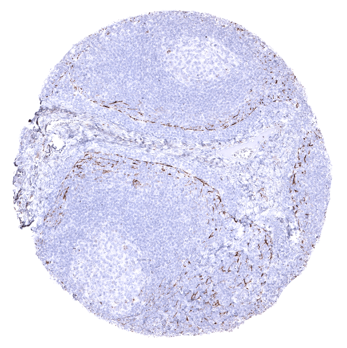
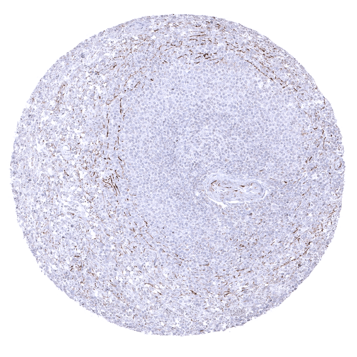
Lymph node - A delicate fibrillar staining caused by fibroblastic reticulum cells is seen, mainly in the interfollicular area.

Ovary, stroma
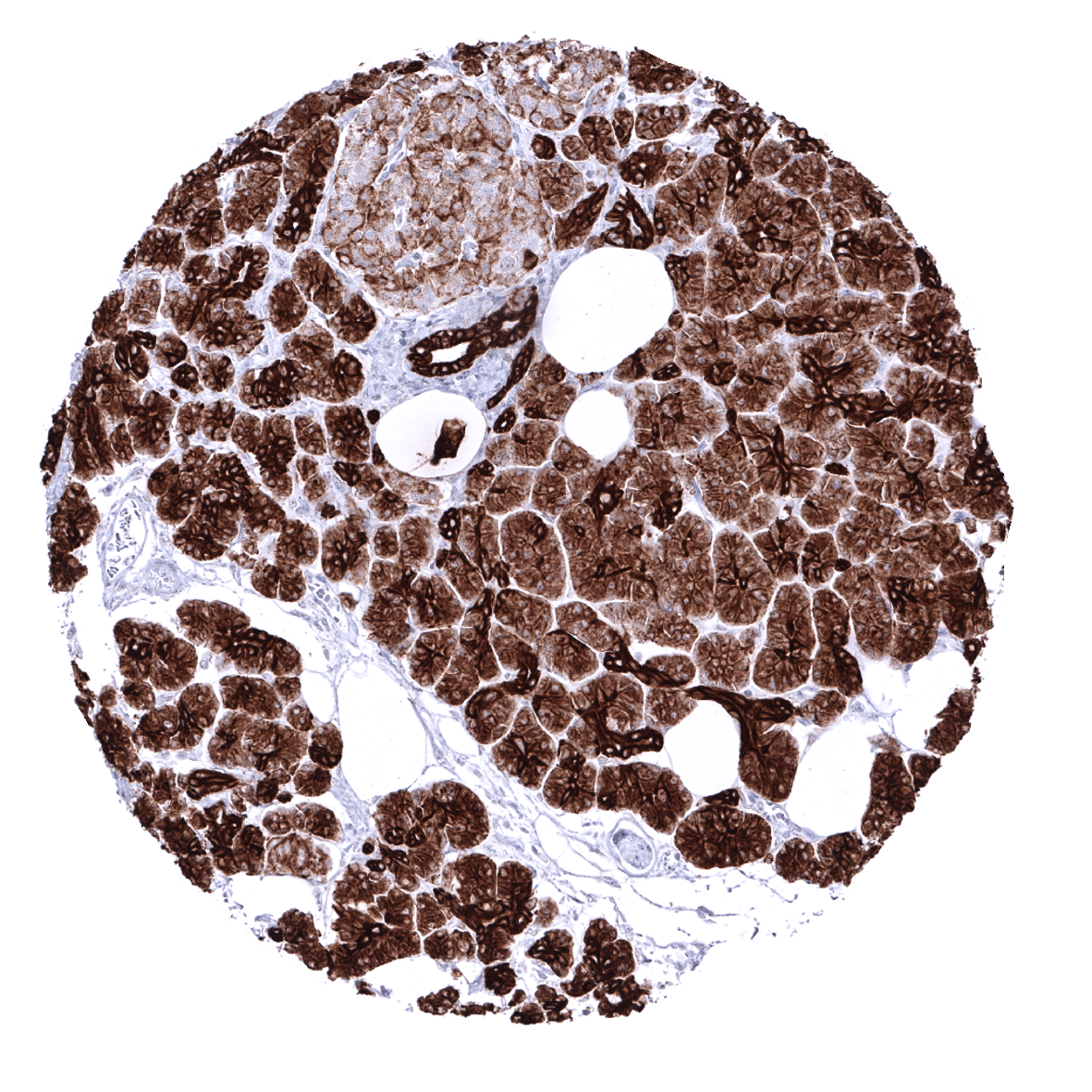
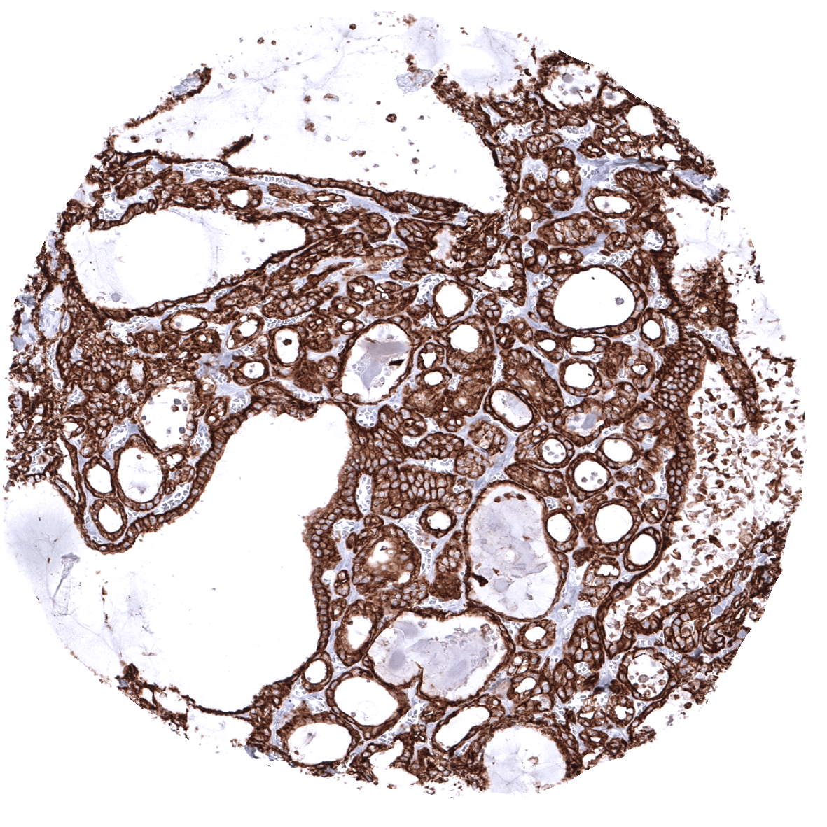
Pancreas - In the pancreas, the intensity of Ckpan immunostaining decreases from intercalated and excretory ducts to acinar cells and is even lower in islet cells.
Parathyroid
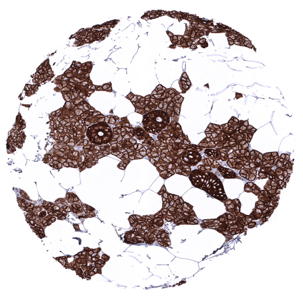
Parotid gland - In the parotid gland, the intensity of Ckpan immunostaining is highest in excretory ducts.

Pituitary gland, anterior lobe
Pituitary gland, posterior lobe
Pregnant uterus (decidua)

Placenta, early

Placenta (amnion and chorion)

Placenta, mature
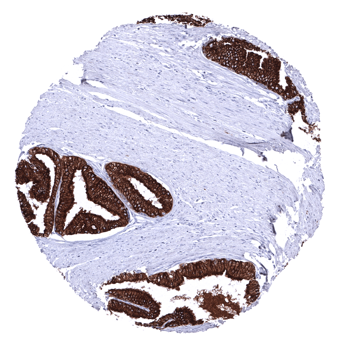
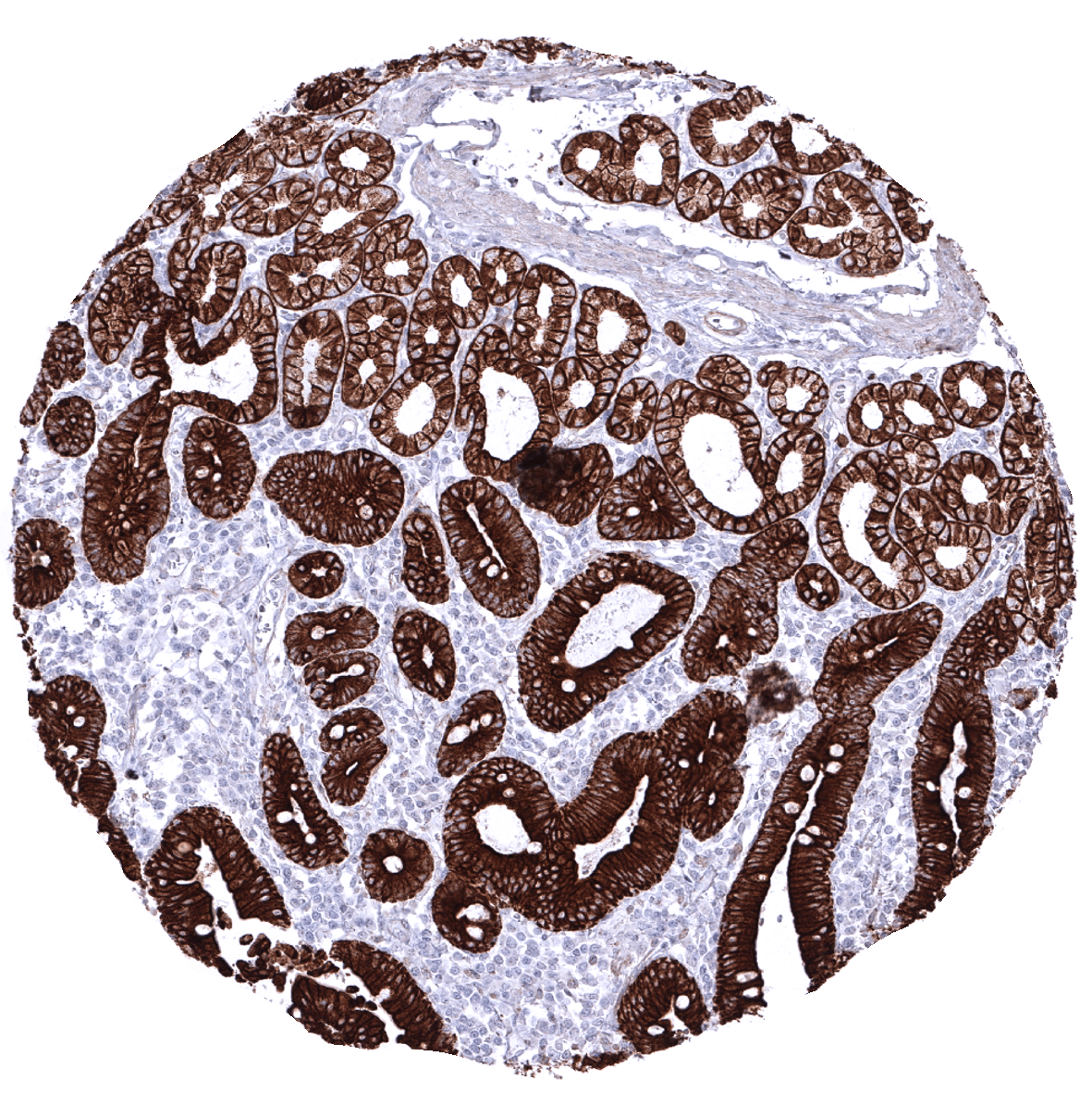
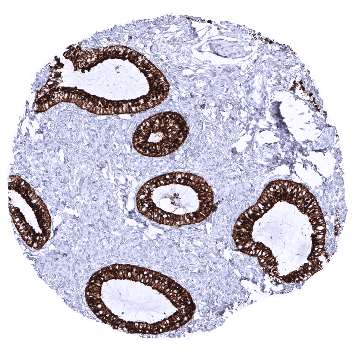
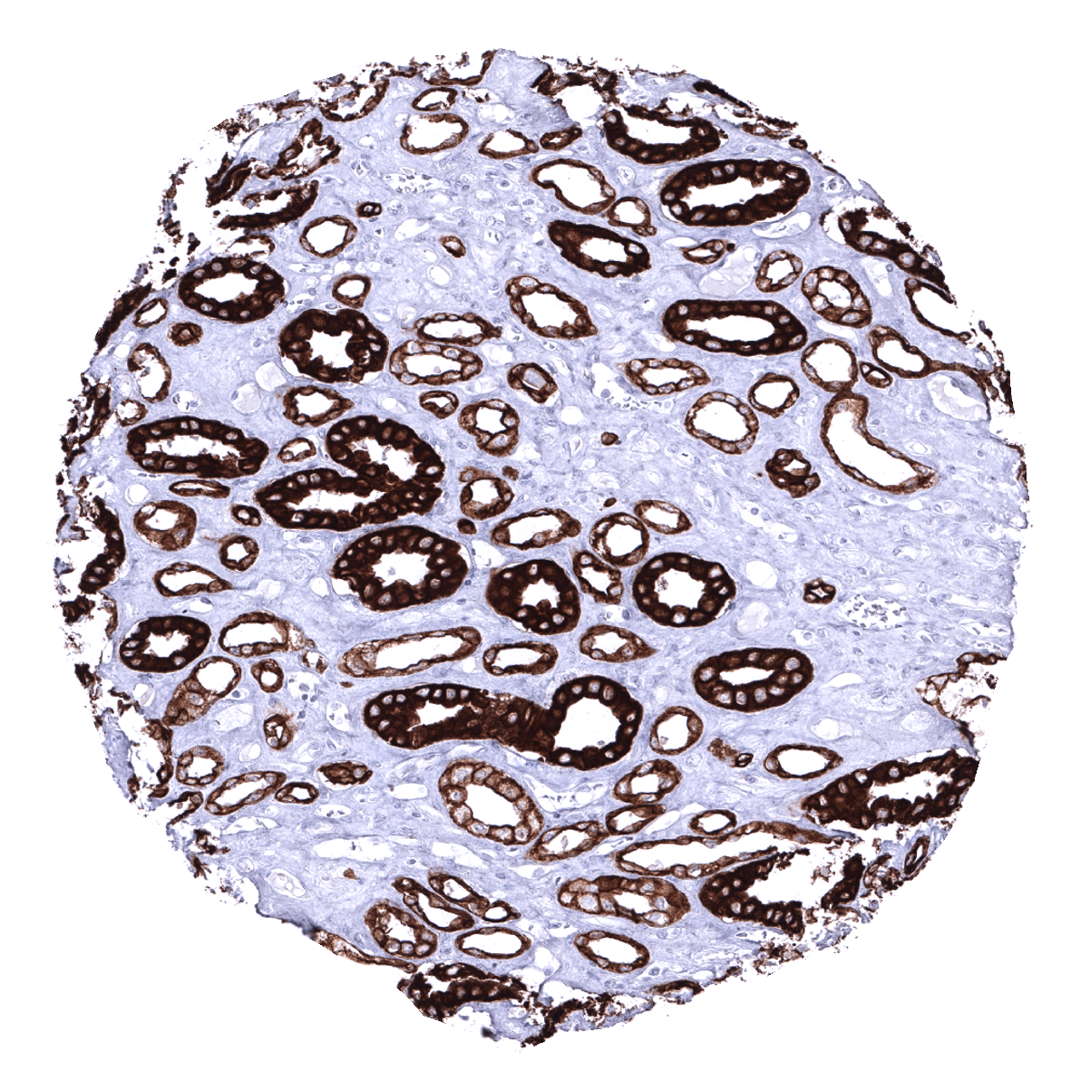
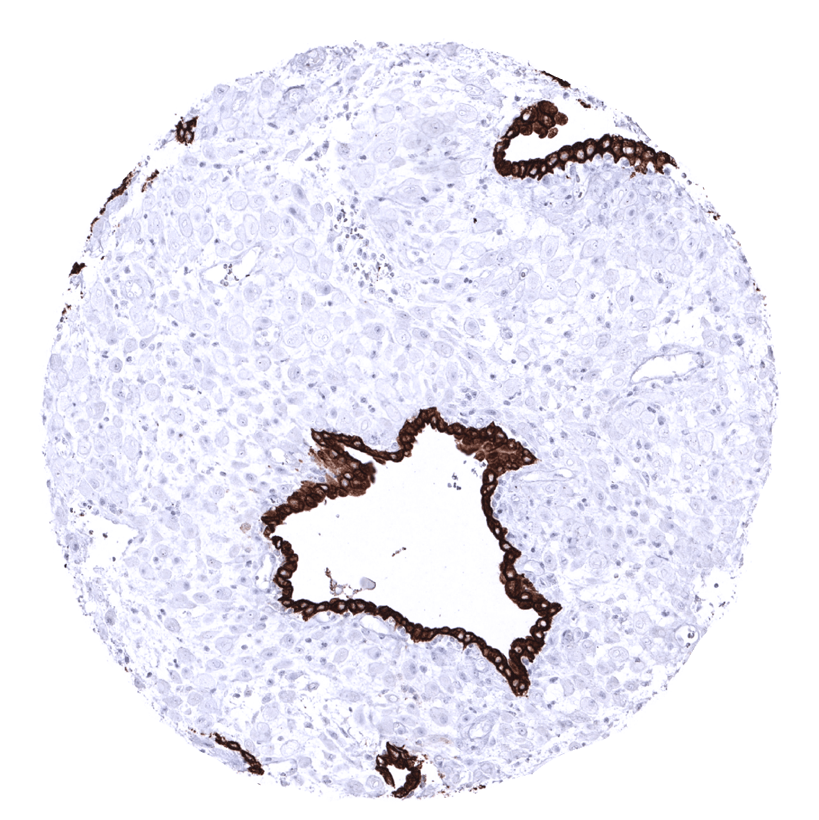
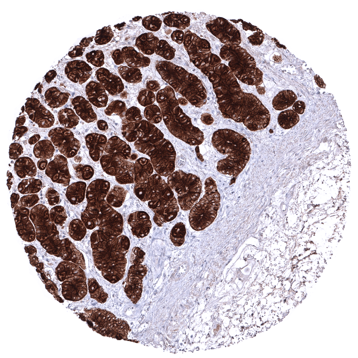
Prostate
Rectum, mucosa

Seminal vesicle
Sinus paranasales
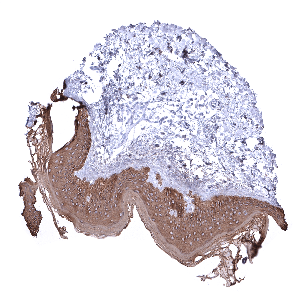
Skin
Spleen - A delicate fibrillar staining is caused by fibroblastic reticulum cells.
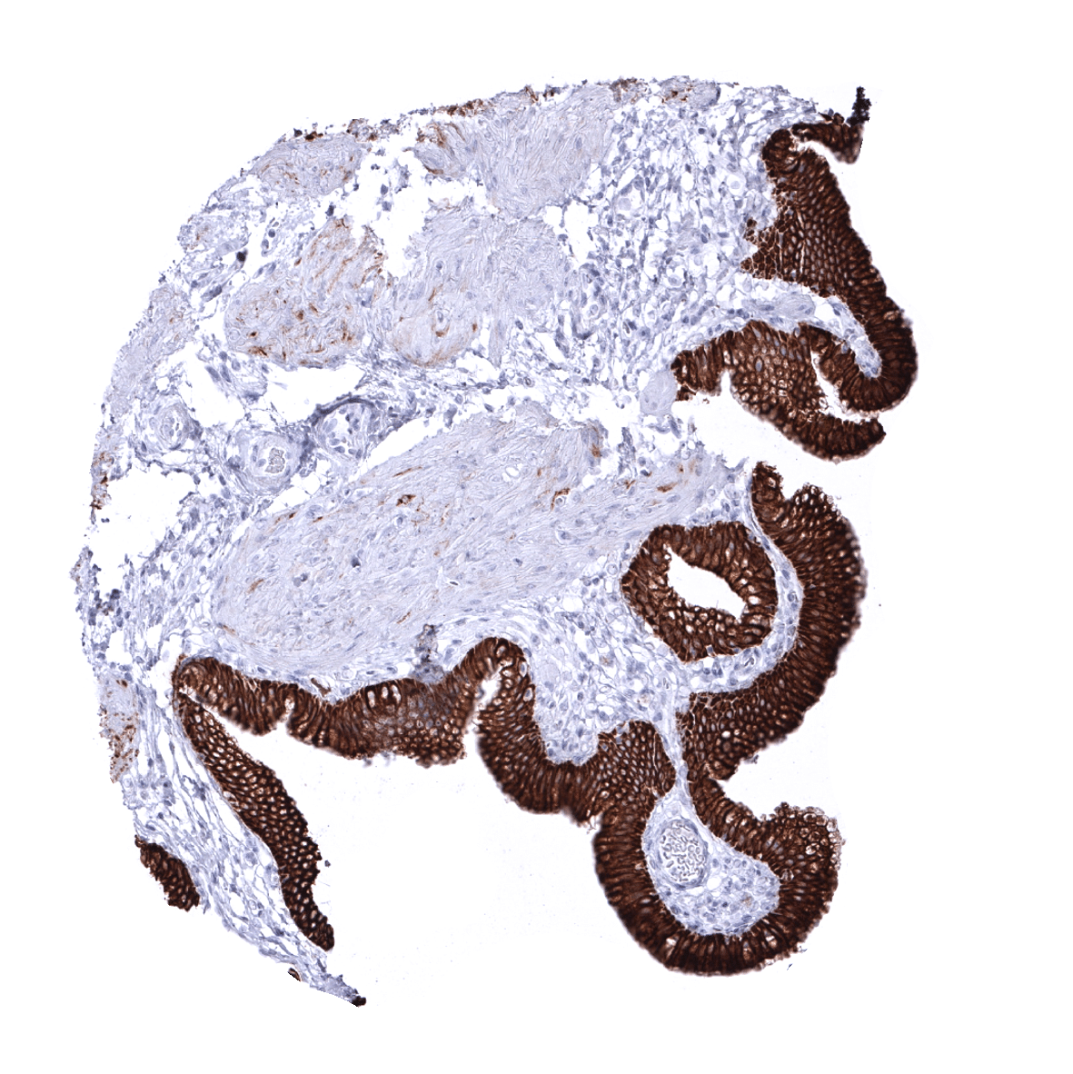
Stomach, antrum
Stomach, corpus
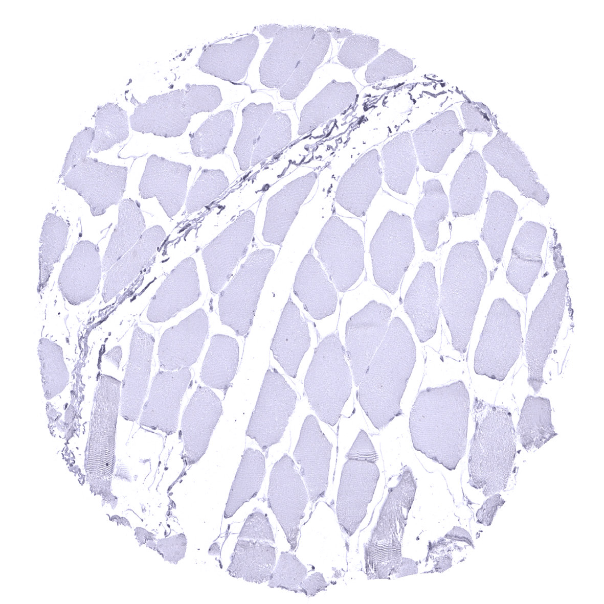
Striated muscle

Testis

Thymus
Thyroid gland
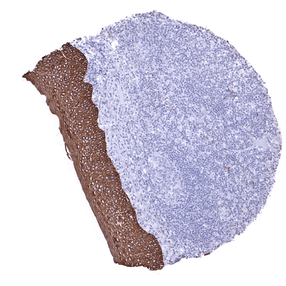
Tonsil, surface epithelium - Squamous epithelial cells are strongly CKpan positive while all lymphocytes are CKpan negative.

Tonsil - CKpan stains all epithelial cells in the tonsil crypts.
Urinary bladder, muscular wall
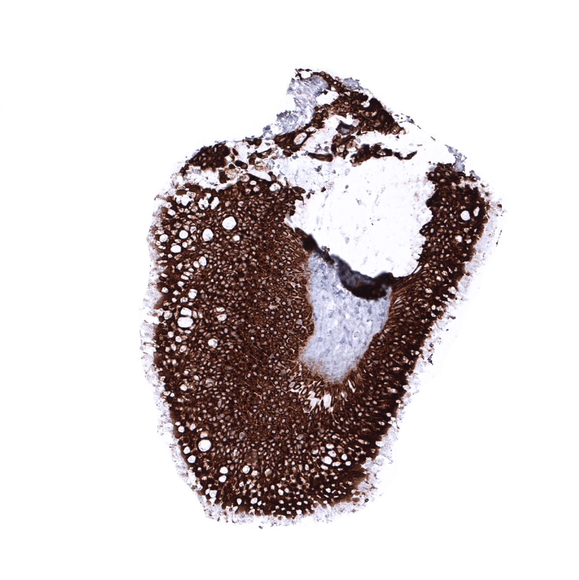
Urinary bladder, urothelium
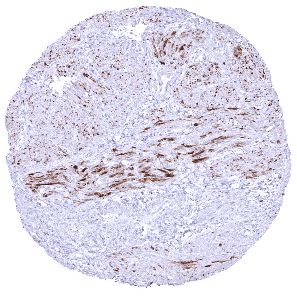
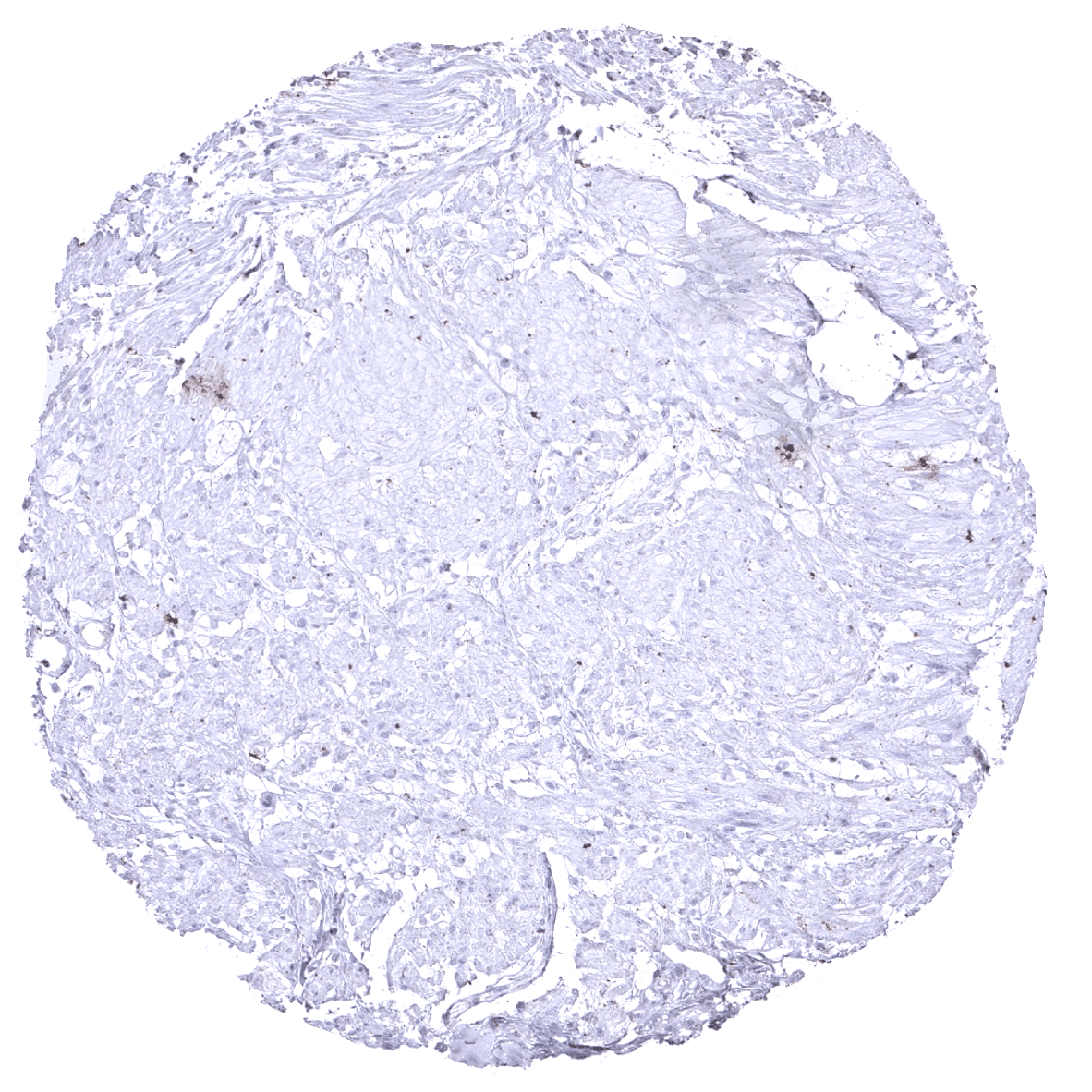
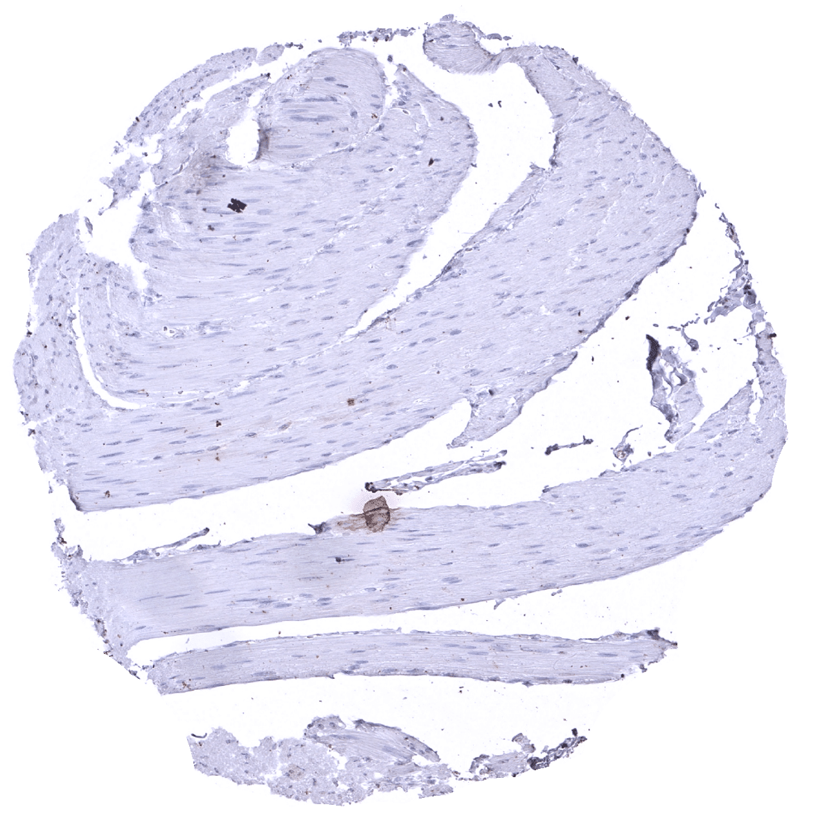
Uterus, myometrium - In the uterus, bundles of smooth muscle cells show weak to moderate Ckpan immunostaining.
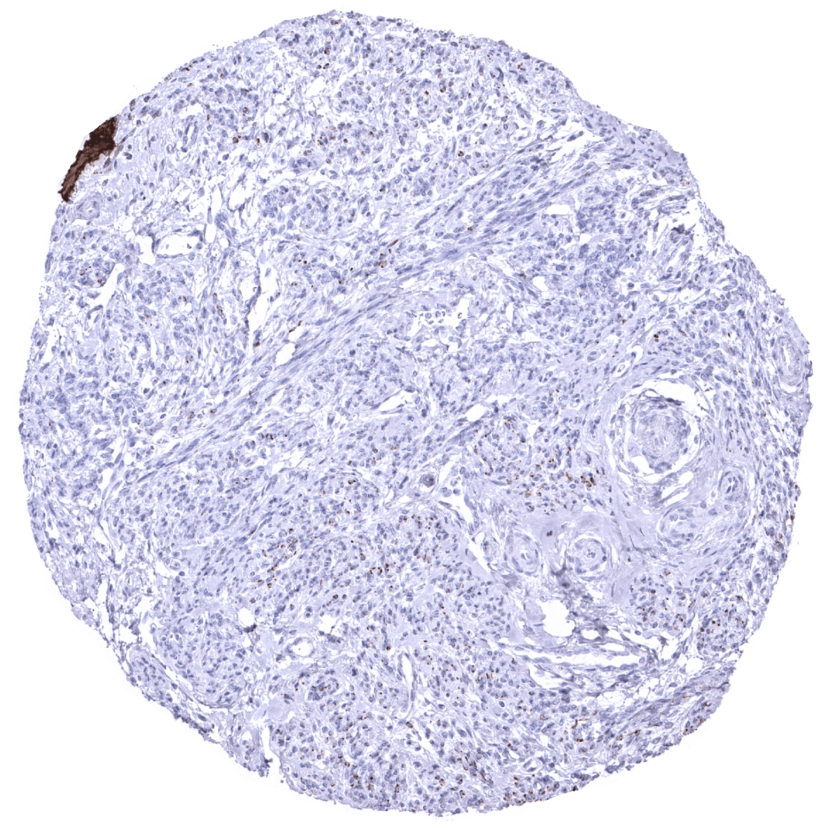
Uterus, myometrium - CKpan staining is not always seen in myometrium samples.