195,00 € – 695,00 €
Product details
Synonyms = k19; k1cs; Keratin 19 Keratin Type i 40kD; krt19
Antibody type = Mouse monoclonal / IgG1
Clone = MSVA-119M
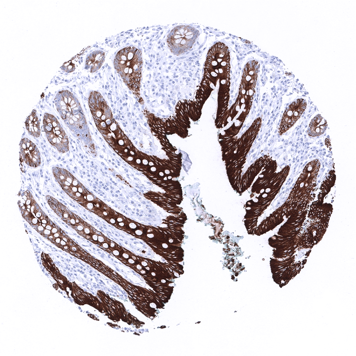
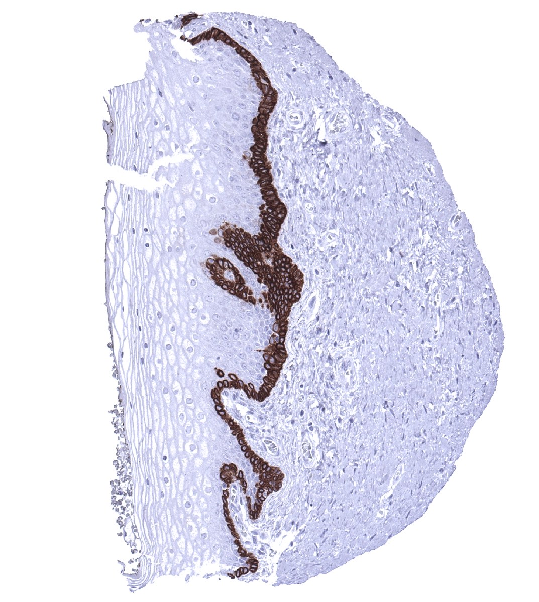
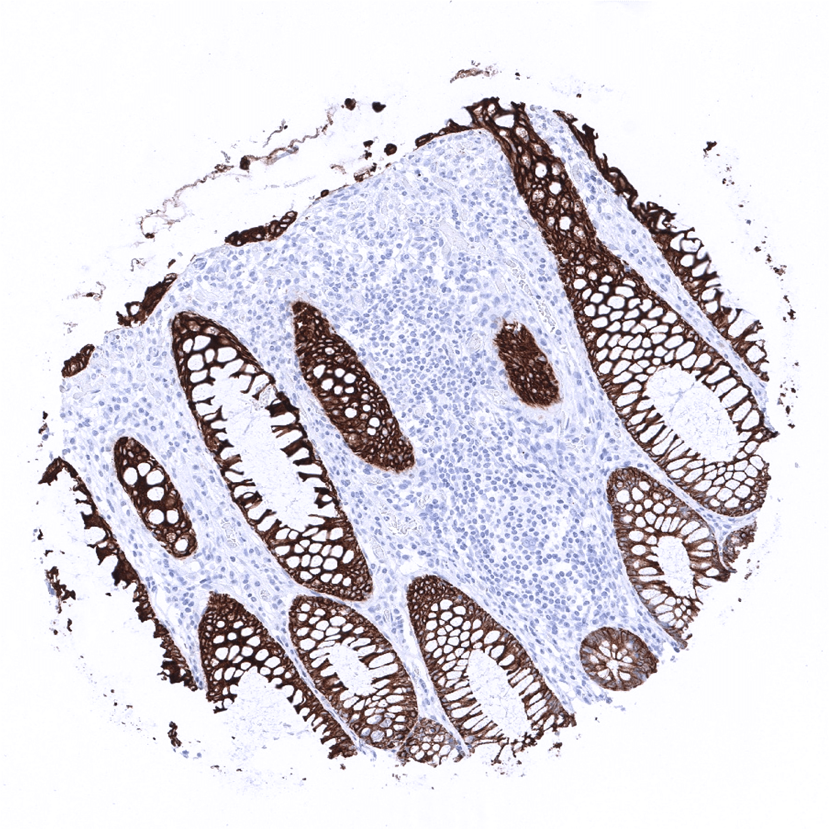
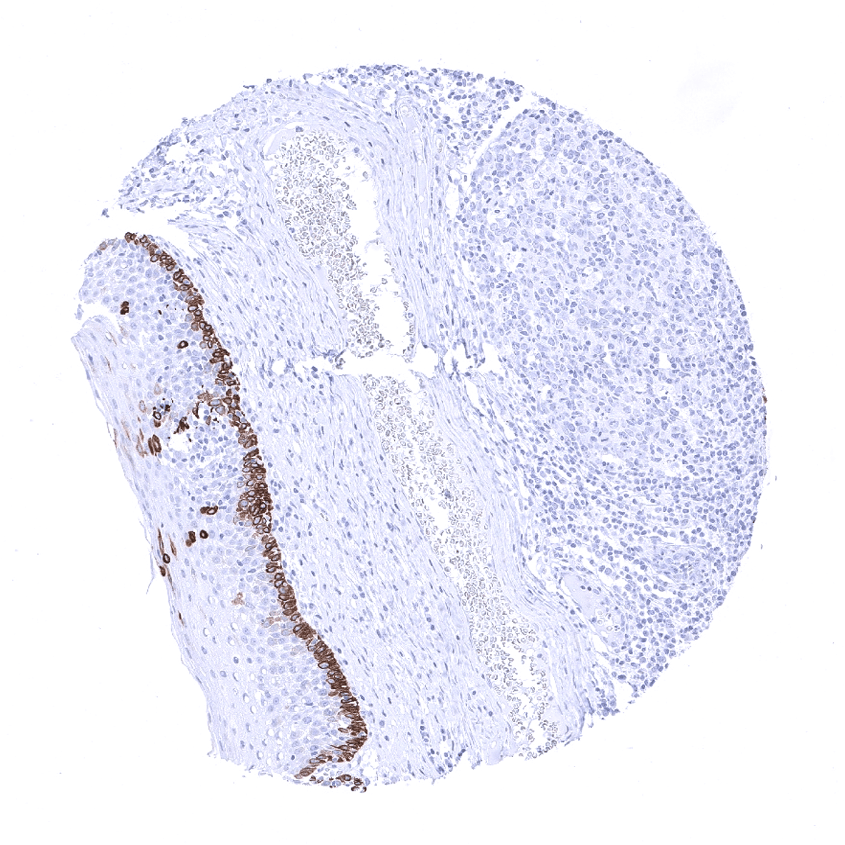
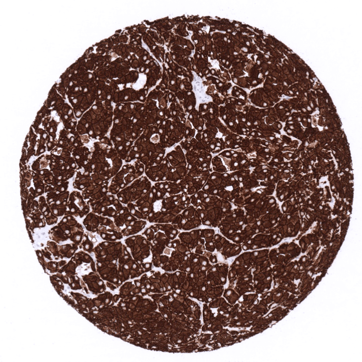
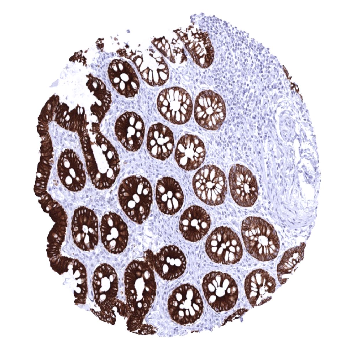
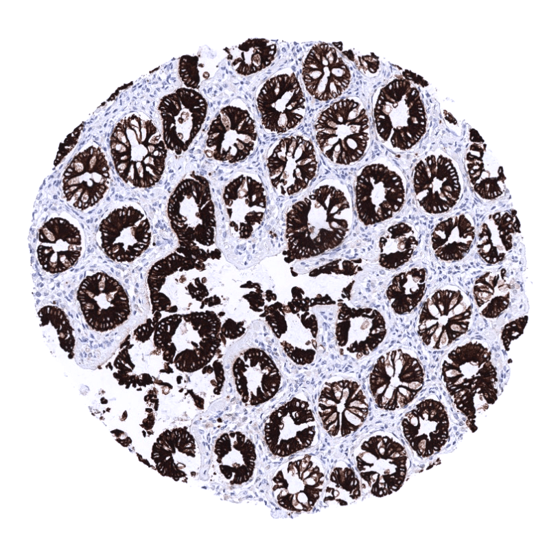
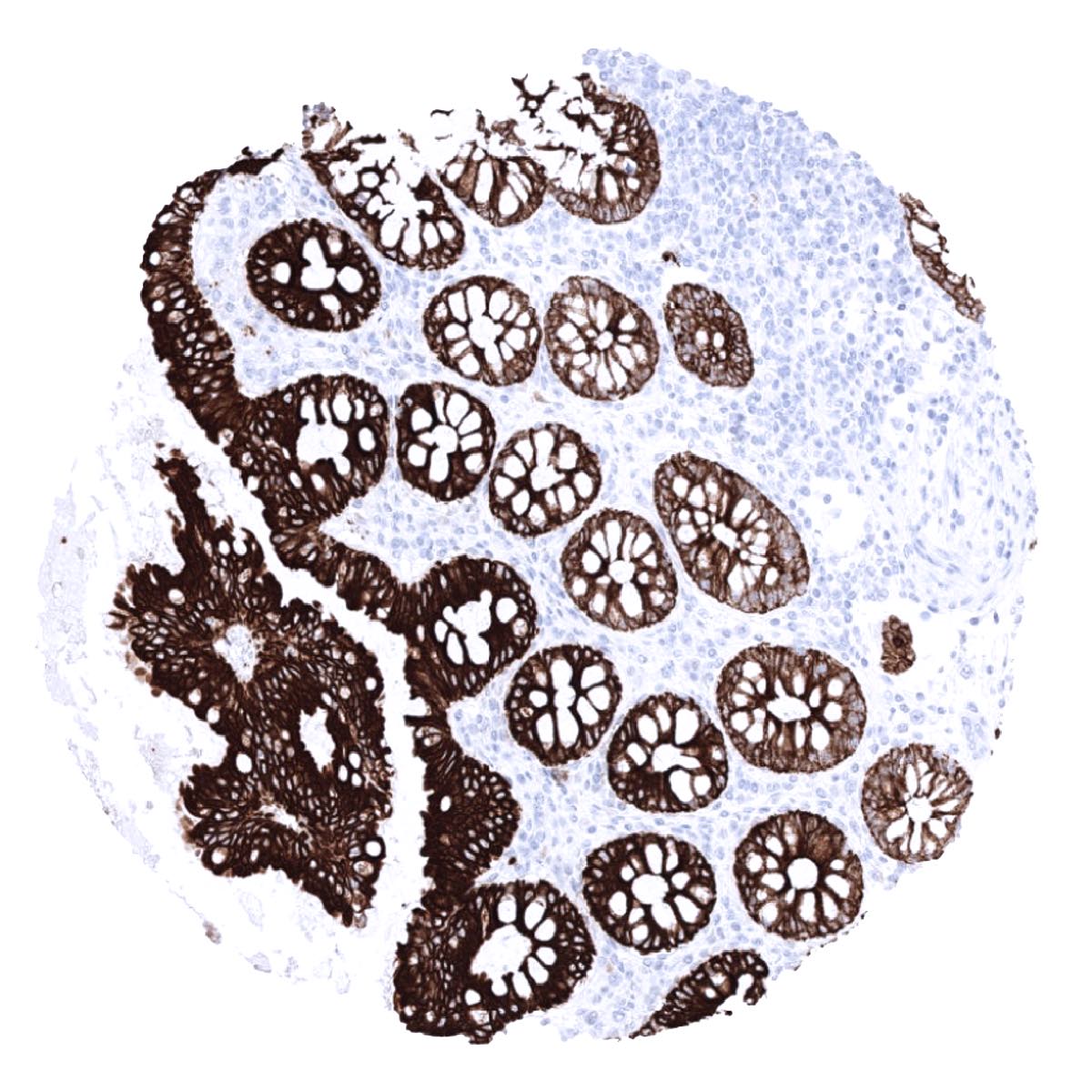
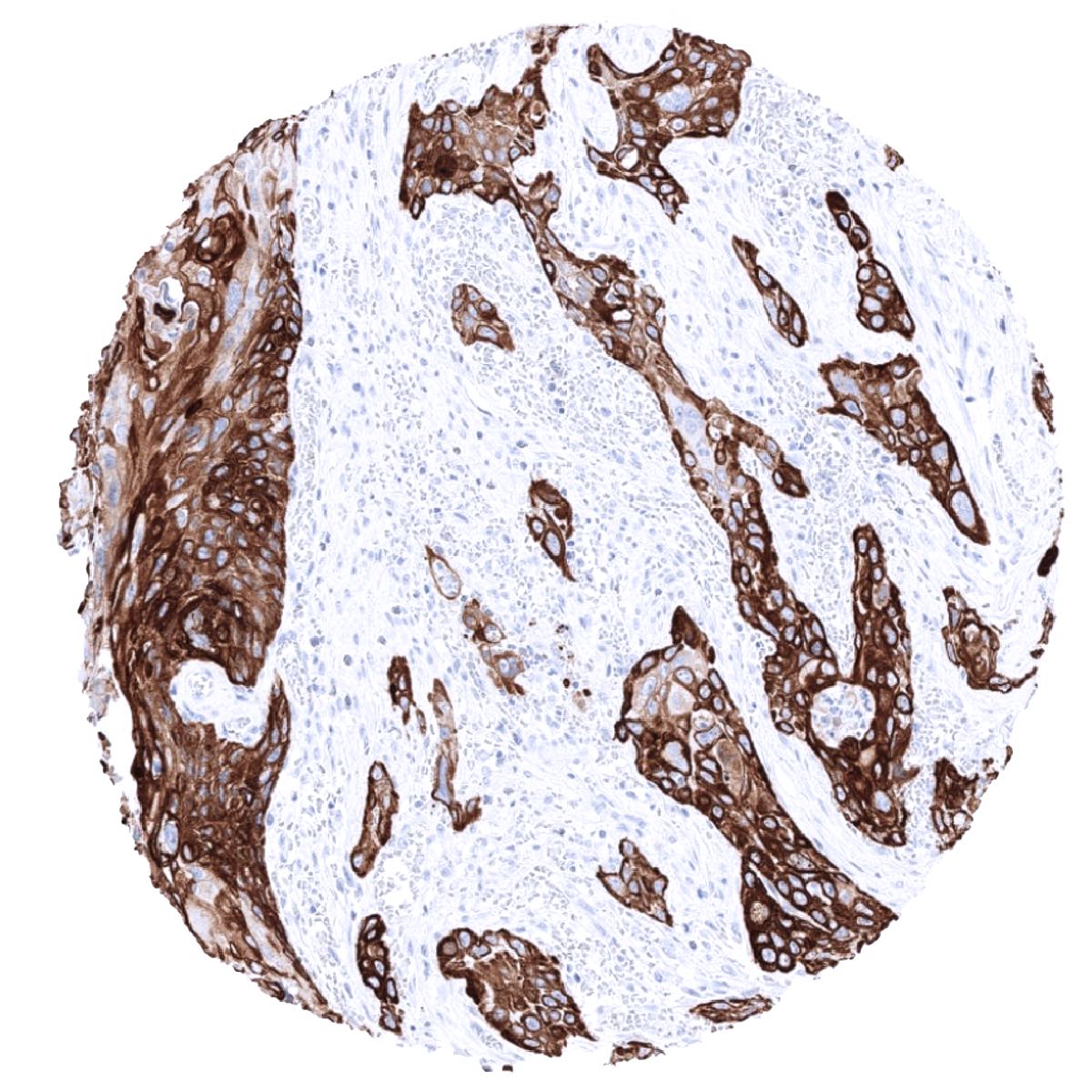
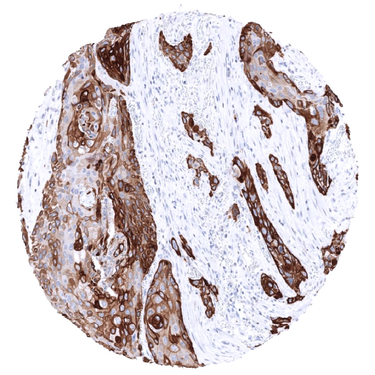
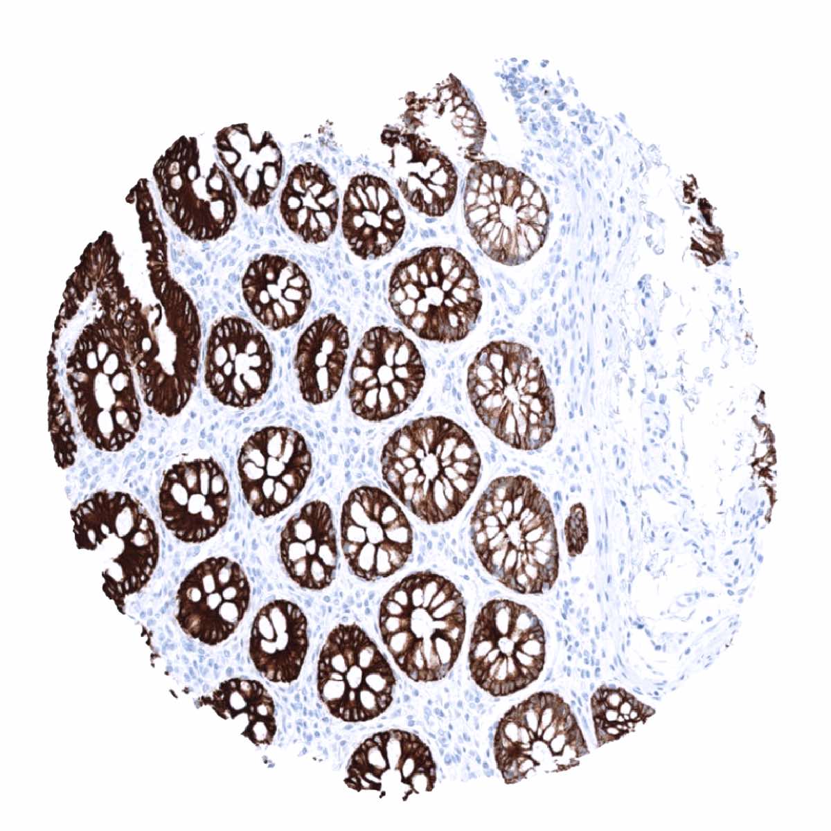
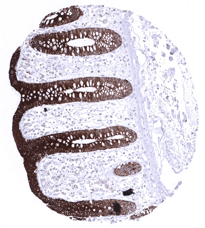
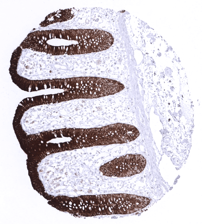
Positive control = Colon: A strong immunostaining is seen in all epithelial cells. Tonsil: A moderate to strong immunostaining occurs in the basal cell layer of the squamous epithelium of the tonsil surface.
Negative control = Colon: All non-epithelial cells are CK19 negative. Tonsil: All non-basal squamous epithelial cells and all non-epithelial cells are CK19 negative.
Cellular localization = Cytoplasmic
Reactivity = Human
Application = Immunohistochemistry
Dilution = 1:100 – 1:200
Intended Use = Research Use Only
Relevance of Antibody
Cytokeratin 19 is highly expressed in many tumor types but virtually absent in hepatocellular carcinoma.
Biology Behind
Cytokeratin 19 (CK19) is an acidic cytokeratin encoded by the KRT19 gene located in a keratin gene cluster at the human chromosome 17q12-q21. With a molecular mass of only 40kD, CK19 is the smallest member of the type 1 family of human keratins. While type 1 acidic cytokeratins typically form dimers with a type 1 basic cytokeratin, CK19 is an exception from this rule. However, like other epithelial cytokeratins, CK19 functions as a cytoskeletal scaffold protein to shape and strengthen the epithelial cell layer. Immunohistochemical analysis of cytokeratin 19 is often applied in diagnostic pathology because the protein is abundantly expressed in the majority of epithelial tumors.[1]
[1] Menz et al. “Diagnostic and prognostic impact of cytokeratin 19 expression analysis in human tumors: a tissue microarray study of 13,172 tumors” Human Pathology: Volume 115, September 2021
Staining Pattern in Normal Tissues
Cytokeratin 19 staining pattern in Normal Tissues with antibody MSVA-119M (images are shown in our “Normal Tissue Gallery”)
| Brain | Cerebrum | Negative. |
| Cerebellum | Negative. | |
| Endocrine Tissues | Thyroid | Negative. |
| Parathyroid | Negative. | |
| Adrenal gland | Negative. | |
| Pituitary gland | Negative. | |
| Respiratory system | Respiratory epithelium | A moderate to strong Upk1b staining occurs in a fraction of cells. |
| Lung | ||
| Gastrointestinal Tract | Salivary glands | A variable (weak to strong) Upk1b positivity can sometimes occur in some serous cells in salivary glands. |
| Esophagus | Usually negative. Scattered Upk1b positive cells can occur. | |
| Stomach | A moderate to strong Upk1b staining is seen in superficial epithelial cells and parietal cells. | |
| Colon | Negative. | |
| Duodenum | Negative. | |
| Rectum | Negative. | |
| Small intestine | Negative. | |
| Liver | A variable (weak to strong) cytoplasmic and membranous Upk1b positivity can sometimes occur in intrahepatic bile ducts. Hepatocytes are negative. | |
| Gallbladder | A variable (weak to strong) cytoplasmic and membranous Upk1b positivity can sometimes occur in gallbladder epithelium | |
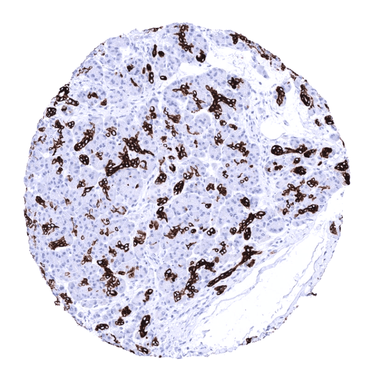
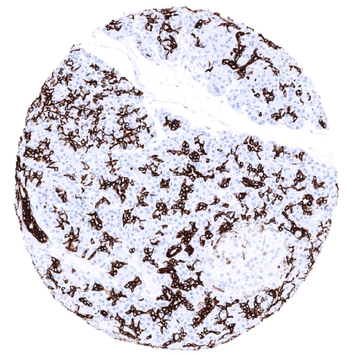
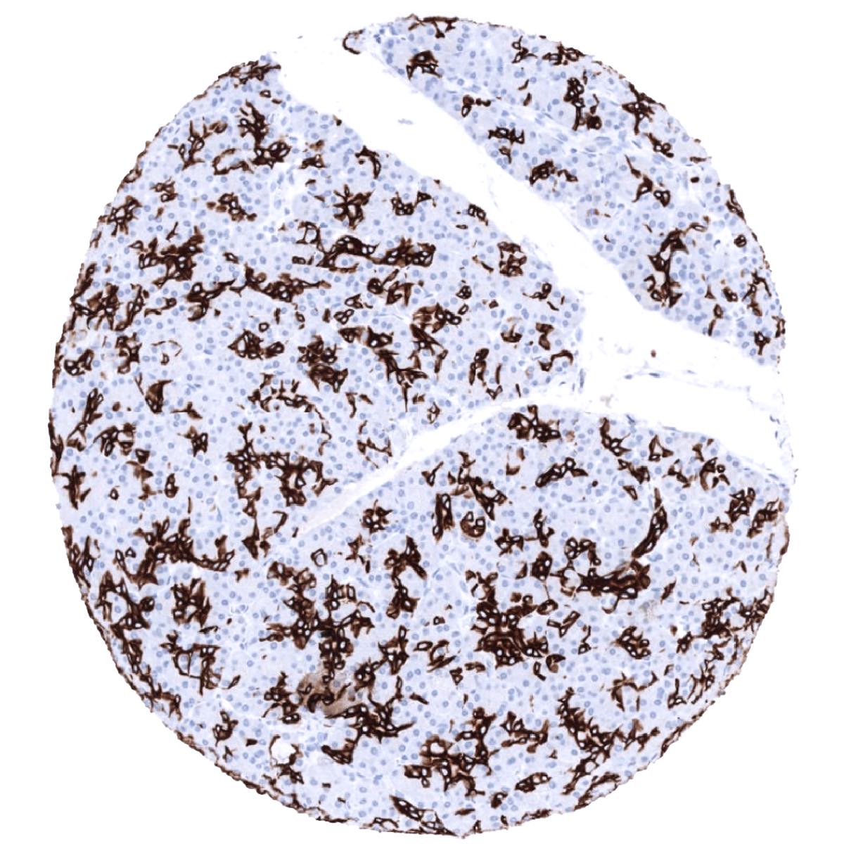
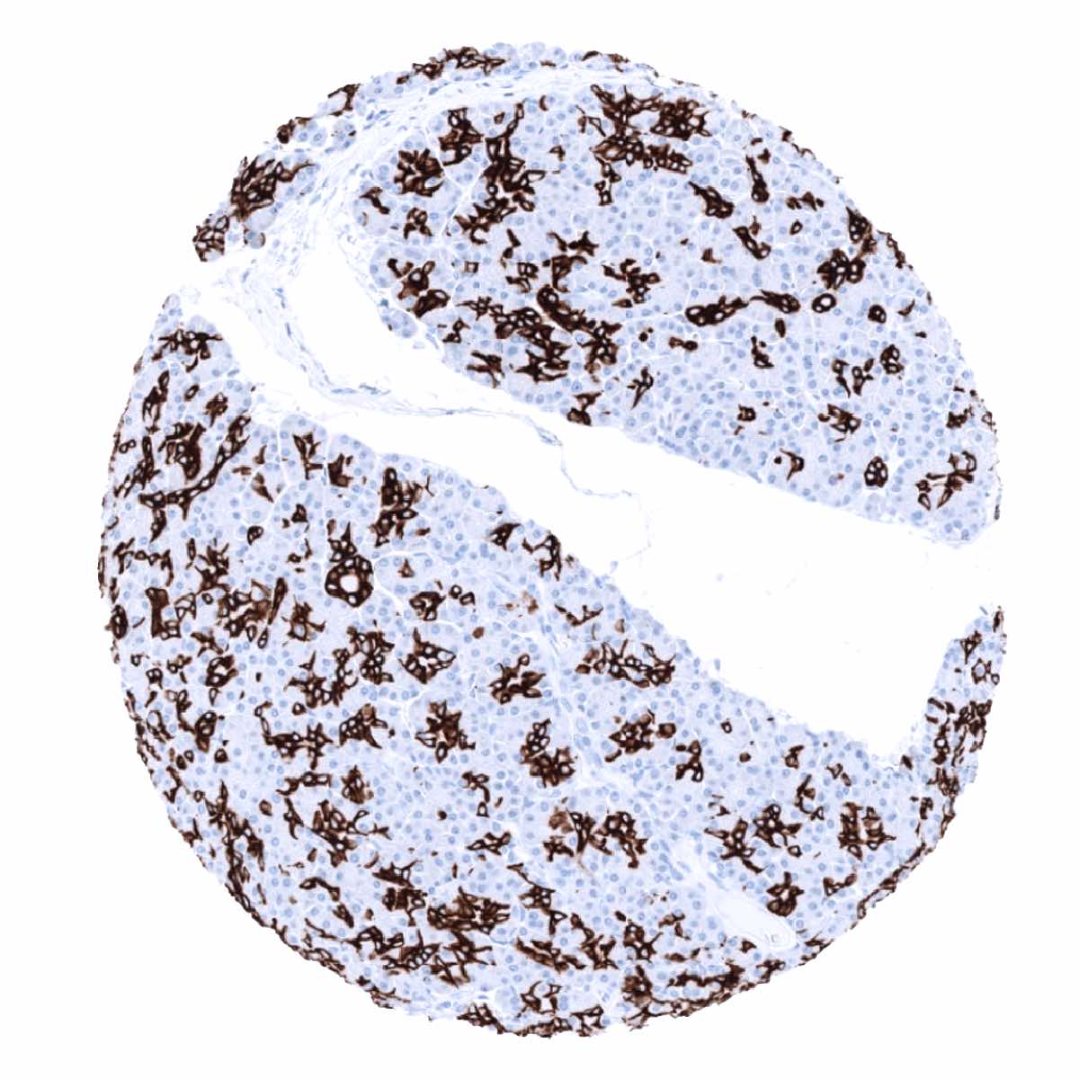
| Pancreas | Weak to moderate staining in intercalated ducts of the pancreas. Excretory ducts of the pancreas can also stain positive. | |
| Genitourinary | Kidney | A variable (weak to strong) cytoplasmic and membranous Upk1b positivity can sometimes occur in the parietal layer of Bowman capsule and in few tubuli in the kidney |
| Urothelium | Variable Upk1b staining. Equally involving all cell layers in the majority of samples but limited to the upper cell layers in some samples. | |
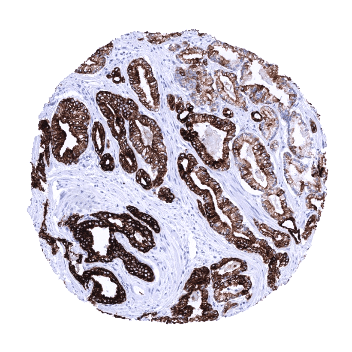
| Male genital | Prostate | Negative. |
| Seminal vesicles | Negative. | |
| Testis | Negative. | |
| Epididymis | Apical membrane of tall columnar cells of the epididymis may show a faint positive staining. | |
| Female genital | Breast | Negative. |
| Uterus, ectocervix | Usually negative. Scattered Upk1b positive cells can occur. | |
| Uterus endocervix | A variable (weak to strong) positivity can sometimes occur in some endocervical glands. | |
| Uterus, endometrium | A variable (weak to strong) Upk1b positivity can sometimes occur in some endometrial glands. | |
| Fallopian Tube | A variable (weak to strong) Upk1b positivity can sometimes occur in a fraction of cells in the fallopian tube. | |
| Ovary | Negative. | |
| Placenta early | A small fraction of the syncytiotrophoblast cells may stain positive. | |
| Placenta mature | A small fraction of the syncytiotrophoblast cells may stain positive. | |
| Amnion | Moderate to strong Upk1b positivity in amnion cells. | |
| Chorion | Moderate to strong Upk1b positivity in chorion cells. | |
| Skin | Epidermis | Usually negative. Scattered Upk1b positive cells can occur. |
| Sebaceous glands | Negative. | |
| Muscle/connective | Heart | Negative. |
| Skeletal | Negative. | |
| Smooth muscle | Negative. | |
| Fat | Negative. | |
| Bone marrow/lymphoid | Bone marrow | Negative. |
| Lymph node | Negative. | |
| Spleen | Negative. | |
| Thymus | Upk1b is positive in a subset of cells in corpuscles of Hassall’s. No staining of inflammatory cells. | |
| Tonsil | Upk1b is regularly positive in the surface epithelium and in the intermediate cell layer of tonsil crypt epithelium. No staining of inflammatory cells. | |
| Remarks | Staining is typically cytoplasmic and membranous. |
These findings fit well to the RNA and protein data described in the Human Protein Atlas (Tissue expression Cytokeratin 19). They add further detail with respect to the cell types responsible for CK19 expression and identify tissues such as the adenohypophysis, which were not described by the protein atlas to be CK19 positive.
Suggested positive tissue control: Colon: A strong immunostaining is seen in all epithelial cells. Tonsil: A moderate to strong immunostaining occurs in the basal cell layer of the squamous epithelium of the tonsil surface.
Suggested negative tissue control: Colon: All non-epithelial cells are CK19 negative. Tonsil: All non-basal squamous epithelial cells and all non-epithelial cells are CK19 negative.

Staining Pattern in Relevant Tumor Types
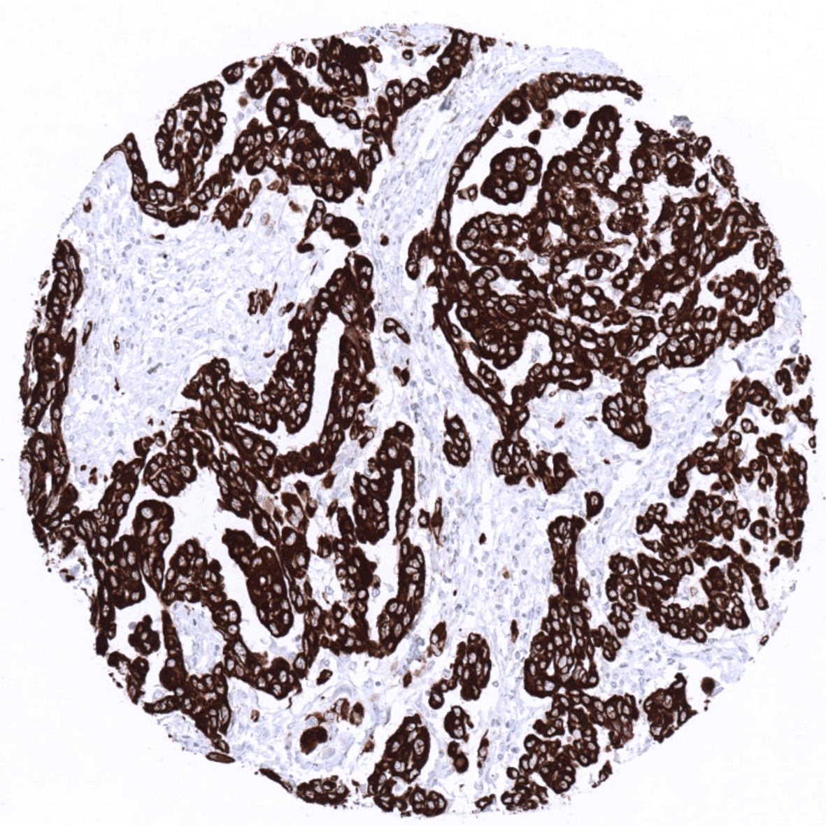
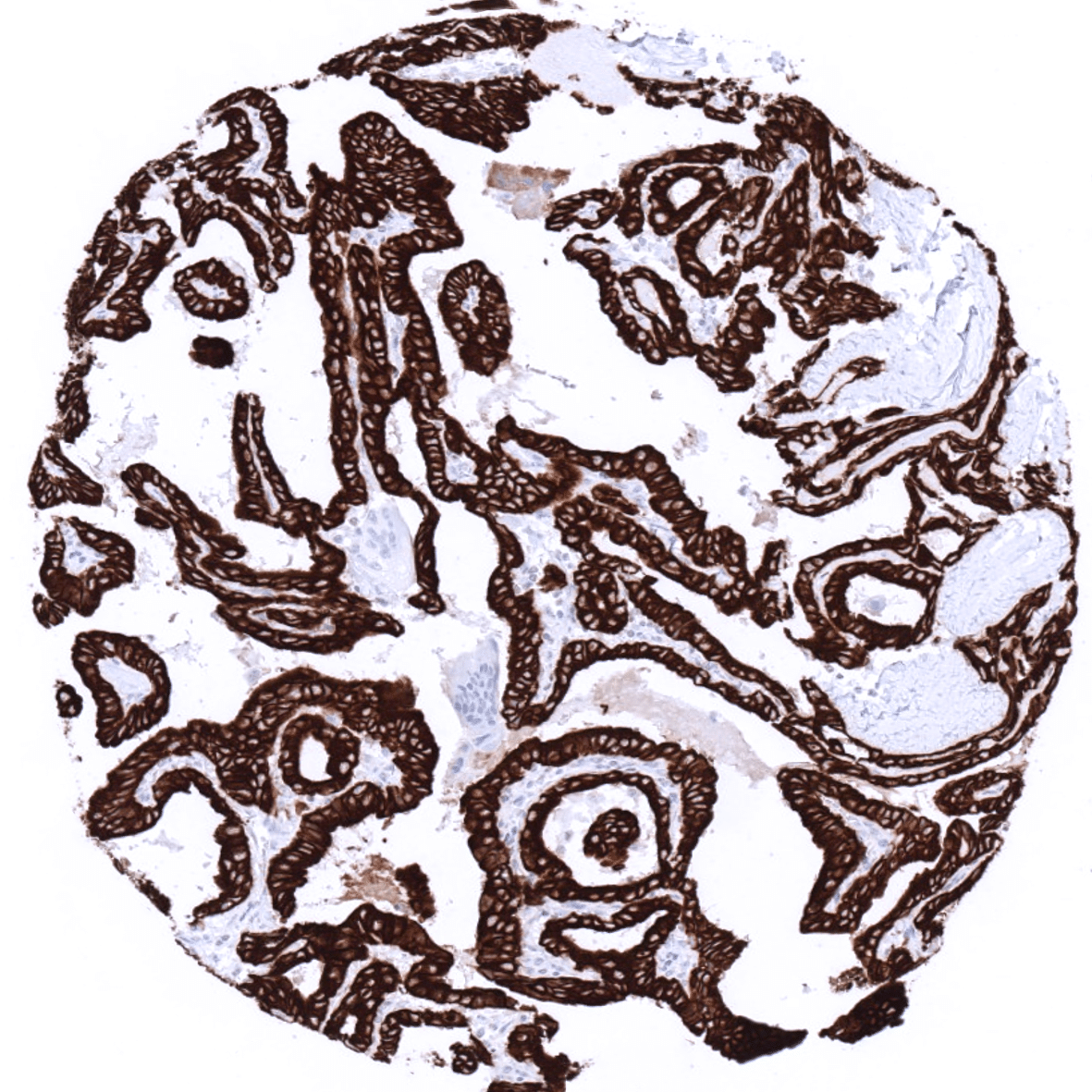
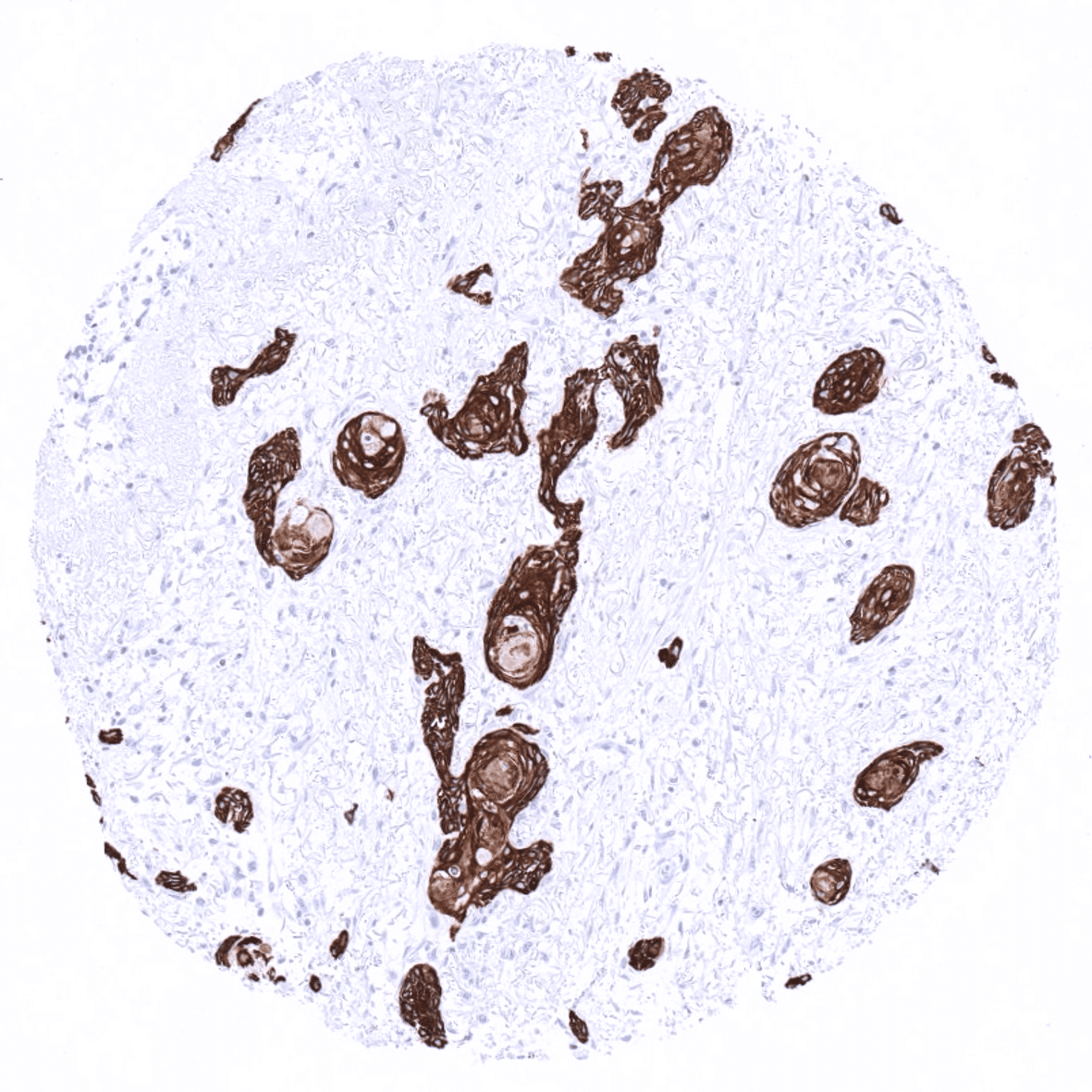
Cytokeratin 19 is almost always expressed in various important cancers such as in adenocarcinomas of the stomach, esophagus, and the colorectum, breast cancers, ovarian cancers, endometrium carcinoma cholangiocarcinomas, and urothelial carcinomas. CK19 immunostaining is usually absent in hepatocellular carcinoma, adrenal adenomas, seminomas, hematological malignancies, and most mesenchymal tumors.
Detailed data on Cytokeratin 19 staining by MSVA-119M obtained from an analysis of 13,172 tumors from 122 different tumors and subtypes have recently been published by Menz et al. “Diagnostic and prognostic impact of cytokeratin 19 expression analysis in human tumors: a tissue microarray study of 13,172 tumors”
The TCGA findings on Cytokeratin 19 RNA expression in different tumor categories have been summarized in the Human Protein Atlas.
Compatibility of Antibodies
Cytokeratin 19 (MSVA-119M) publication summary:
Menz et al.: “Diagnostic and prognostic impact of cytokeratin 19 expression analysis in human tumors: a tissue microarray study of 13,172 tumors” Human Pathology: Volume 115, September 2021
A total of 13,172 tumors were analyzed from 122 different tumor categories by using the following protocol: Heat-induced antigen retrieval for 5 minutes in an autoclave at 121°C in pH7,8 Target Retrieval Solution buffer. MSVA-119M at a dilution of 1:150 at 37°C for 60 minutes. Visualization of bound antibody by the EnVision Kit (Dako, Agilent). This protocol was also used for all stainings depicted in our tumor and normal tissue galleries.
At least one case with a positive cytokeratin 19 immunostaining was seen in 89 (73%) and at least one case with a strong cytokeratin 19 immunostaining was seen in 87 (71,3%) of 122 tumor categories. The distribution of positive staining results is shown “organ-systematic” and in a “ranking order” figure below (images based on data from Menz et al.). Results on possible associations with histopathological and clinical parameters of tumor aggressiveness in several cancer types are also summarized below (table based on data from Menz et al.).
Authors conclusions on diagnostic utility with respect to the distinction of benign versus malignant (Menz et al.):
- A positive CK19 immunostaining in squamous epithelium (usually CK19 negative) may herald neoplastic transformation (more studies needed).
Authors conclusions on diagnostic utility with respect to the distinction of different tumor entities (Menz et al.):
- CK19 is usually negative in hepatocellular carcinoma but often positive in metastases to the liver, especially in case of adenocarcinomas.
- CK19 IHC is usually negative in seminoma but mostly positive in embryonal carcinoma of the testis.
- Among epithelial tumors, clear cell renal cell carcinomas (58,2%), adrenocortical adenomas (0%) and adrenocortical carcinomas (12,5%) are particularly often CK19 negative.
- Others have proposed that high CK19 and expression may be suggestive of papillary thyroid cancer: Although the rate of strong positivity was high in papillary thyroid cancer (93,9%) it must be noted that 57,4% of thyroid adenomas also showed strong CK19 staining. This limits the use of CK19 IHC for this purpose.
Authors conclusions on prognostic/predictive role of CK19 expression (Menz et al.):
- Loss of CK19 expression in tumors derived from CK19 expressing precursor cells (i.e. urothelial carcinoma, breast cancer) is often linked to unfavorable tumor features and/or poor prognosis.
- Upregulation/neo-expression of CK19 in tumors derived from CK19 negative precursor cells (i.e. clear cell renal cell carcinoma, neuroendocrine tumors) is often linked to unfavorable tumor features and/or poor prognosis.
Data from the publication “Diagnostic and prognostic impact of cytokeratin 19 expression analysis in human tumors: a tissue microarray study of 13,172 tumors” Published by Menz et al. in Human Pathology: Volume 115, September 2021
Summarized in own graphics:
1. CK19 staining in tumors “organ-specific” with antibody MSVA-119M

2.CK19 staining in tumors “ranking order” by positivity with antibody MSVA-119M

Protocol Recommendations
IHC users have different preferences on how the stains should look like. Some prefer high staining intensity of the target stain and even accept some background. Others favor absolute specificity and lighter target stains. Factors that invariably lead to more intense staining include higher concentration of the antibody and visualization tools, longer incubation time, higher temperature during incubation, higher temperature and longer duration of the heat induced epitope retrieval (slide pretreatment). The impact of the pH during slide pretreatment has variable effects and depends on the antibody and the target protein.
All images and data shown here and in our product galleries are obtained by the manual protocol described below. Other protocols resulting in equivalent staining are described as well.
Manual protocol
Freshly cut sections should be used (less than 10 days between cutting and staining). Heat-induced antigen retrieval for 5 minutes in an autoclave at 121°C in pH 7,8 Target Retrieveal Solution buffer. Apply MSVA-119M at a dilution of 1:150 at 37°C for 60 minutes. Visualization of bound antibody by the EnVision Kit (Dako, Agilent) according to the manufacturer’s directions.
Agilent / Dako – Omnis
Unmasking IHC EnV FLEX TRS High pH 97°C ; FLEX peroxidase blocking for 3 minutes (32°C), MSVA-119M 1:150 for 30 minutes at 32°C, FLEX+ mouse/rabbit (LINKER) for 10 minutes at 32°C, horseradish peroxidase (HRP) for 30 minutes (32°C), FLEX DAB+Sub-Chromo for 5 minutes (32°C), FLEX hematoxylin for 5 minutes (room temperature).
These images reflect stainings by the protocol described above. It is of note that a comparable staining result can also be obtained by different protocols. In general, a longer pretreatment, a longer incubation time of the primary antibody, a higher antibody concentration, and a longer incubation time of FLEX+LINKER result in stronger staining, potentially at the cost of more background staining. Modifications of the protocol with a strengthening effect on staining intensity in combination with changes of other parameters that result in lower staining intensity can result in a comparable result as shown above.
Agilent / Dako – Autostainer Link 48
Pretreatment in PT-Link for 30 minutes at 95°C (pH high); FLEX peroxidase blocking for 5 minutes (room temperature), MSVA-119M 1:150 for 20 minutes (room temperature), FLEX+ mouse/rabbit (LINKER) for 15 minutes (room temperature), horseradish peroxidase (HRP) for 20 minutes (room temperature), FLEX DAB+Sub-Chromo for 10 minutes (room temperature), FLEX hematoxylin for 5 minutes (room temperature).
These images reflect stainings by the protocol described above. It is of note that a comparable staining result can also be obtained by different protocols. In general, a longer pretreatment, a longer incubation time of the primary antibody, a higher antibody concentration, and a longer incubation time of FLEX+LINKER result in stronger staining, potentially at the cost of more background staining. Modifications of the protocol with a strengthening effect on staining intensity in combination with changes of other parameters that result in lower staining intensity can result in a comparable result as shown above.
Leica – BOND RX
Dewax at 72°C for 30 seconds; Pretreatment in Bond Epitope Retrieval Solution (ER2 – EDTA pH9) for 20 minutes at 100°C; Peroxidase blocking for 5 minutes (room temperature), MSVA-119M 1:150 for 15 minutes (room temperature), Post primary (rabbit anti mouse) for 8 minutes (room temperature), Polymer (goat anti rabbit) for 8 minutes (room temperature), mixed DAB refine for 10 minutes (room temperature), hematoxylin for 5 minutes (room temperature).

These images reflect stainings by the protocol described above. It is of note that a comparable staining result can also be obtained by different protocols. In general, a longer pretreatment, a longer incubation time of the primary antibody, a higher antibody concentration, a higher temperature during incubation, and a longer incubation time of Post primary and or the Polymer result in stronger staining, potentially at the cost of more background staining. Modifications of the protocol with a strengthening effect on staining intensity in combination with changes of other parameters that result in lower staining intensity can result in a comparable result as shown above.
Roche – Ventana Discovery ULTRA
Pretreatment for 64 minutes at 100°C (pH 8,4); CM peroxidase blocking for 12 minutes (room temperature), MSVA-119M 1:150 for 20 minutes at 36°C, secondary antibody (anti-mouse HQ) for 12 minutes at 36°C, anti-HQ HRP for 12 minutes at room temperature, DAB at room temperature, hematoxylin II at room temperature for 8 minutes, bluing reagent at room temperature for 4 minutes.

These images depict staining results obtained by the protocol described above. It is of note, that the Ventana machines generally require higher antibody concentrations than other commonly used autostainers because the antibodies are automatically diluted during the procedure. Various other protocols can result in an identical result as shown above. A longer pretreatment, a longer incubation time of the primary antibody, a higher antibody concentration, a higher temperature during incubation, and a longer incubation time of secondary antibody and or the anti-HQ HRP result in stronger staining, potentially at the cost of more background staining.
Impact of pH
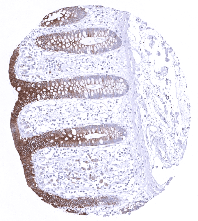
pH7,8 is optimal but pH6 results in only slightly weaker stainings. At pH9, Cytokeratin 19 immunostaining is markedly reduced.
Potential Research Applications
- The diagnostic utility of KRT19 expression analysis should be investigated in a large cohort of tumors from different entities.
Evidence for Antibody Specificity in IHC
Specificity of MSVA-119M is documented by strong positive staining in cell types that are well documented to express KRT19 such as a large variety of epithelial cell types and absence of staining in all tissues known to not express KRT19 including hepatocytes, adrenal gland, testis, mesenchymal tissues, lymphatic tissues and the brain.
