245,00 € – 895,00 €
Product details
Synonyms = CAK1; Megakaryocyte potentiating factor; Mesothelin; MSLN; SMR; SMRP
Antibody type = Mouse monoclonal / IgG2
Clone = MSVA-235M
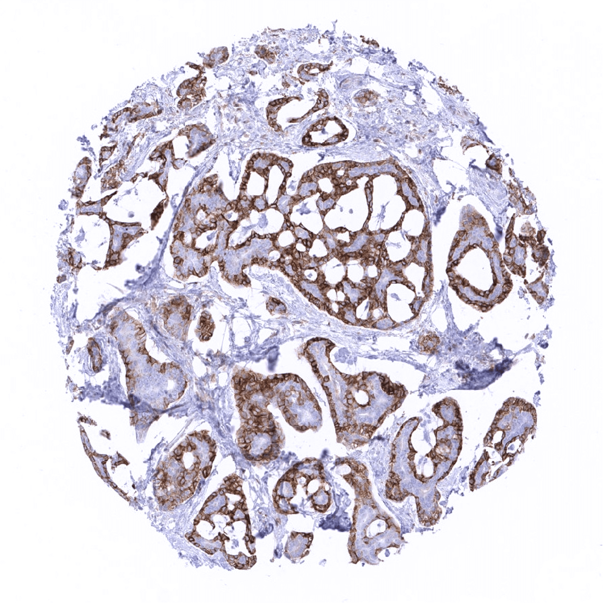
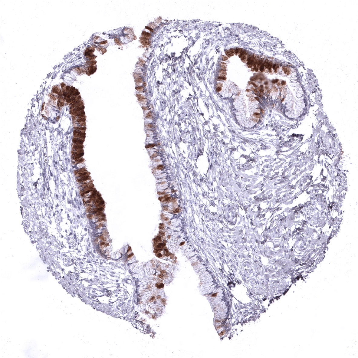
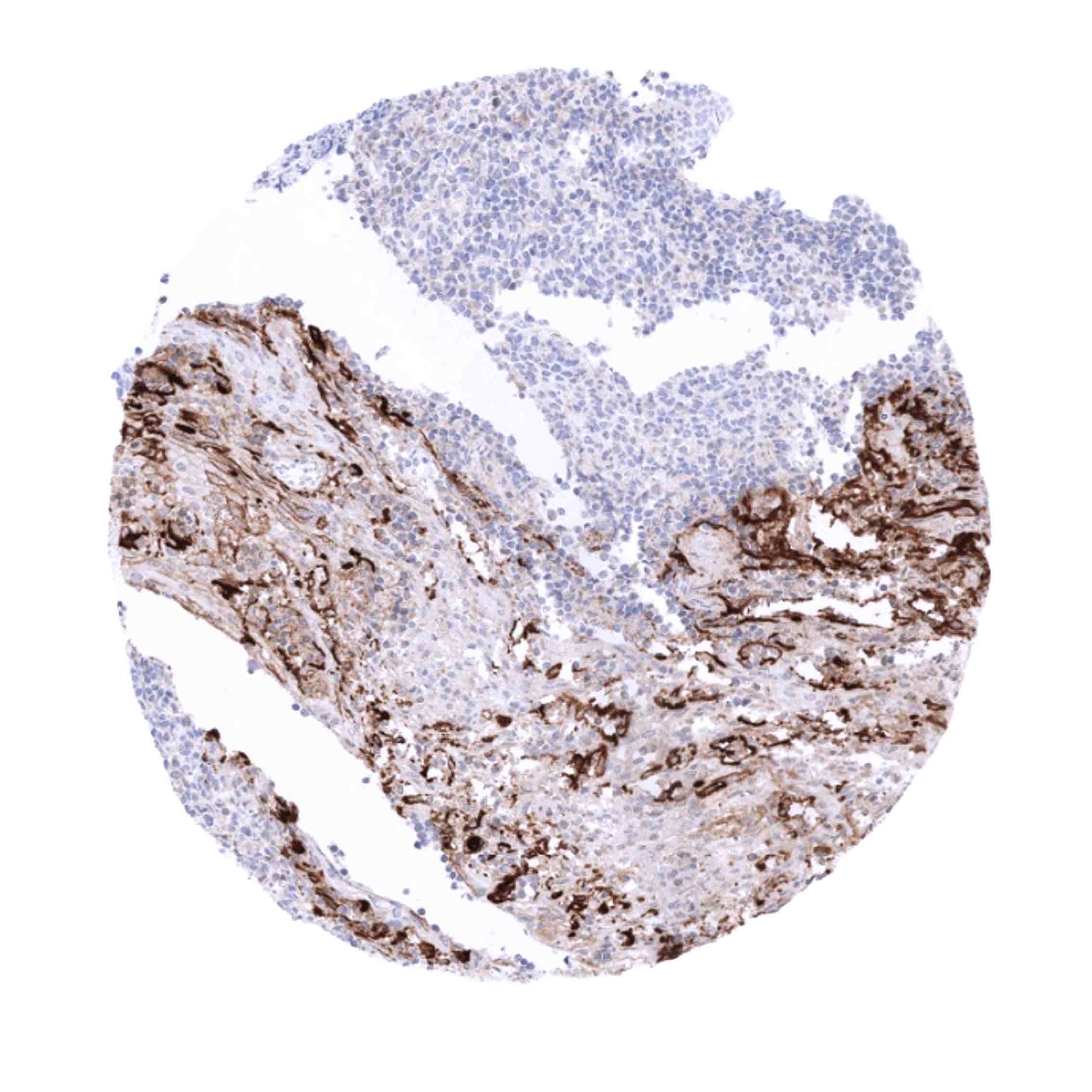
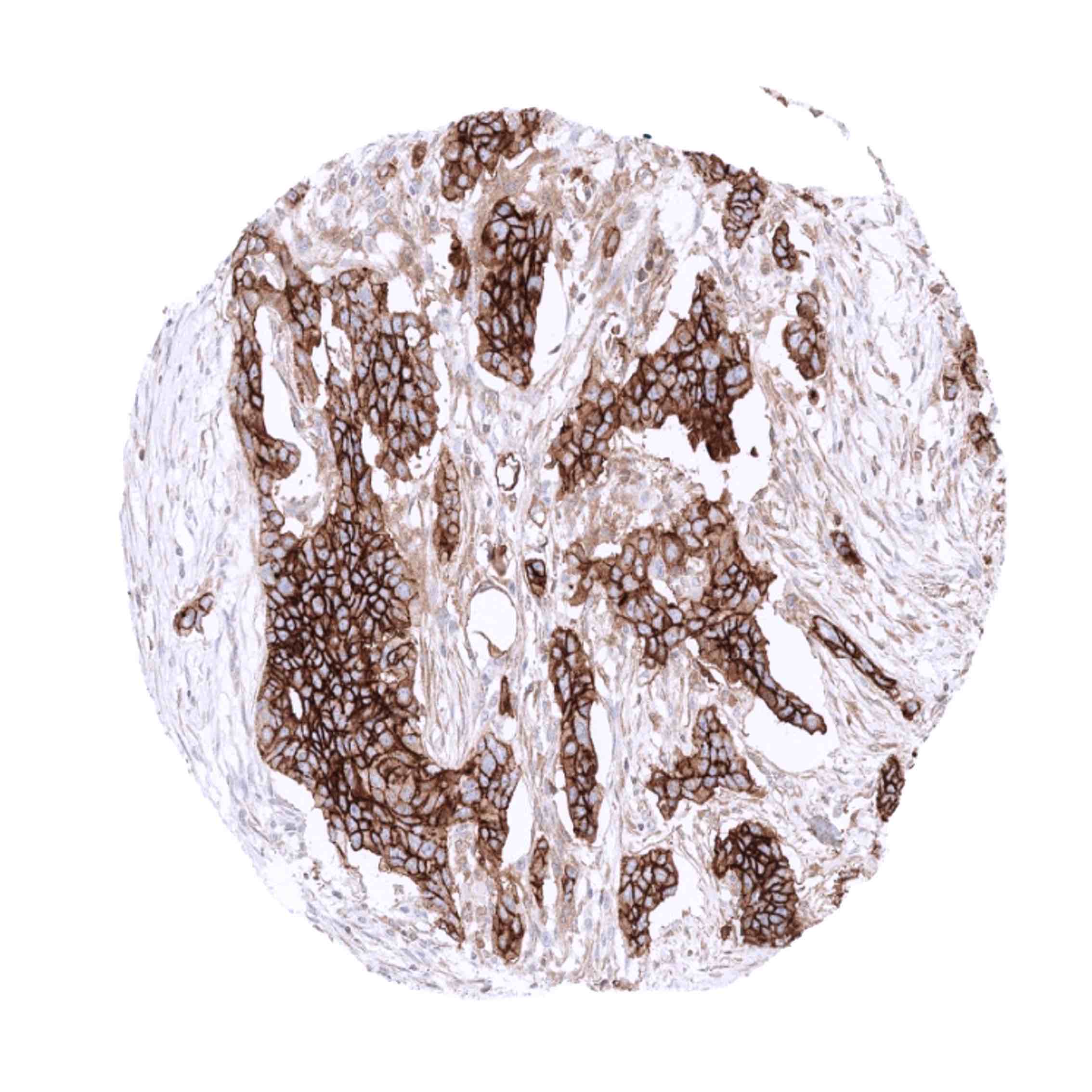
Positive control = Tonsil: A fraction of squamous epithelial cells of the tonsil crypts should show strong mesothelin immunostaining. Fallopian tube: An at least moderate mesothelin positivity should be seen at the apical membrane of epithelial cells.
Negative control = Tonsil: Squamous epithelial cells of the tonsil surface and all lymphocytes must not show mesothelin immunostaining.
Cellular localization = Cell Surface and Secreted
Reactivity = Human
Application = Immunohistochemistry
Dilution = 1:100 – 1:200
Intended Use = Research Use Only
Relevance of Antibody
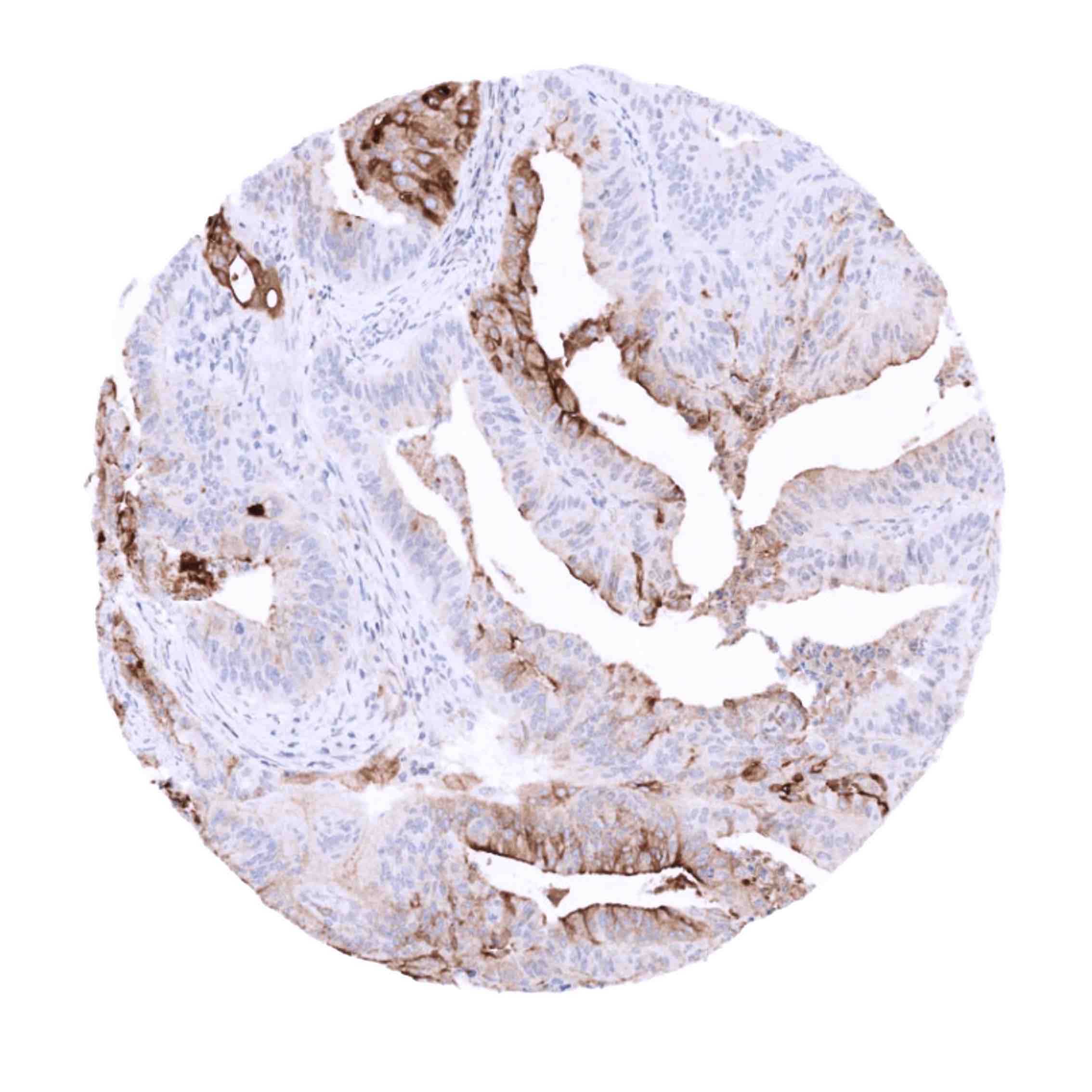
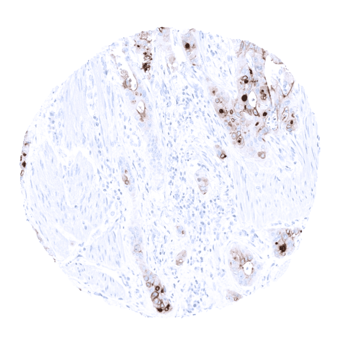
Mesothelin is expressed in various epithelial and mesothelial tumor types.
Biology Behind
Mesothelin (MSLN) is membranous glycoprotein of 40kDa that is processed by proteolytic cleavage of a larger preprotein encoded by the mesothelin at human chromosome 16p13. MSLN may function as a cell adhesion protein as it is it anchored to the cell membrane and can bind to known cell adhesion molecules such as CA125. However, its exact function has not been clarified as to yet. Mesothelin was originally named after its expression in mesothelial cells. Subsequent work demonstrated that MSLN expression is retained in epithelial mesotheliomas, and that it may also be expressed de-novo in several other cancer types, including ovarian and some kinds of squamous cell carcinomas, making it an attractive target for targeted anti-cancer therapies.[1]
[1] Weidemann et al. “Mesothelin Expression in Human Tumors: A Tissue Microarray Study on 12,679 Tumors” Biomedicines 2021, 9, 397.
Staining Pattern in Normal Tissues
Mesothelin staining pattern in Normal Tissues with antibody MSVA-235M (images are shown in our “Normal Tissue Gallery”)
| Brain | Cerebrum | Negative. |
| Cerebellum | Negative. | |
| Endocrine Tissues | Thyroid | Negative. |
| Parathyroid | Negative. | |
| Adrenal gland | Negative. | |
| Pituitary gland | Weak to moderate predominantly cytoplasmic mesothelin staining in the cytoplasm of few cells of the adenohypophysis. | |
| Respiratory system | Respiratory epithelium | Weak to moderate mesothelin staining in goblet cells of respiratory epithelium. |
| Lung | Negative. | |
| Gastrointestinal Tract | Salivary glands | Weak to moderate mesothelin staining in some scattered glands in the sublingual gland. |
| Esophagus | Negative. | |
| Stomach | Weak to moderate mesothelin staining in some intermediate (neck) cells of the stomach antrum. | |
| Colon | Negative. | |
| Duodenum | Weak to moderate mesothelin staining in some scattered glands in Brunner glands. | |
| Rectum | Strong mesothelin positivity can be seen of some individual cells and of small cell groups of the rectal mucosa epithelium. | |
| Small intestine | Negative. | |
| Liver | Negative. | |
| Gallbladder | Negative. | |
| Pancreas | Negative. | |
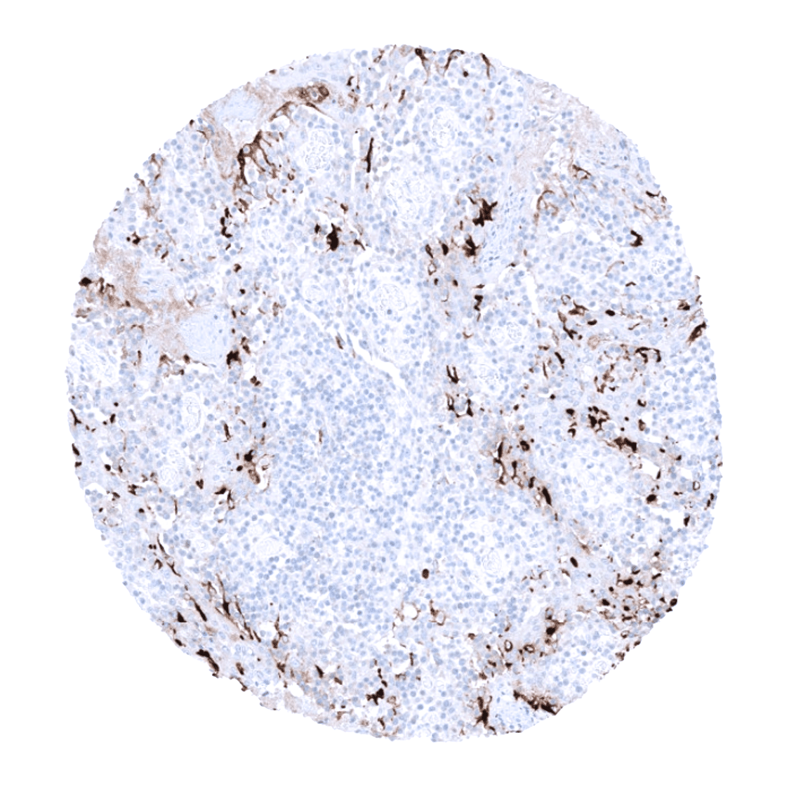
| Genitourinary | Kidney | Negative. |
| Urothelium | Negative. | |
| Male genital | Prostate | Negative. |
| Seminal vesicles | A subset of epithelial cells may show some mesothelin staining. | |
| Testis | Negative. | |
| Epididymis | Weak to moderate apical mesothelin staining may occur in some epithelial cells of the caput epididymis. | |
| Female genital | Breast | Negative. |
| Uterus, ectocervix | Negative. | |
| Uterus endocervix | Weak to moderate mesothelin staining in scattered individual cells or in groups of cells of endocervical mucosa epithelium. | |
| Uterus, endometrium | Weak to moderate mesothelin staining in scattered individual cells or in groups of cells of endometrial epithelium. | |
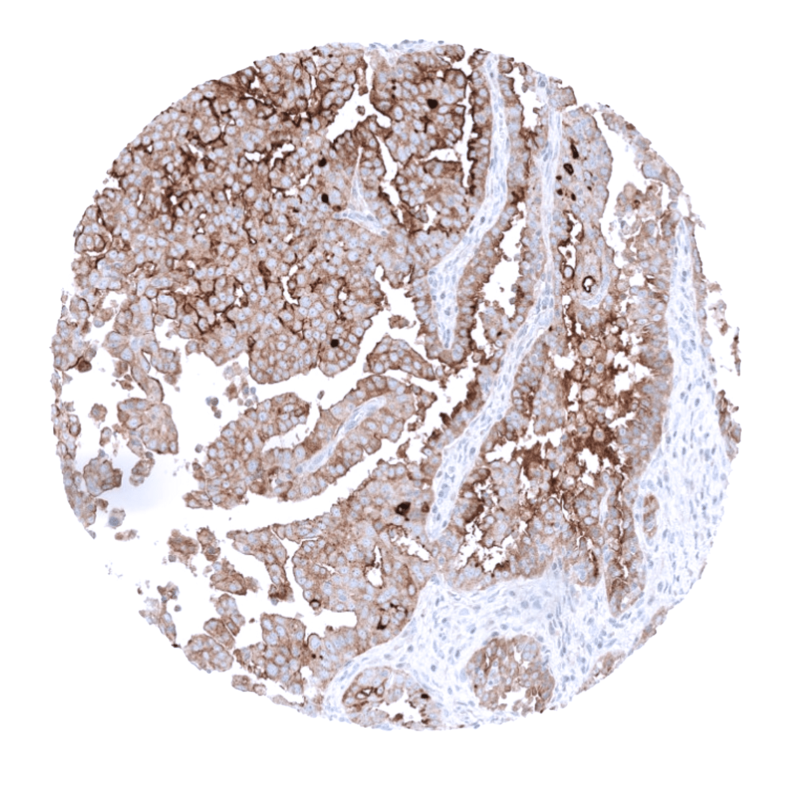
| Fallopian Tube | Moderate mesothelin staining in the fallopian tube epithelium (apical cell border and cilia). | |
| Ovary | Negative. | |
| Placenta early | Negative. | |
| Placenta mature | Negative. | |
| Amnion | Strong staining of all amnion cells. | |
| Chorion | Moderate staining of a subset of chorion cells. | |
| Skin | Epidermis | Negative. |
| Sebaceous glands | Negative. | |
| Muscle/connective tissue | Heart | Negative. |
| Skeletal muscle | Negative. | |
| Smooth muscle | Negative. | |
| Fat | Negative. | |
| Bone marrow/lymphoid | Bone marrow | Negative. |
| Lymph node | Negative. | |
| Spleen | Negative. | |
| Thymus | Strong staining of a subset of cells of corpuscles of Hassall’s. Lymphocytes do not stain. | |
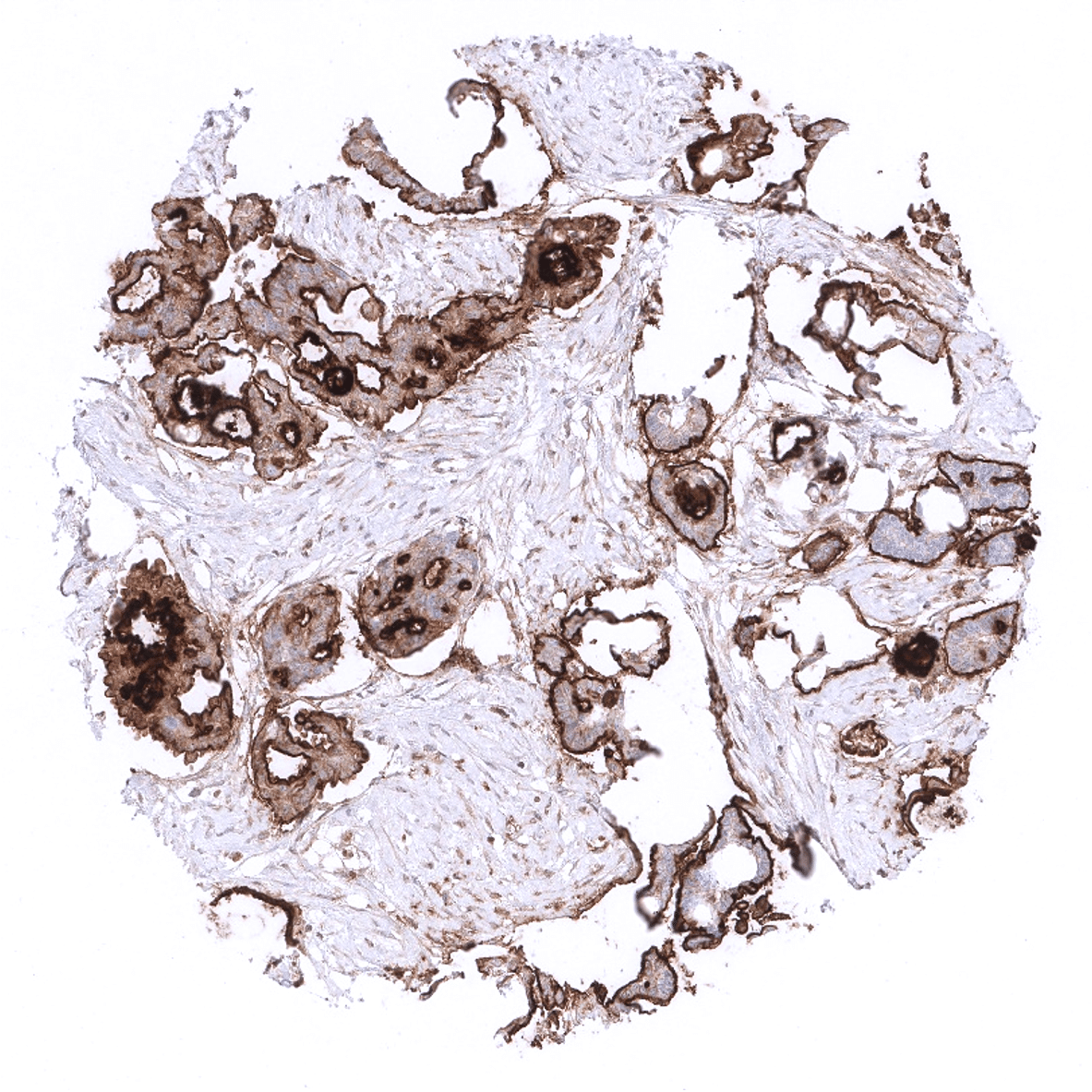
| Tonsil | Strong staining in fraction of cells of the squamous epithelium (intermediate to superficial cell layers) of tonsil crypts. Lymphocytes do not stain. | |
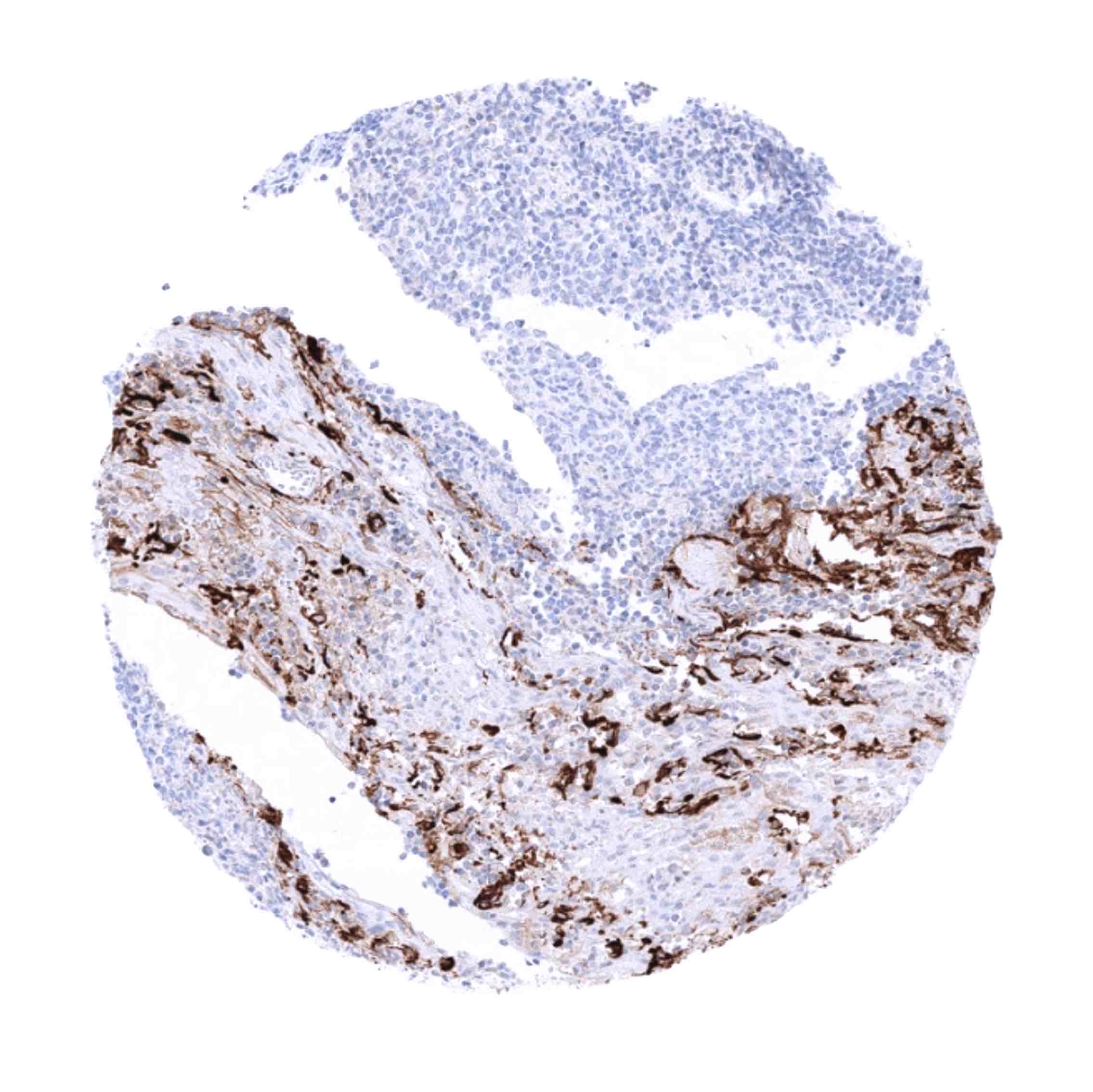
| Remarks | Staining is predominnatly membranous but also cytoplasmic. |
These findings fit well with the RNA and protein data described in the Human Protein Atlas (Tissue expression Mesothelin).
Suggested positive tissue control: Tonsil: A fraction of squamous epithelial cells of the tonsil crypts should show strong mesothelin immunostaining. Fallopian tube: An at least moderate mesothelin positivity should be seen at the apical membrane of epithelial cells.
Suggested negative tissue control: Tonsil: Squamous epithelial cells of the tonsil surface and all lymphocytes should not show mesothelin immunostaining.

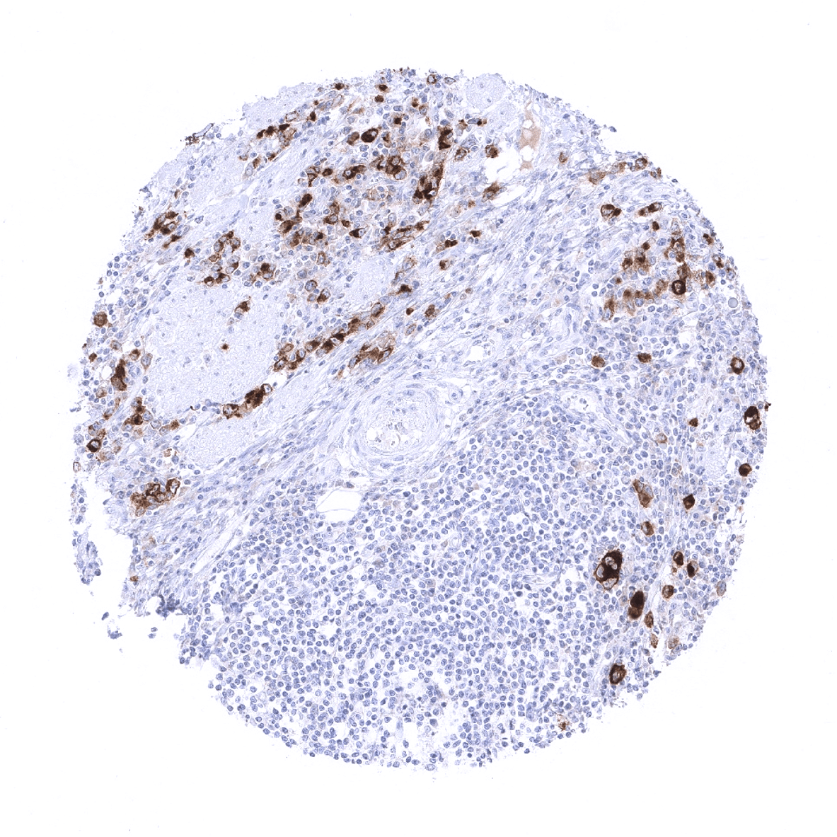
Staining Pattern in Relevant Tumor Types
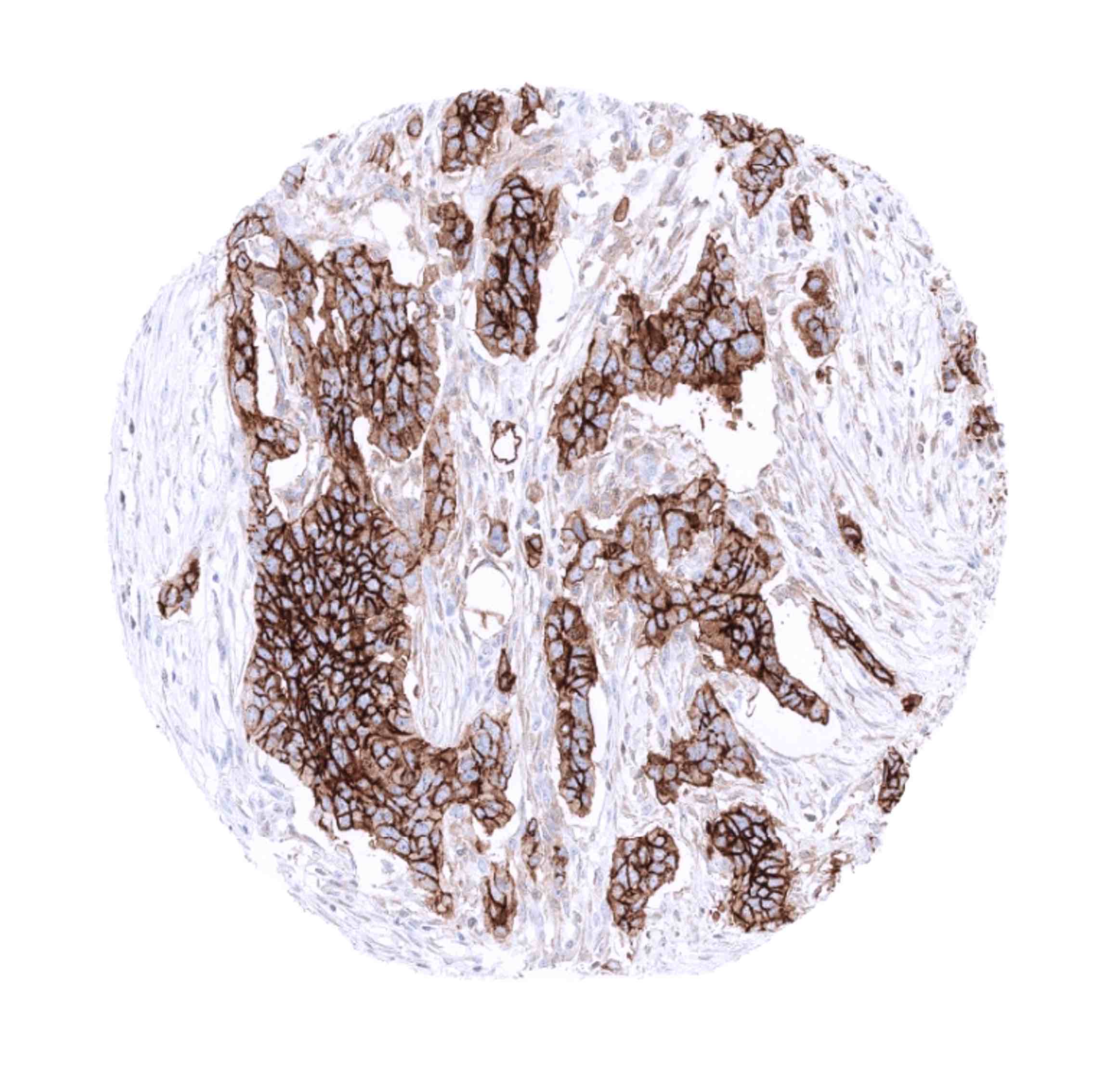
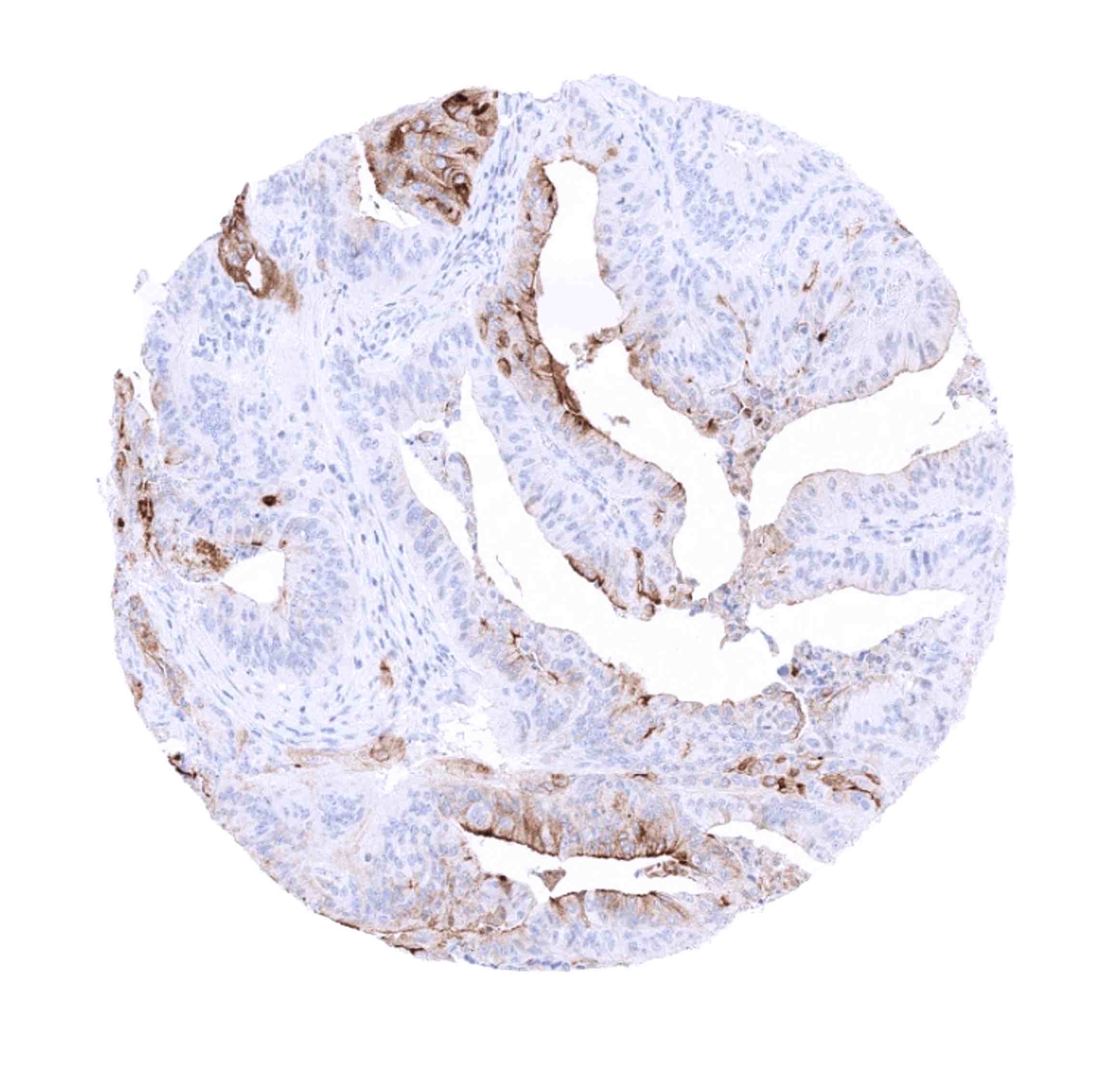
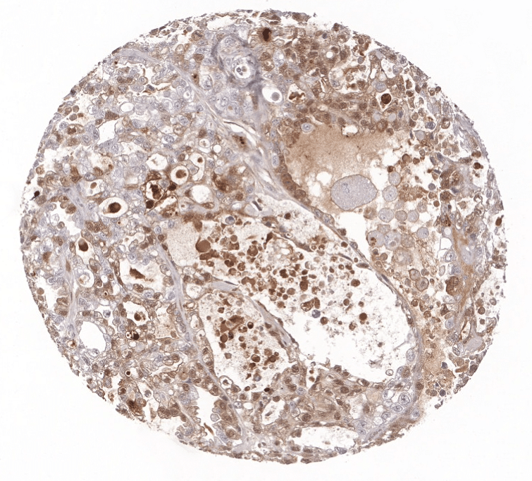
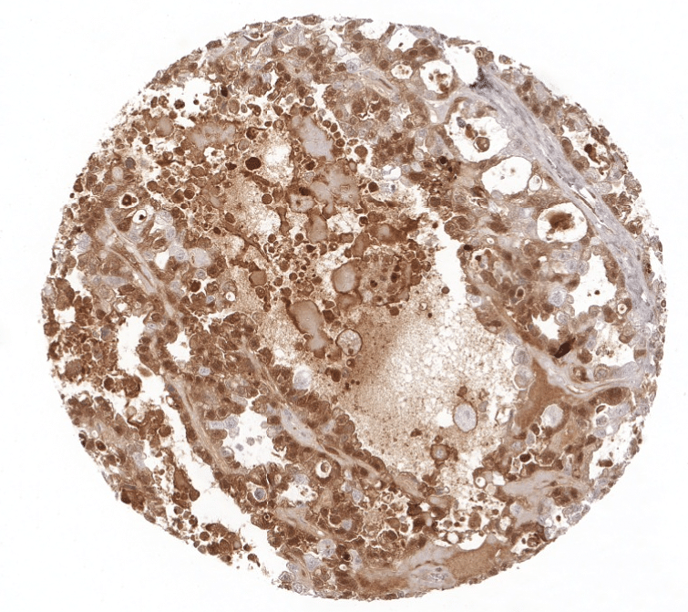
Mesothelin can be found expressed in many different tumor types. The highest rates of positivity (>50% of positive cases) are seen in carcinomas of the ovary, pancreas, endometrium, lung (adenocarcinoma) as well as in malignant mesothelioma. Mesothelin positivity is particularly rare or absent (<5%) carcinomas of the breast, kidney, prostate, and the thyroid as well as in most subtypes of soft tissue tumors.
Detailed data on Mesothelin staining by MSVA-235M obtained from an analysis of 12,679 tumors from 122 different tumor types and subtypes have recently been published by Weidemann et at “Mesothelin Expression in Human Tumors: A Tissue Microarray Study on 12,679 Tumors”
The TCGA findings on Mesothelin RNA expression in different tumor categories have been summarized in the Human Protein Atlas.


Compatibility of Antibodies
Mesothelin (MSVA-235M) publication summary
Paper used for meta analysis: Weidemann et al “Mesothelin Expression in Human Tumors: A Tissue Microarray Study on 12,679 Tumors” in Biomedicines 2021, 9, 397.
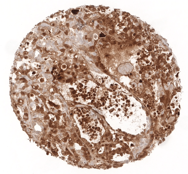
A total of 13218 tumors were analyzed from 122 different tumor categories by using the following protocol: Heat-induced antigen retrieval for 5 minutes in an autoclave at 121°C in pH9 Target Retrieval Solution buffer. MSVA-235M at a dilution of 1:150 at 37°C for 60 minutes. Visualization of bound antibody by the EnVision Kit (Dako, Agilent). This protocol was also used for all stainings depicted in our cancer tissue gallery & normal tissue gallery.
At least one case with a positive mesothelin immunostaining was seen in 66 (54%) and at least one case with a strong mesothelin immunostaining was seen in 50 (41%) of 122 tumor categories. The distribution of positive staining results is shown in an “organ-systematic” and in a “ranking order” figure below (images based on data from Weidemann et al). Results on possible associations with histopathological and clinical parameters of tumor aggressiveness in several cancer types are also summarized below (table based on data from Weidemann et al).
Authors conclusions on diagnostic utility of mesothelin immunohistochemistry with respect to the distinction of benign versus malignant (Weidemann et al):
- Mesothelin expression is often seen in pancreatic adenocarcinomas (89%) and adenocarcinomas of the ampulla Vateri (79%), but is infrequent in pancreatitis and absent in acinus cell carcinomas and in the normal pancreas.
- Mesothelin immunohistochemistry may represent a suitable tool for supporting the diagnosis pancreatic cancer in surgical pathology.
Authors conclusions on diagnostic utility of mesothelin immunohistochemistry with respect to the distinction of different tumor entities (Weidemann et al):
- Low diagnostic utility because of many tumor types can show mesothelin positivity.
- A positive MSLN immunostaining might be considered an argument against a tumor origin from organs that never or very rarely gave rise to MSLN positive cancer cells, such as the prostate, thyroid, kidney, germ cell tumors, adrenal tumors, melanoma, many soft tissue tumor types, and hematologic neoplasms.
Authors conclusions on prognostic/predictive role of mesothelin expression (Weidemann et al.):
- High mesothelin expression may be linked to a favorable response towards cancer drugs that target mesothelin drugs such as amatuximab, anetumab and others.
- High mesothelin expression is associated with unfavorable tumor features in breast and colorectal cancer.
- Reduced mesothelin expression is associated with tumor invasiveness in urothelial cancer.
- The absence of statistical associations between MSLN expression and tumor phenotype and/or prognosis in carcinomas of the bladder, breast, ovary, endometrium, kidney, lung, and stomach argues against a general/major prognostic role of MSLN expression levels in cancer.
Data from the publication: “Mesothelin Expression in Human Tumors: A Tissue Microarray Study on 12,679 Tumors”. Published by Weidemann et al. in Biomedicines 2021, 9, 397.
Summarized in own graphics:
1. Mesothelin staining in tumors “organ-specific” with antibody MSVA-235M

2. Mesothelin staining in tumors “ranking order” by positivity with antibody MSVA-235M

Protocol Recommendations
IHC users have different preferences on how the stains should look like. Some prefer high staining intensity of the target stain and even accept some background. Others favor absolute specificity and lighter target stains. Factors that invariably lead to more intense staining include higher concentration of the antibody and visualization tools, longer incubation time, higher temperature during incubation, higher temperature and longer duration of the heat induced epitope retrieval (slide pretreatment). The impact of the pH during slide pretreatment has variable effects and depends on the antibody and the target protein.
All images and data shown here and in our image galleries are obtained by the manual protocol described below. Other protocols resulting in equivalent staining are described as well.
Manual protocol
Slides are deparaffinized and exposed to heat-induced antigen retrieval for 5 minutes in an autoclave at 121°C in pH9 buffer. Apply MSVA-235M at a dilution of 1:150 at 37°C for 60 minutes. Visualization of bound antibody by the EnVision Kit (Dako, Agilent) according to the manufacturer’s directions.


Agilent / Dako – Autostainer Link 48
Pretreatment in PT-Link for 30 minutes at 95°C (pH high); FLEX peroxidase blocking for 5 minutes (room temperature), MSVA-235M 1:150 for 20 minutes (room temperature), FLEX+ mouse/rabbit (LINKER) for 15 minutes (room temperature), horseradish peroxidase (HRP) for 20 minutes (room temperature), FLEX DAB+Sub-Chromo for 10 minutes (room temperature), FLEX hematoxylin for 5 minutes (room temperature).
These images reflect stainings by the protocol described above. It is of note that a comparable staining result can also be obtained by different protocols. In general, a longer pretreatment, a longer incubation time of the primary antibody, a higher antibody concentration, and a longer incubation time of FLEX+LINKER result in stronger staining, potentially at the cost of more background staining. Modifications of the protocol with a strengthening effect on staining intensity in combination with changes of other parameters that result in lower staining intensity can result in a comparable result as shown above.
Leica – BOND RX
Dewax at 72°C for 30 seconds; Pretreatment in Bond Epitope Retrieval Solution (ER2 – EDTA pH9) for 20 minutes at 100°C; Peroxidase blocking for 5 minutes (room temperature), MSVA-235M 1:150 for 15 minutes (room temperature), Post primary (rabbit anti mouse) for 8 minutes (room temperature), Polymer (goat anti rabbit) for 8 minutes (room temperature), mixed DAB refine for 10 minutes (room temperature), hematoxylin for 5 minutes (room temperature).
These images reflect stainings by the protocol described above. It is of note that a comparable staining result can also be obtained by different protocols. In general, a longer pretreatment, a longer incubation time of the primary antibody, a higher antibody concentration, a higher temperature during incubation, and a longer incubation time of Post primary and or the Polymer result in stronger staining, potentially at the cost of more background staining. Modifications of the protocol with a strengthening effect on staining intensity in combination with changes of other parameters that result in lower staining intensity can result in a comparable result as shown above.
Roche – Ventana Discovery ULTRA
Pretreatment for 64 minutes at 100°C (pH 8,4); CM peroxidase blocking for 12 minutes (room temperature), MSVA-235M 1:150 for 20 minutes at 36°C, secondary antibody (anti-mouse HQ) for 12 minutes at 36°C, anti-HQ HRP for 12 minutes at room temperature, DAB at room temperature, hematoxylin II at room temperature for 8 minutes, bluing reagent at room temperature for 4 minutes.
These images depict staining results obtained by the protocol described above. It is of note, that the Ventana machines generally require higher antibody concentrations than other commonly used autostainers because the antibodies are automatically diluted during the procedure. Various other protocols can result in an identical result as shown above. A longer pretreatment, a longer incubation time of the primary antibody, a higher antibody concentration, a higher temperature during incubation, and a longer incubation time of secondary antibody and or the anti-HQ HRP result in stronger staining, potentially at the cost of more background staining.
Impact of pH
pH9 is optimal but pH7,8 results in only slightly weaker stainings. At pH6, Mesothelin immunostaining is markedly reduced.
Potential Research Applications
- Mesothelin is a highly promising therapeutic target for which a variety of drugs are under development.
- A comprehensive study analyzing mesothelin expression in various tumor entities would be helpful to assess the diagnostic significance of mesothelin IHC.
- Mesothelin expression occurs in a variable fraction of cases in many different tumor types but the prognostic and predictive relevance of mesothelin expression is unclear.
- The function of mesothelin in cancers is not yet fully elucidated.
Evidence for Antibody Specificity in IHC
Specificity of MSVA-235M is documented by strong positive staining in cell types that are well documented to express mesothelin such as tonsil crypts or fallopian tube and absence of staining in all tissues known to not express mesothelin including tissues notorious for non-specific IHC background such as kidney, colonic mucosa, and epidermis.
