295,00 € – 995,00 €
Product details
Synonyms = Cortical thymocyte antigen CD1A, Epidermal dendritic cell marker CD1a antibody, FCB6, HTA1, T cell surface antigen T6 / Leu 6, T-Cell Surface Glycoprotein CD1A
Antibody type = Recombinant Mouse monoclonal / IgG
Clone = MSVA-001M
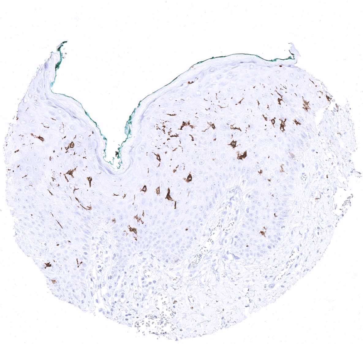
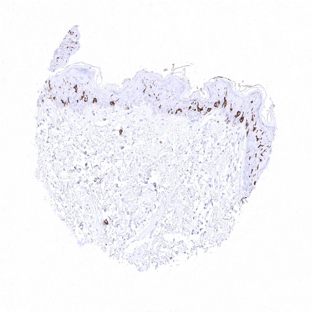
Positive control = Skin: A strong CD1a staining should be seen in Langerhans cells.
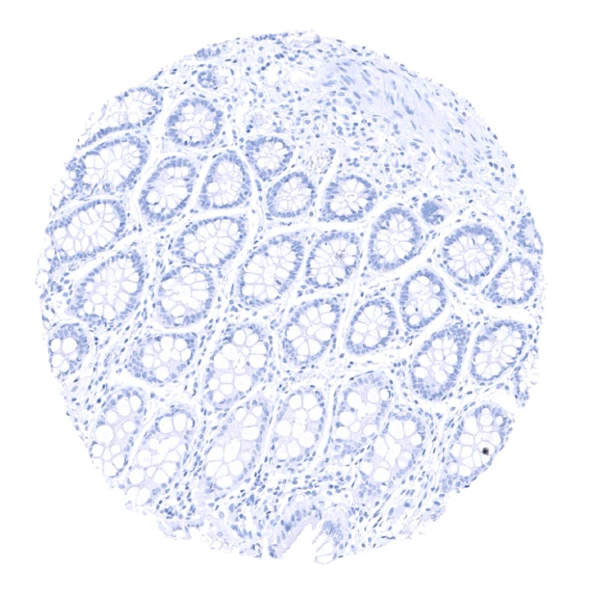
Negative control = Skin: CD1a staining must be absent in squamous epithelial cells and sebaceous glands cells.
Cellular localization = Cell surface
Reactivity = Human
Application = Immunohistochemistry
Dilution = 1:100 – 1:200
Intended Use = Research Use Only
Relevance of Antibody
Biology Behind
The CD1a protein is a transmembrane glycoprotein encoded by the CD1A gene on 1q23.1. It is a member of a family of 5 CD1 proteins that are structurally related to the major histocompatibility complex (MHC) proteins. It is the main role of CD1 proteins to present lipid and glycolipid antigens of self or microbial origin to T cells. The CD1 family members differ slightly in their specificity for particular lipid ligands in their cellular localization.
Staining Pattern in Normal Tissues
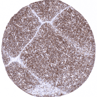
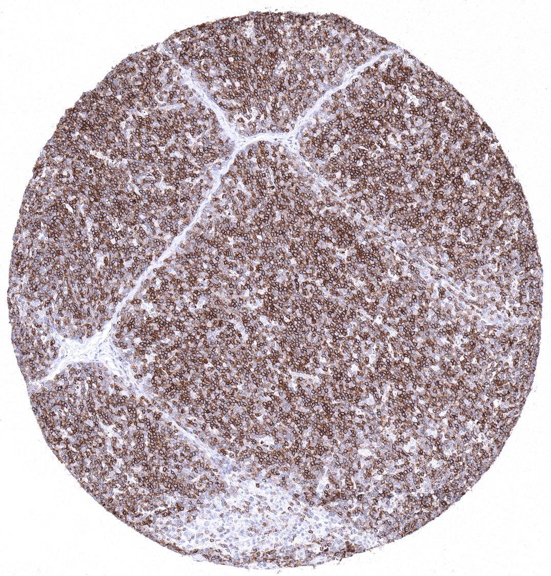
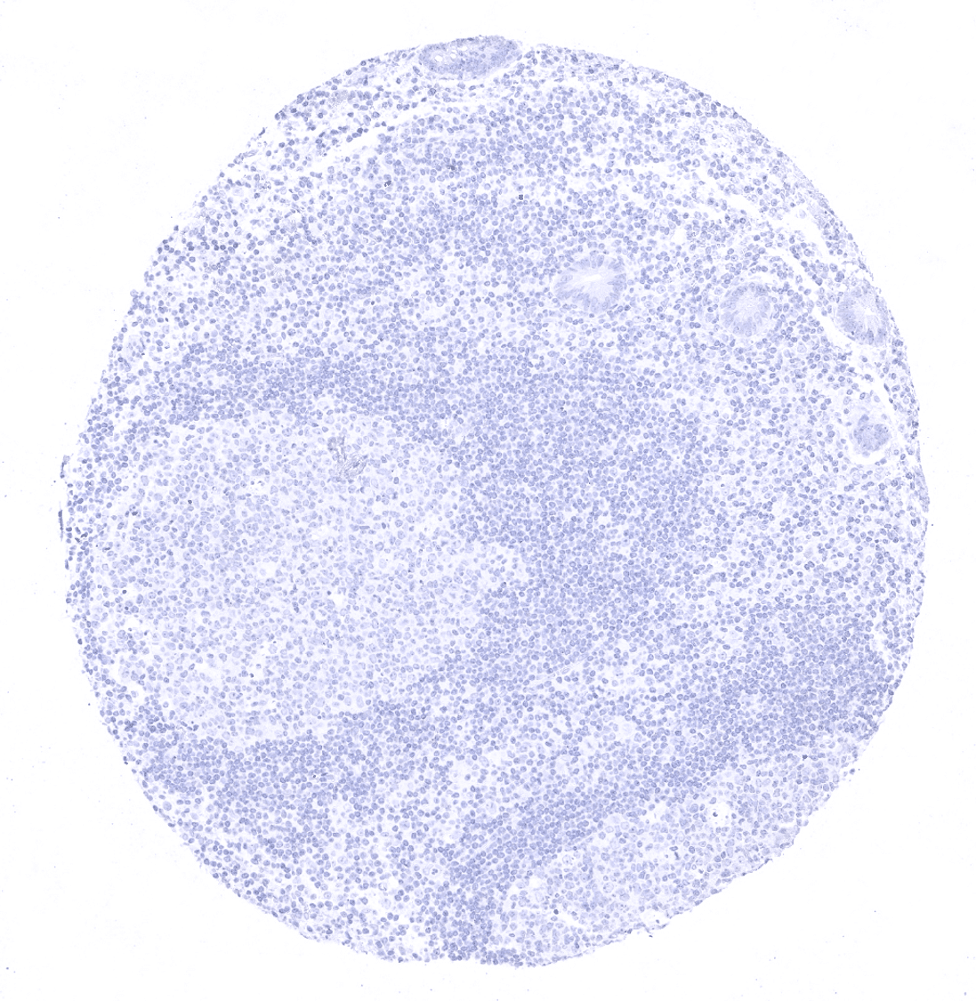
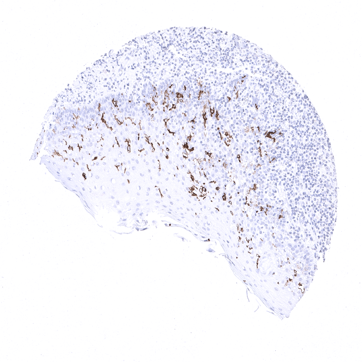
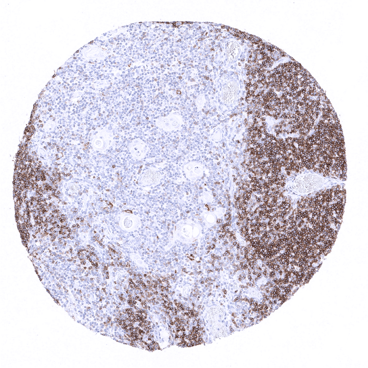
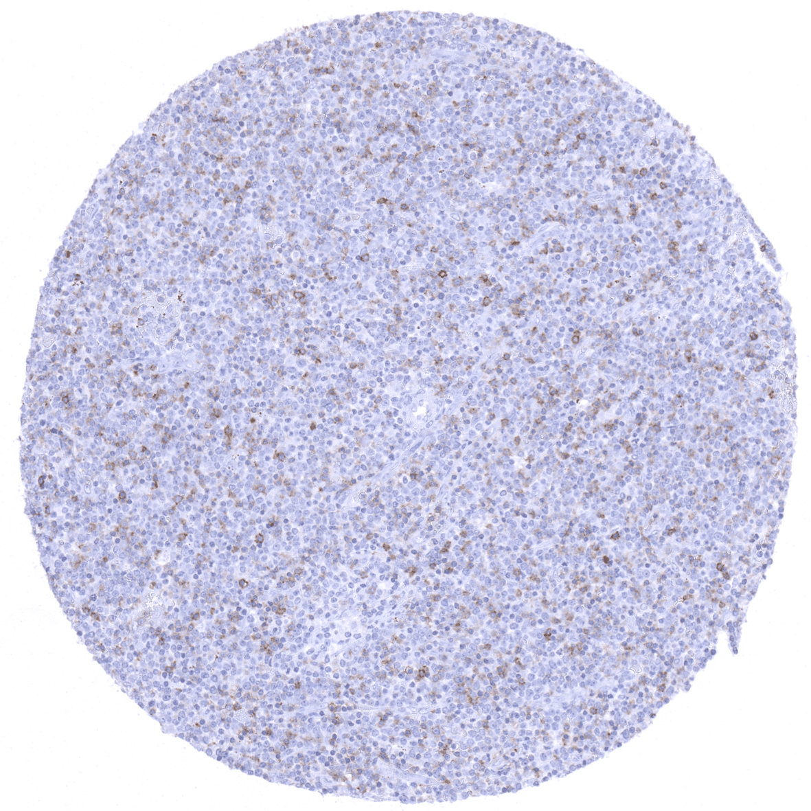
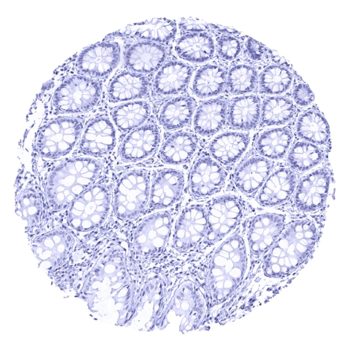
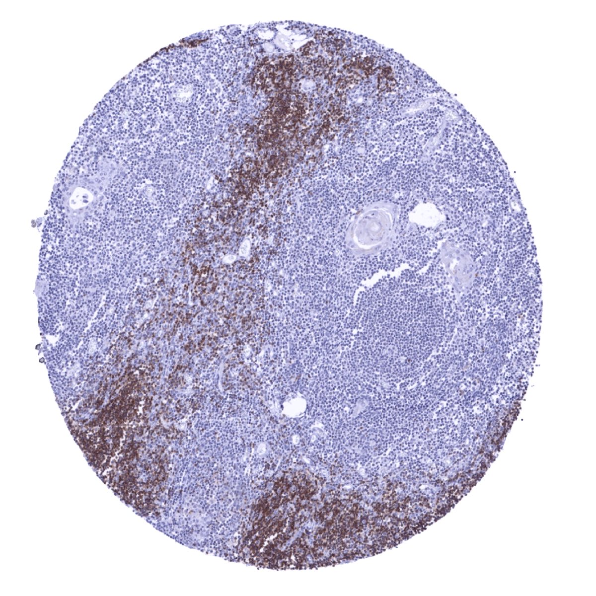
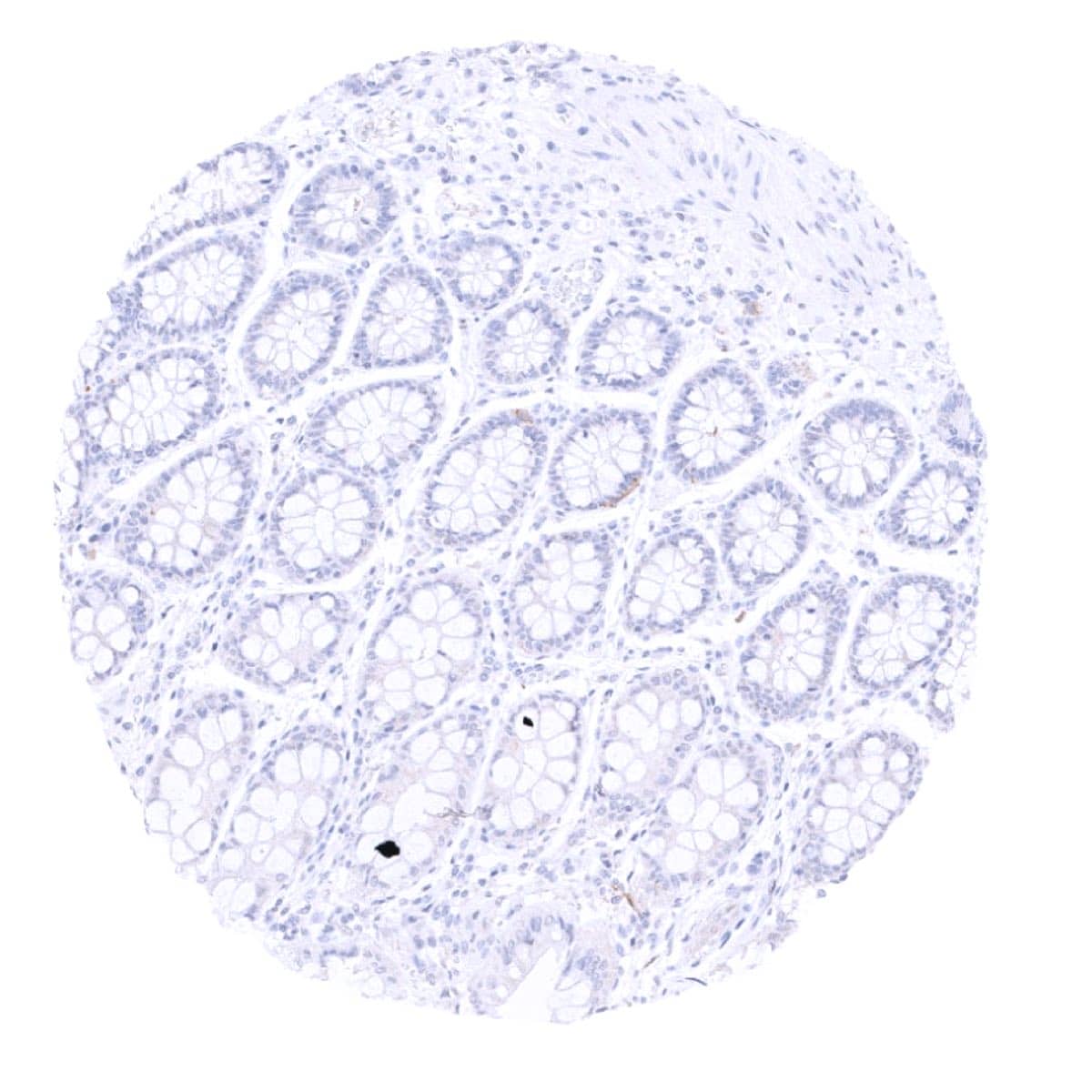
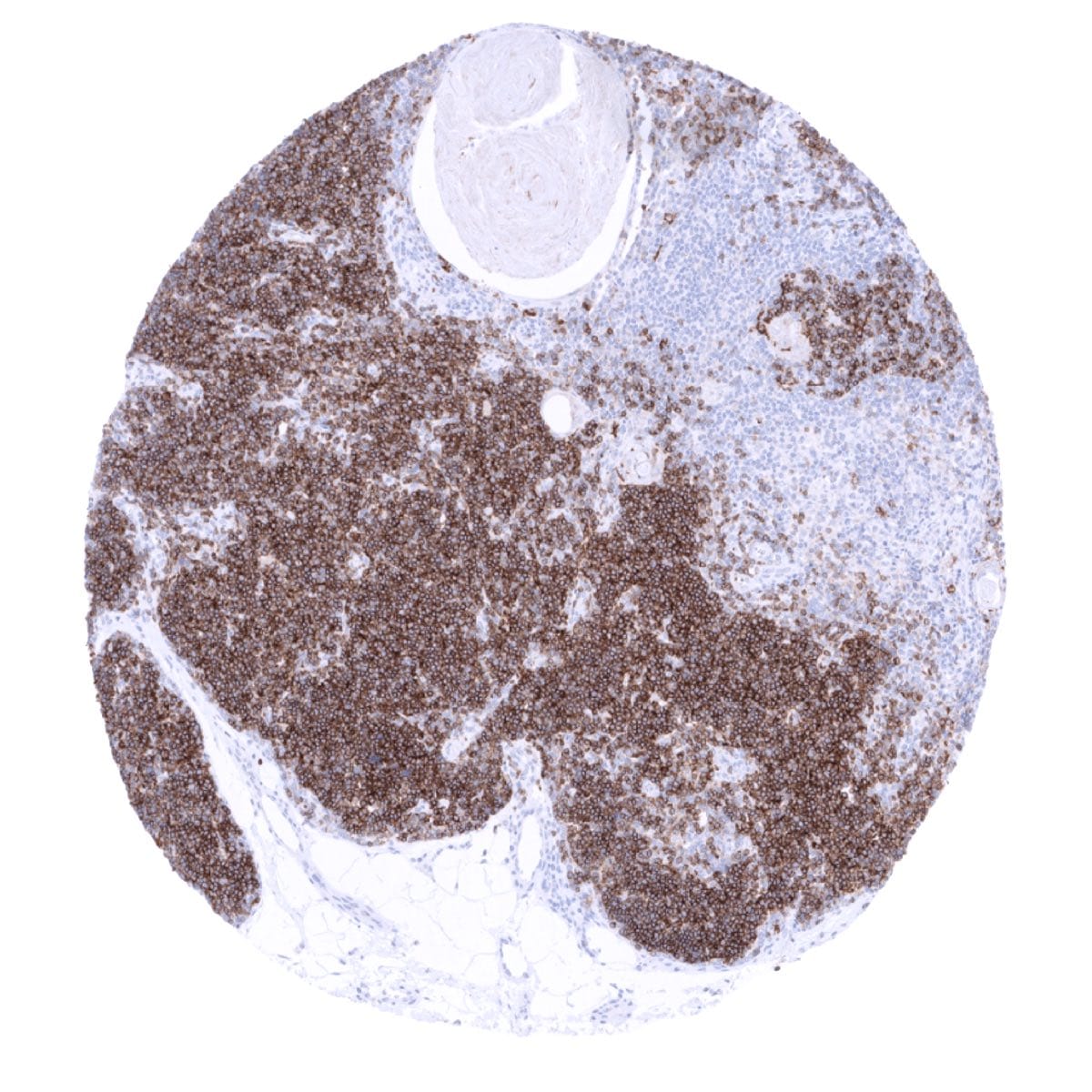
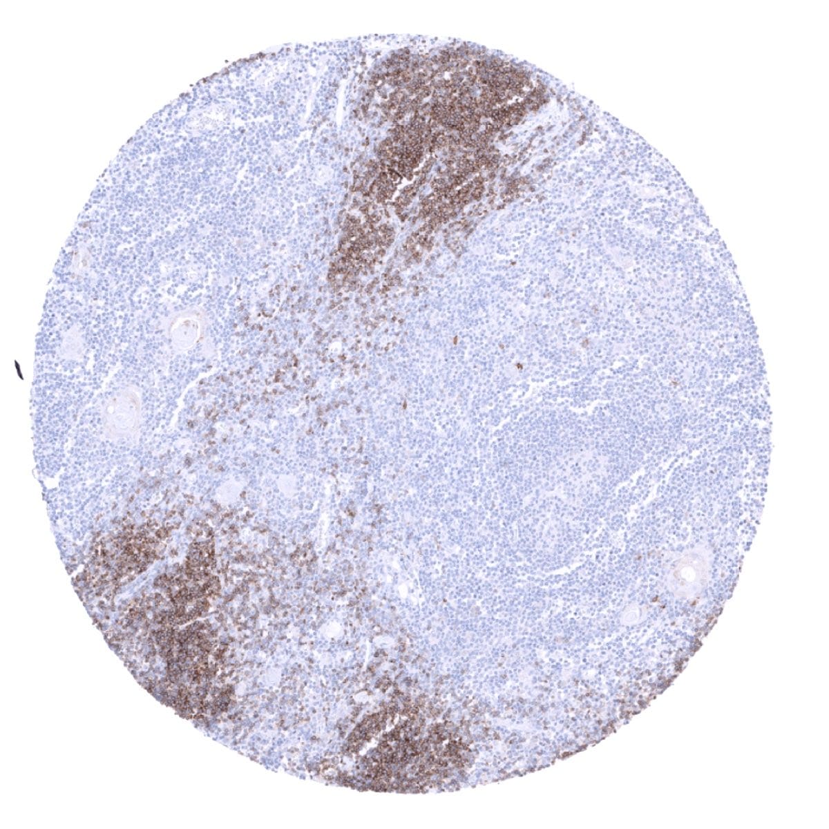
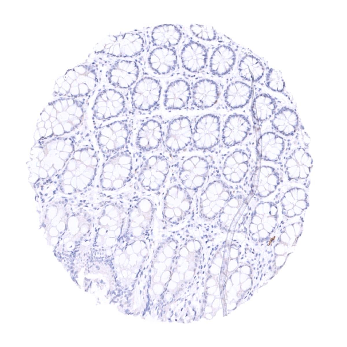
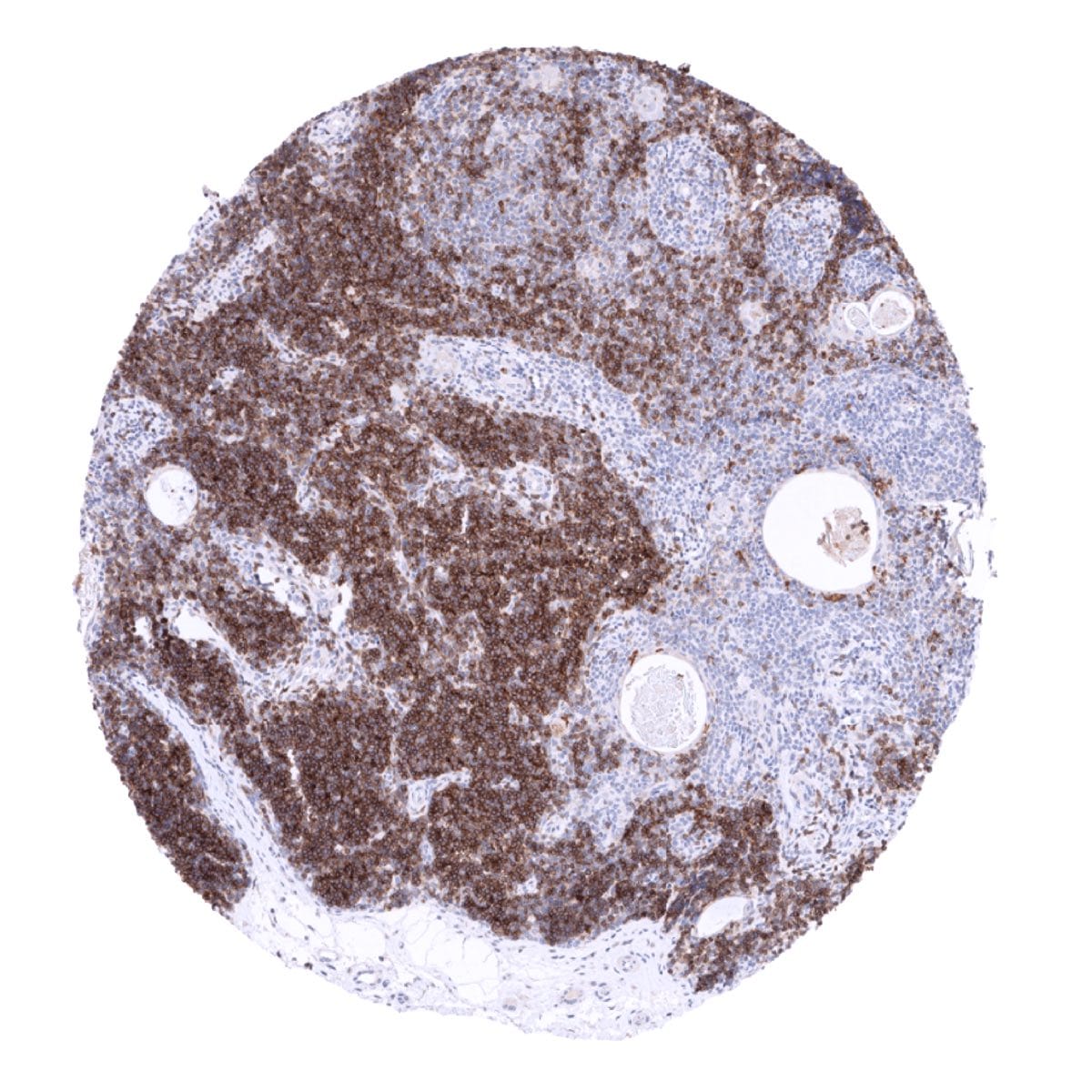
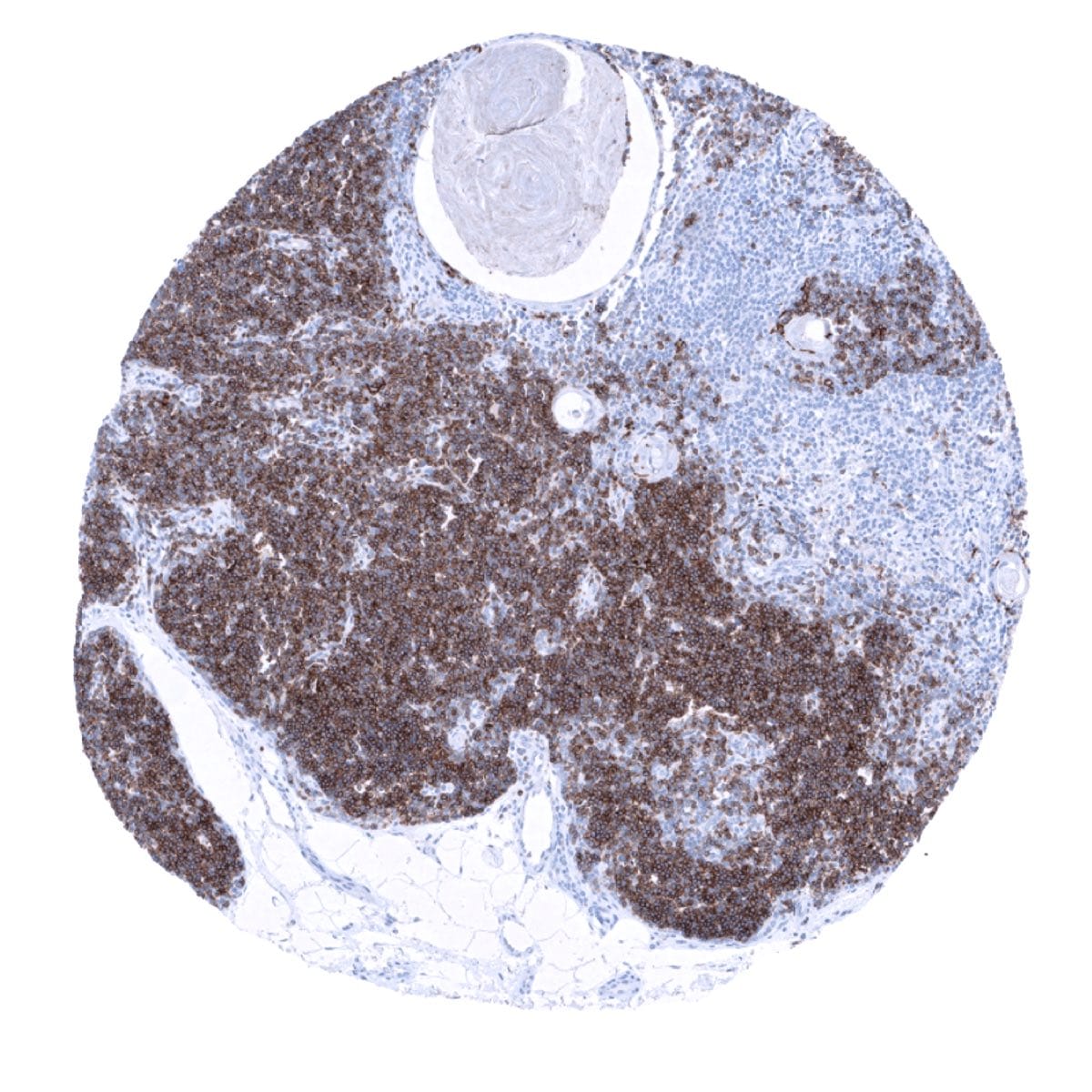
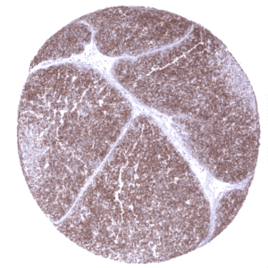
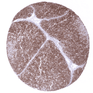
CD1a is strongly expressed in Langerhans cells of the skin including hair follicles and sebaceous glands and in non-keratinizing squamous epithelial tissues. A strong CD1a immunostaining is also seen in >80% of the small lymphocytes of the thymic cortex but CD1a is virtually absent in any other lymphatic cells under normal conditions.
These findings are largely consistent with RNA and protein data summarized in the Human Protein Atlas (Tissue expression CD1a).
A very faint staining of the surface epithelium of fallopian tube and of endometrium can occasionally be seen and reflects a cross-reactivity.
Suggested positive tissue control: Skin: A strong CD1a staining should be seen in Langerhans cells.
Suggested negative tissue control: Skin: CD1a staining must be absent in squamous epithelial cells and sebaceous glands cells.
Staining Pattern in Relevant Tumor Types
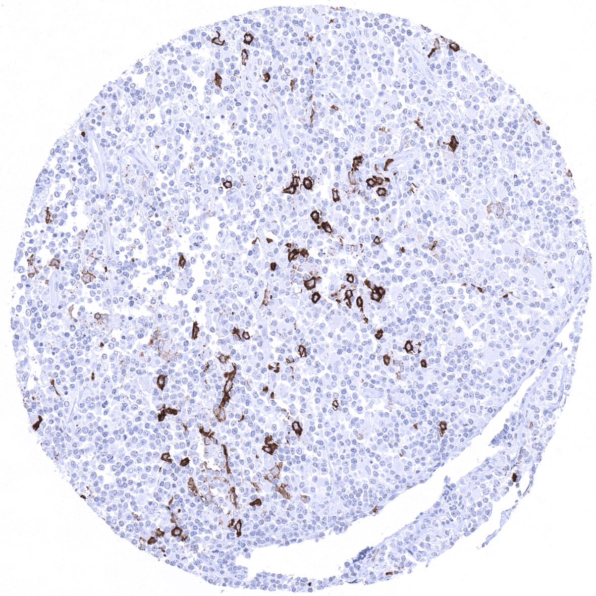
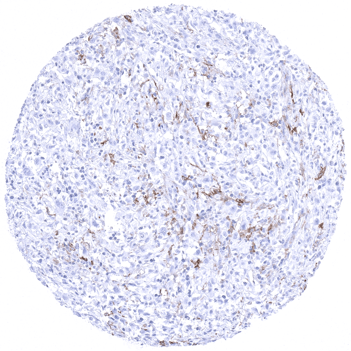
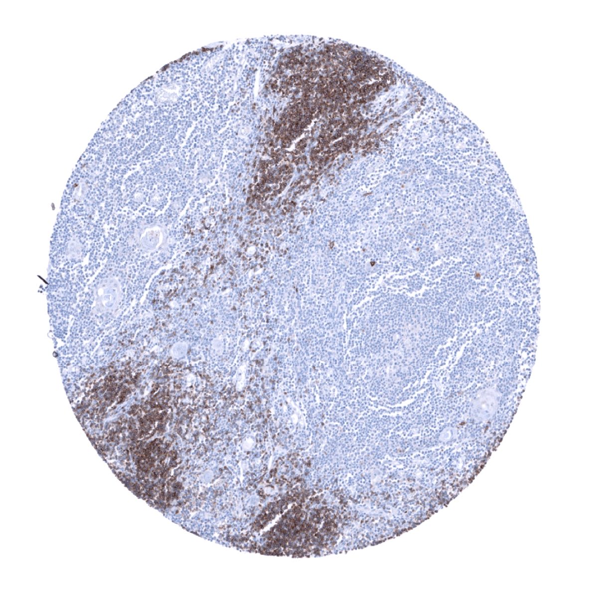
CD1a expression regularly occurs in Langerhans cell histiocytosis but it can also be seen in T cell lymphoma (predominantly cutaneous) and in myeloid leukemia. CD1a positive cells can also be found in regionary lymph nodes where they can migrate.
The TCGA findings on CD1a RNA expression in different tumor categories have been summarized in the Human Protein Atlas.
Compatibility of Antibodies
No data available at the moment
Protocol Recommendations
IHC users have different preferences on how the stains should look like. Some prefer high staining intensity of the target stain and even accept some background. Others favor absolute specificity and lighter target stains. Factors that invariably lead to more intense staining include higher concentration of the antibody and visualization tools, longer incubation time, higher temperature during incubation, higher temperature and longer duration of the heat induced epitope retrieval (slide pretreatment). The impact of the pH during slide pretreatment has variable effects and depends on the antibody and the target protein. Accordingly, multiple different protocols can generate identical staining results.
All images and data shown here and in our image galleries are obtained by the manual protocol described below. Other protocols resulting in equivalent staining are described as well.
Manual protocol
Freshly cut sections should be used (less than 10 days between cutting and staining). Heat-induced antigen retrieval for 5 minutes in an autoclave at 121°C in pH 7,8 Target Retrieval Solution buffer. Apply MSVA-001M at a dilution of 1:150 at 37°C for 60 minutes. Visualization of bound antibody by the EnVision Kit (Dako, Agilent) according to the manufacturer’s directions.

Agilent / Dako – Autostainer Link 48
Pretreatment in PT-Link for 30 minutes at 95°C (pH high); FLEX peroxidase blocking for 5 minutes (room temperature), MSVA-001M 1:100 for 20 minutes (room temperature), FLEX+ mouse/rabbit (LINKER) for 15 minutes (room temperature), horseradish peroxidase (HRP) for 20 minutes (room temperature), FLEX DAB+Sub-Chromo for 10 minutes (room temperature), FLEX hematoxylin for 5 minutes (room temperature).
These images reflect stainings by the protocol described above. It is of note that a comparable staining result can also be obtained by different protocols. In general, a longer pretreatment, a longer incubation time of the primary antibody, a higher antibody concentration, and a longer incubation time of FLEX+LINKER result in stronger staining, potentially at the cost of more background staining. Modifications of the protocol with a strengthening effect on staining intensity in combination with changes of other parameters that result in lower staining intensity can result in a comparable result as shown above.
Leica – BOND RX
Dewax at 72°C for 30 seconds; Pretreatment in Bond Epitope Retrieval Solution (ER2 – EDTA pH9) for 20 minutes at 100°C; Peroxidase blocking for 5 minutes (room temperature), MSVA-001M 1:150 for 15 minutes (room temperature), Post primary (rabbit anti mouse) for 8 minutes (room temperature), Polymer (goat anti rabbit) for 8 minutes (room temperature), mixed DAB refine for 10 minutes (room temperature), hematoxylin for 5 minutes (room temperature).

These images reflect stainings by the protocol described above. It is of note that a comparable staining result can also be obtained by different protocols. In general, a longer pretreatment, a longer incubation time of the primary antibody, a higher antibody concentration, a higher temperature during incubation, and a longer incubation time of Post primary and or the Polymer result in stronger staining, potentially at the cost of more background staining. Modifications of the protocol with a strengthening effect on staining intensity in combination with changes of other parameters that result in lower staining intensity can result in a comparable result as shown above.
Roche – Ventana Discovery ULTRA
Pretreatment for 64 minutes at 100°C (pH 8,4); CM peroxidase blocking for 12 minutes (room temperature), MSVA-001M 1:100 for 20 minutes at 36°C, secondary antibody (anti-rabbit HQ) for 12 minutes at 36°C, anti-HQ HRP for 12 minutes at room temperature, DAB at room temperature, hematoxylin II at room temperature for 8 minutes, bluing reagent at room temperature for 4 minutes.
These images depict staining results obtained by the protocol described above. It is of note, that the Ventana machines generally require higher antibody concentrations than other commonly used autostainers because the antibodies are automatically diluted during the procedure. Various other protocols can result in an identical result as shown above. A longer pretreatment, a longer incubation time of the primary antibody, a higher antibody concentration, a higher temperature during incubation, and a longer incubation time of secondary antibody and or the anti-HQ HRP result in stronger staining, potentially at the cost of more background staining.
Impact of pH

pH9 is optimal but pH7,8 results in only slightly weaker stainings. At pH6, CD1a immunostaining is markedly reduced.
Potential Research Applications
- The role of CD1a positive cells in inflammatory and allergic disease is not fully understood.
- The frequency of CD1a immunostaining in Langerhans cell histiocytosis, other hematological and non-hematological neoplasms should be evaluated.
Evidence for Antibody Specificity in IHC
Specificity and utility of MSVA-001M is documented by strong positive staining in thymocytes and Langerhans cells, the only cell types known to express CD1a and the absence of CD1a immunostaining in virtually all other types. A very faint staining of the surface epithelium of fallopian tube and of endometrium can occasionally be seen and reflects a cross-reactivity which appears to be negligeable.