
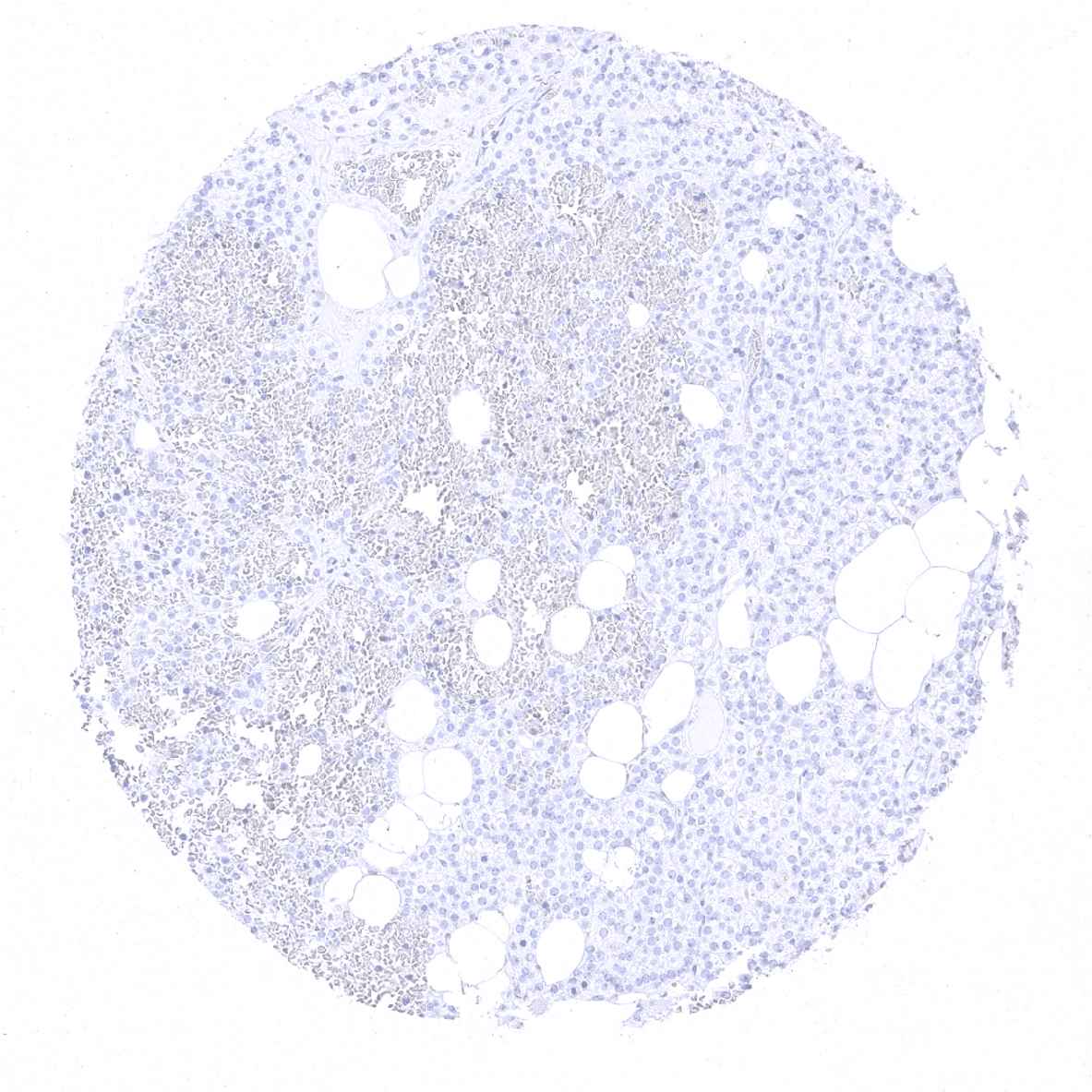
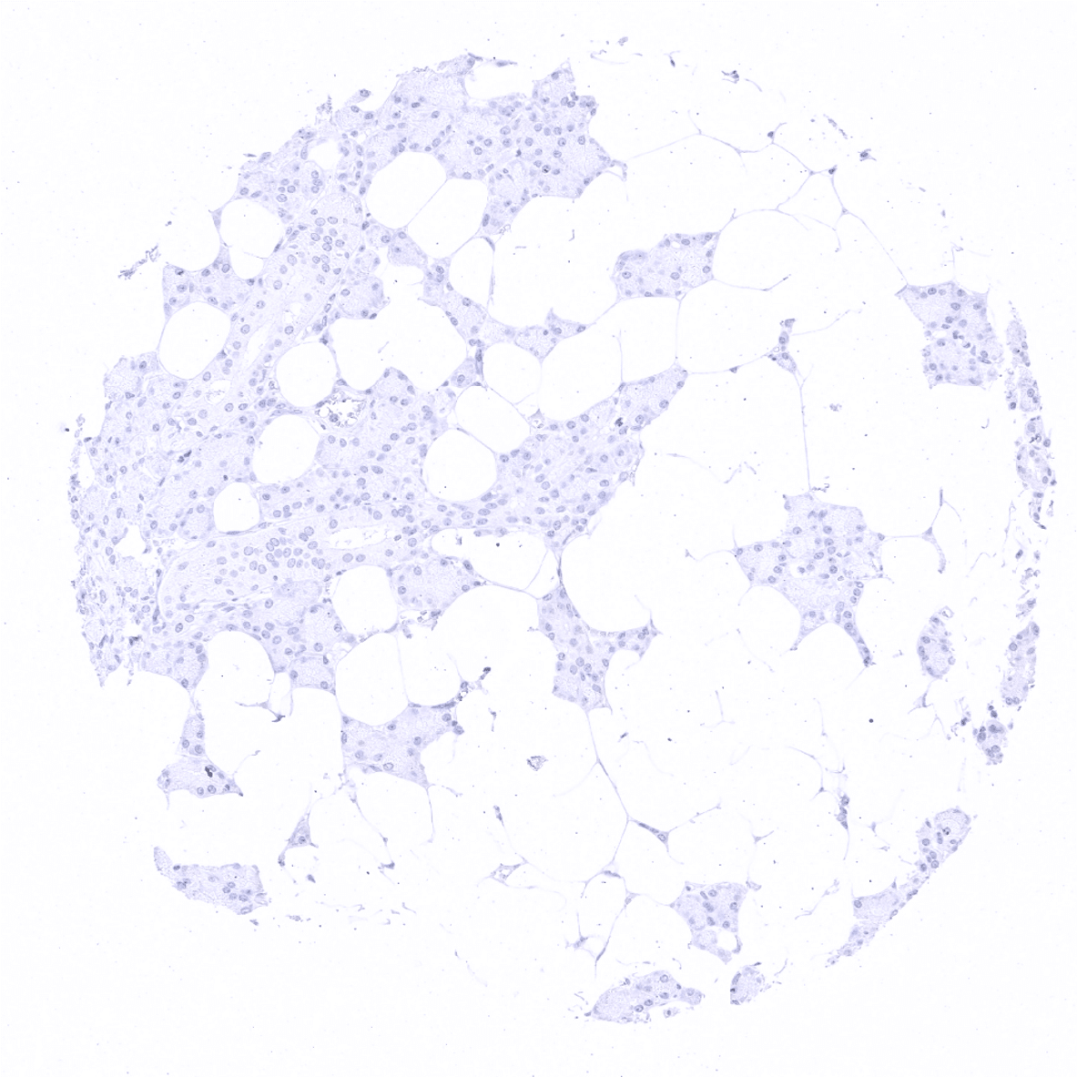
Adrenal gland
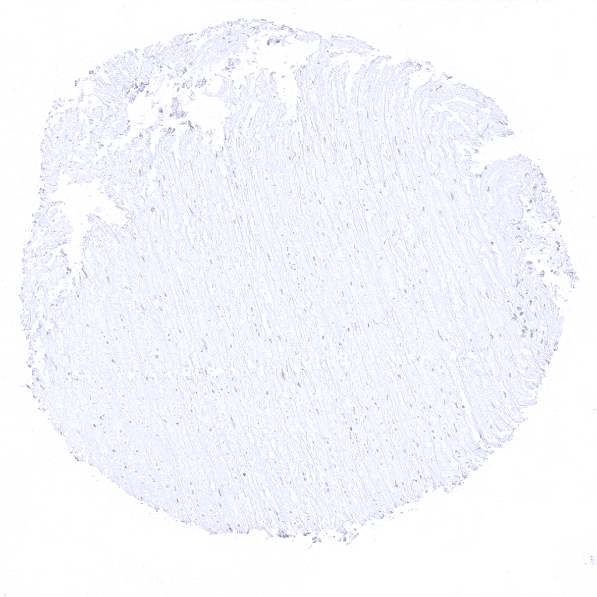
Aorta, media
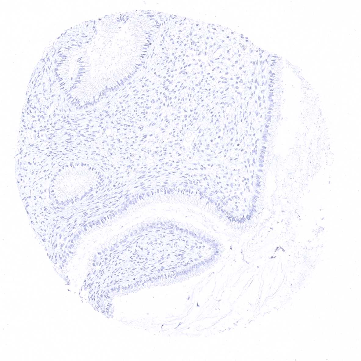
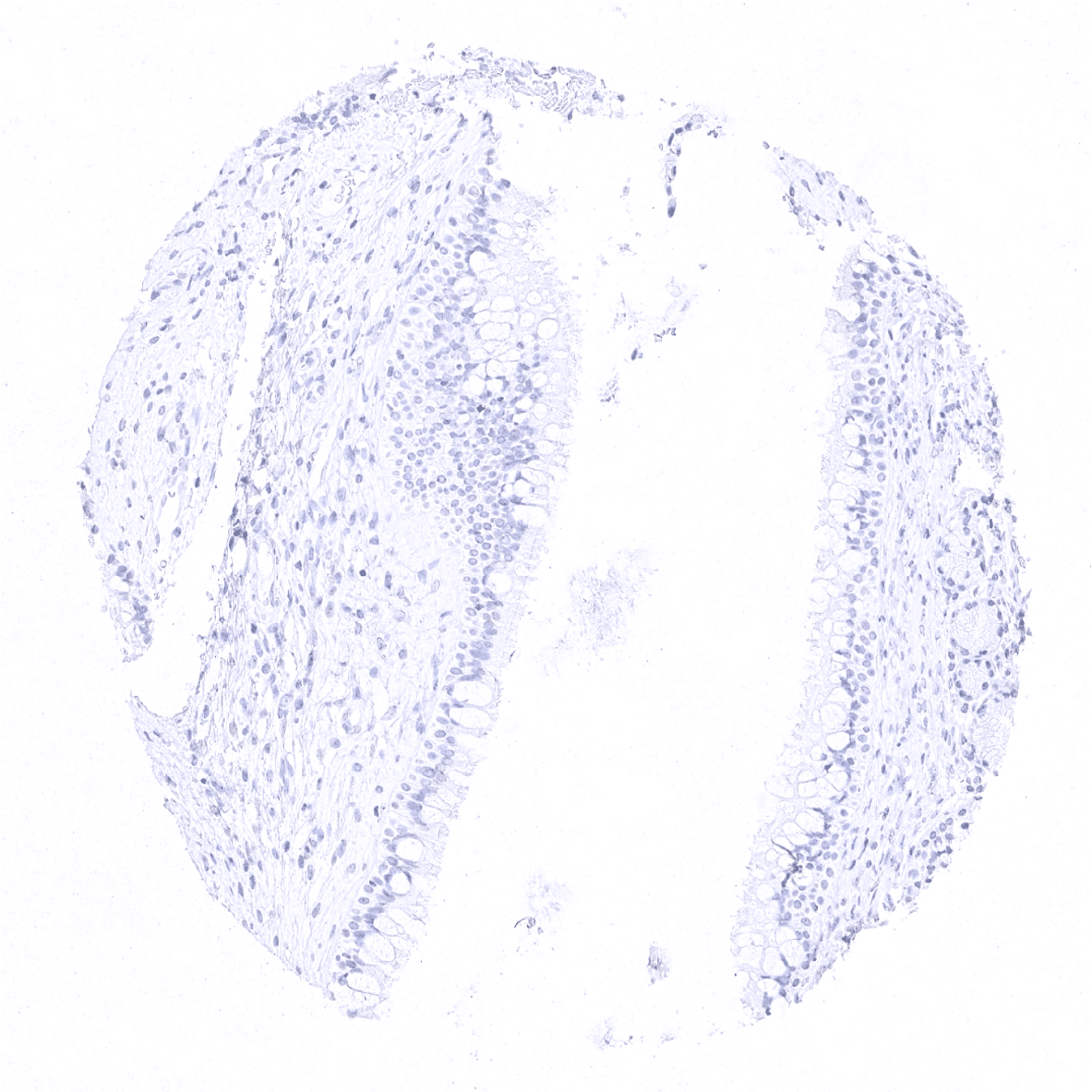
Appendix, mucosa
Appendix, muscular wall
Bone marrow
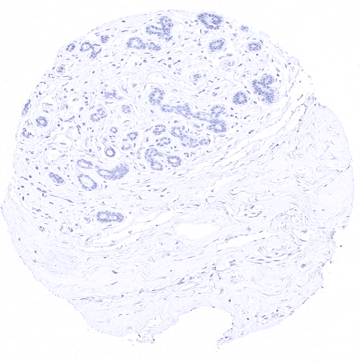
Breast

Bronchus, mucosa
Cerebellum (molecular layer, Purkinje cell layer, granule cell layer, white matter)

Cerebellum (granule cell layer, white matter)

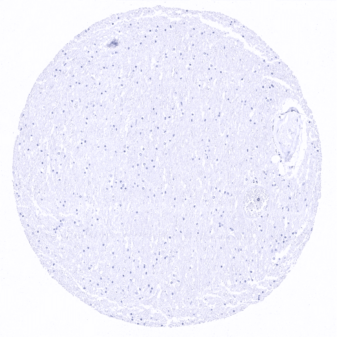
Cerebrum, grey matter
Cerebrum, white matter
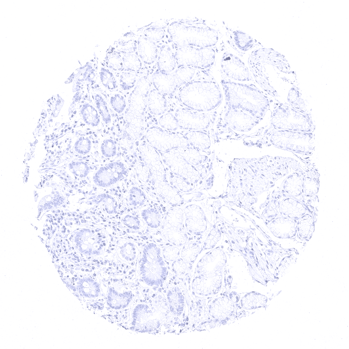
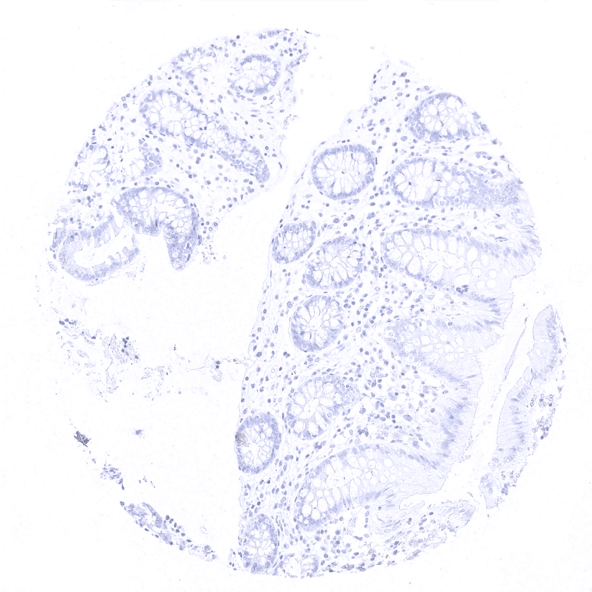
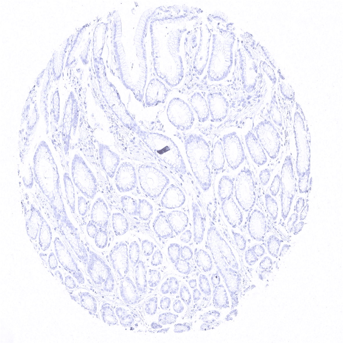
Colon descendens, mucosa
Colon descendens, muscular wall
Duodenum, Brunner gland
Duodenum, mucosa
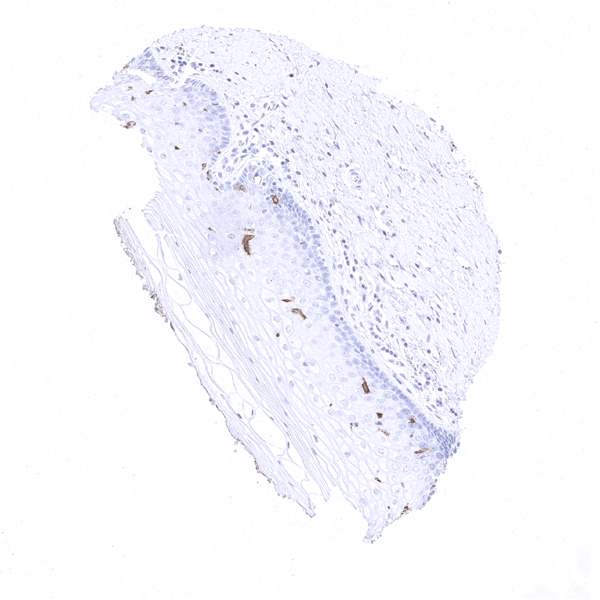
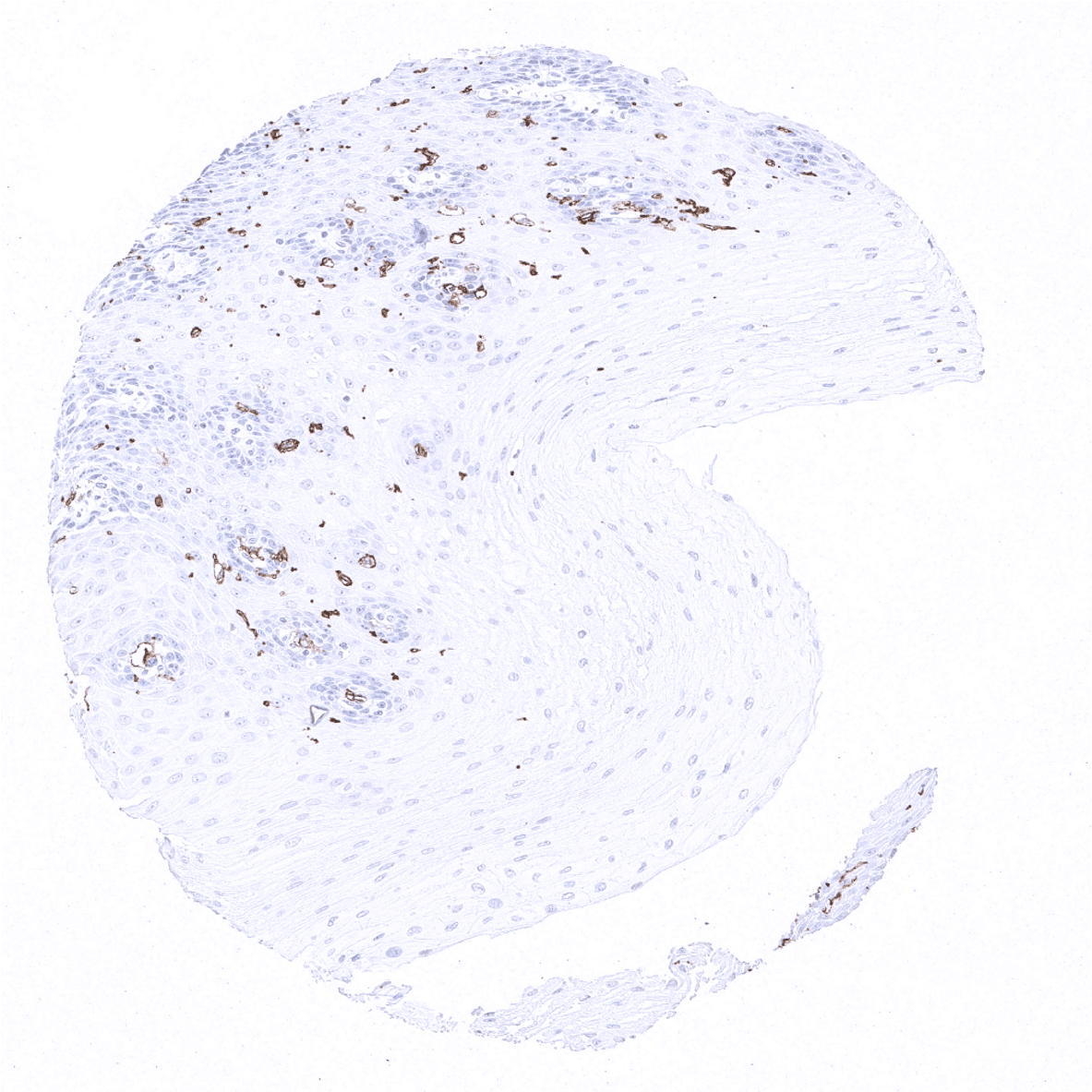
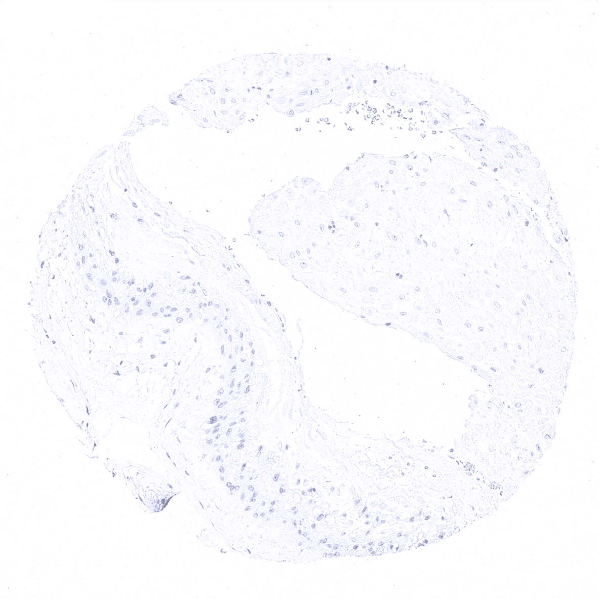
Ectocervix - Moderate CD1a positivity in Langerhans cells of the squamous epithelium of the ectocervix.
Endocervix

Endometrium, proliferation
Endometrium, secretion
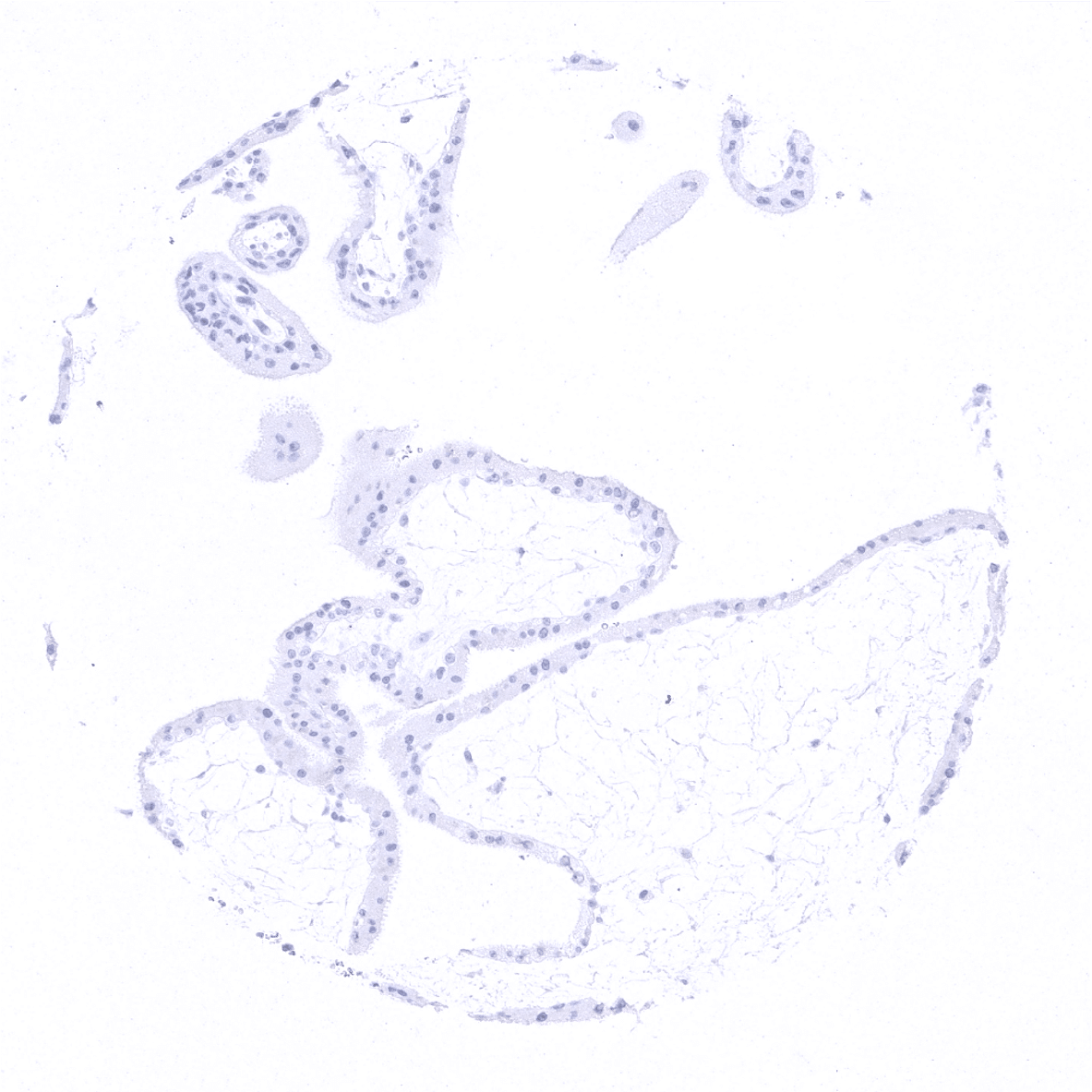
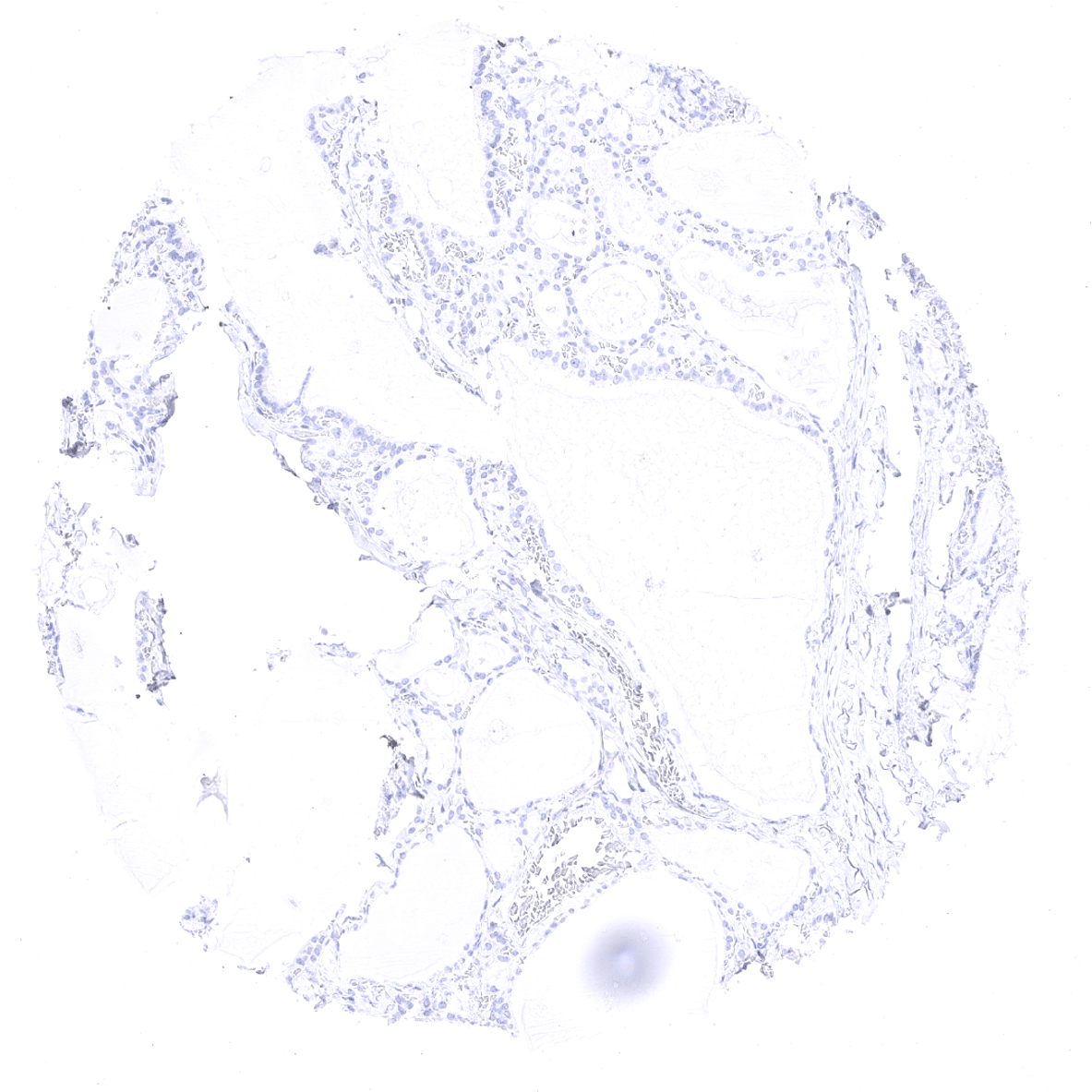
Epididymis
Esophagus, squamous epithelium

Fallopian tube, mucosa
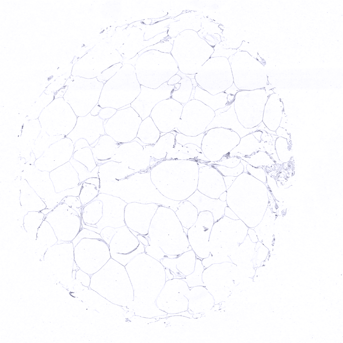
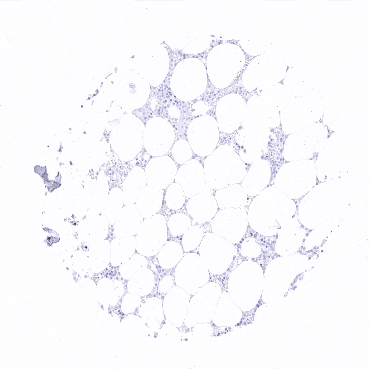
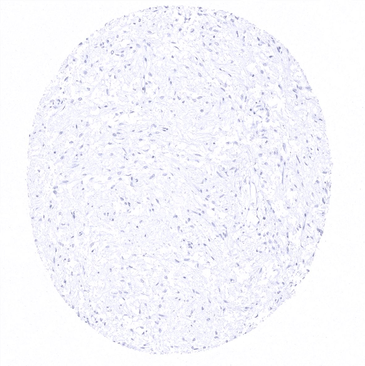
Fat
Gallbladder, epithelium
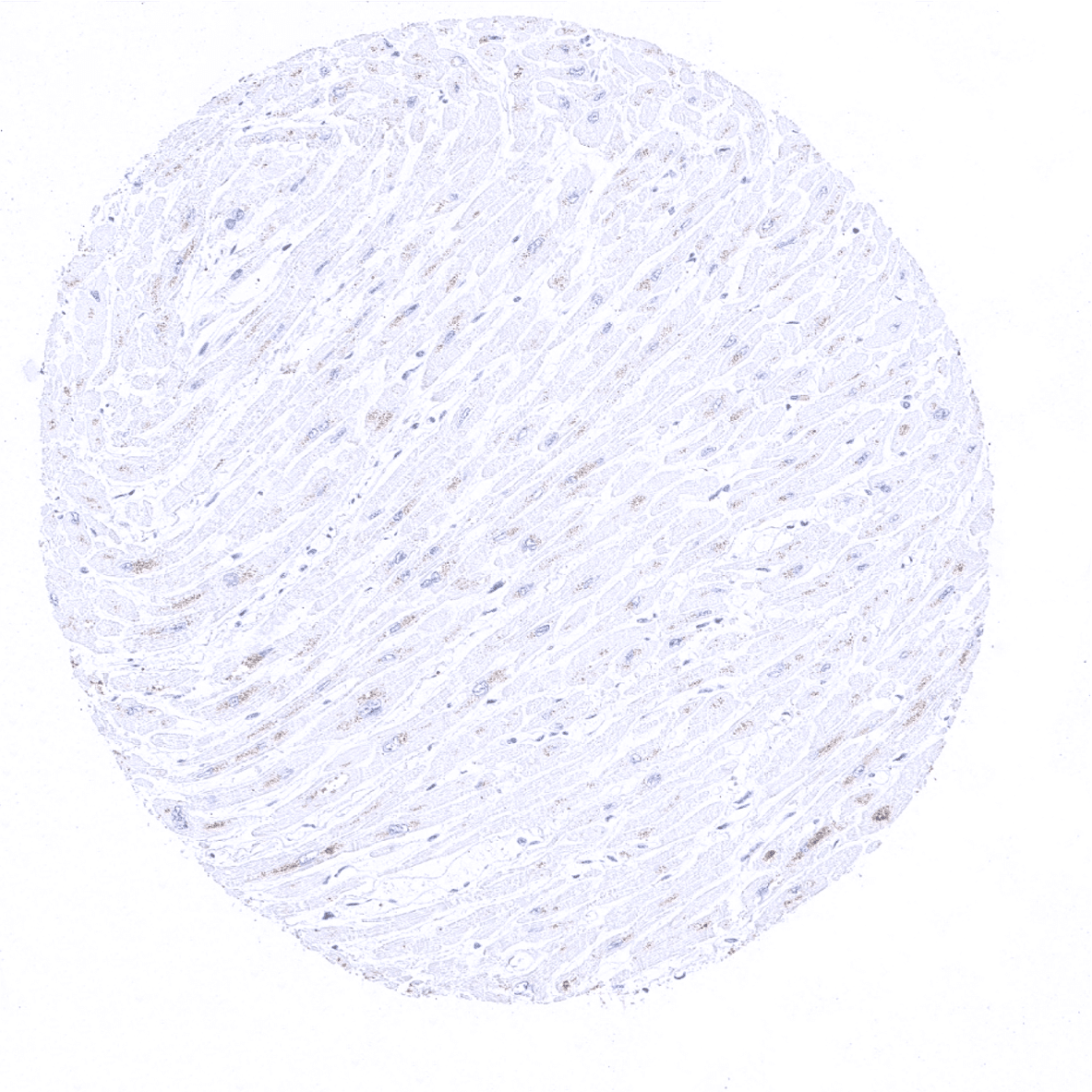
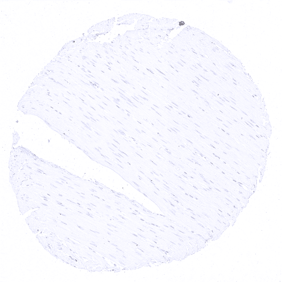
Heart
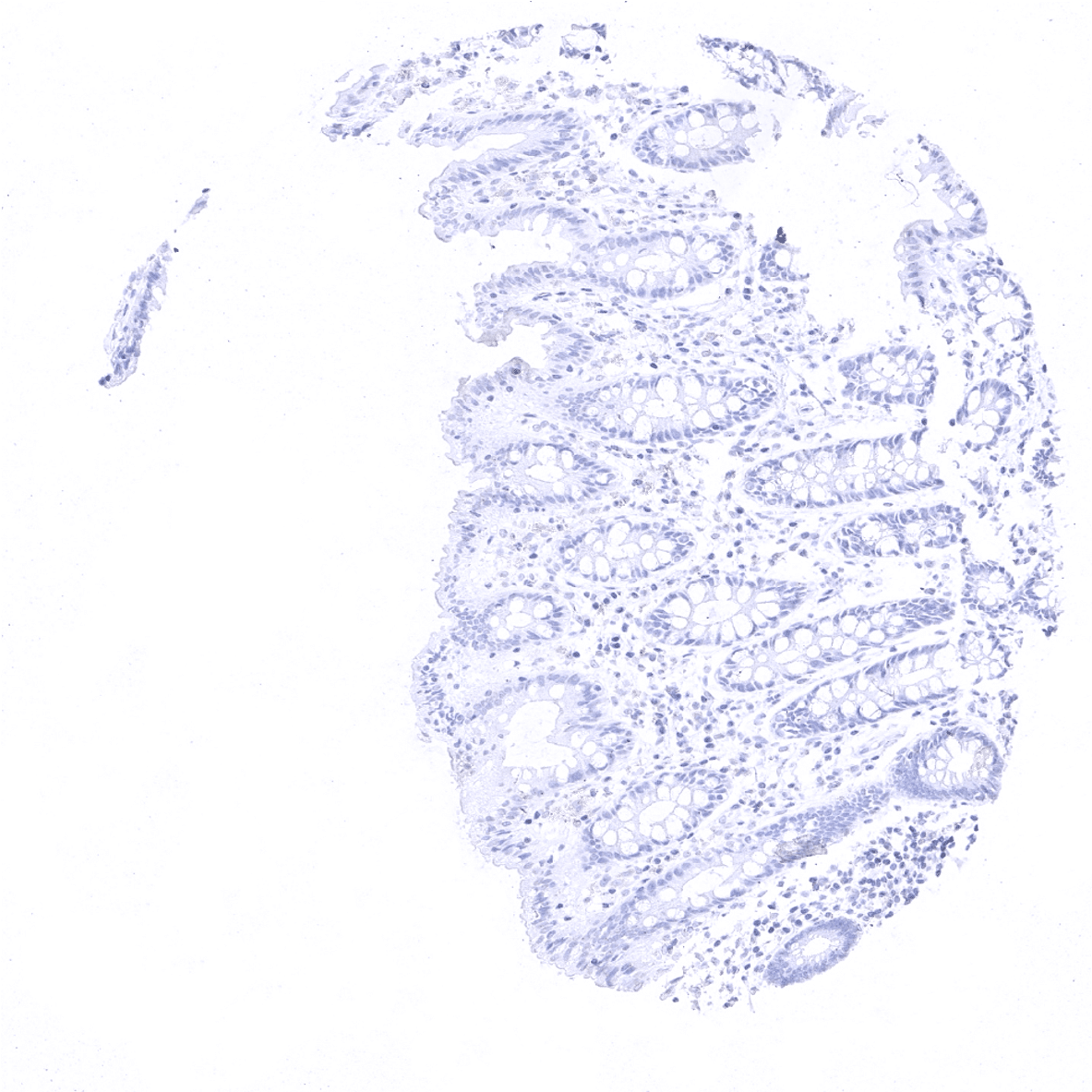
Ileum, mucosa
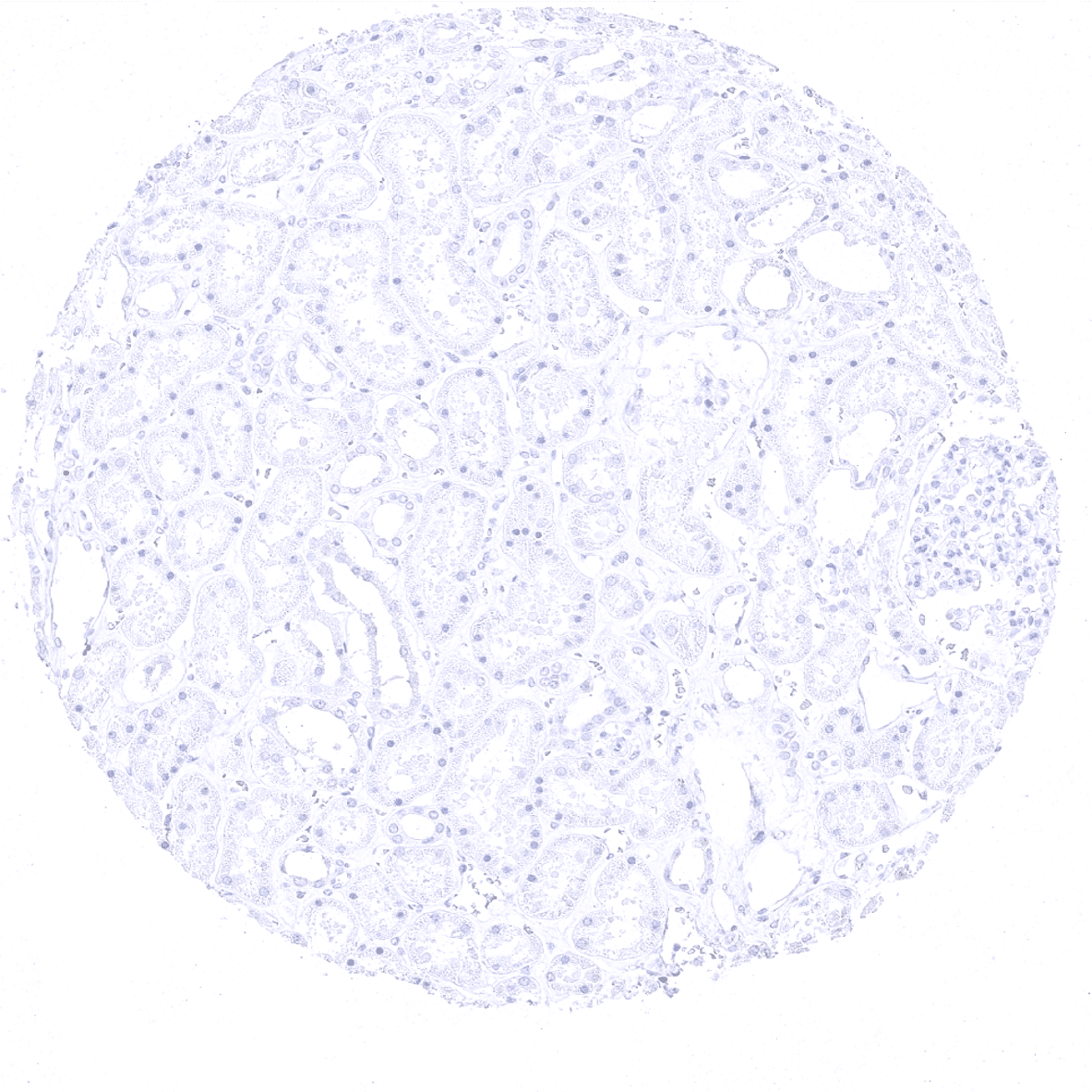
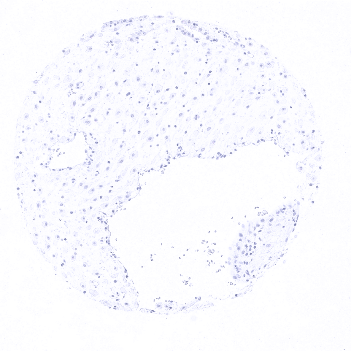
Kidney, cortex
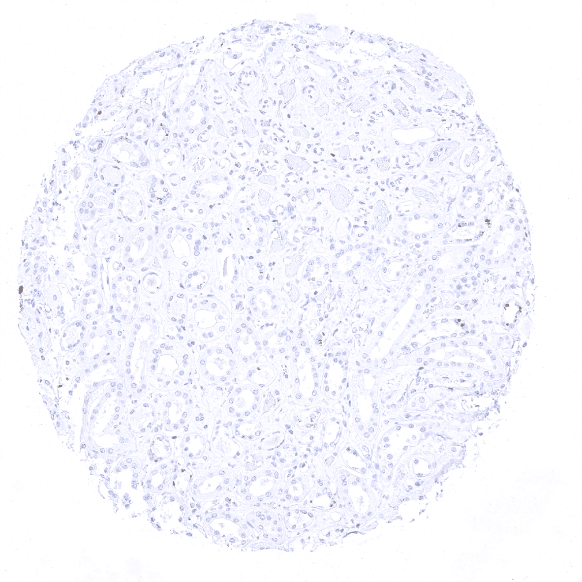
Kidney, medulla
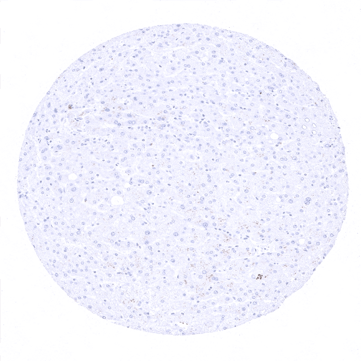
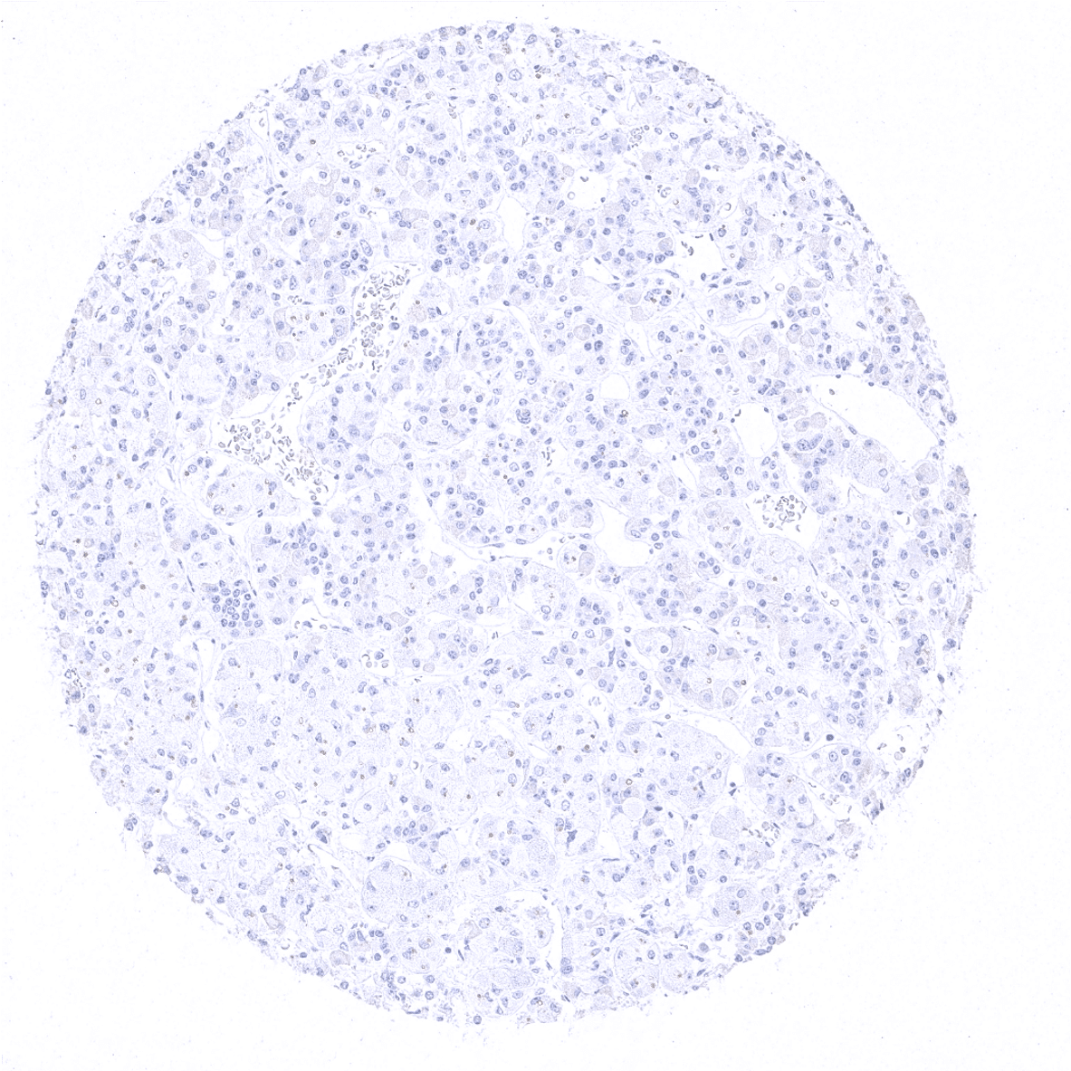
Liver
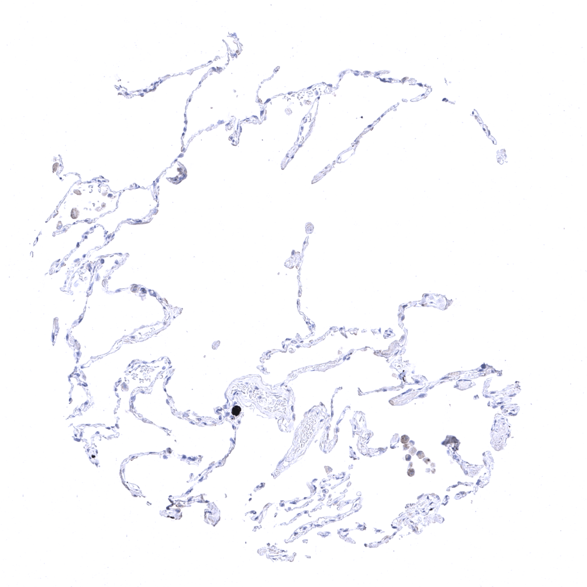
Lung

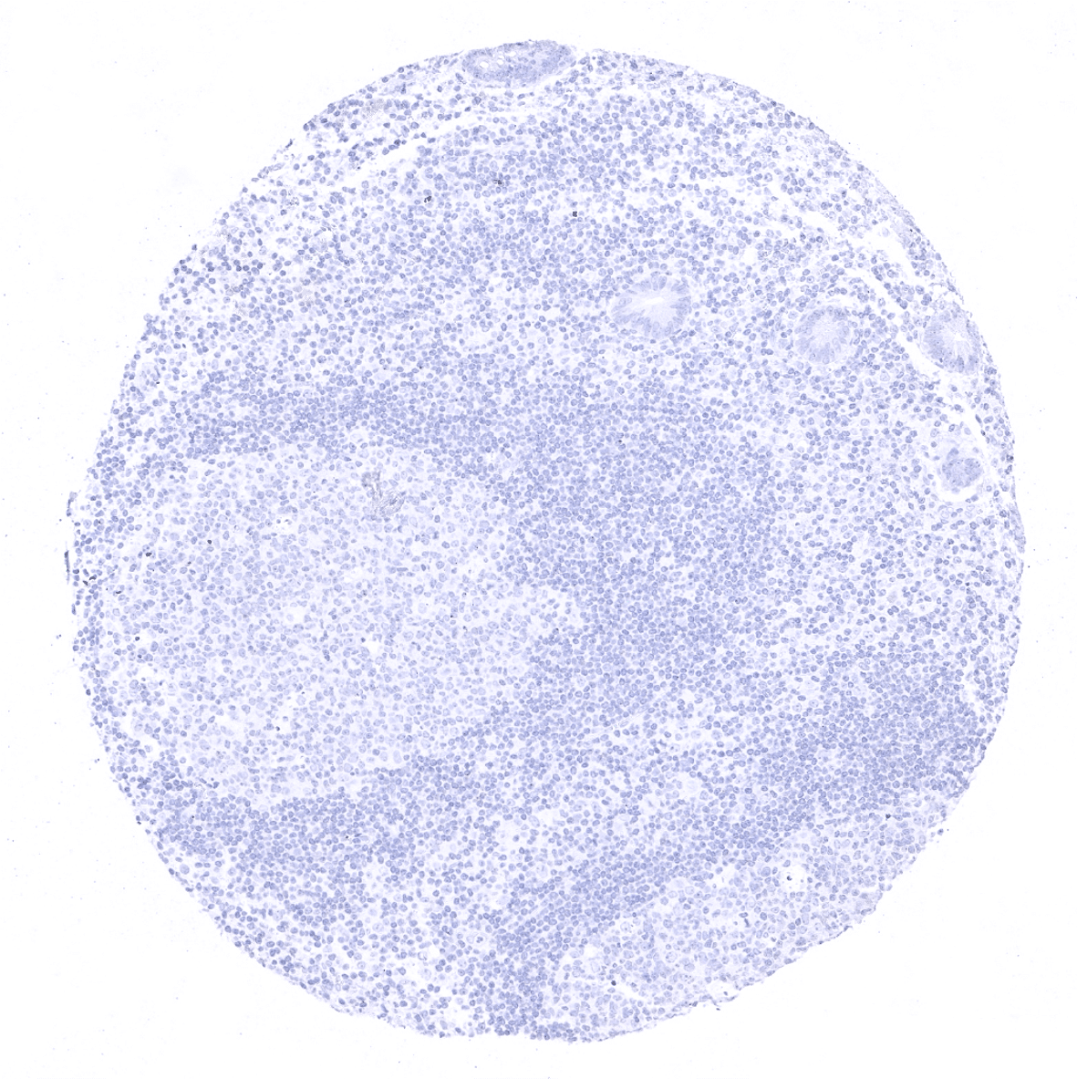
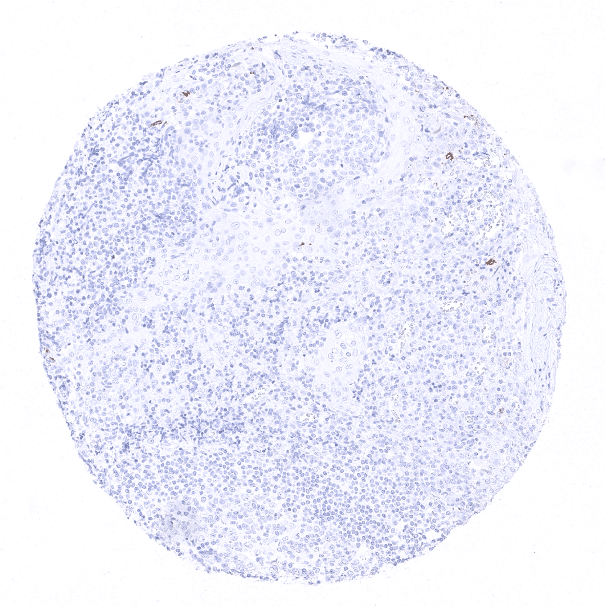
Lymph node - CD1a immunostaining is absent in normal lymph nodes.
Ovary, stroma
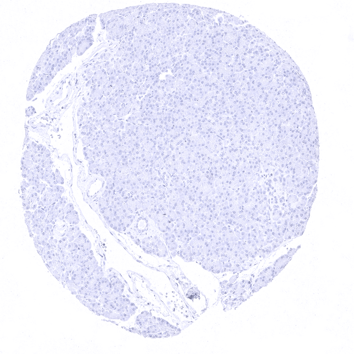
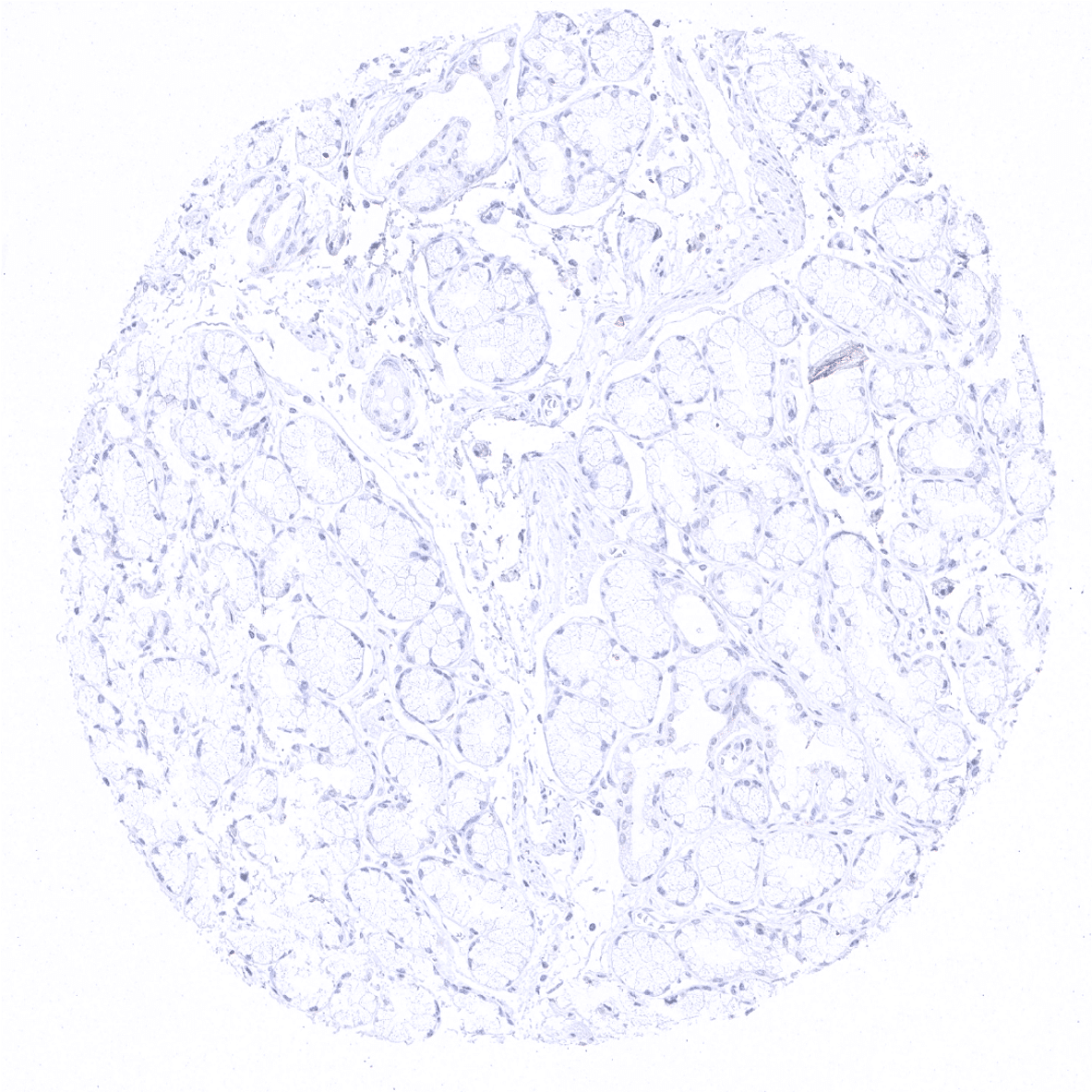
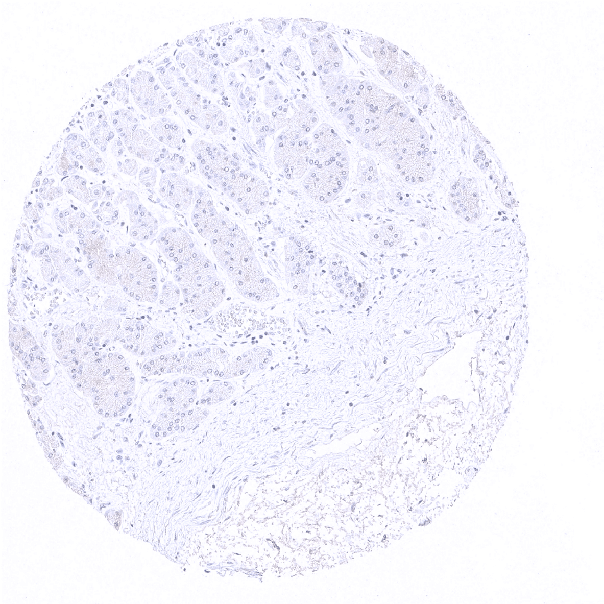
Pancreas
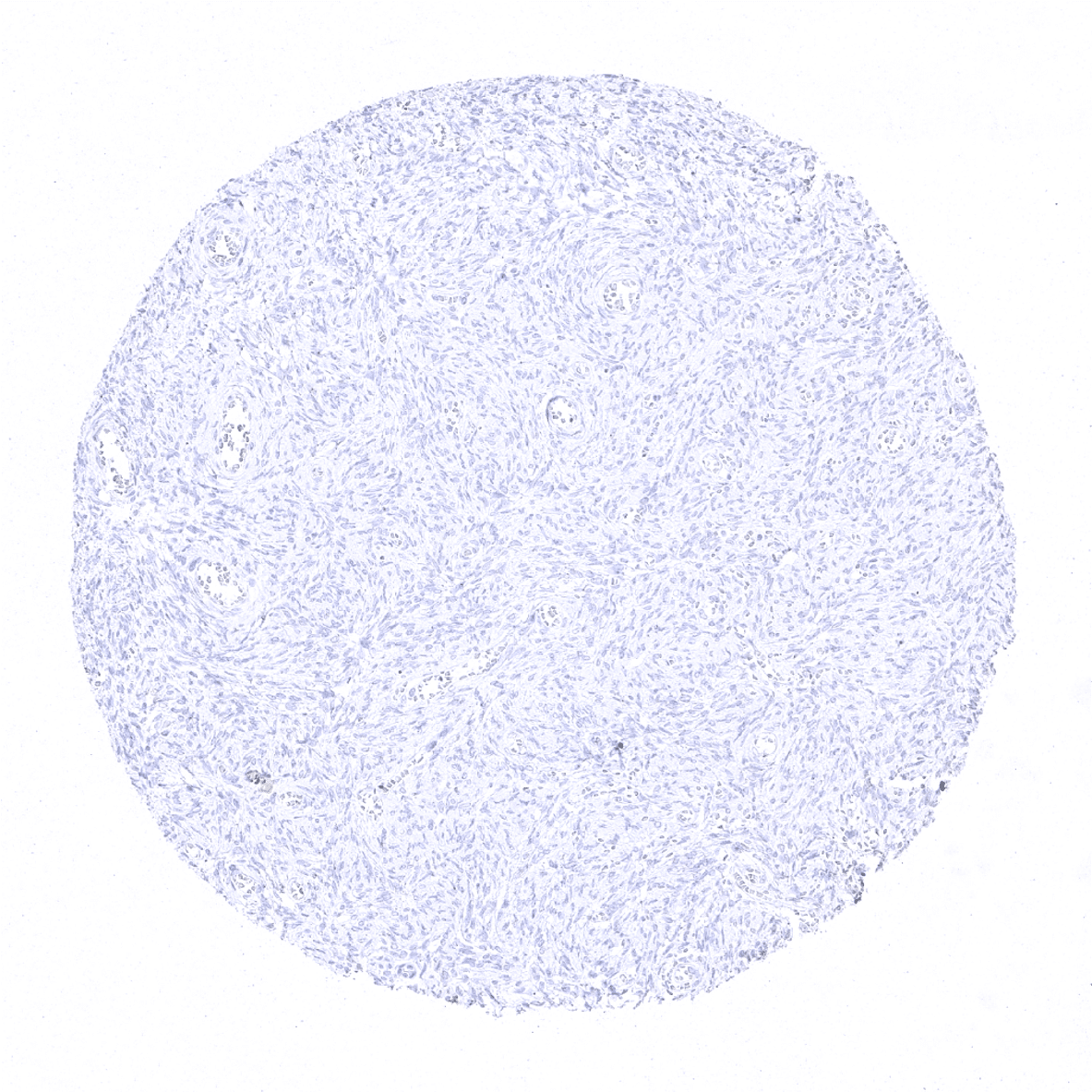
Parathyroid
Parotid gland
Pituitary gland, anterior lobe
Pituitary gland, posterior lobe
Pregnant uterus (decidua)
Placenta, early

Placenta, mature
Placenta (amnion and chorion)
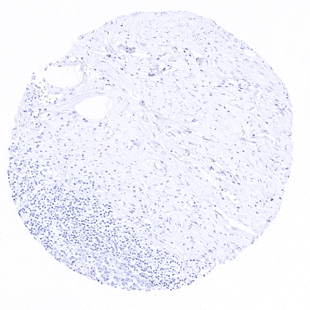
Prostate
Rectum, mucosa
Seminal vesicle
Sinus paranasales
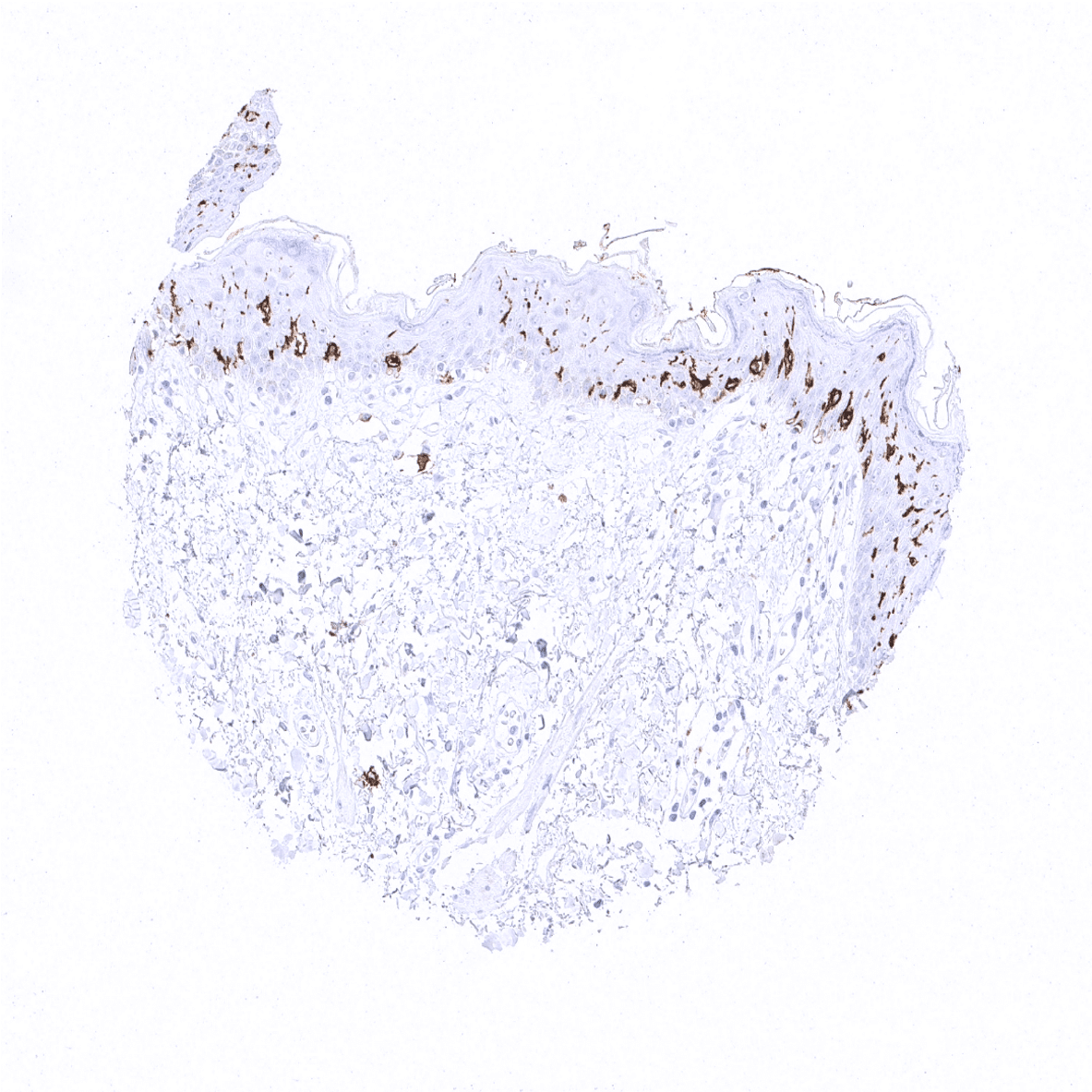
Skin - CD1a is strongly expressed in Langerhans cells of the skin.

Spleen
Stomach, antrum
Stomach, corpus

Striated muscle
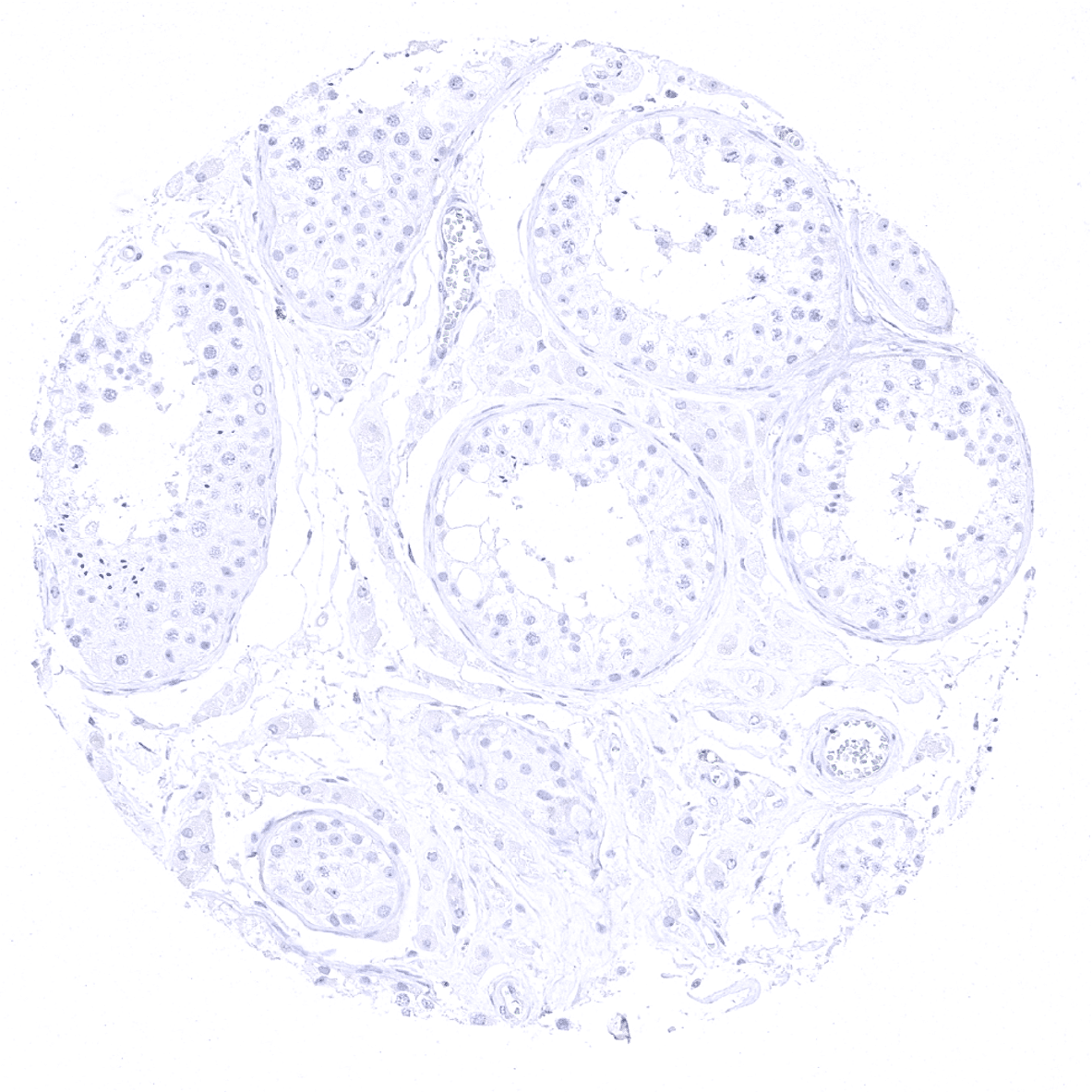
Testis
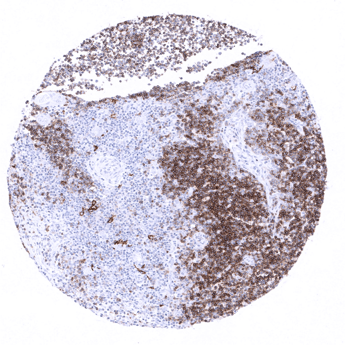
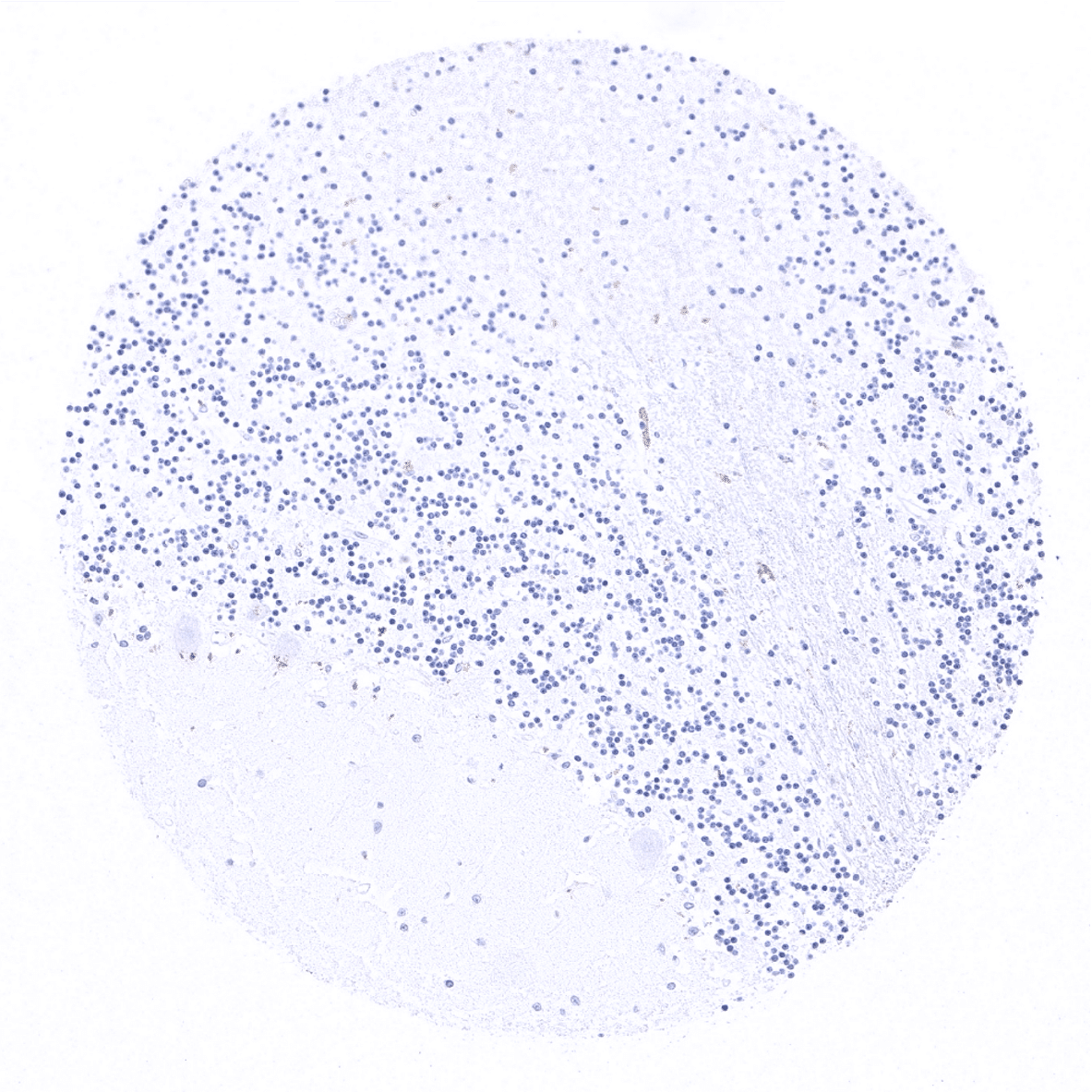
Thymus - A strong CD1a positivity is seen in the majority of lymphocytes in cortex but in only few cells of the medulla.
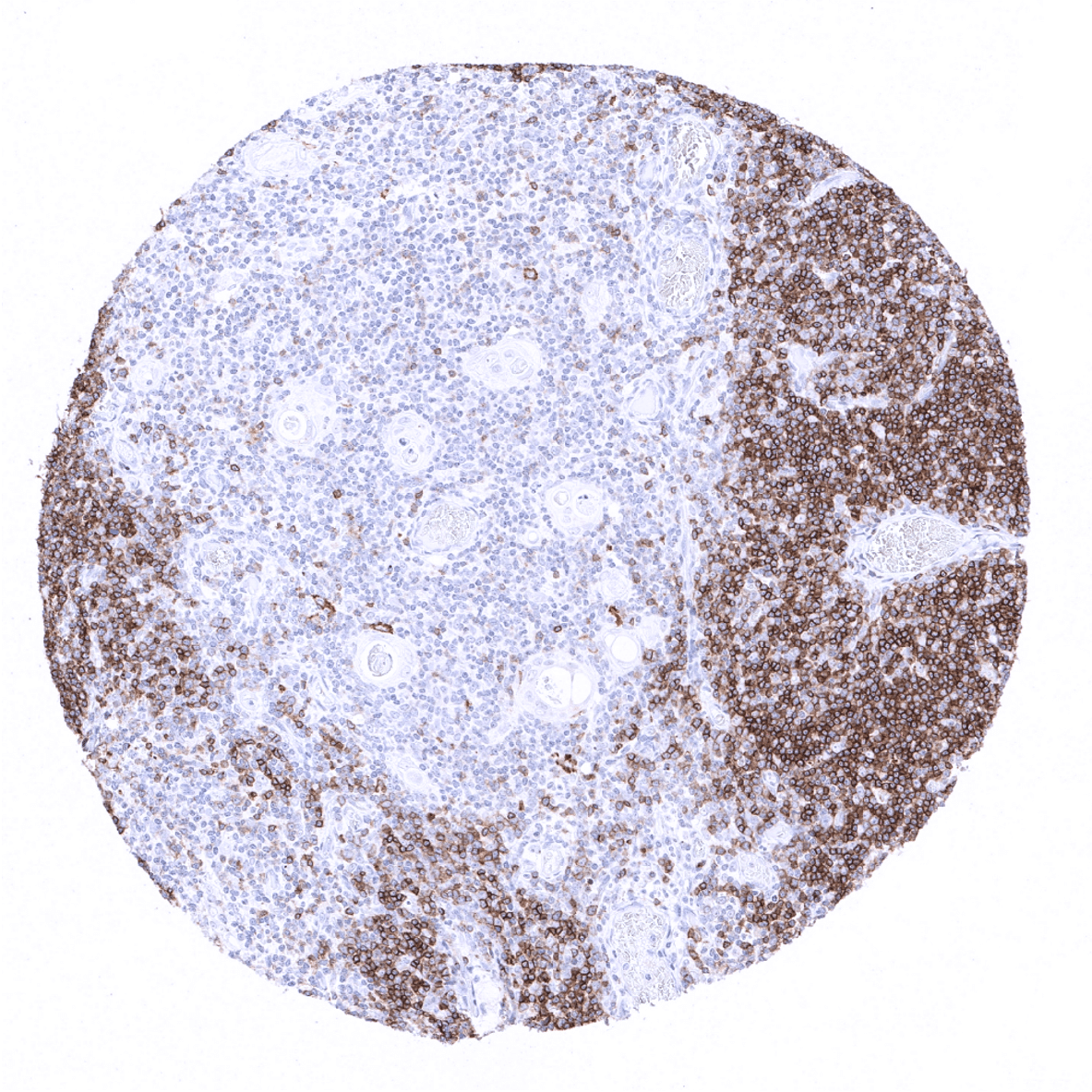
Thymus - CD1a immunostaining is seen in >80% of cortical thymocytes but is largely absent in the medulla.
Thyroid gland
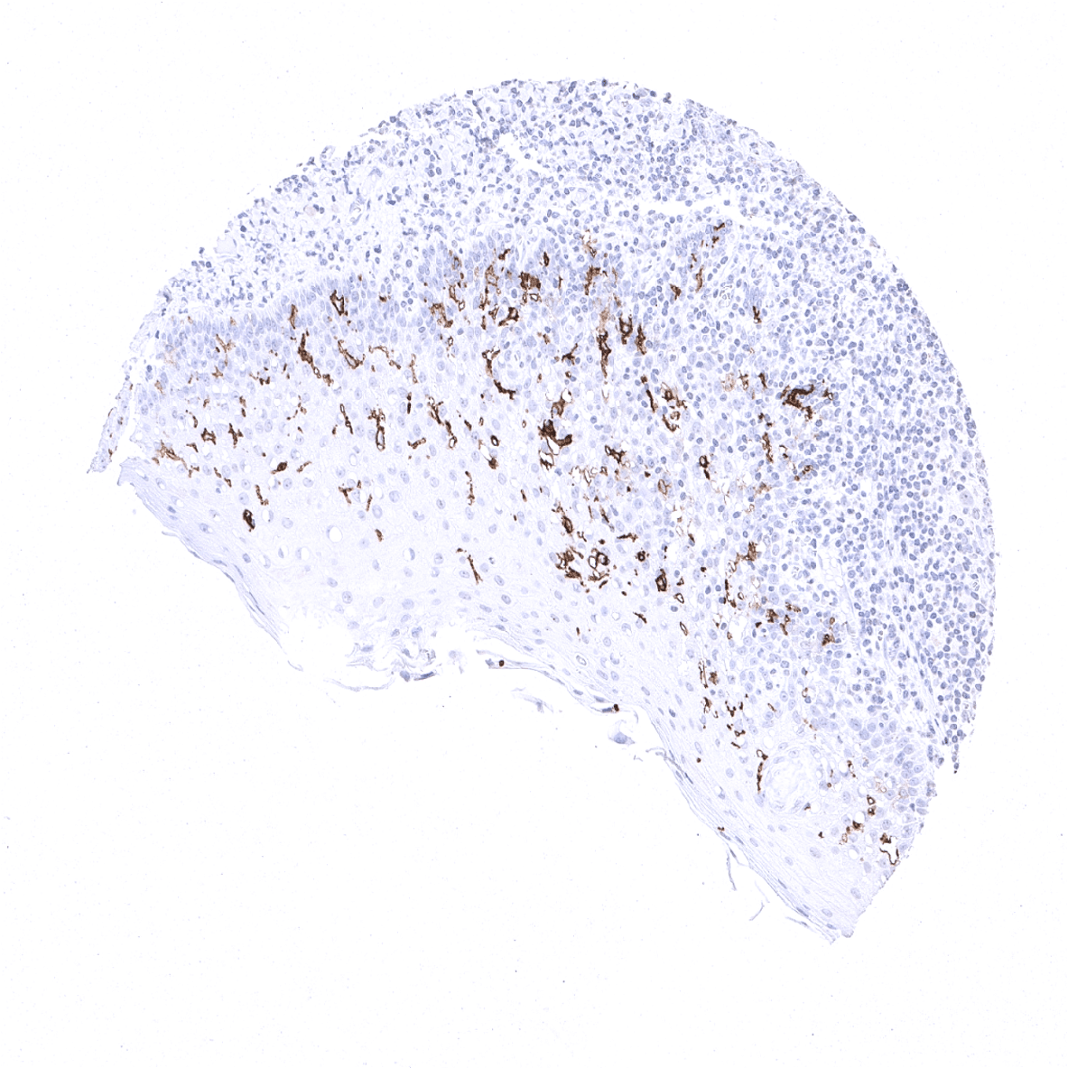
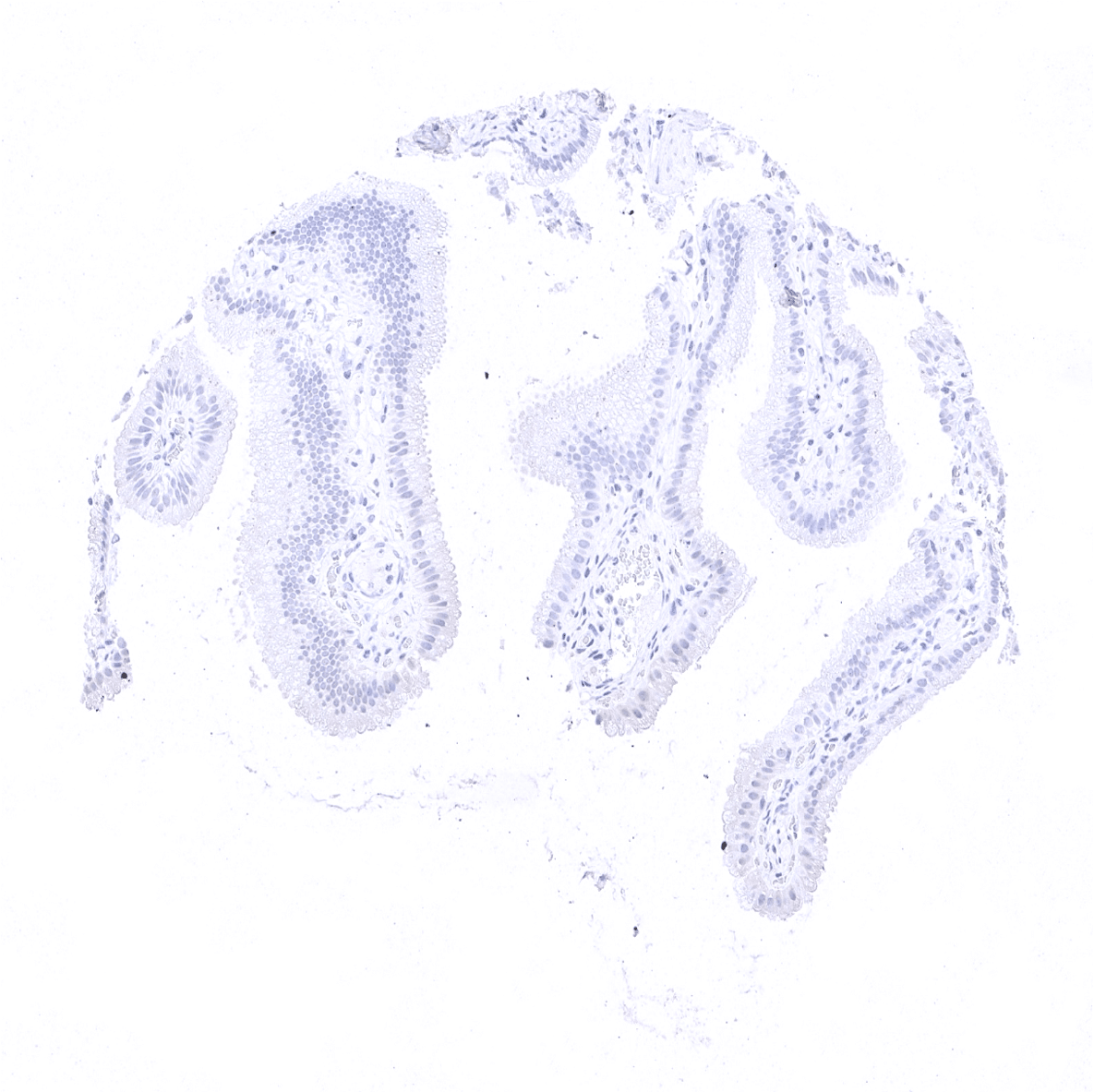
Tonsil, surface epithelium - Strong CD1a immunostaining in Langerhans cells of the squamous epithelium of the tonsil surface.
Tonsil
Urinary bladder, muscular wall
Urinary bladder, urothelium
Uterus, myometrium