145,00 € – 595,00 €
Product details
Synonyms = B220, CD45R, GP180, Leukocyte common antigen (LCA), Loc, Ly-5, Lyt-4, Protein tyrosine phosphatase receptor type C (PTPRC), Receptor-type tyrosine-protein phosphatase C, T200 glycoprotein
Antibody type = Mouse monoclonal / IgG2a
Clone = MSVA-472M
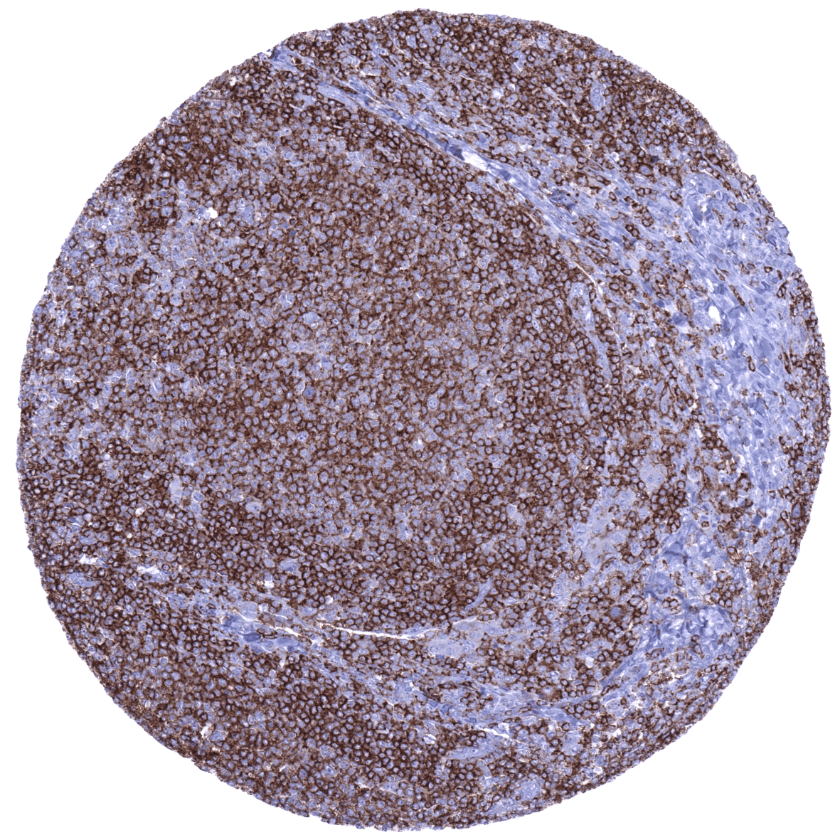
Positive control = Tonsil: A strong membranous CD45RA immunostaining should be seen in the majority of lymphocytes.
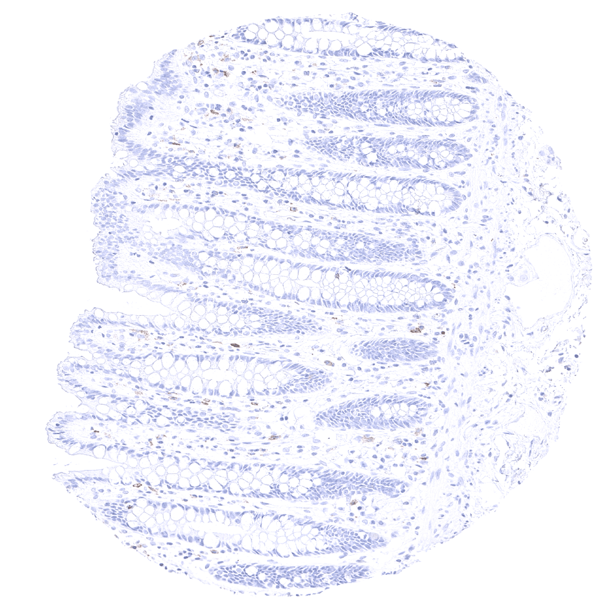
Negative control = Tonsil: CD45RA immunostaining should be absent in epithelial cells.
Cellular localization = Cell Surface
Reactivity = Human
Application = Immunohistochemistry
Dilution = 1:100 – 1:200
Intended Use = Research Use Only
Relevance of Antibody
CD45RA is expressed on naive T lymphocytes.
Biology Behind
CD45 is a type I transmembrane protein that is present in various isoforms on all differentiated hematopoietic cells except erythrocytes and plasma cells. The gene is located at 1q31.3-q32.1 and contains 34 exons coding for an unusually large protein with extracellular and cytoplasmic domains. The exons 4, 5, and 6 (corresponding to protein regions A, B, and C) are alternatively spliced to generate up to eight different protein products featuring combinations of zero, one, two, or all three exons. All these CD45 isoforms are essential regulators of T- and B-cell antigen receptor signaling. They function through either direct interaction with components of the antigen receptor complexes via its extracellular domain or by activating various Src family kinases required for the antigen receptor signaling via its cytoplasmic domain. Antibodies against specific CD45 isoforms are thus used in immunohistochemistry to differentiate between important immune cell types. Naive T lymphocytes are typically positive for CD45RA, which includes only the A protein region.
Staining Pattern in Normal Tissues
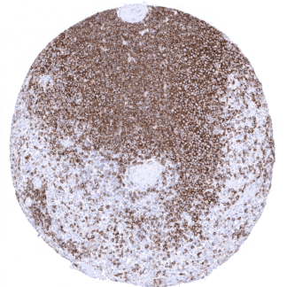
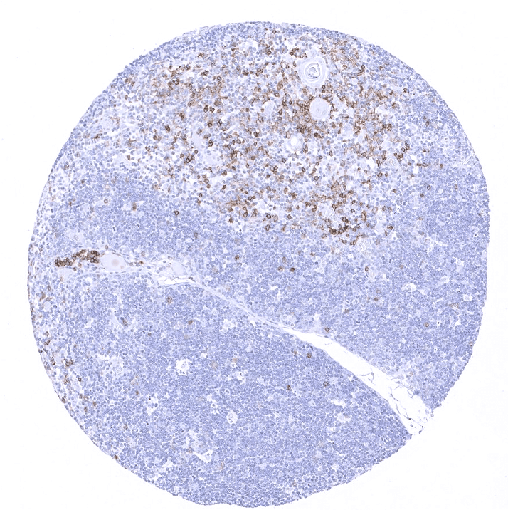
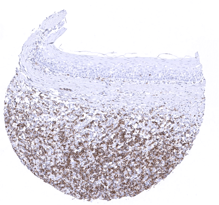
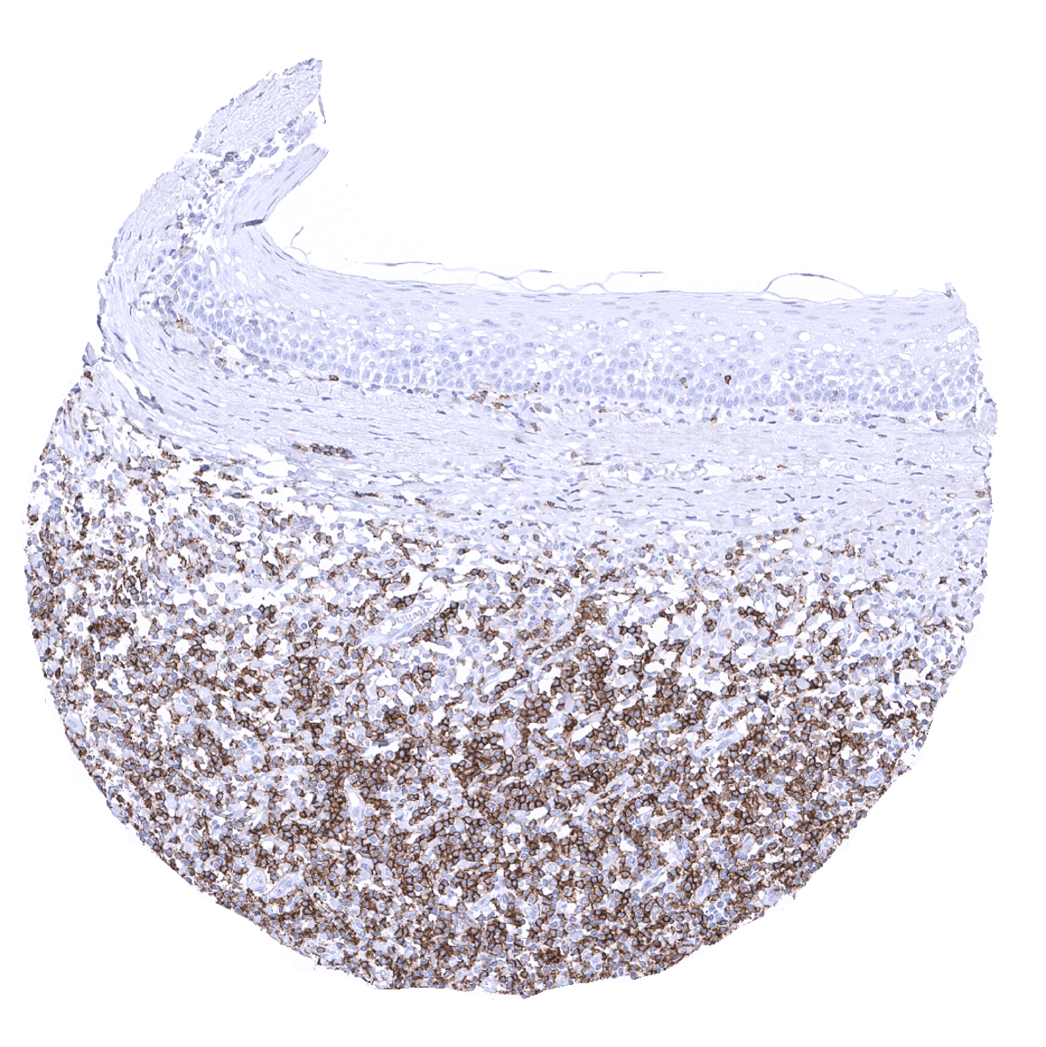
CD45RA immunostaining is seen in a variable fraction of T- and B-lymphocytes. The fraction of CD45RA positive lymphocytes is lowest in the cortex of the thymus (<1%) but is higher (>20%) in the thymic medulla. In lymph nodes, the white pulpa of the spleen and in the tonsil, the vast majority of lymphocytes are CD45RA positive. In non-lymphatic organs, lymphoid follicles often show a high percentage of CD45RA positive lymphocytes, while the rate of CD45RA positivity is more variable among scattered lymphocytes. A faint CD45RA immunostaining is also seen on the apical membrane of amnion cells.
These findings are largely comparable to the RNA and protein data described in the Human Protein Atlas (Tissue expression CD45RA)
Suggested positive tissue control: Tonsil: A strong membranous CD45RA immunostaining should be seen in the majority of lymphocytes.
Suggested negative tissue control:Tonsil: CD45RA immunostaining should be absent in epithelial cells.
Staining Pattern in Relevant Tumor Types
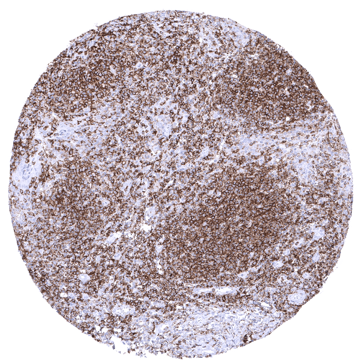
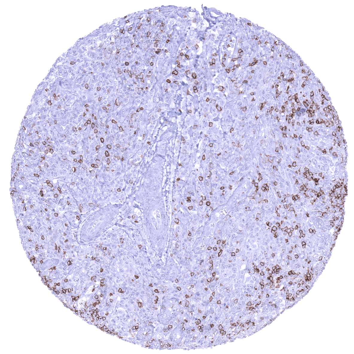
CD45RA is often expressed in most lymphoma subtypes. CD45RA positive cells are also seen in virtually every other tumor type as a part of the tumor infiltrating lymphocytes.
The TCGA findings on CD45RA RNA expression in different tumor categories have been summarized in the Human Protein Atlas.

Compatibility of Antibodies
No data available at the moment
Protocol Recommendations
IHC users have different preferences on how the stains should look like. Some prefer high staining intensity of the target stain and even accept some background. Others favor absolute specificity and lighter target stains. Factors that invariably lead to more intense staining include higher concentration of the antibody and visualization tools, longer incubation time, higher temperature during incubation, higher temperature and longer duration of the heat induced epitope retrieval (slide pretreatment). The impact of the pH during slide pretreatment has variable effects and depends on the antibody and the target protein. Accordingly, multiple different protocols can generate identical staining results.
All images and data shown here and in our image galleries are obtained by the manual protocol described below. Other protocols resulting in equivalent staining are described as well.
-Manual protocol
Freshly cut sections should be used (less than 10 days between cutting and staining). Heat-induced antigen retrieval for 5 minutes in an autoclave at 121°C in pH 7.8 Target Retrieveal Solution buffer. Apply MSVA-472M at a dilution of 1:200 at 37°C for 60 minutes. Visualization of bound antibody by the EnVision Kit (Dako, Agilent) according to the manufacturer’s directions.
Potential Research Applications
- Identification of naive T cells (CD45RA+) together with other markers.
- Naive T- (and B-) cells are of high importance for any type of immune response. Accordingly, CD45RA is part of most large panels for immune cell characterization by multicolor immunofluorescence.
Evidence for Antibody Specificity in IHC
Specificity of MSVA-472M is documented by strong positive staining in various types of lymphocytes that are well known to express CD45RA while CD45RA staining is largely absent in the cortex of the thymus and in virtually all epithelial cell types.