


395,00 € – 1.495,00 €
Product details
Synonyms = Cadherin-16 (CDH16); Kidney-specific cadherin; Ksp-cadherin antibody
Antibody type = Recombinant Rabbit monoclonal / Rabbit IgG
Clone = MSVA-516R
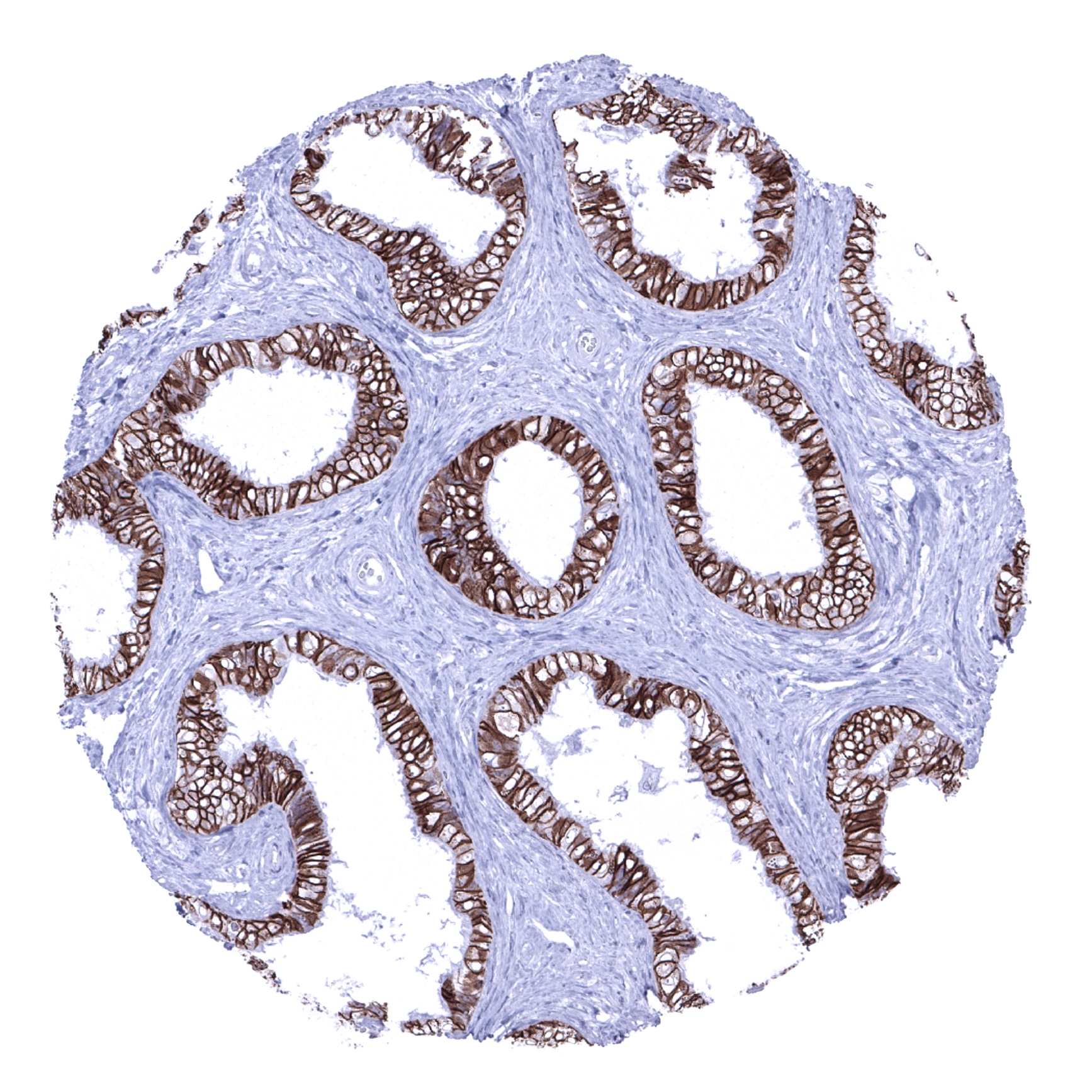
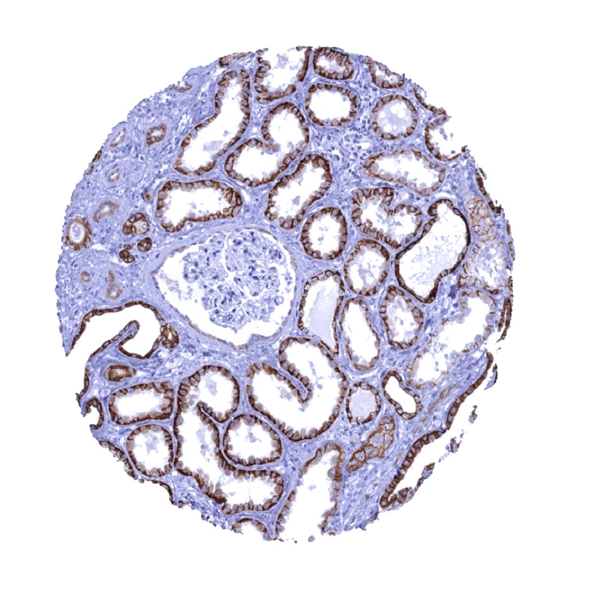
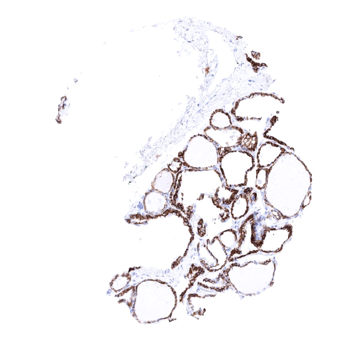
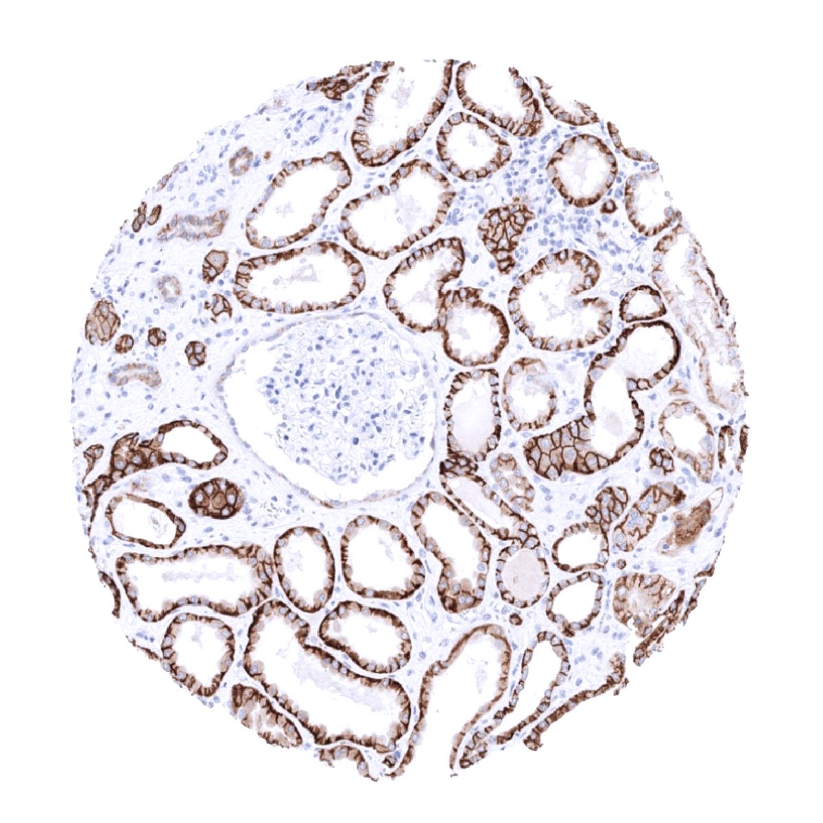
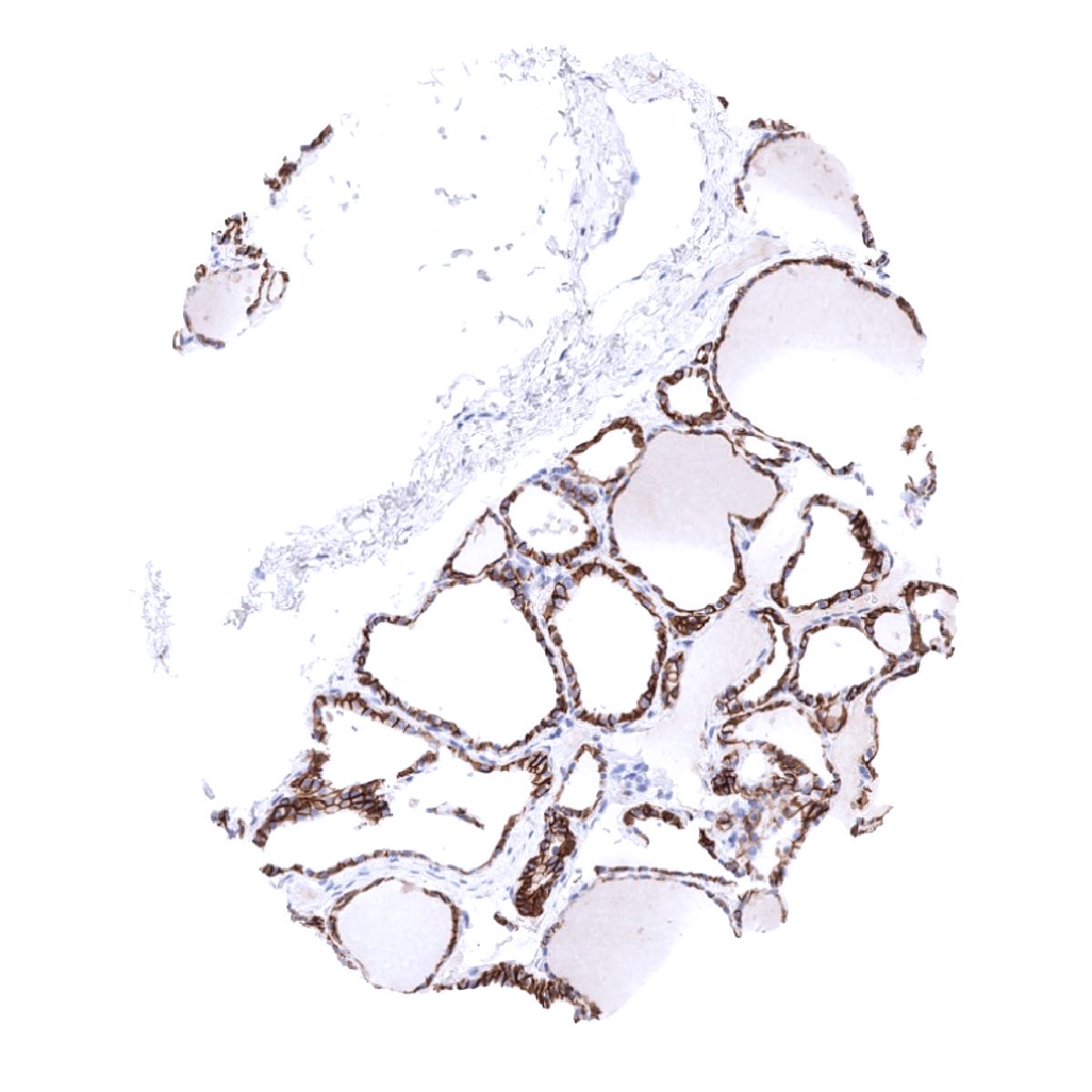
Positive control = Kidney: A strong staining should be seen in distal tubuli and collecting ducts while the staining is at least moderate in proximal tubuli.
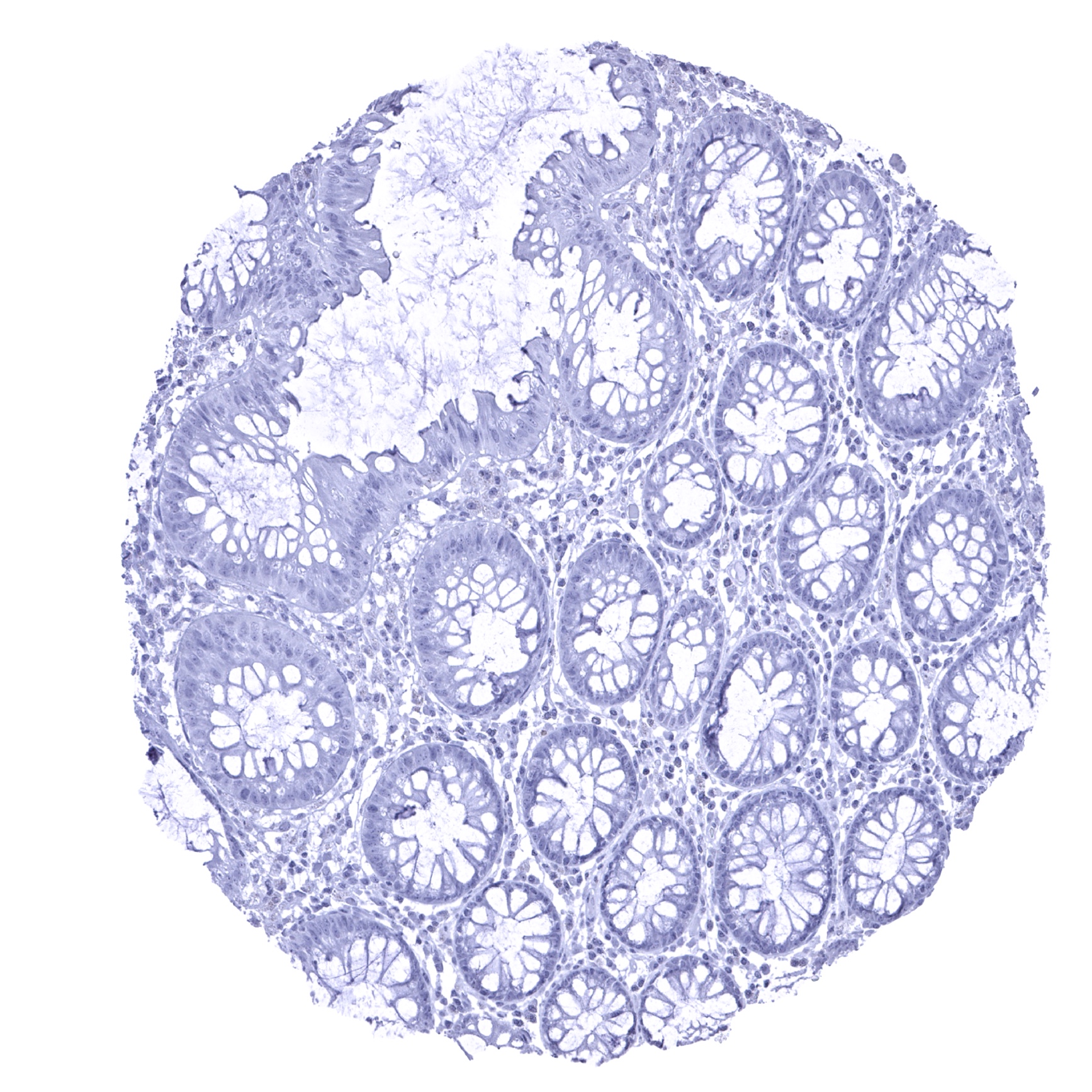
Negative control = Colon: All cells should be CDH16 negative.
Cellular localization = Cell Surface and Cytoplasmic
Reactivity = Human
Application = Immunohistochemistry
Dilution = 1:100 – 1:200
Intended Use = Research Use Only
Relevance of Antibody
CDH16 is a marker for kidney and thyroid epithelial cells.
Biology Behind
Cadherin-16, coded by the CDH16 gene at 16q22.1, belongs to the cadherin superfamily which is composed of calcium-dependent, membrane-associated glycoproteins with a role in cell-adhesion. CDH16 is involved in embryonal development and cell growth. CDH16 supports the formation of tubuli during renal development and remains expressed in distal tubuli of adult kidneys. CDH16 is also relevant for the development of follicular thyroid cells and thyroid follicular polarity.
Staining Pattern in Normal Tissues
CDH16 staining pattern in Normal Tissues with antibody MSVA-516R (images are shown in our “Normal Tissue Gallery”)
| Brain | Cerebrum | Negative. |
| Cerebellum | Negative. | |
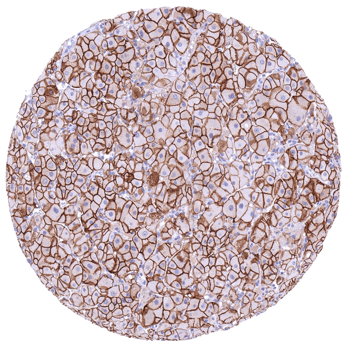
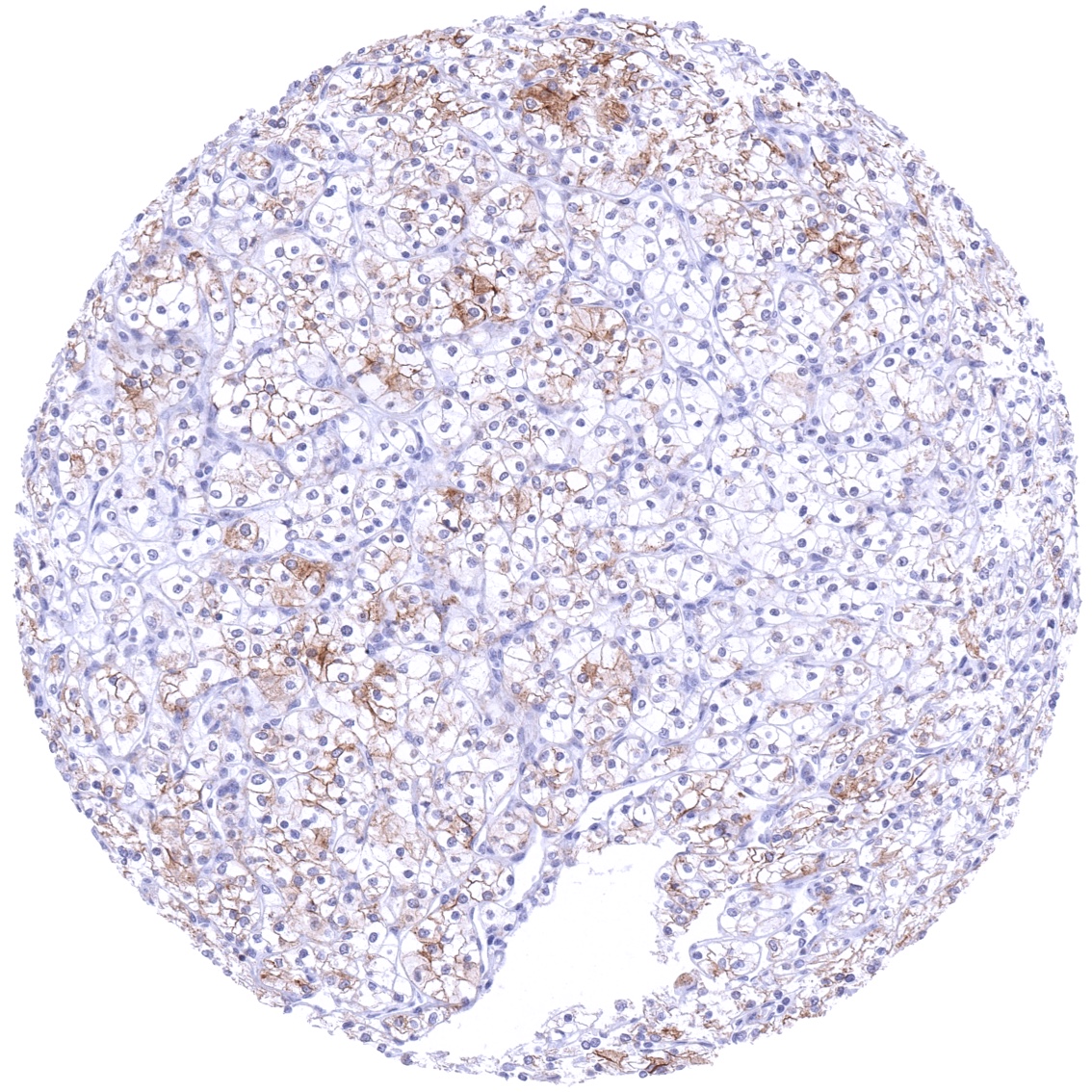
| Endocrine Tissues | Thyroid | Strong predominantly membranous CDH16 staining of follicular cells. |
| Parathyroid | Negative. | |
| Adrenal gland | Negative. | |
| Pituitary gland | Negative. | |
| Respiratory system | Respiratory epithelium | Negative. |
| Lung | Negative. | |
| Gastrointestinal Tract | Salivary glands | Negative. |
| Esophagus | Negative. | |
| Stomach | Negative. | |
| Duodenum | Negative. | |
| Small intestine | Negative. | |
| Appendix | Negative. | |
| Colon | Negative. | |
| Rectum | Negative. | |
| Liver | Negative. | |
| Gallbladder | Moderate to strong, predominantly membranous CDH16 staining of a small fraction of epithelial cells which are often arranged in groups. | |
| Pancreas | Negative. | |
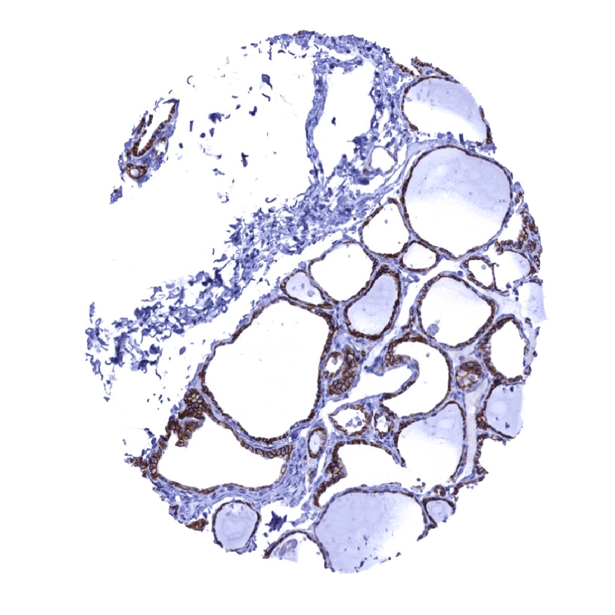
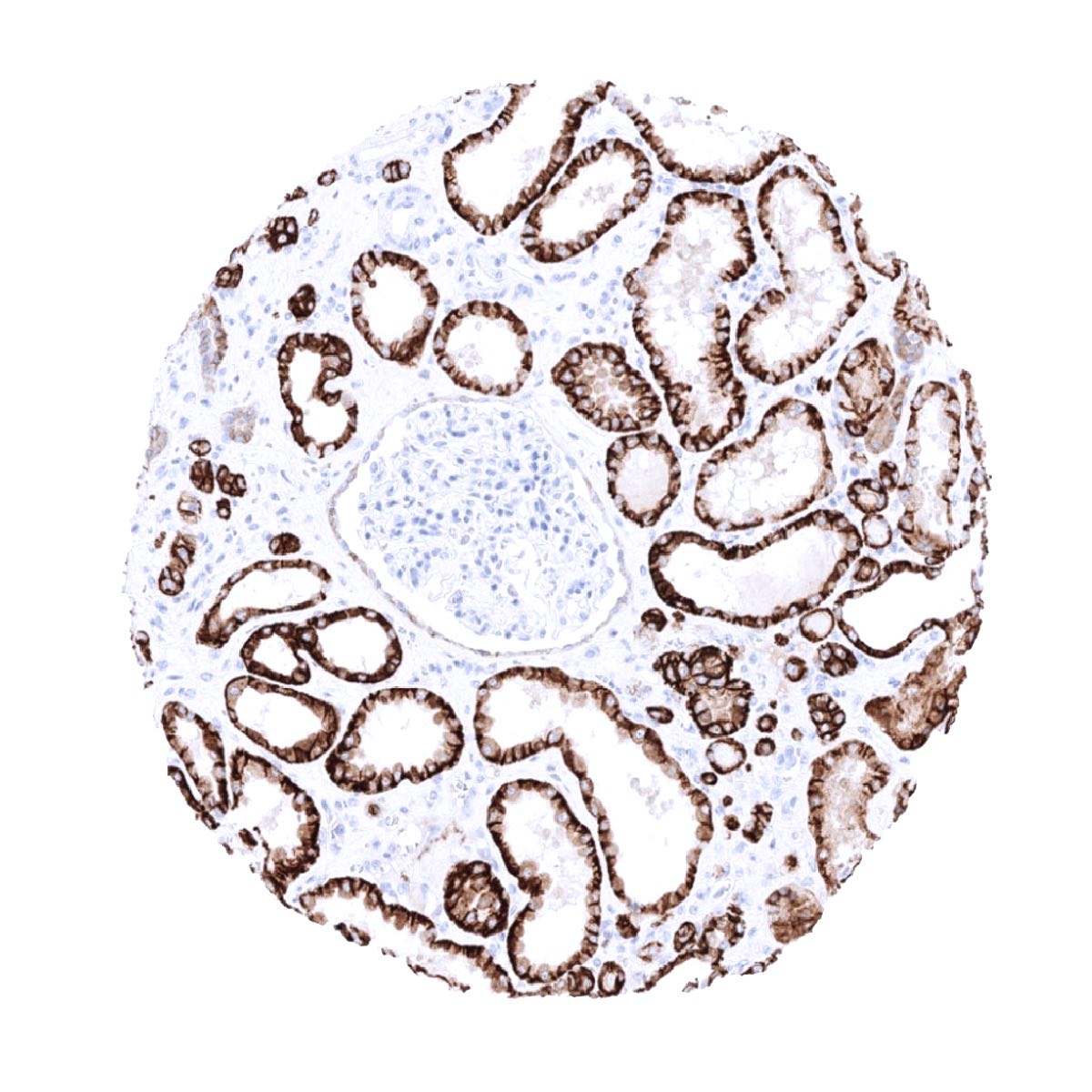
| Genitourinary | Kidney | Strong predominantly membranous but also cytoplasmic CDH16 staining of distal tubuli and collecting ducts. Staining is somewhat weaker and predominantly seen in basal and lateral membranes of proximal tubuli. |
| Urothelium | Negative. | |
| Male genital | Prostate | Negative. |
| Seminal vesicles | Moderate to strong, predominantly membranous CDH16 staining of a small fraction of epithelial cells which are often arranged in groups. | |
| Testis | Negative. | |
| Epididymis | Strong, predominantly membranous but also cytoplasmic CDH16 staining in all epithelial cells of the cauda epididymis. CDH16 is very weak or absent in epithelial cells of the caput. | |
| Female genital | Breast | Negative. |
| Uterus, myometrium | Negative. | |
| Uterus, ectocervix | Negative. | |
| Uterus endocervix | Moderate to strong, predominantly membranous CDH16 staining of a small fraction of epithelial cells which are often arranged in groups. | |
| Uterus, endometrium | Moderate to strong, predominantly membranous CDH16 staining of a small fraction of epithelial cells which are often arranged in groups. | |
| Fallopian Tube | Moderate to strong, predominantly membranous CDH16 staining of a small fraction of epithelial cells which are often arranged in groups. | |
| Ovary | Negative. | |
| Placenta early | Negative. | |
| Placenta mature | Negative. | |
| Amnion | Negative. | |
| Chorion | Negative. | |
| Skin | Epidermis | Negative. |
| Sebaceous glands | Negative. | |
| Muscle/connective tissue | Heart muscle | Negative. |
| Skeletal muscle | Negative. | |
| Smooth muscle | Negative. | |
| Vessel walls | Negative. | |
| Fat | Negative. | |
| Stroma | Negative. | |
| Endothelium | Negative. | |
| Bone marrow/ lymphoid tissue | Bone marrow | Negative. |
| Lymph node | Negative. | |
| Spleen | Negative. | |
| Thymus | Negative. | |
| Tonsil | Negative. | |
| Remarks |
The findings described above are this consistent with the RNA data described in the Human Protein Atlas (Tissue expression CDH16)
Positive control = Kidney: A strong staining should be seen in distal tubuli and collecting ducts while the staining is at least moderate in proximal tubuli.
Negative control = Colon: All cells should be CDH16 negative.

Staining Pattern in Relevant Tumor Types
A positive CDH16 immunostaining is most commonly seen in kidney cancer. CDH16 expression also occurs in cancers of the thyroid, uterine cervix, endometrium and the ovary.
The TCGA findings on CDH16 RNA expression in different tumor categories have been summarized in the Human Protein Atlas.
Compatibility of Antibodies
CDH16 (MSVA-516R) publication summary
Papers used for data compilation:
-Paper 1: Lennartz et al.: “Reduced CDH16 expression is linked to poor prognosis in clear cell renal cell carcinoma” Published in Urol Oncol. 2022 Jul;40(7) (PMID: 35606285).
-Paper 2: Lennartz et al.: “Cadherin-16 (CDH16) immunohistochemistry: a useful diagnostic tool for renal cell carcinoma and papillary carcinomas of the thyroid” Published in Sci Rep. 2023 Aug 9;13(1) (PMID: 37558687).
In these two studies, a total of 14’734 tumors were analyzed from 155 different tumor categories by using the following protocol: Heat-induced antigen retrieval for 5 minutes in an autoclave at 121°C in pH 7.8 Target Retrieveal Solution buffer. MSVA-516R at a dilution of 1:150 at 37°C for 60 minutes. Visualization of bound antibody by the EnVision Kit (Dako, Agilent). This protocol was also used for all stainings depicted in our tumor and normal tissue galleries.
At least one case with a positive CDH16 immunostaining was seen in 49 (31.6%) and at least one case with a strong CDH16 staining was seen in 26 (16.8%) of 155 tumor categories. The distribution of positive staining results is shown in an “organ-systematic” and in a “ranking order” figure below (images based on a compilation of data from Lennartz et al., some subcategories were merged or omitted for display purposes). Results on possible associations with histopathological and clinical parameters of tumor aggressiveness are also summarized below (table based on data from Lennartz et al.).
Authors conclusions on diagnostic utility with respect to the distinction of benign versus malignant (Lennartz et al.)
- A loss of CDH16 expression in biopsies, resections or cells (cytology) from the thyroid is a strong argument for neoplastic transformation and is most frequently seen in papillary thyroid cancer (≥75% negative). Normal thyroid always shows strong CDH16 positivity.
Authors conclusions on diagnostic utility with respect to the distinction of different tumor entities (Lennartz et al.):
- In metastases of unknown origin, CDH16 positivity argues for a metastatic renal cell carcinoma.
- In the urinary bladder or the prostate, CDH16 positivity of a glandular formation argues for a benign nephrogenic adenoma.
Authors conclusions on prognostic role of CDH16 expression (Lennartz et al.):
- A reduced CDH16 expression is associated with advanced pT stage, unfavorable tumor grade and poor prognosis of clear cell renal cell carcinoma.
Data from the publication: “Cadherin-16 (CDH16) immunohistochemistry: a useful diagnostic tool for renal cell carcinoma and papillary carcinomas of the thyroid”By Lennartz et al.: Published in Sci Rep. 2023 Aug 9;13(1) (PMID: 37558687). Summarized in own graphics.
Figure 1. Cadherin-16 staining in cancer (“organ-systematic”; according to Lennartz et al.)

Figure 2. Cadherin-16 staining in cancer (“ranking list”; according to Lennartz et al.)

Protocol Recommendations
IHC users have different preferences on how the stains should look like. Some prefer high staining intensity of the target stain and even accept some background. Others favor absolute specificity and lighter target stains. Factors that invariably lead to more intense staining include higher concentration of the antibody and visualization tools, longer incubation time, higher temperature during incubation, higher temperature and longer duration of the heat induced epitope retrieval (slide pretreatment). The impact of the pH during slide pretreatment has variable effects and depends on the antibody and the target protein.
All images and data shown here and in our image galleries are obtained by the manual protocol described below. Other protocols resulting in equivalent staining are described as well.
Manual protocol
Freshly cut sections should be used (less than 10 days between cutting and staining). Heat-induced antigen retrieval for 5 minutes in an autoclave at 121°C in pH 7,8 Target Retrieval Solution buffer. Apply MSVA-516R at a dilution of 1:150 at 37°C for 60 minutes. Visualization of bound antibody by the EnVision Kit (Dako, Agilent) according to the manufacturer’s directions.
Agilent / Dako – Autostainer Link 48
Pretreatment in PT-Link for 30 minutes at 95°C (pH high); FLEX peroxidase blocking for 5 minutes (room temperature), MSVA-516R 1:150 for 20 minutes (room temperature), FLEX+ mouse/rabbit (LINKER) for 15 minutes (room temperature), horseradish peroxidase (HRP) for 20 minutes (room temperature), FLEX DAB+Sub-Chromo for 10 minutes (room temperature), FLEX hematoxylin for 5 minutes (room temperature).
These images reflect stainings by the protocol described above. It is of note that a comparable staining result can also be obtained by different protocols. In general, a longer pretreatment, a longer incubation time of the primary antibody, a higher antibody concentration, and a longer incubation time of FLEX+LINKER result in stronger staining, potentially at the cost of more background staining. Modifications of the protocol with a strengthening effect on staining intensity in combination with changes of other parameters that result in lower staining intensity can result in a comparable result as shown above.
Leica – BOND RX
Dewax at 72°C for 30 seconds; Pretreatment in Bond Epitope Retrieval Solution (ER2 – EDTA pH9) for 20 minutes at 100°C; Peroxidase blocking for 5 minutes (room temperature), MSVA-516R 1:150 for 15 minutes (room temperature), Post primary (rabbit anti mouse) for 8 minutes (room temperature), Polymer (goat anti rabbit) for 8 minutes (room temperature), mixed DAB refine for 10 minutes (room temperature), hematoxylin for 5 minutes (room temperature).
These images reflect stainings by the protocol described above. It is of note that a comparable staining result can also be obtained by different protocols. In general, a longer pretreatment, a longer incubation time of the primary antibody, a higher antibody concentration, a higher temperature during incubation, and a longer incubation time of Post primary and or the Polymer result in stronger staining, potentially at the cost of more background staining. Modifications of the protocol with a strengthening effect on staining intensity in combination with changes of other parameters that result in lower staining intensity can result in a comparable result as shown above.
Potential Research Applications
- The diagnostic utility of CDH16 immunostaining should be evaluated in studies comparing CDH16 staining in a broad range of different cancers.
- The prognostic role of CDH16 expression loss should be investigated in cancers of the kidney and of the thyroid.
