295,00 € – 995,00 €
Product details
Synonyms = ATP-dependent DNA helicase II 80kDa subunit; CTC box-binding factor 85kDa subunit (CTC85); CTCBF; DNA repair protein XRCC5; KARP1; Ku autoantigen 80kDa; Ku80; Ku86; KUB2; Lupus Ku autoantigen protein p86; Nuclear factor IV (NFIV); Thyroid-lupus autoantigen; TLAA; X-ray repair cross-complementing protein 5 (XRCC5)
Antibody type = Mouse monoclonal / IgG
Clone = MSVA-880M
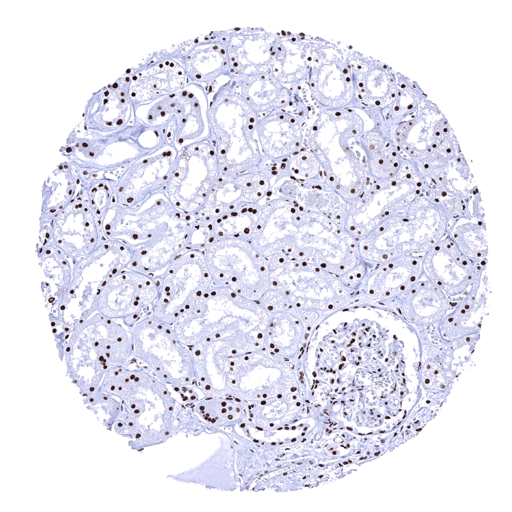
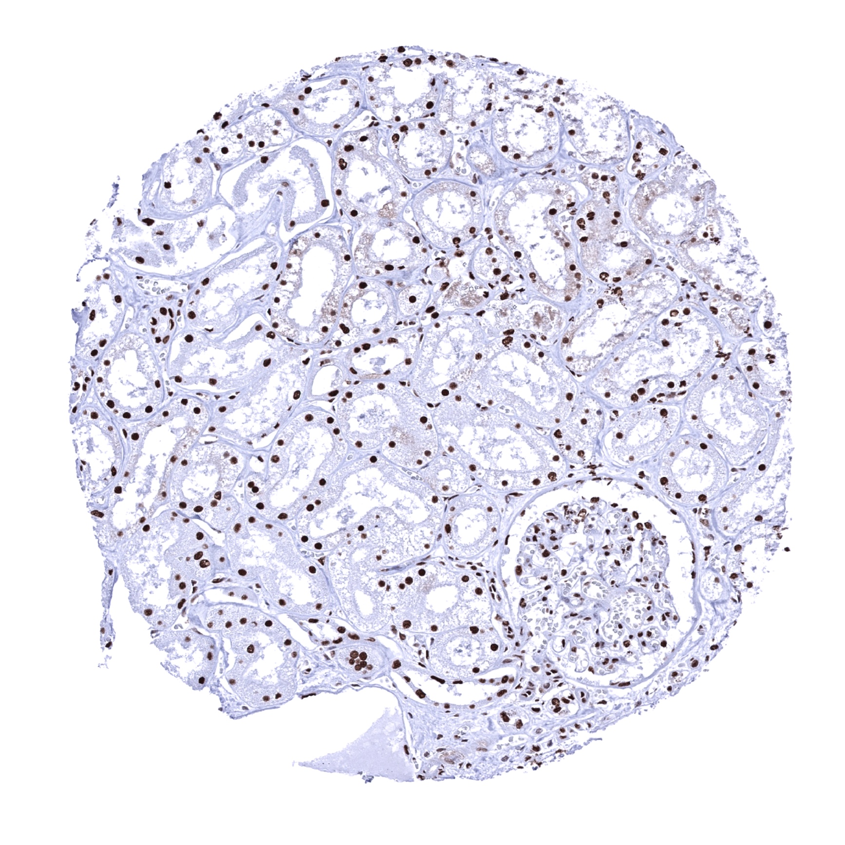
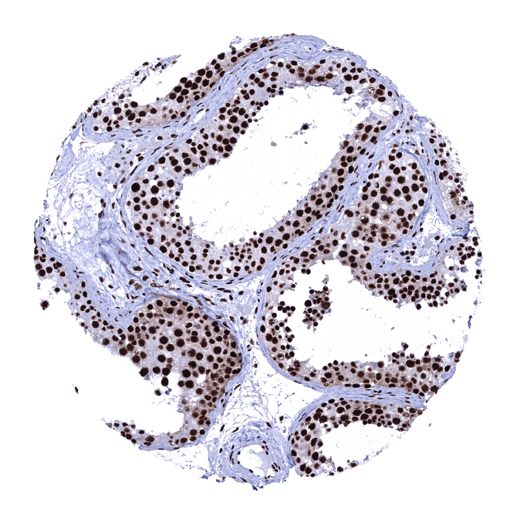
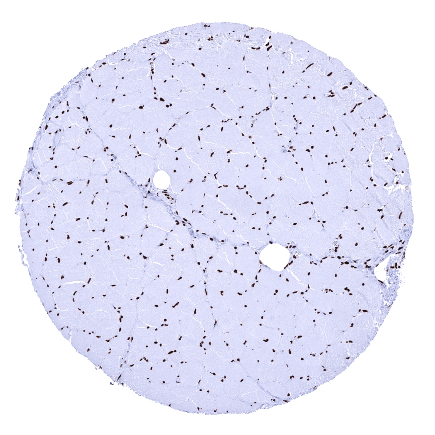
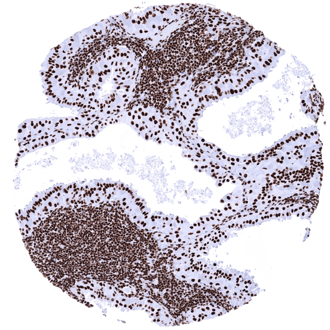
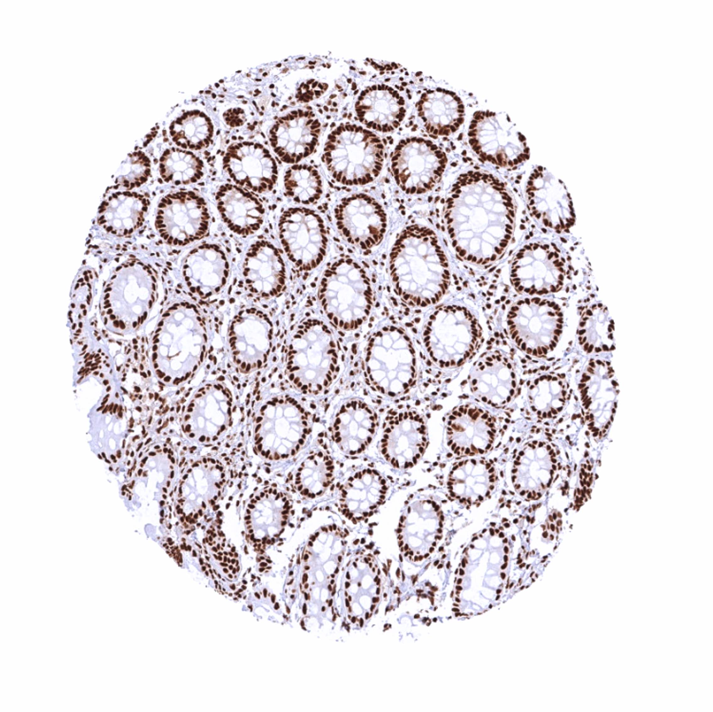
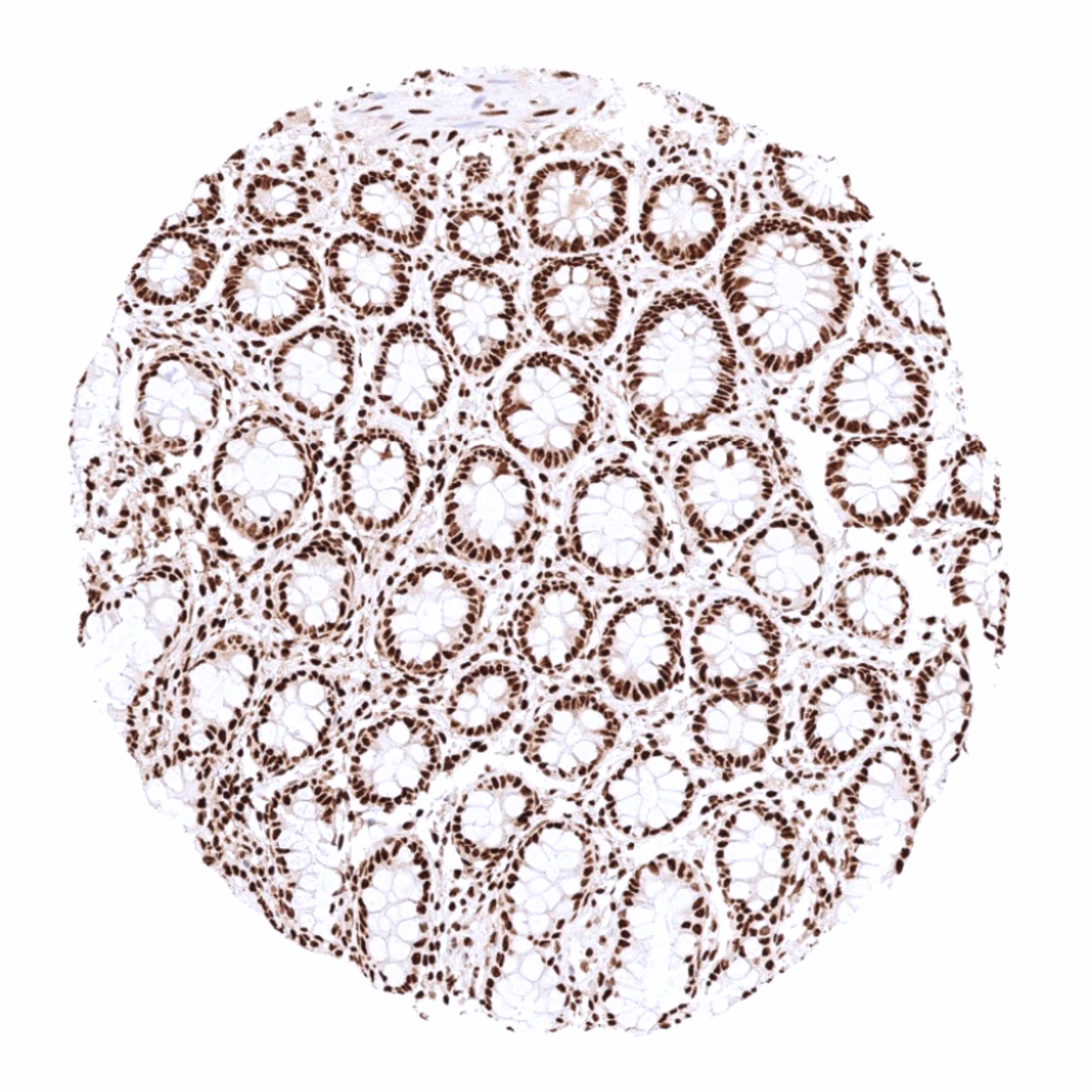
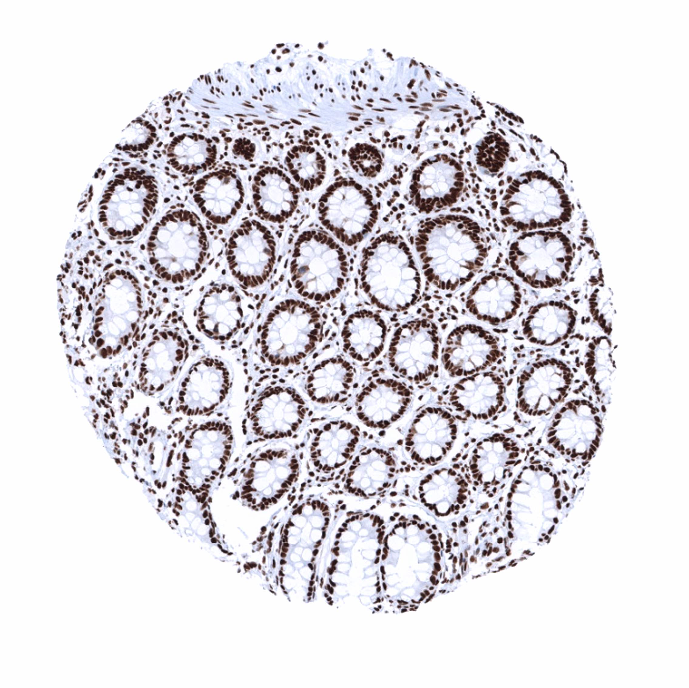
Positive control = Tonsil: A strong staining should be seen in the nuclei of all cells.
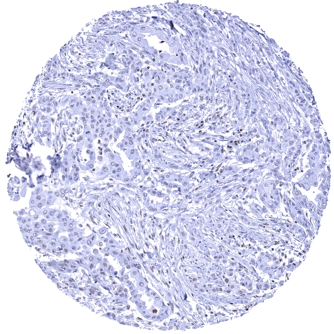
Negative control = A tumor or a cell line with documented loss of Ku80 expression.
Cellular localization = Nucleus. Nucleoplasm.
Reactivity = Human
Application = Immunohistochemistry
Dilution = 1:100 – 1:200
Intended Use = Research Use Only
Relevance of Antibody
XRCC5 is a critical gene for radiosensitivity and senescence.
Biology Behind
Ku80 is an 80 kDa protein that is coded by the XRCC5 gene on chromosome 2q35. Together with Ku70 it forms the Ku heterodimer, which binds to DNA double-strand break ends. The Ku heterodimer is essential for the non-homologous end joining (NHEJ) pathway of DNA repair and V(D)J recombination as well as for telomere length maintenance and subtelomeric gene silencing. In cancer, deletion or mutation of the Ku80 (or Ku70) genes results in a highly radiosensitive phenotype. Low function of Ku80 leads to accelerating aging. Ku80(-/-) mice exhibit early onset of senescence. The quantity of Ku80 protein varies greatly between species and is strongly linked to longevity. Ku80 acts as an autoantigen in systemic lupus erythematosus.
Staining Pattern in Normal Tissues
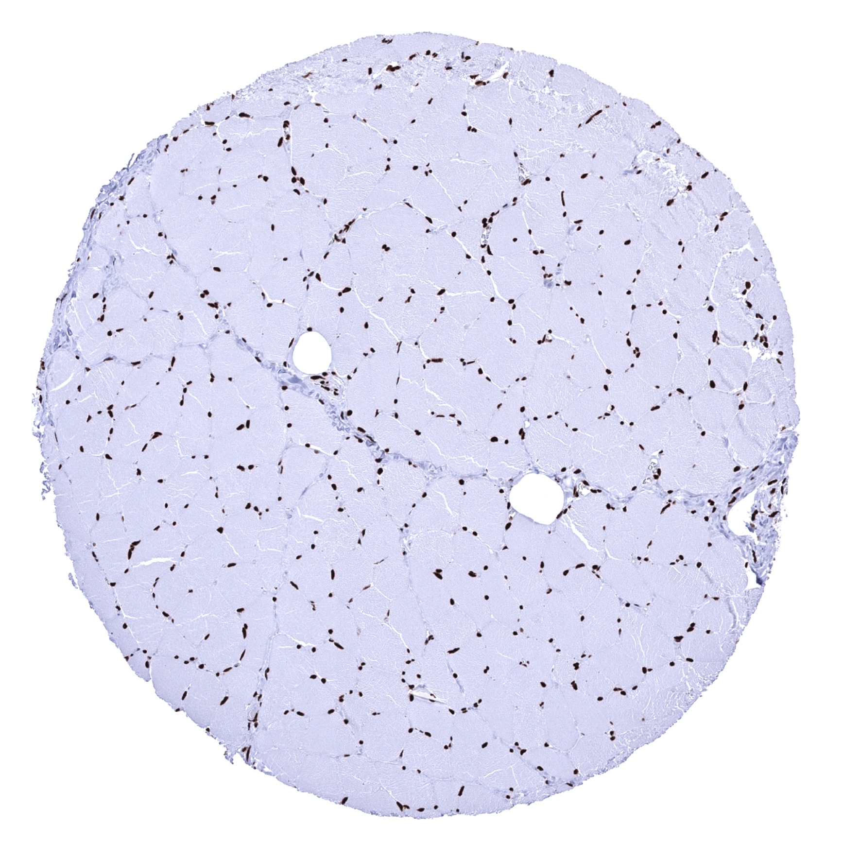
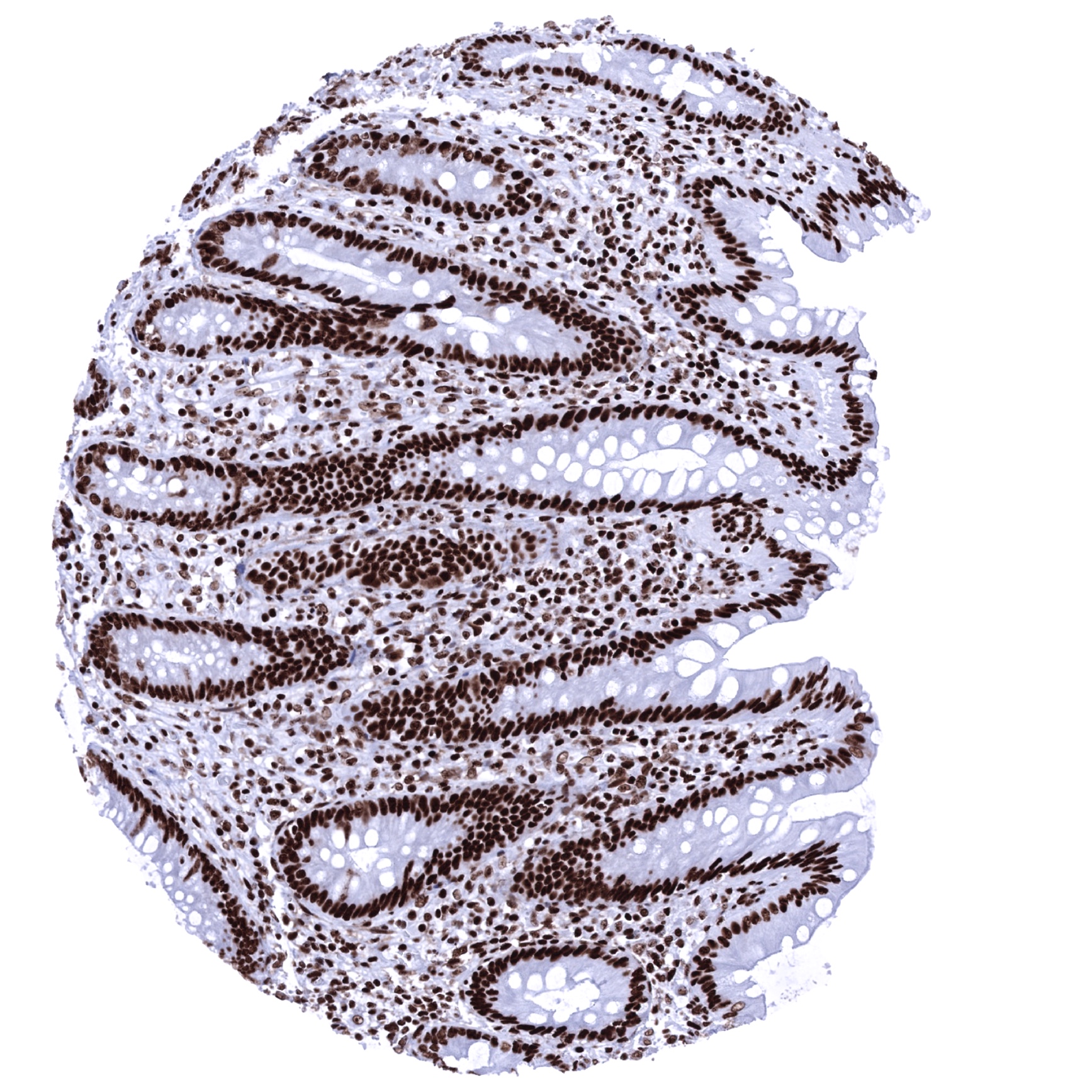
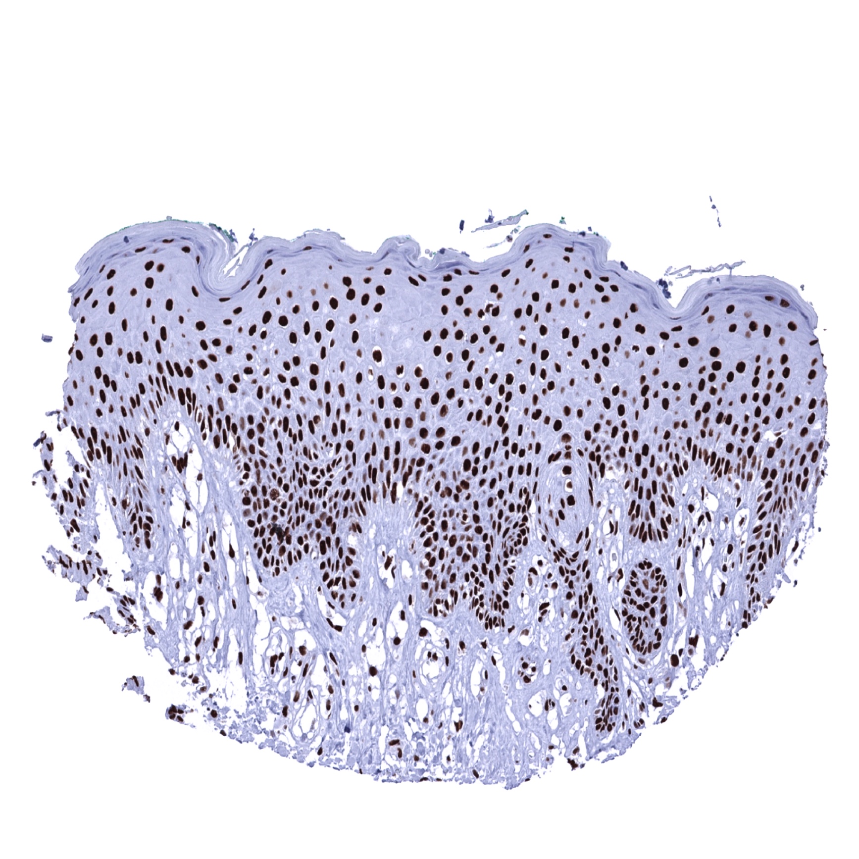
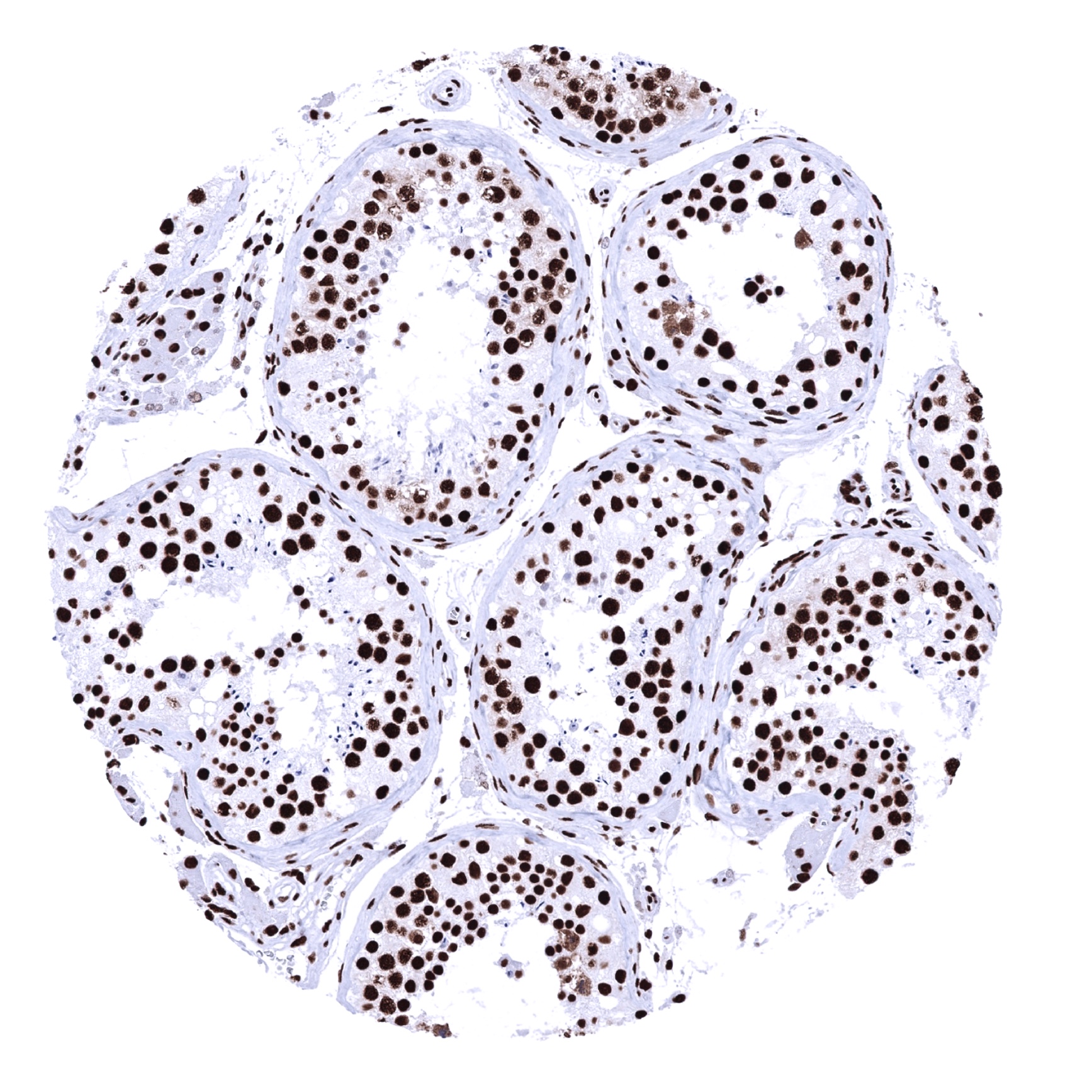
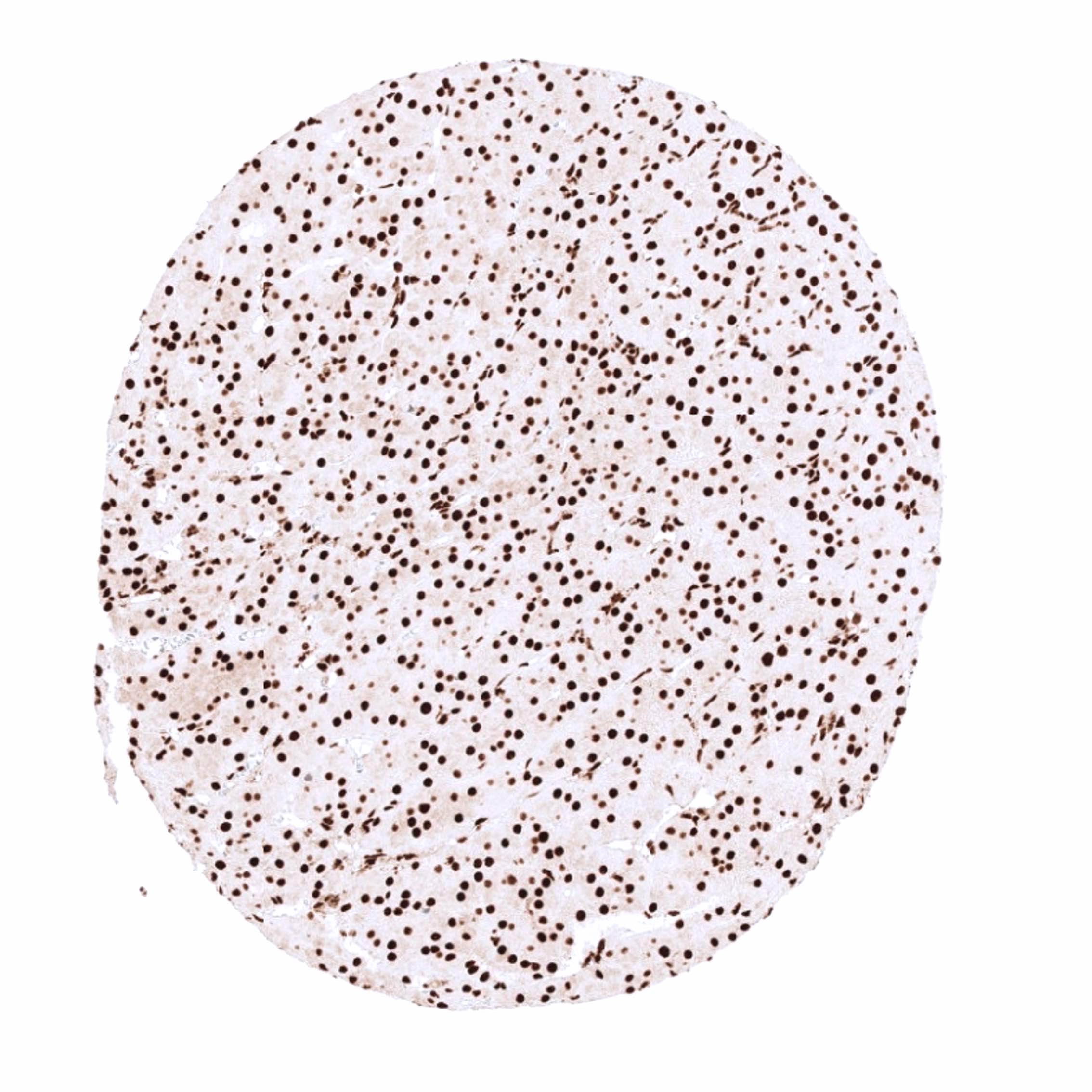
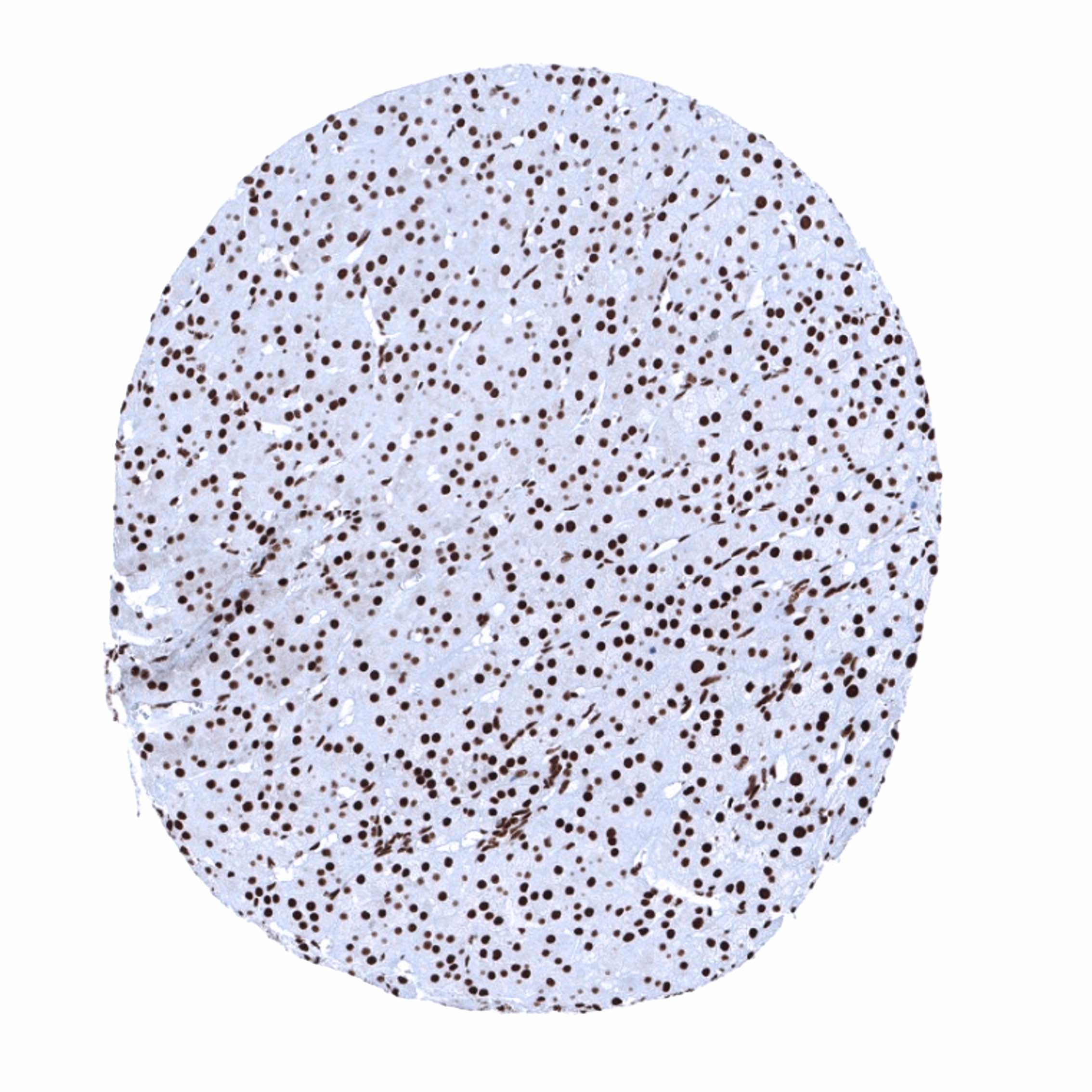
Ku80 immunostaining occurs in virtually all nuclei of all cells in all tissues. Variations of the staining intensity are hardly discernible. A reduced level of expression is, however, seen in hepatocytes and spermatids. Cytoplasmic and/or membranous Ku80 immunostaining is not seen in any normal striated muscle, heart muscle, smooth muscle, myometrium of the uterus, corpus spongiosum of the penis, ovarian stroma, fat, skin (including hair follicle and sebaceous glands), oral mucosa of the lip, oral cavity, surface epithelium of the tonsil, and transitional mucosa of the anal canal, ectocervix, squamous epithelium of the esophagus, urothelium of the renal pelvis and urinary bladder, decidua, placental trophoblastic cells, lymph node, spleen, thymus, tonsil, mucosa of the stomach, duodenum, ileum, appendix, colon, rectum and gall bladder, pancreas, liver, parotid gland, submandibular gland, sublingual gland, Brunner gland of the duodenum, cortex and medulla of the kidney, prostate, seminal vesicle, epididymis, testis, respiratory epithelium and glands of bronchi and paranasal sinus, lung, breast, endocervix, endometrium, fallopian tube, corpus luteum and follicular cyst of the ovary, adrenal gland, parathyroid gland, cerebellum, cerebrum and pituitary gland.
The findings described above are this consistent with the RNA data described in the Human Protein Atlas (Tissue expression XRCC5)
Positive control = Tonsil: A strong staining should be seen in the nuclei of all cells.
Negative control = A tumor or a cell line with documented loss of Ku80 expression.
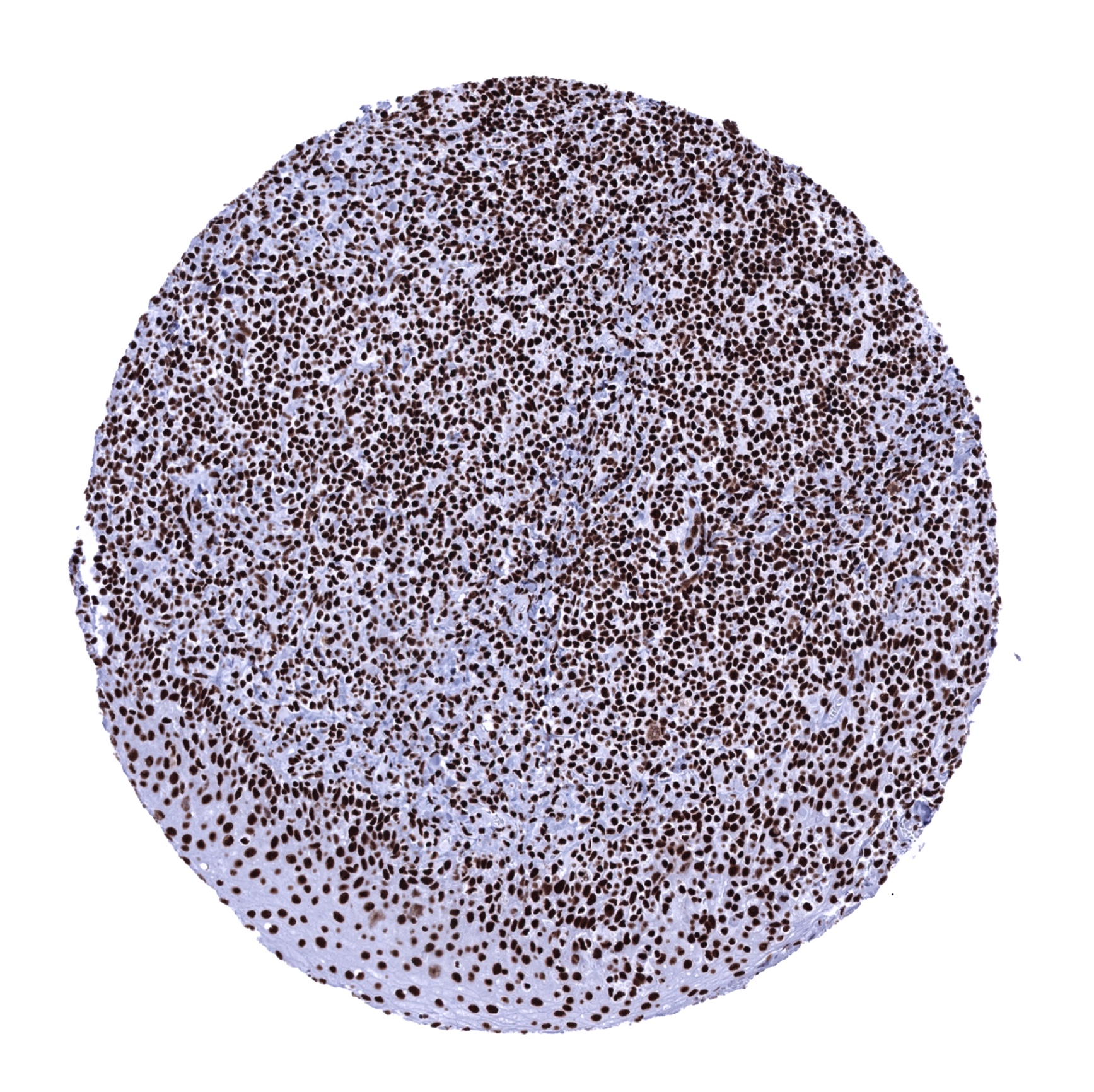
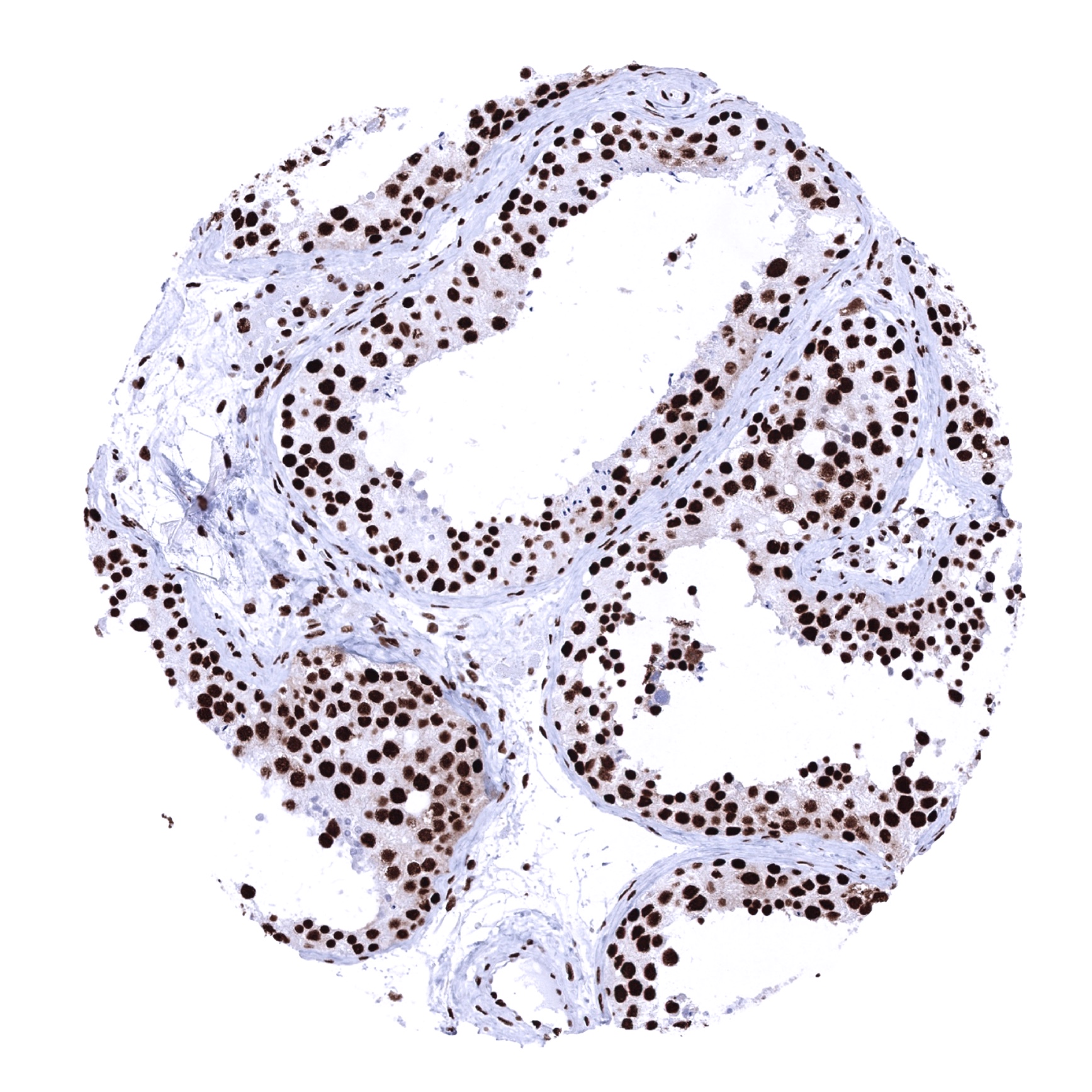
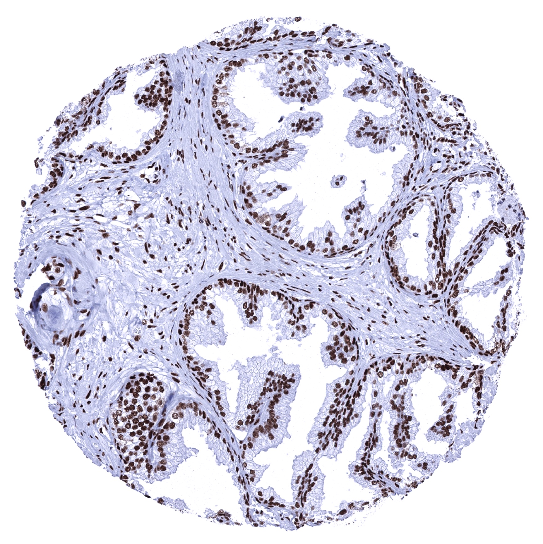
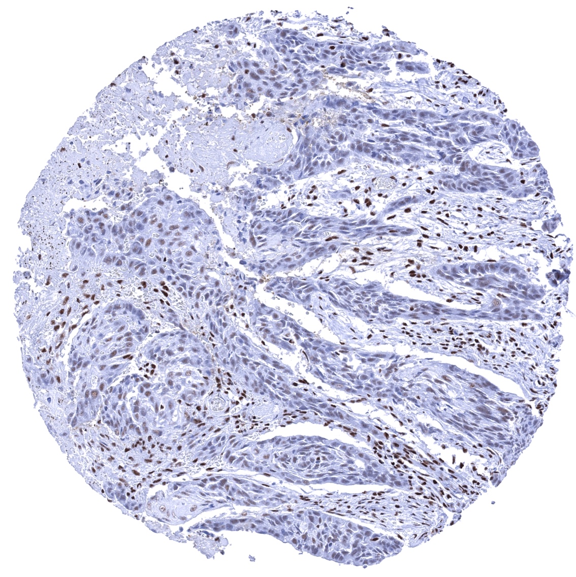
Staining Pattern in Relevant Tumor Types
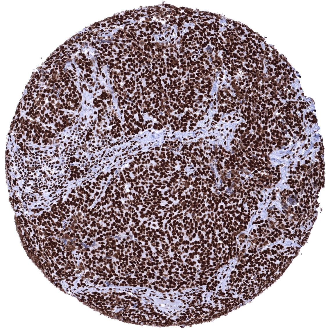
Nuclear Ku80 immunostaining is seen in the vast majority of tumors although the intensity may vary considerably between individual cases.
The TCGA findings on XRCC5 RNA expression in different tumor categories have been summarized in the Human Protein Atlas.
Compatibility of Antibodies
No data available at the moment
Protocol Recommendations
IHC users have different preferences on how the stains should look like. Some prefer high staining intensity of the target stain and even accept some background. Others favor absolute specificity and lighter target stains. Factors that invariably lead to more intense staining include higher concentration of the antibody and visualization tools, longer incubation time, higher temperature during incubation, higher temperature and longer duration of the heat induced epitope retrieval (slide pretreatment). The impact of the pH during slide pretreatment has variable effects and depends on the antibody and the target protein.
All images and data shown here and in our image galleries are obtained by the manual protocol described below. Other protocols resulting in equivalent staining are described as well.
Manual protocol
Freshly cut sections should be used (less than 10 days between cutting and staining). Heat-induced antigen retrieval for 5 minutes in an autoclave at 121°C in pH 7,8 Target Retrieval Solution buffer. Apply MSVA-880M at a dilution of 1:150 at 37°C for 60 minutes. Visualization of bound antibody by the EnVision Kit (Dako, Agilent) according to the manufacturer’s directions.

Agilent / Dako – Autostainer Link 48
Pretreatment in PT-Link for 30 minutes at 95°C (pH high); FLEX peroxidase blocking for 5 minutes (room temperature), MSVA-880M 1:150 for 20 minutes (room temperature), FLEX+ mouse/rabbit (LINKER) for 15 minutes (room temperature), horseradish peroxidase (HRP) for 20 minutes (room temperature), FLEX DAB+Sub-Chromo for 10 minutes (room temperature), FLEX hematoxylin for 5 minutes (room temperature).


These images reflect stainings by the protocol described above. It is of note that a comparable staining result can also be obtained by different protocols. In general, a longer pretreatment, a longer incubation time of the primary antibody, a higher antibody concentration, and a longer incubation time of FLEX+LINKER result in stronger staining, potentially at the cost of more background staining. Modifications of the protocol with a strengthening effect on staining intensity in combination with changes of other parameters that result in lower staining intensity can result in a comparable result as shown above.
Leica – BOND RX
Dewax at 72°C for 30 seconds; Pretreatment in Bond Epitope Retrieval Solution (ER2 – EDTA pH9) for 20 minutes at 100°C; Peroxidase blocking for 5 minutes (room temperature), MSVA-880M 1:150 for 15 minutes (room temperature), Post primary (rabbit anti mouse) for 8 minutes (room temperature), Polymer (goat anti rabbit) for 8 minutes (room temperature), mixed DAB refine for 10 minutes (room temperature), hematoxylin for 5 minutes (room temperature).

These images reflect stainings by the protocol described above. It is of note that a comparable staining result can also be obtained by different protocols. In general, a longer pretreatment, a longer incubation time of the primary antibody, a higher antibody concentration, a higher temperature during incubation, and a longer incubation time of Post primary and or the Polymer result in stronger staining, potentially at the cost of more background staining. Modifications of the protocol with a strengthening effect on staining intensity in combination with changes of other parameters that result in lower staining intensity can result in a comparable result as shown above.
Roche – Ventana Discovery ULTRA
Pretreatment for 64 minutes at 100°C (pH 8,4); CM peroxidase blocking for 12 minutes (room temperature), MSVA-880M 1:150 for 20 minutes at 36°C, secondary antibody (anti-MOUSE HQ) for 12 minutes at 36°C, anti-HQ HRP for 12 minutes at room temperature, DAB at room temperature, hematoxylin II at room temperature for 8 minutes, bluing reagent at room temperature for 4 minutes.

These images depict staining results obtained by the protocol described above. It is of note, that the Ventana machines generally require higher antibody concentrations than other commonly used autostainers because the antibodies are automatically diluted during the procedure. Various other protocols can result in an identical result as shown above. A longer pretreatment, a longer incubation time of the primary antibody, a higher antibody concentration, a higher temperature during incubation, and a longer incubation time of secondary antibody and or the anti-HQ HRP result in stronger staining, potentially at the cost of more background staining.
Potential Research Applications
- The prognostic and predictive relevance of the expression levels of Ku80 needs to be investigated. For this purpose it will be necessary to as much as possible quantitate the level of expression in individual tumors.
Evidence for Antibody Specificity in IHC
There are two ways how the specificity of antibodies can be documented for immunohistochemistry on formalin fixed tissues. These are: 1. Comparison with a second independent method for target expression measurement across a large number of different tissue types (orthogonal strategy), and 2. Comparison with one or several independent antibodies for the same target and showing that all positive staining results are also seen with other antibodies for the same target (independent antibody strategy).
Orthogonal validation is, however, not suited for validation of antibodies for proteins that are expressed in every cell of every organ.
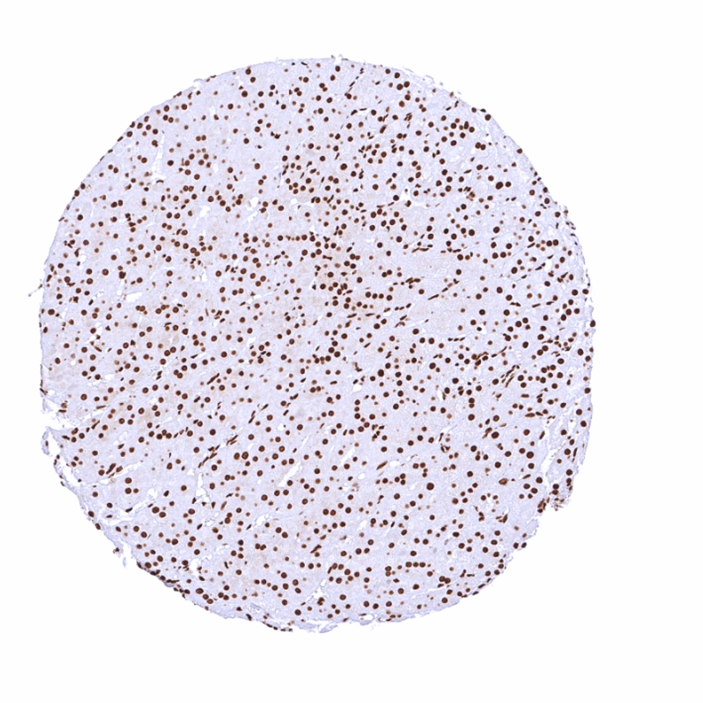
Comparison of antibodies: Specific Ku80 staining by MSVA-880M is supported by the perfect match with the expected staining pattern of a purely nuclear staining in normal tissues. This staining pattern also matches the stainings observed by the antibody HPA025813 used in the analyses described in the Human Protein Atlas (Tissue expression XRCC5). HPA025813 also results in a markedly reduced staining of hepatocytes as seen for MSVA-880M. A similar pattern of staining is also found by the independent MSVA product candidate called “Validation antibody 1f8”. Comparative images are shown below.
Antibody Comparison: MSVA-880M vs another commercially available XRCC5 antibody called “Validation Antibody 1f8”