
195,00 € – 695,00 €
Product details
Synonyms = GDB; Golli MBP; myelin basic protein; Hemopoietic MBP; HMBPR; HUGO; MLD; Myelin A1 Protein, basic; Myelin Deficient; Myelin membrane encephalitogenic protein; SHI; Shiverer; SP
Antibody type = Recombinant Rabbit monoclonal / IgG
Clone = MSVA-390R
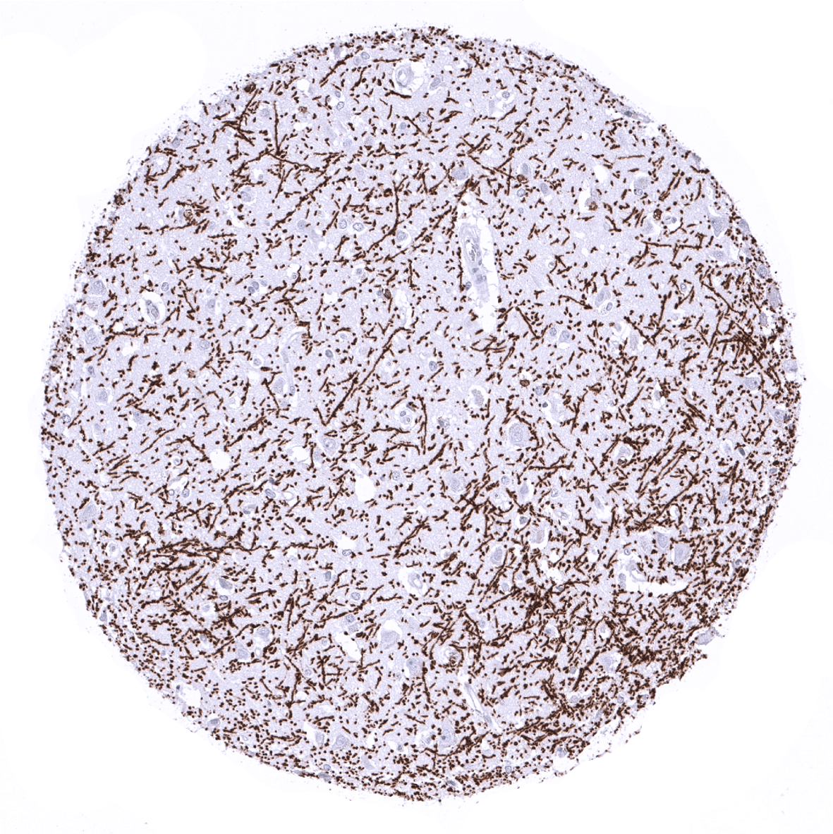
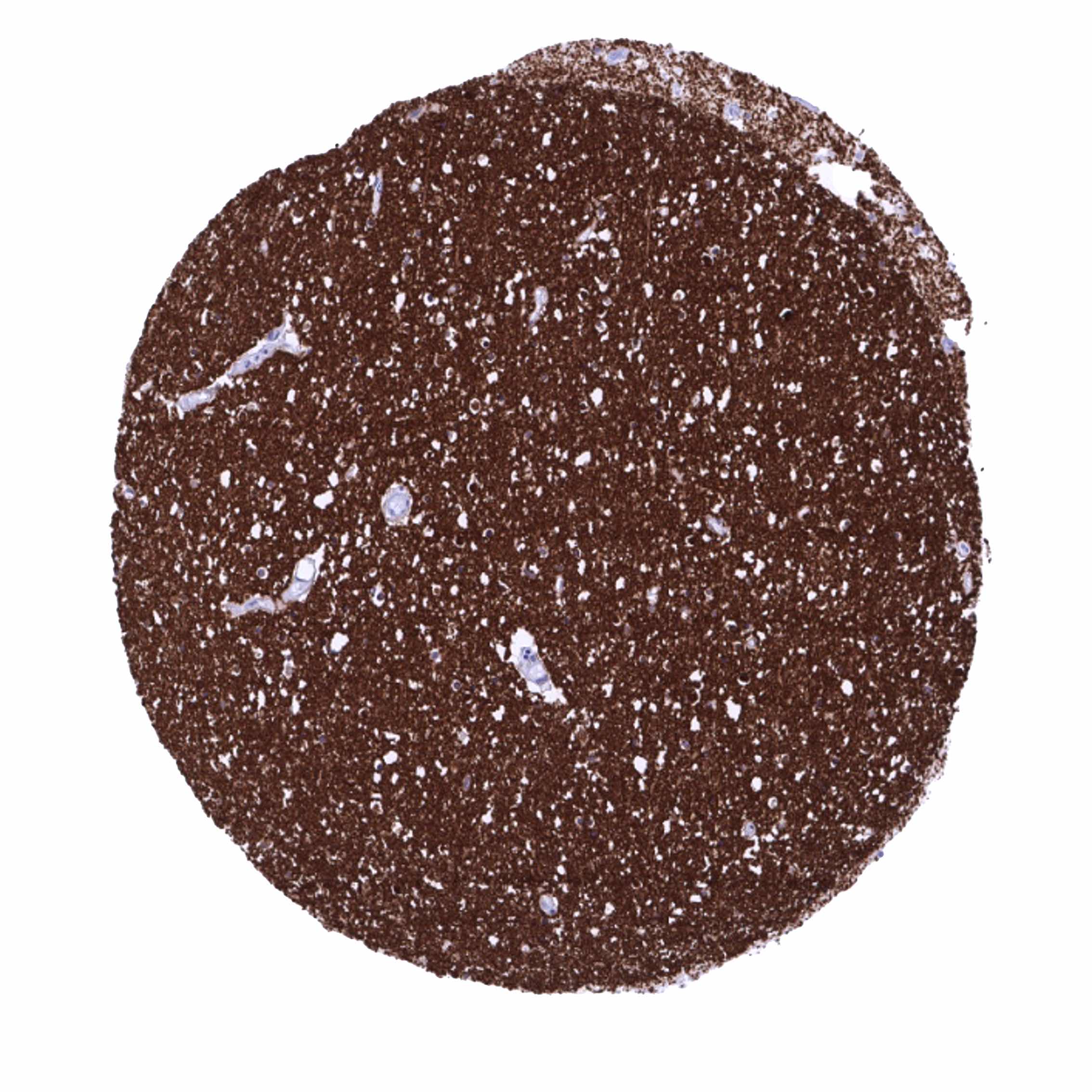
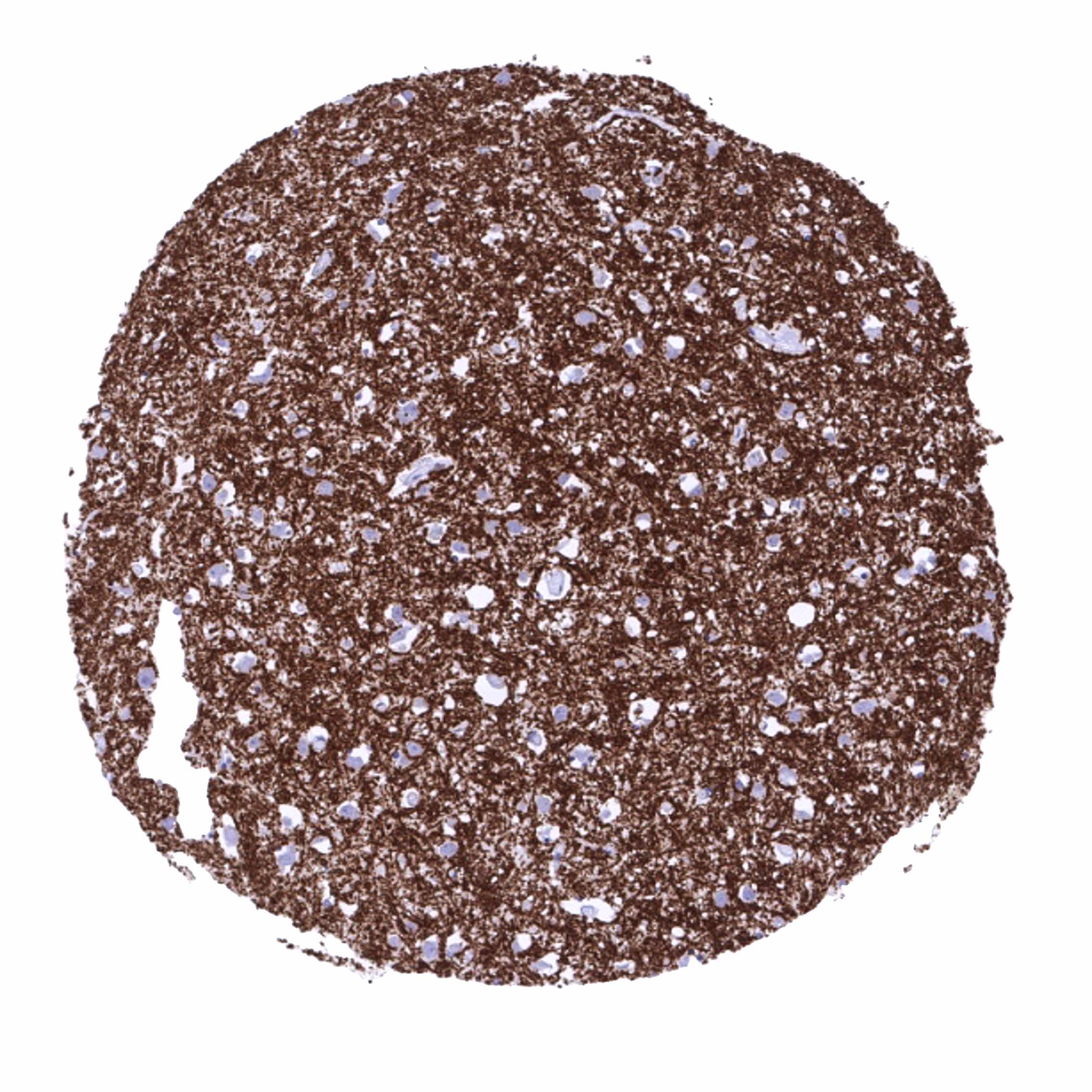
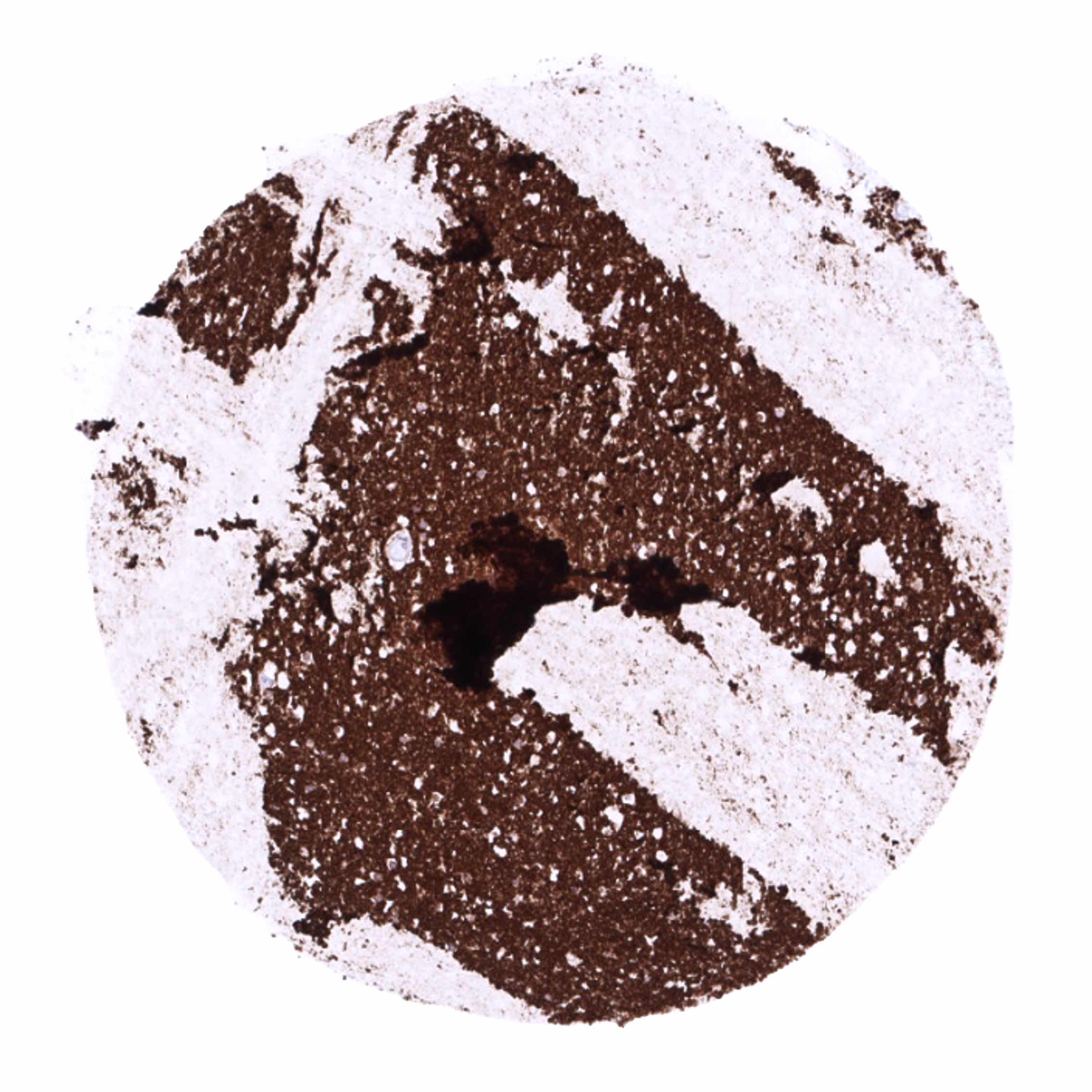
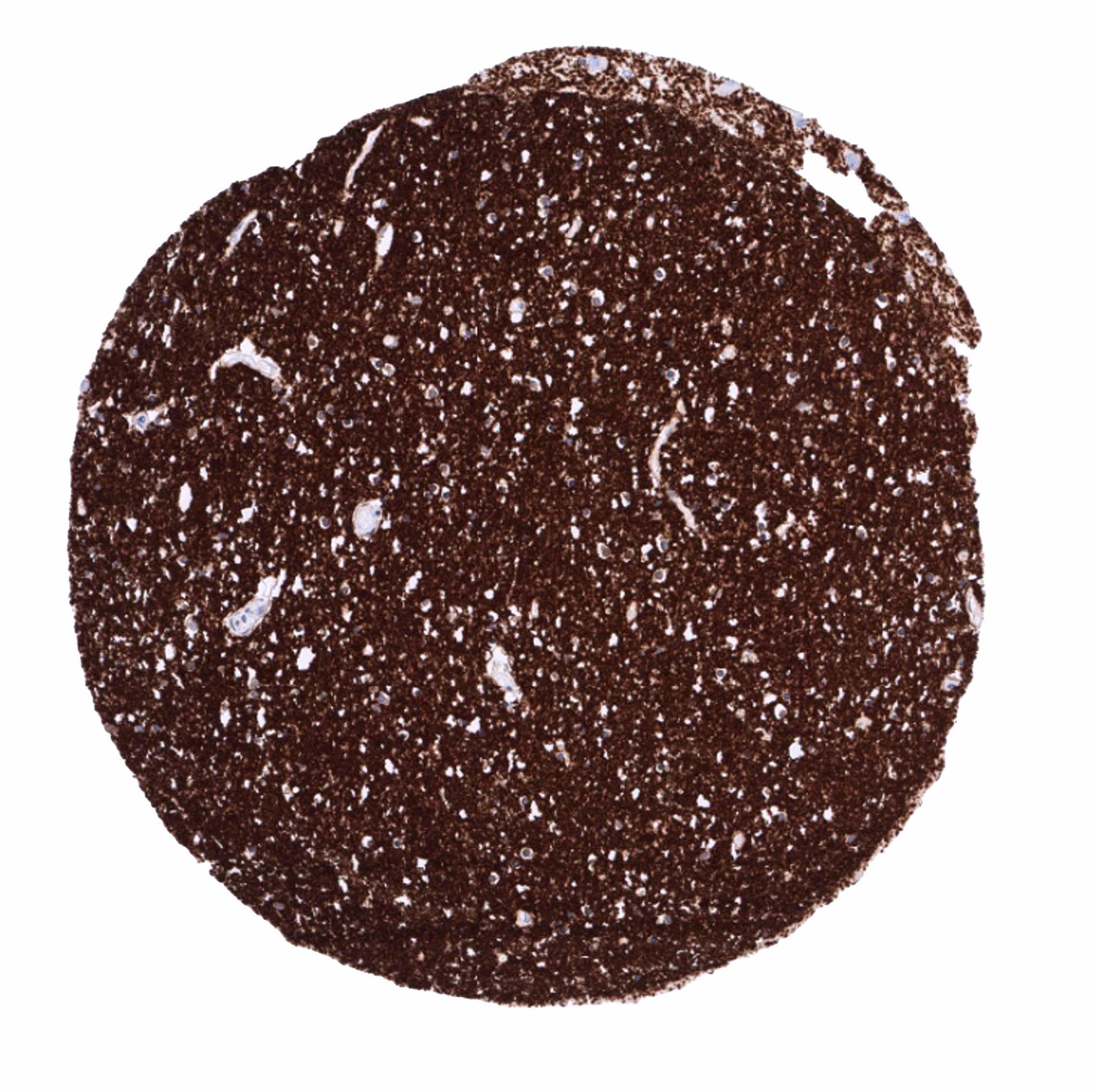
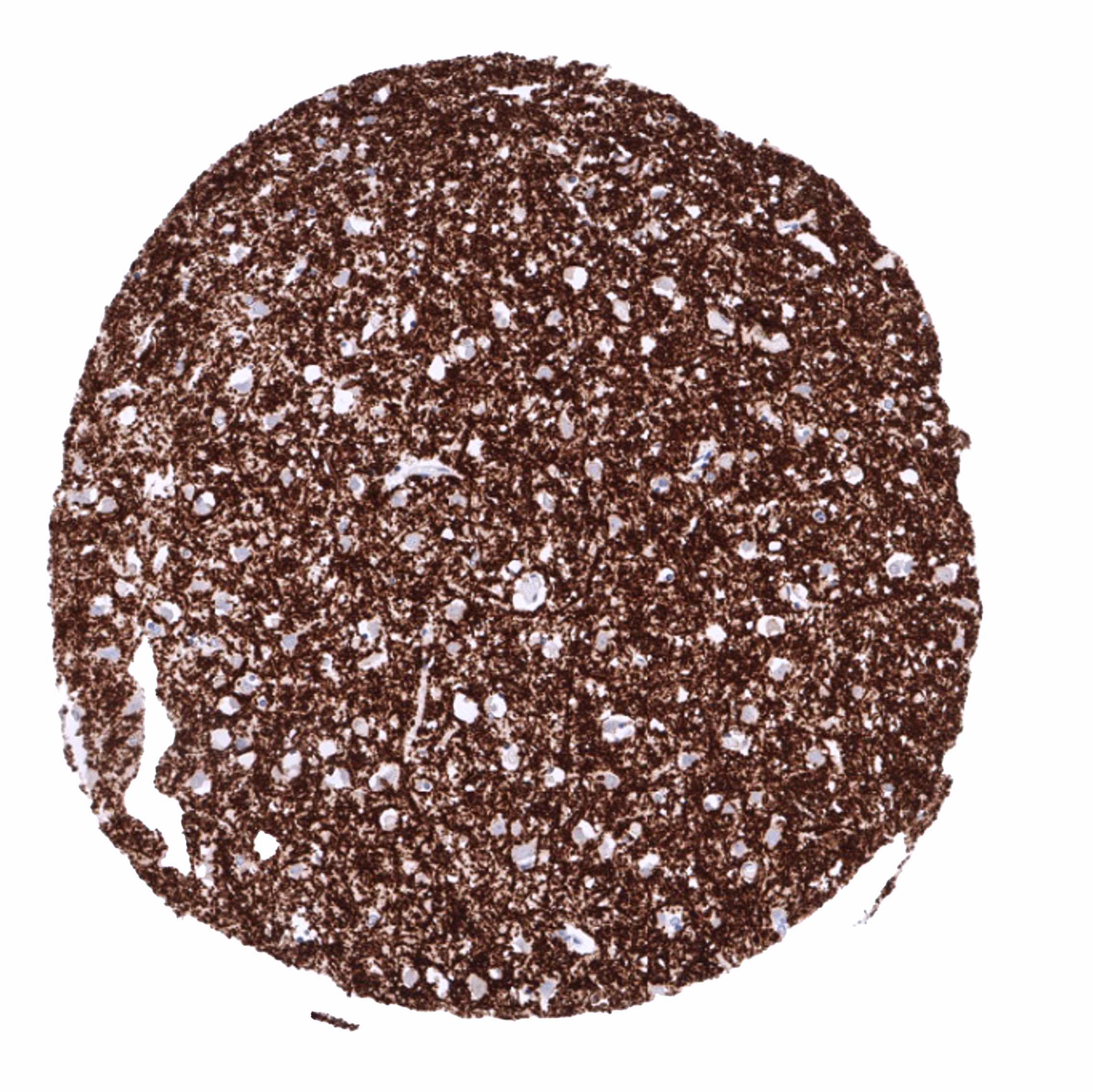
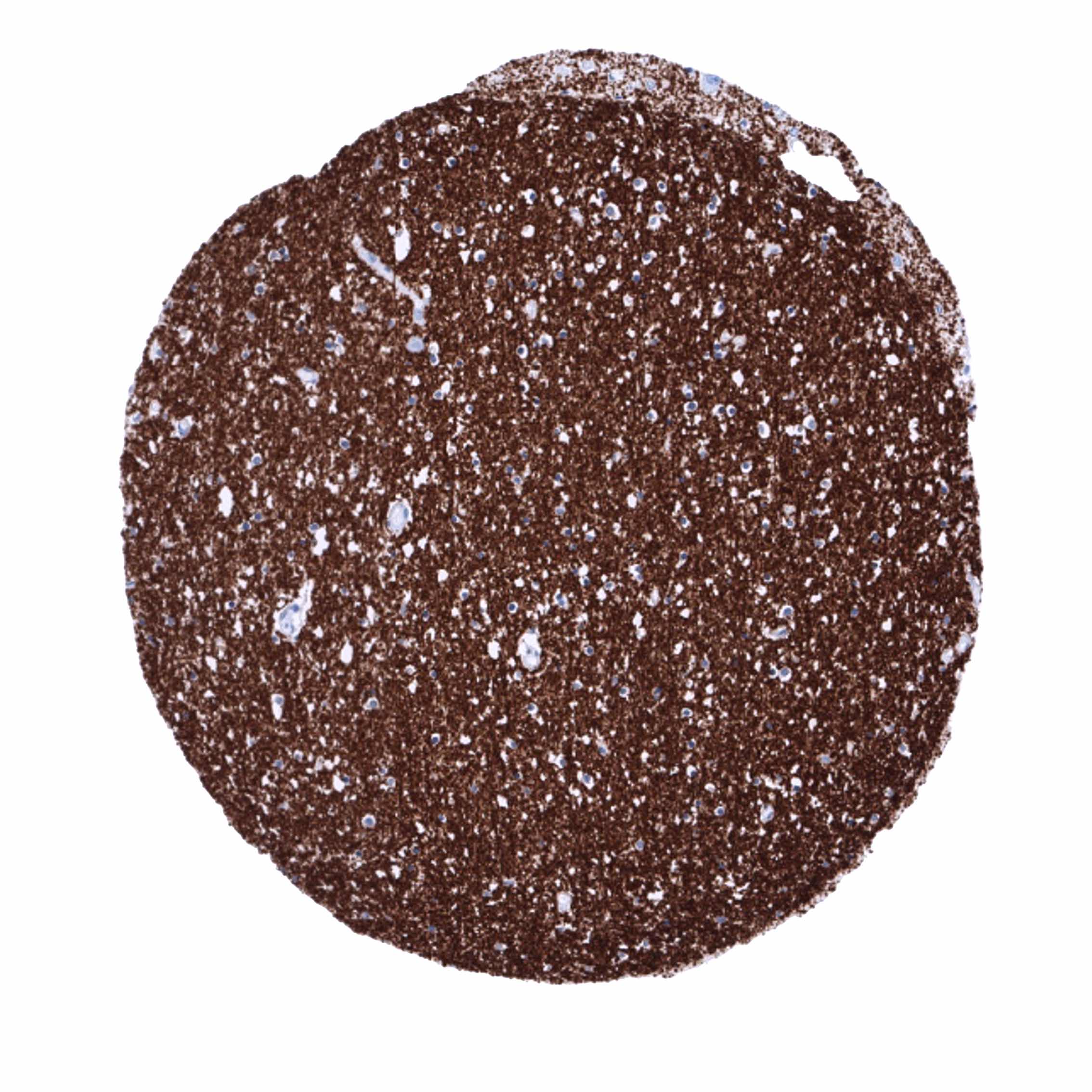
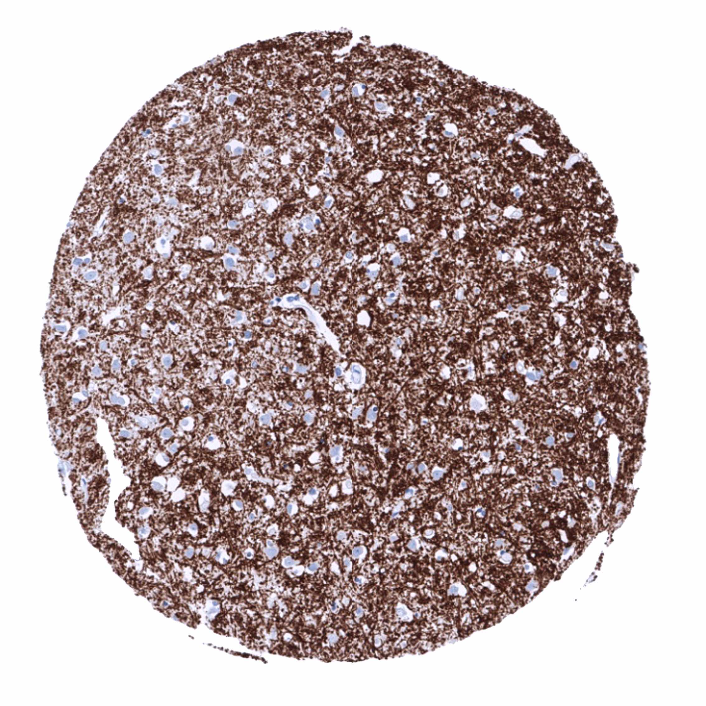
Positive control = Brain: A fibrillar staining pattern should be seen.
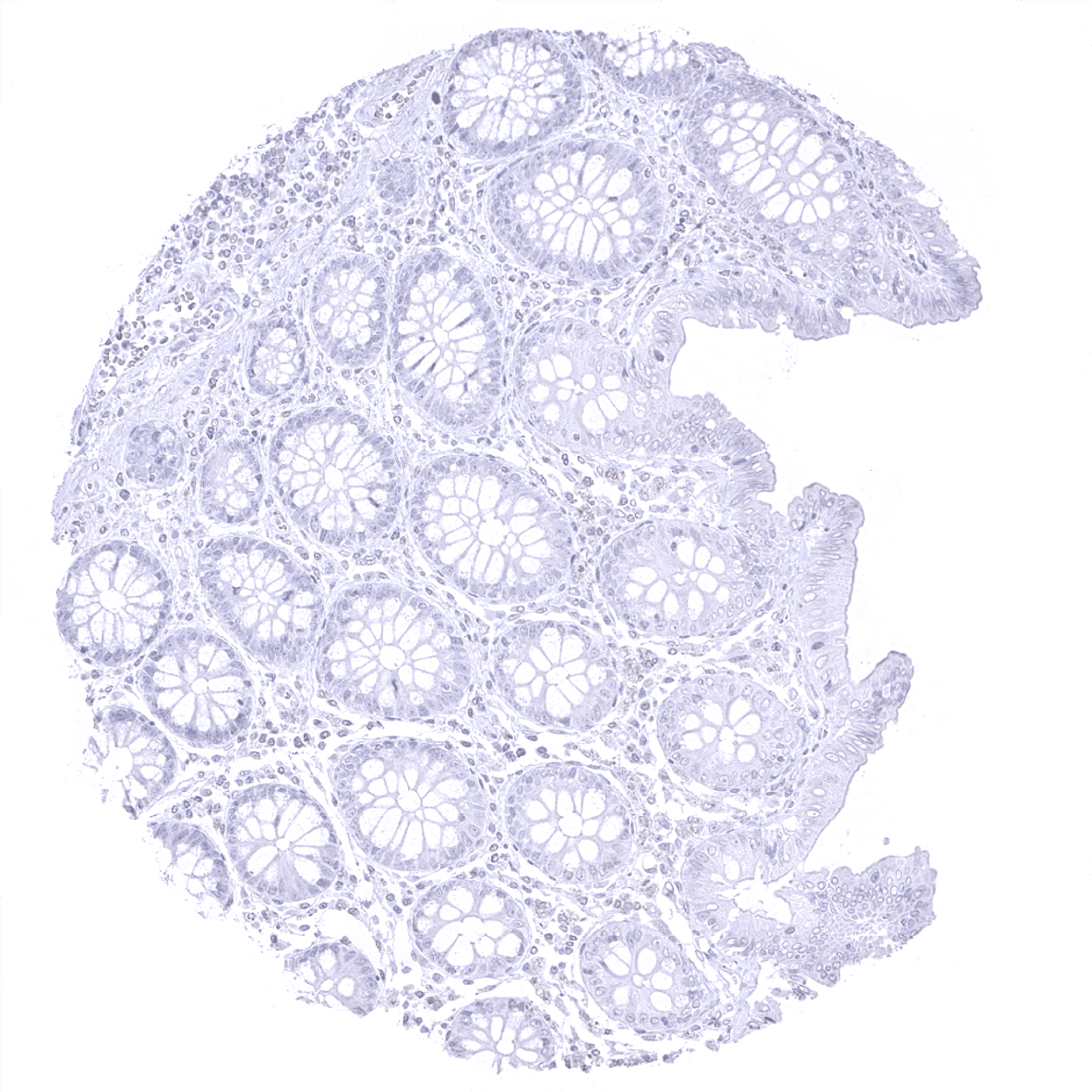
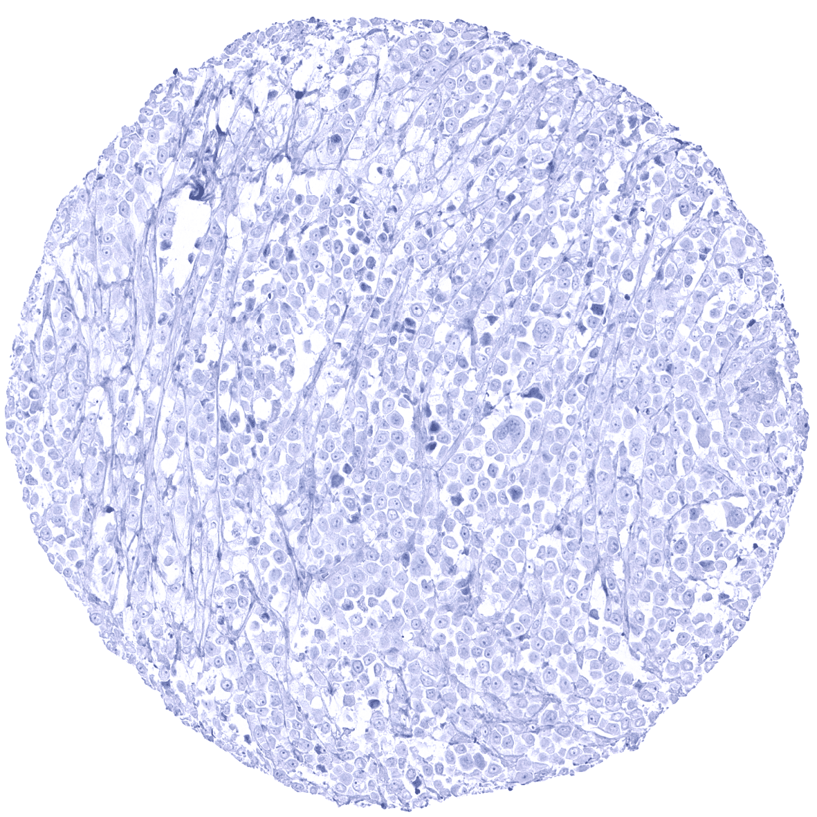
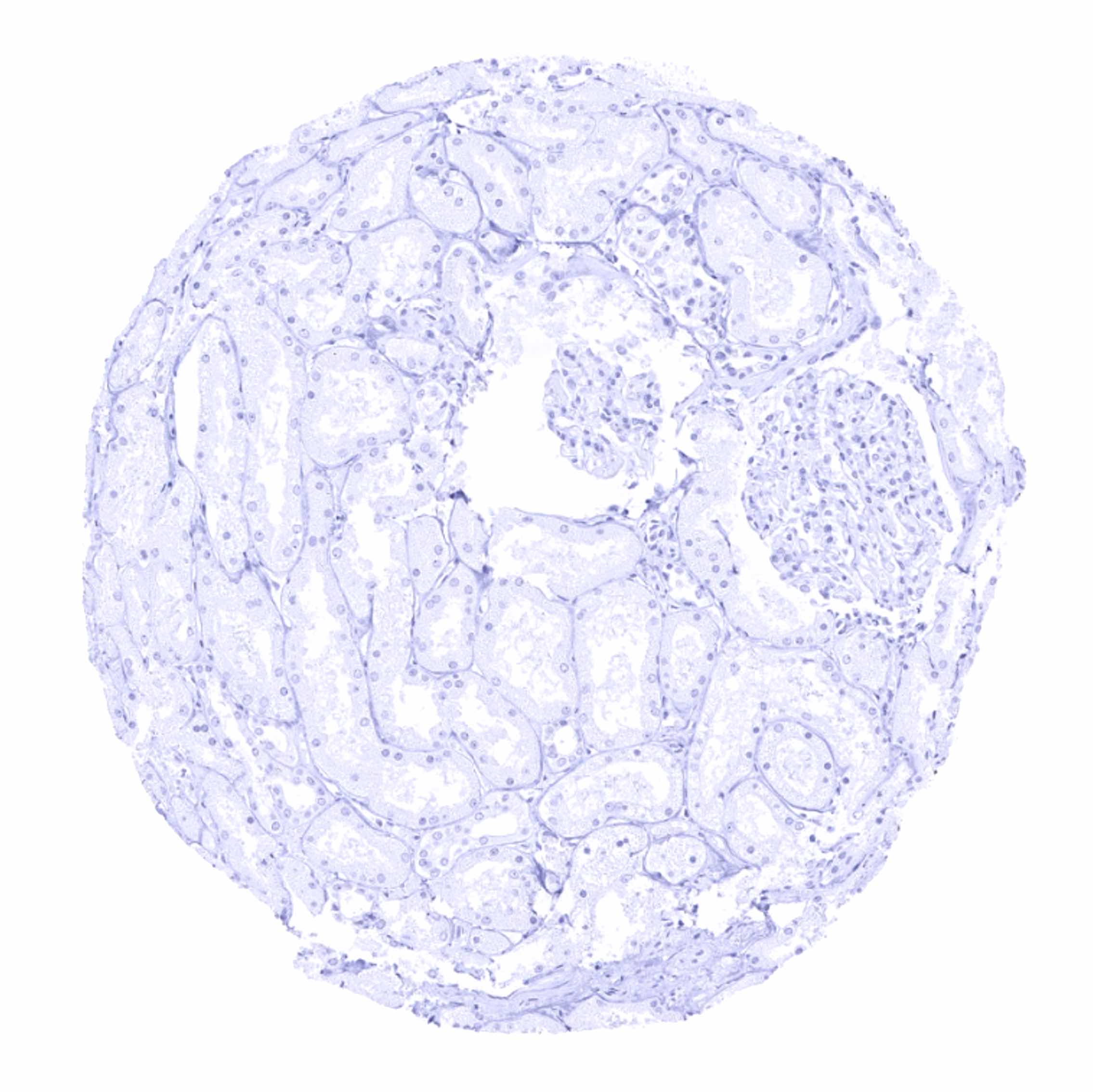
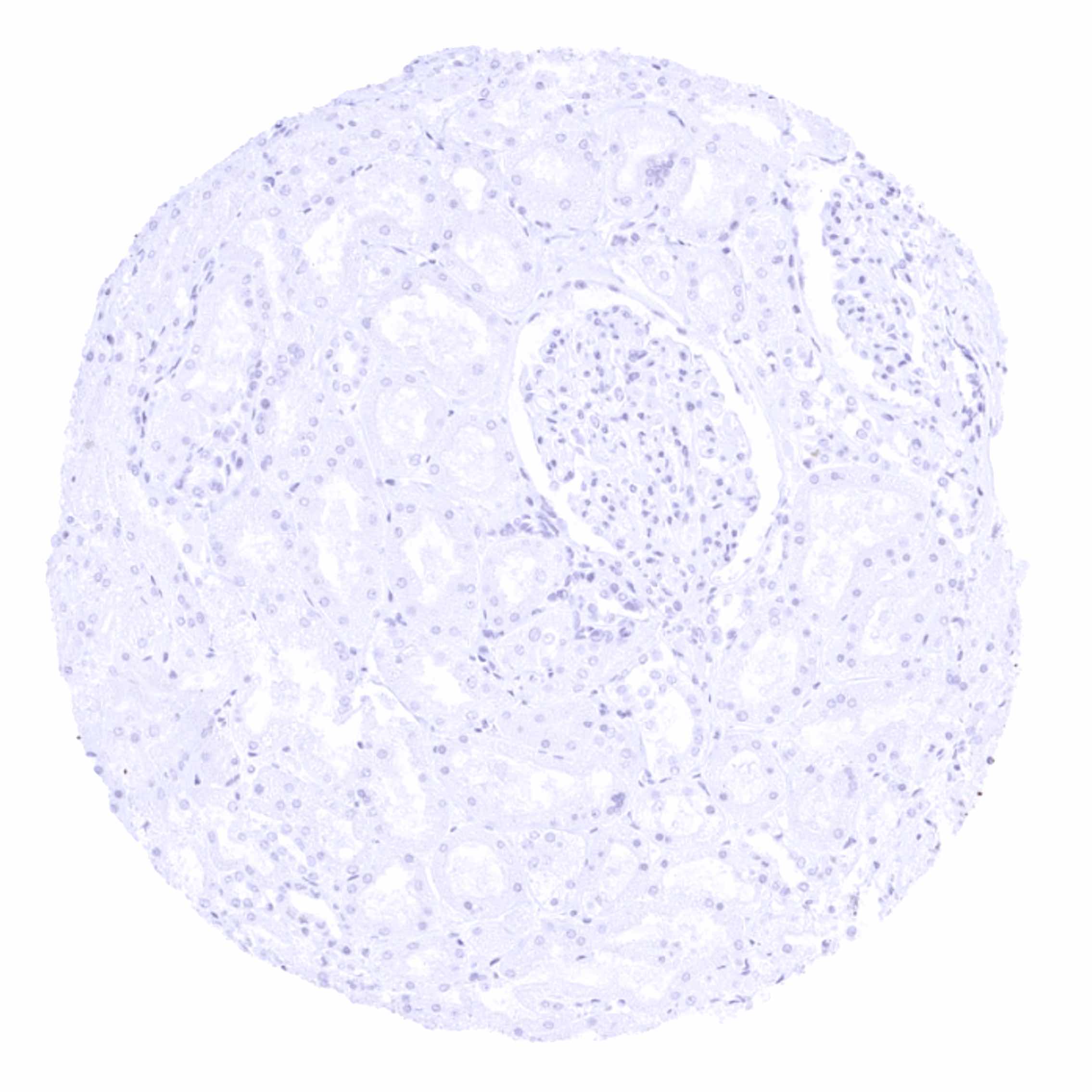
Negative control = Colon: MBP staining should be completely absent.
Cellular localization = Cell surface and cytoplasm.
Reactivity = Human
Application = Immunohistochemistry
Dilution = 1:100 – 1:200
Intended Use = Research Use Only
Relevance of Antibody
MBP is a pivotal protein of myelinated nerves.
Biology Behind
Myelin basic protein (MBP) is coded by the MBP gene on 18q22. It is produced by oligodendrocytes and plays a relevant role in the myelination of nerves in the nervous system by adhering the cytoplasmic leaflets of the oligodendrocyte (OLG) membrane to each other. MBP maintains the correct structure of myelin, interacts with the lipids in the myelin membrane and forms a ‘molecular sieve’ that restricts diffusion of some membrane proteins from paranodal loops into compact myelin. The gene is characterized by multiple splice variants and a large number of post-translational modifications of the protein, including phosphorylation, methylation, deamidation, citrullination, ADP-ribosylation, and N-terminal acylation. The most common form of MBP is about 18.5 kDa. MBP interacts with various small ligands and proteins. Alterations in these post-translational modifications have been associated with demyelinating diseases. MBP isolated from individuals with multiple sclerosis have had a higher degree of citrullination and a smaller positive charge.
Staining Pattern in Normal Tissues
MBP is only seen in the brain where MBP immunostaining results in a fibrillar pattern along intracerebral axons. MBP is expressed in oligodendrocytes at a late stage of cell differentiation.
These findings are largely consistent with the RNA and protein data described in the Human Protein Atlas (Tissue expression MBP)
Positive control: Brain: A fibrillar staining pattern should be seen.
Negative control: Colon: MBP staining should be completely absent.

Staining Pattern in Relevant Tumor Types
A positive MBP immunostaining is particularly seen in oligodendroglioma and oligoastrocytomas. MBP has been described to be occasionally expressed in teratomas but is otherwise not seen in extracranial tumors.
The TCGA findings on MBP RNA expression in different tumor categories have been summarized in the Human Protein Atlas.
Compatibility of Antibodies
No data available at the moment
Protocol Recommendations
IHC users have different preferences on how the stains should look like. Some prefer high staining intensity of the target stain and even accept some background. Others favor absolute specificity and lighter target stains. Factors that invariably lead to more intense staining include higher concentration of the antibody and visualization tools, longer incubation time, higher temperature during incubation, higher temperature and longer duration of the heat induced epitope retrieval (slide pretreatment). The impact of the pH during slide pretreatment has variable effects and depends on the antibody and the target protein.
All images and data shown here are obtained by the manual protocol described below. Other protocols resulting in equivalent staining are described as well.
Manual protocol
Freshly cut sections should be used (less than 10 days between cutting and staining). Heat-induced antigen retrieval for 5 minutes in an autoclave at 121°C in pH 7,8 Target Retrieval Solution buffer. Apply MSVA-390R at a dilution of 1:150 at 37°C for 60 minutes. Visualization of bound antibody by the EnVision Kit (Dako, Agilent) according to the manufacturer’s directions.
Agilent / Dako – Autostainer Link 48
Pretreatment in PT-Link for 30 minutes at 95°C (pH high); FLEX peroxidase blocking for 5 minutes (room temperature), MSVA-390R 1:150 for 20 minutes (room temperature), FLEX+ mouse/rabbit (LINKER) for 15 minutes (room temperature), horseradish peroxidase (HRP) for 20 minutes (room temperature), FLEX DAB+Sub-Chromo for 10 minutes (room temperature), FLEX hematoxylin for 5 minutes (room temperature).

These images reflect stainings by the protocol described above. It is of note that a comparable staining result can also be obtained by different protocols. In general, a longer pretreatment, a longer incubation time of the primary antibody, a higher antibody concentration, and a longer incubation time of FLEX+LINKER result in stronger staining, potentially at the cost of more background staining. Modifications of the protocol with a strengthening effect on staining intensity in combination with changes of other parameters that result in lower staining intensity can result in a comparable result as shown above.
Leica – BOND RX
Dewax at 72°C for 30 seconds; Pretreatment in Bond Epitope Retrieval Solution (ER2 – EDTA pH9) for 20 minutes at 100°C; Peroxidase blocking for 5 minutes (room temperature), MSVA-390R 1:150 for 15 minutes (room temperature), Post primary (rabbit anti mouse) for 8 minutes (room temperature), Polymer (goat anti rabbit) for 8 minutes (room temperature), mixed DAB refine for 10 minutes (room temperature), hematoxylin for 5 minutes (room temperature).

These images reflect stainings by the protocol described above. It is of note that a comparable staining result can also be obtained by different protocols. In general, a longer pretreatment, a longer incubation time of the primary antibody, a higher antibody concentration, a higher temperature during incubation, and a longer incubation time of Post primary and or the Polymer result in stronger staining, potentially at the cost of more background staining. Modifications of the protocol with a strengthening effect on staining intensity in combination with changes of other parameters that result in lower staining intensity can result in a comparable result as shown above.
Roche – Ventana Discovery ULTRA
Pretreatment for 64 minutes at 100°C (pH 8,4); CM peroxidase blocking for 12 minutes (room temperature), MSVA-390R 1:150 for 20 minutes at 36°C, secondary antibody (anti-rabbit HQ) for 12 minutes at 36°C, anti-HQ HRP for 12 minutes at room temperature, DAB at room temperature, hematoxylin II at room temperature for 8 minutes, bluing reagent at room temperature for 4 minutes.

These images depict staining results obtained by the protocol described above. It is of note, that the Ventana machines generally require higher antibody concentrations than other commonly used autostainers because the antibodies are automatically diluted during the procedure. Various other protocols can result in an identical result as shown above. A longer pretreatment, a longer incubation time of the primary antibody, a higher antibody concentration, a higher temperature during incubation, and a longer incubation time of secondary antibody and or the anti-HQ HRP result in stronger staining, potentially at the cost of more background staining.
Potential Research Applications
- MBP is pivotal in research projects that involve an assessment of the status of myelinization of intracranial axons.
Evidence for Antibody Specificity in IHC
There are two ways how the specificity of antibodies can be documented for immunohistochemistry on formalin fixed tissues. These are: 1. comparison with a second independent method for target expression measurement across a large number of different tissue types (orthogonal strategy), and 2. Comparison with one or several independent antibodies for the same target and showing that all positive staining results are also seen with other antibodies for the same target (independent antibody strategy).
Orthogonal validation: For the antibody MSVA-390R specificity is suggested by the strong concordance of the immunostaining with RNA expression data derived fromthe Human Protein Atlas (HPA) RNA-seq tissue dataset, the FANTOM5 project, and the Genotype-Tissue Expression (GTEx) project which are all summarized in Human Protein Atlas (Tissue expression MBP). Immunostaining by using MSVA-390R was almost exclusively detected in the brain, which is the only organ with documented RNA expression.
Comparison of antibodies: The validity of immunostainings produced by MSVA-390R is also supported by identical (fibrillar) staining patterns in the brain as obtained by the use of the antibody CAB002300.
The respective images are depicted in the protein atlas for:
