295,00 € – 995,00 €
Product details
Synonyms = solute carrier family 6 (neurotransmitter transporter, GABA), member 1 , A730043E01 , GABATHG , GABATR , GAT-1 , Gabt , Gabt1 , Gat1 , XT-1 , Xtrp1
Antibody type = Recombinant Rabbit monoclonal / IgG
Clone = HMV4721
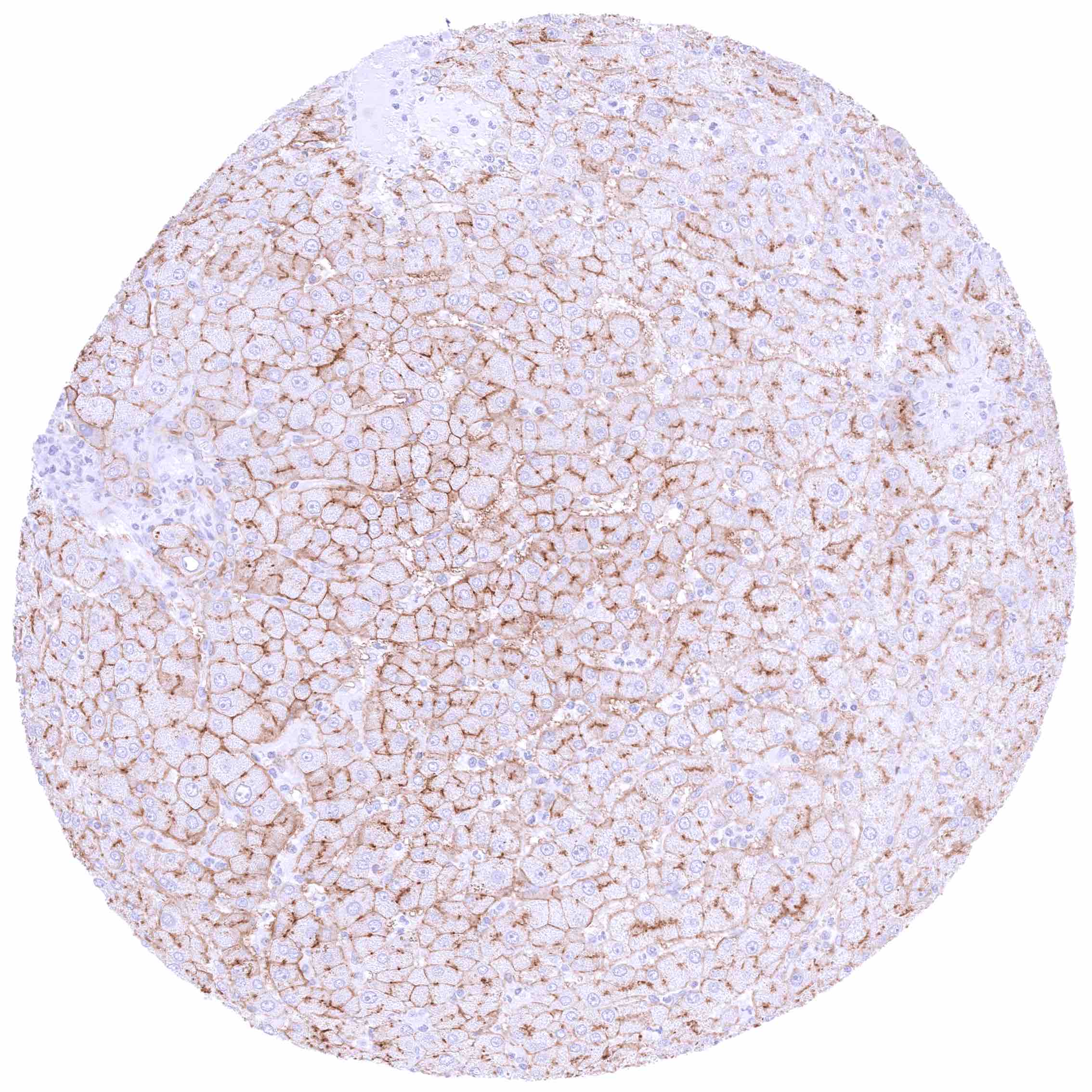
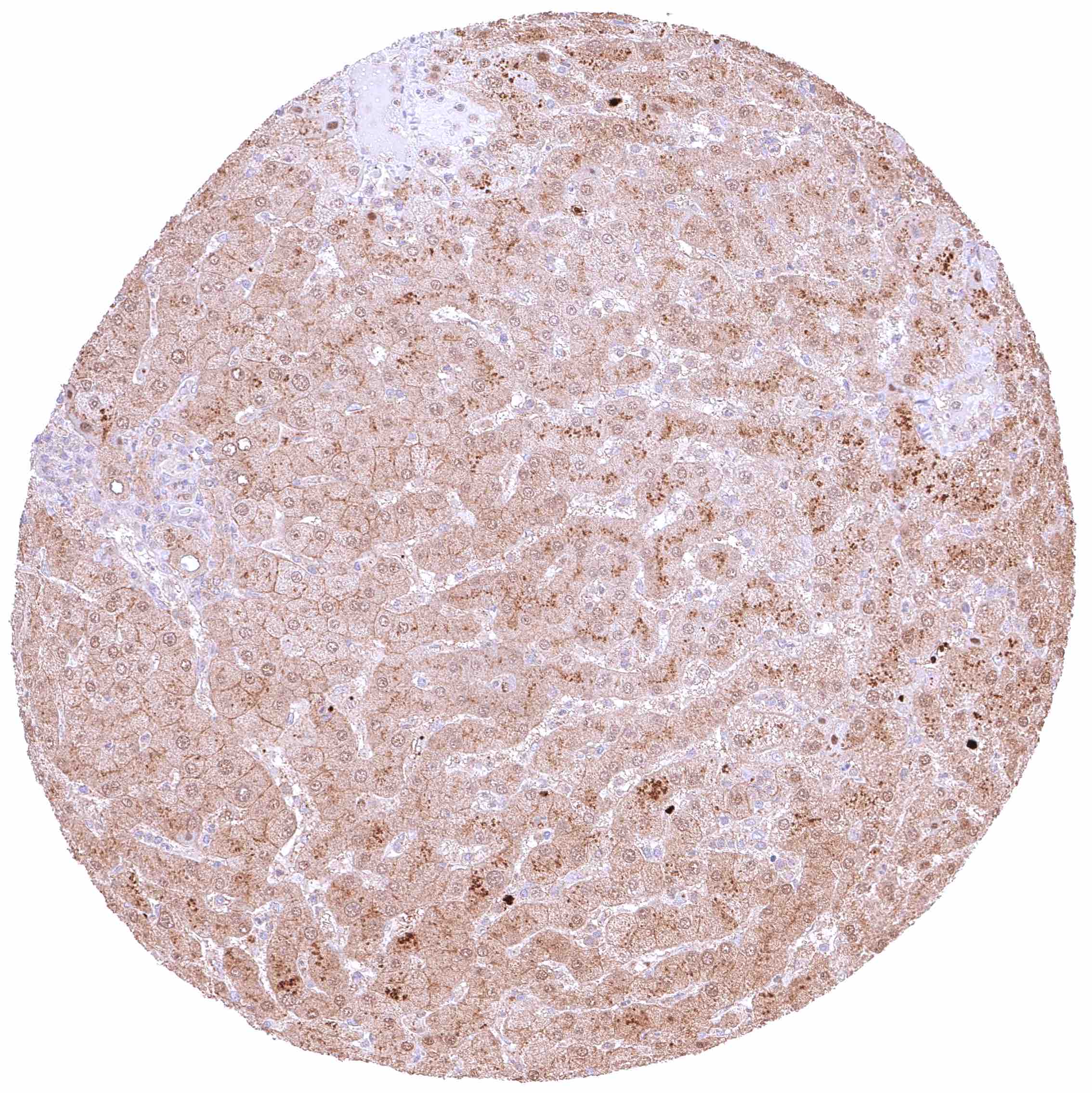
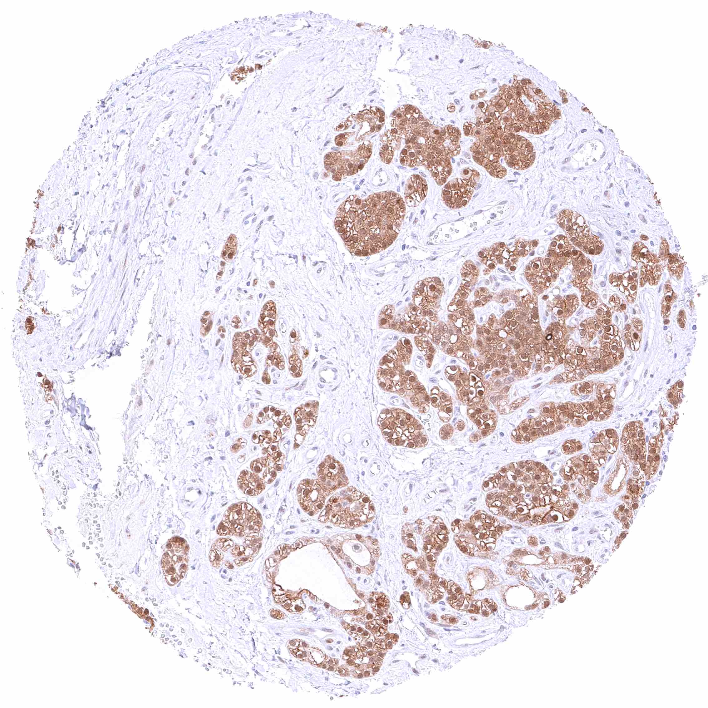
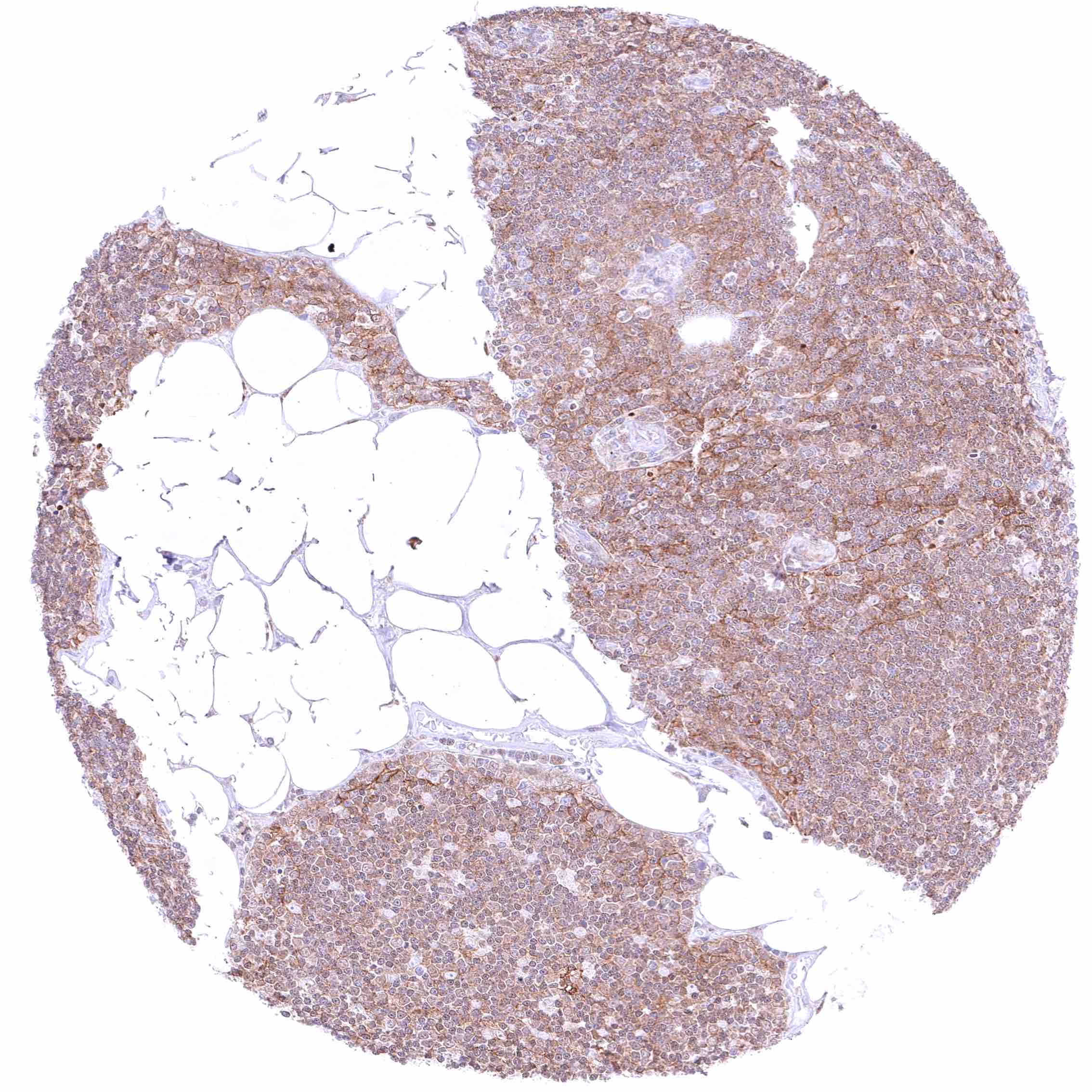
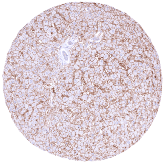
Positive control = Liver: A variable, weak to strong, membranous SLC6A1 staining must be seen in hepatocytes.
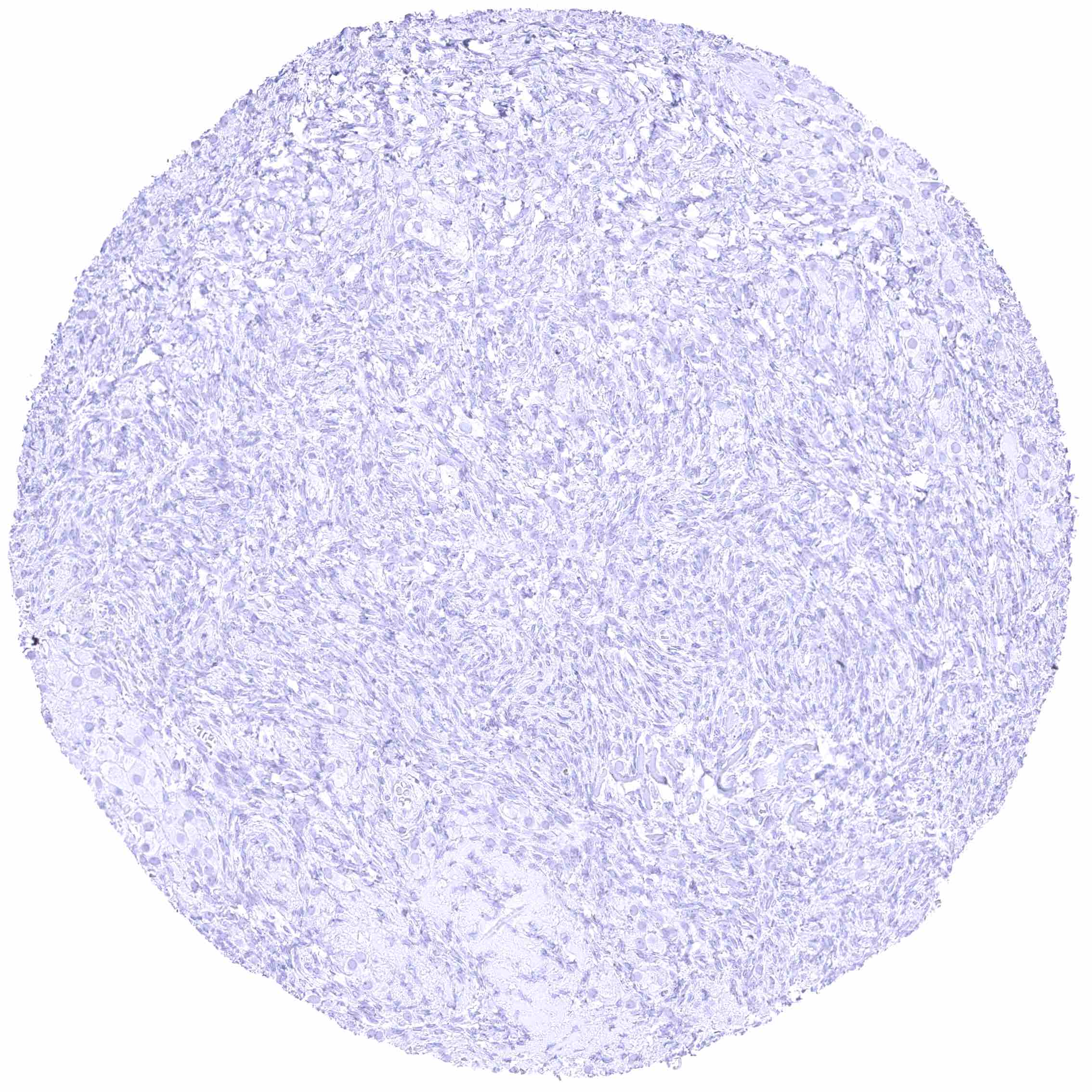
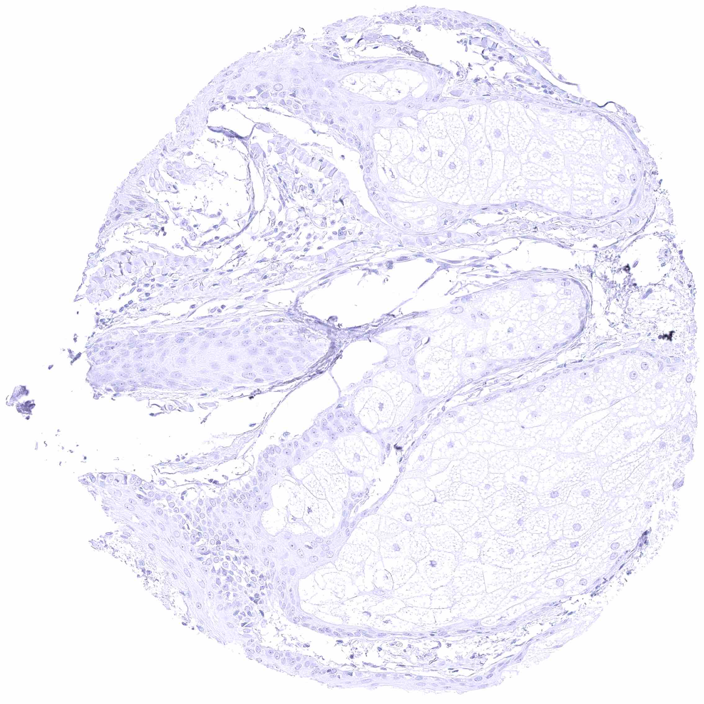
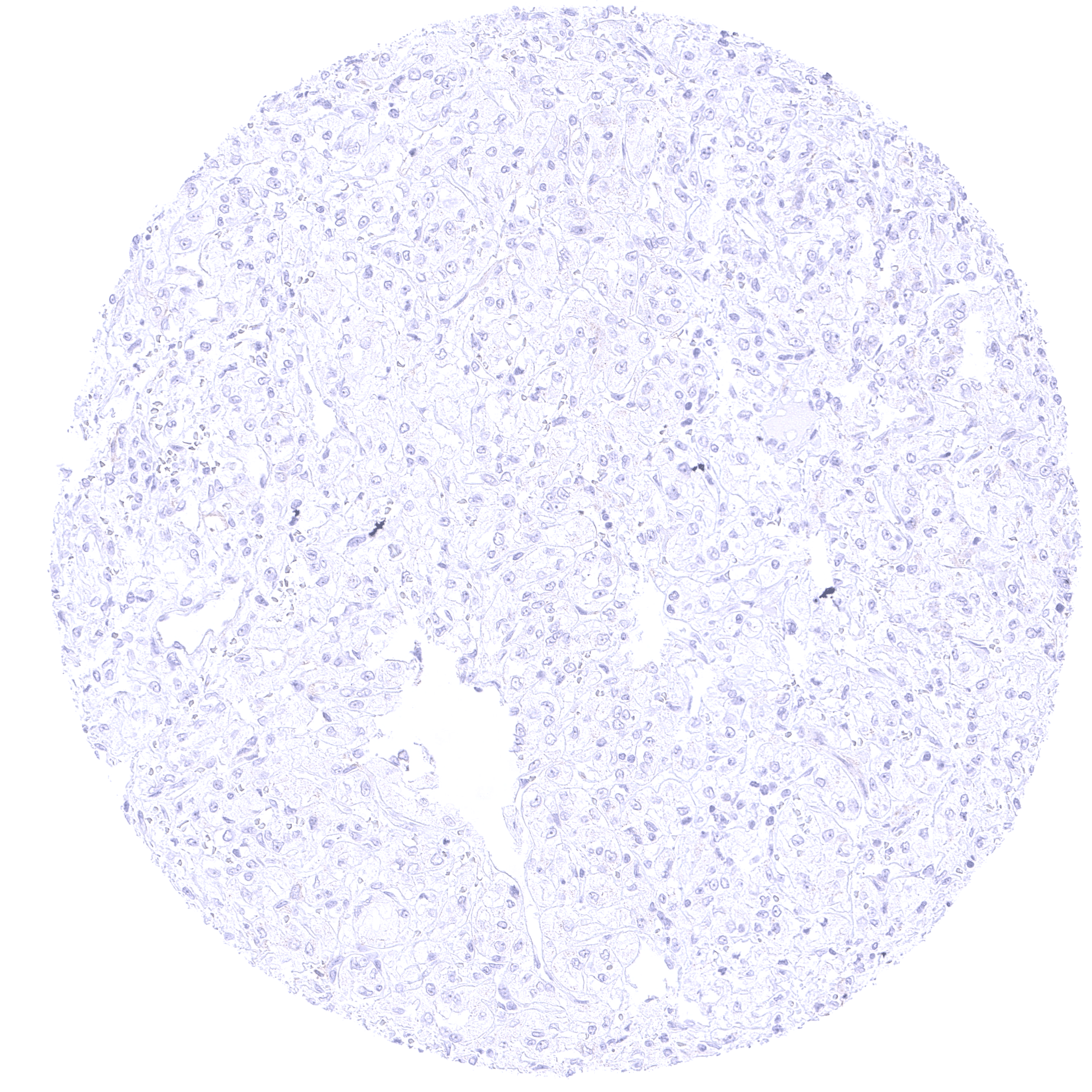
Negative control = Lymph node: SLC6A1 staining must be absent in all cell types.
Cellular localization = Membranous
Reactivity = Human
Application = Immunohistochemistry
Dilution = 1:100 – 1:200
Intended Use = Research Use Only
Relevance of Antibody
SLC6A1 is a regulator of synaptic transmission in the central nervous system.
Biology Behind
The Solute carrier family 6 member 1 (SLC6A1) gene is located on chromosome 3 at position 3p25.3 and codes for the 67kDa SLC6A1 protein which acts as a sodium-dependent neurotransmitter transporter for glycine. It plays a crucial role in the regulation of synaptic transmission and modulation of excitatory signals within the central nervous system. Accordingly, SLC6A1 is primarily expressed in the brain, notably in the spinal cord, cerebellum, and various regions of the cerebral cortex. Germline mutations have been linked to neurodevelopmental disorders, particularly epilepsy syndromes, such as severe myoclonic epilepsy in infancy. SLC6A1 knock-out mice exhibit behavioral changes, increased susceptibility to seizures, and neurodevelopmental deficits. SLC6A1-associated neurodevelopmental disorders are among the top ten most common monogenic causes for both autism and epilepsy. Several SLC6A1 variants exhibit reduced transmembranous trafficking which might be rescuable by pharmacochaperoning. SLC6A1 is also found in several extra-cranial tissues like the liver, the thymus and the parathyroid, indicating a broader physiological role. SLC6A1 has been suggested to play a role in various cancers. SLC6A1 expression levels have been found to correlate with cancer stage and prognosis in several tumor types.
Staining Pattern in Normal Tissues
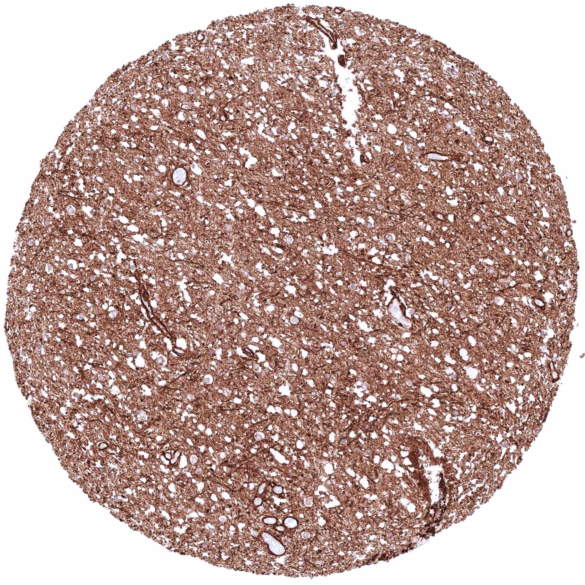
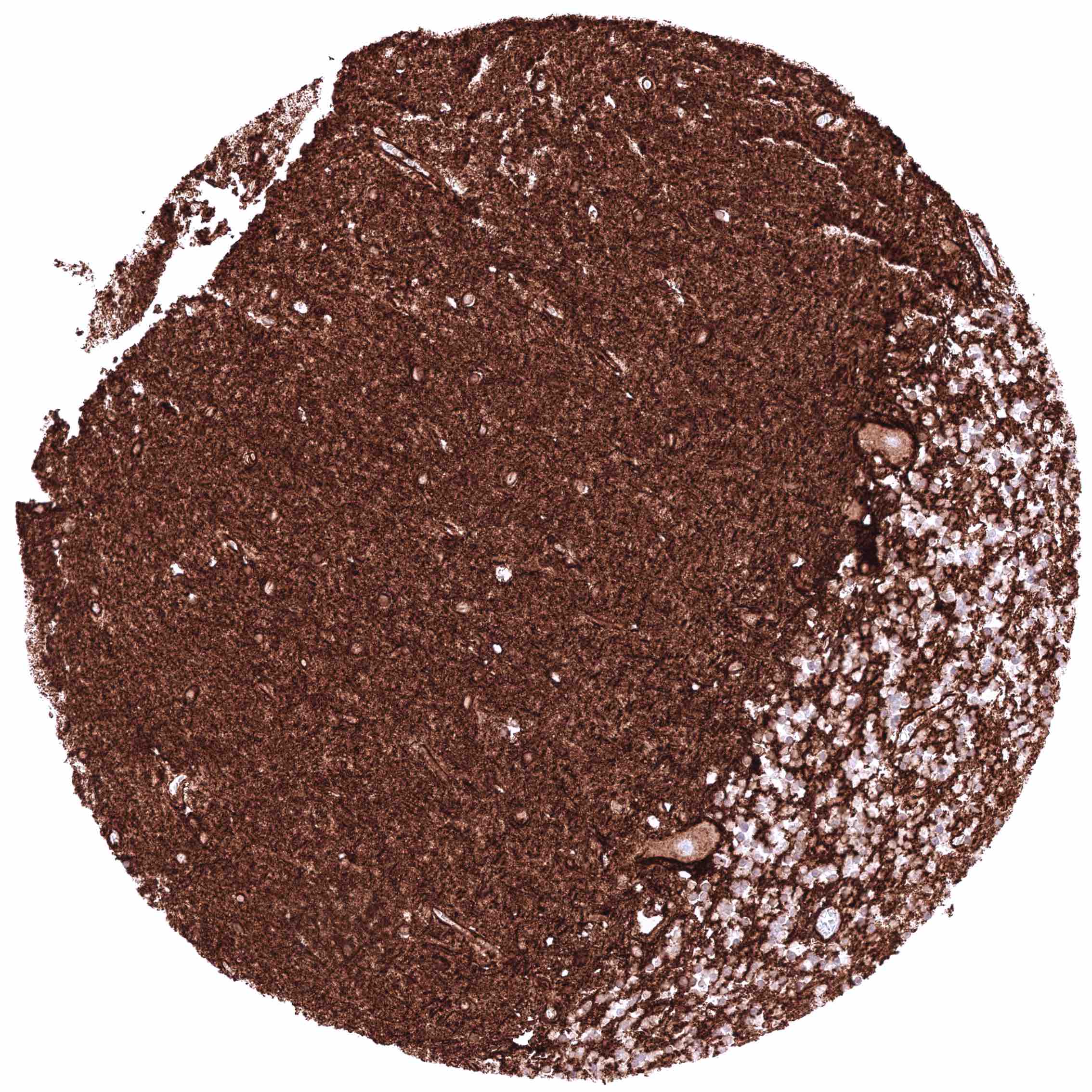
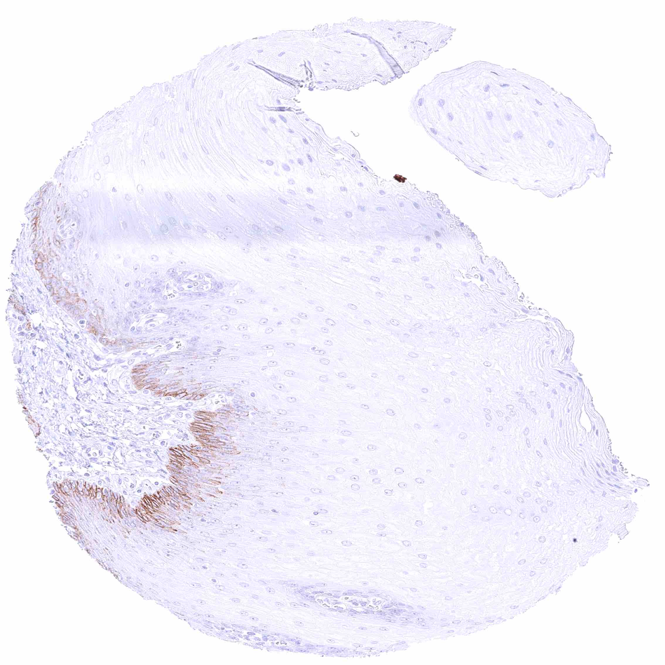
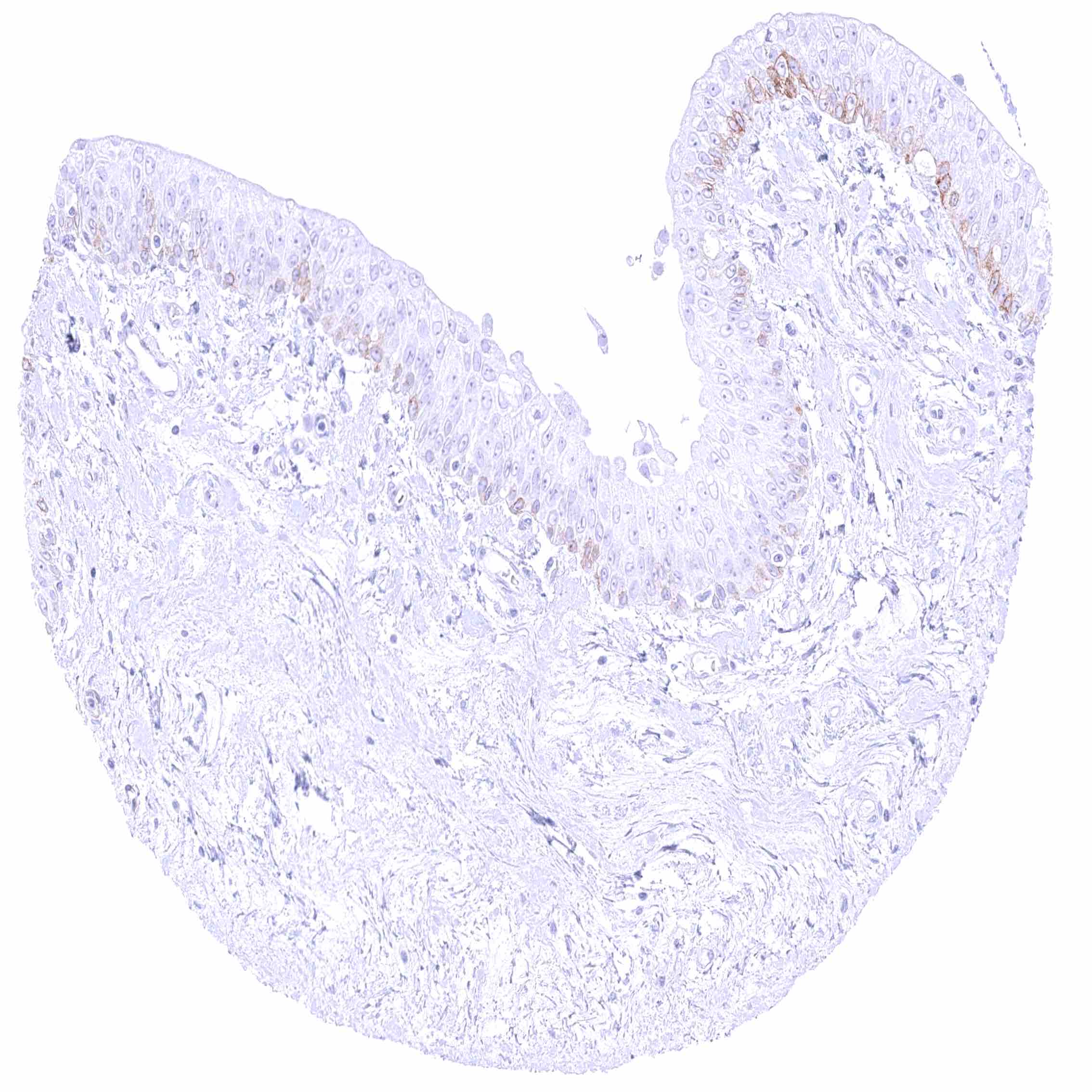
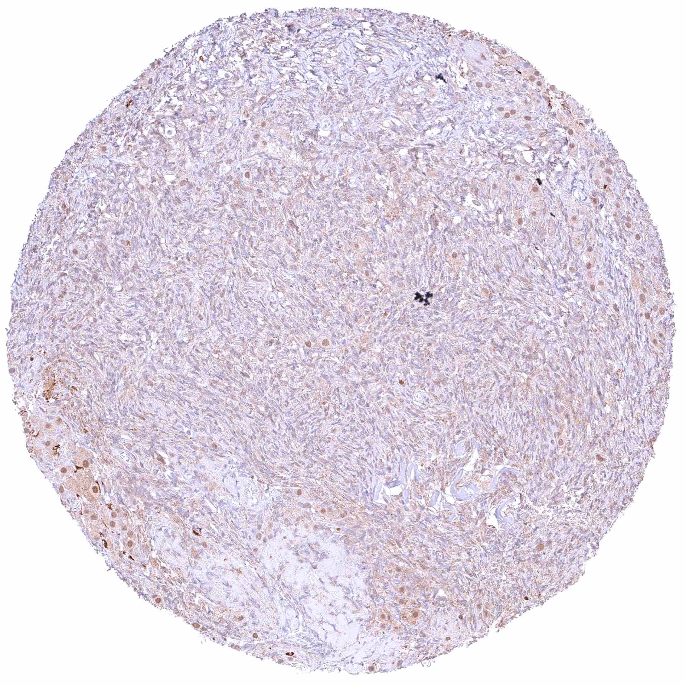
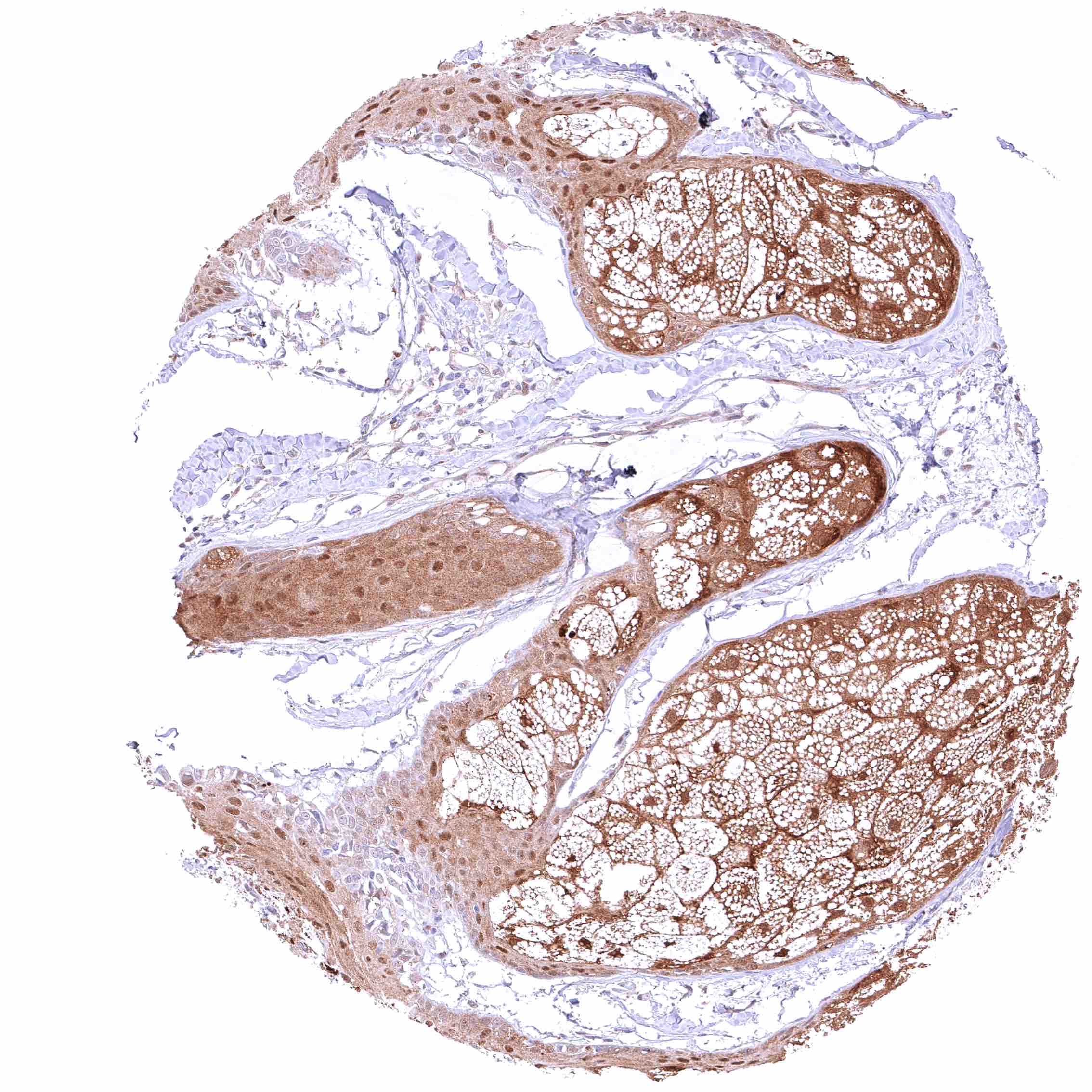
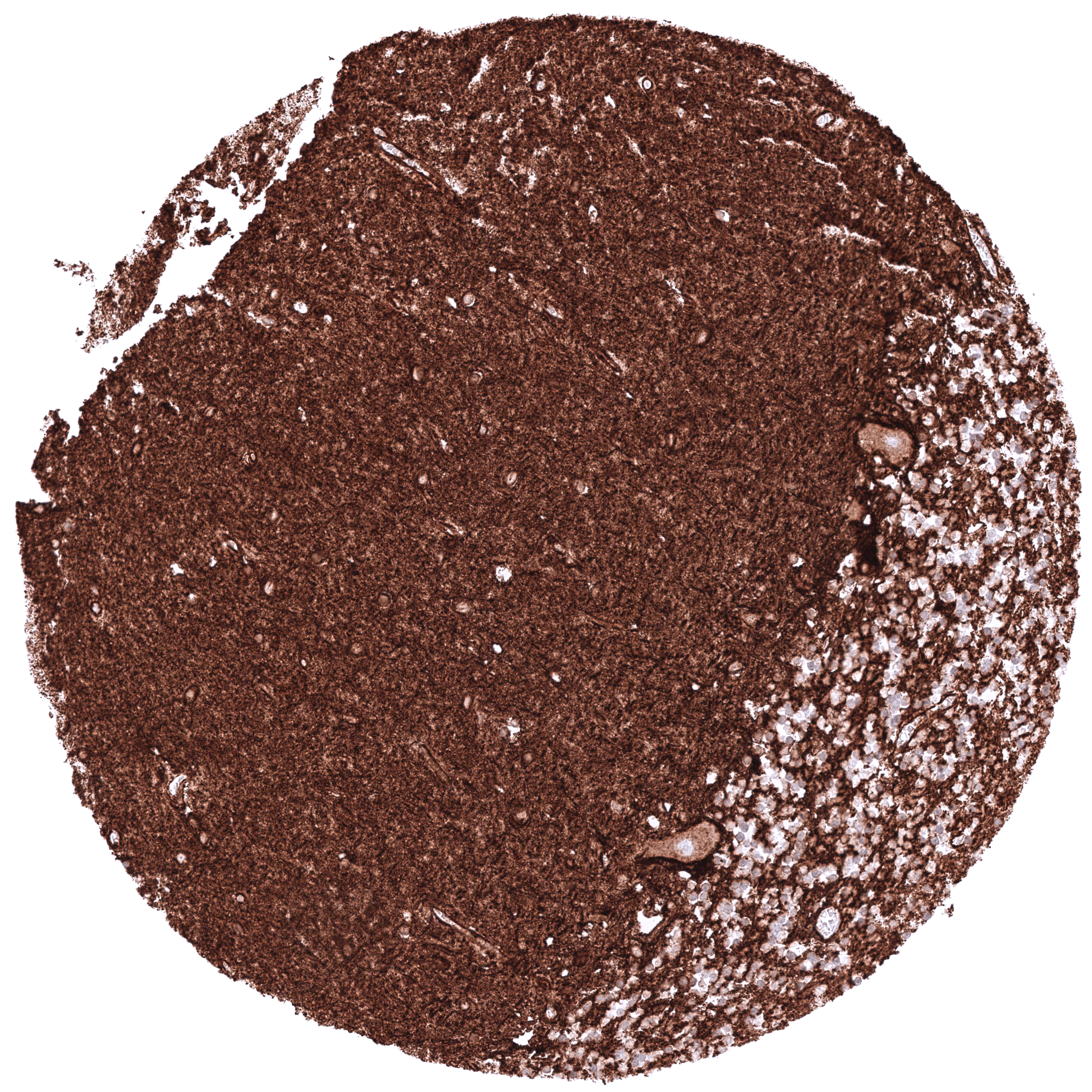
SLC6A1 staining in normal tissues predominantly occurs in the brain. SLC6A1 can also be found in the parathyroid gland, the thymus and few other tissues such as basal cells in the squamous epithelium and the urothelium. Images describing the SLC6A1 staining pattern in normal tissues obtained by the antibody HMV4271 are shown in our “Normal Tissue Gallery”.
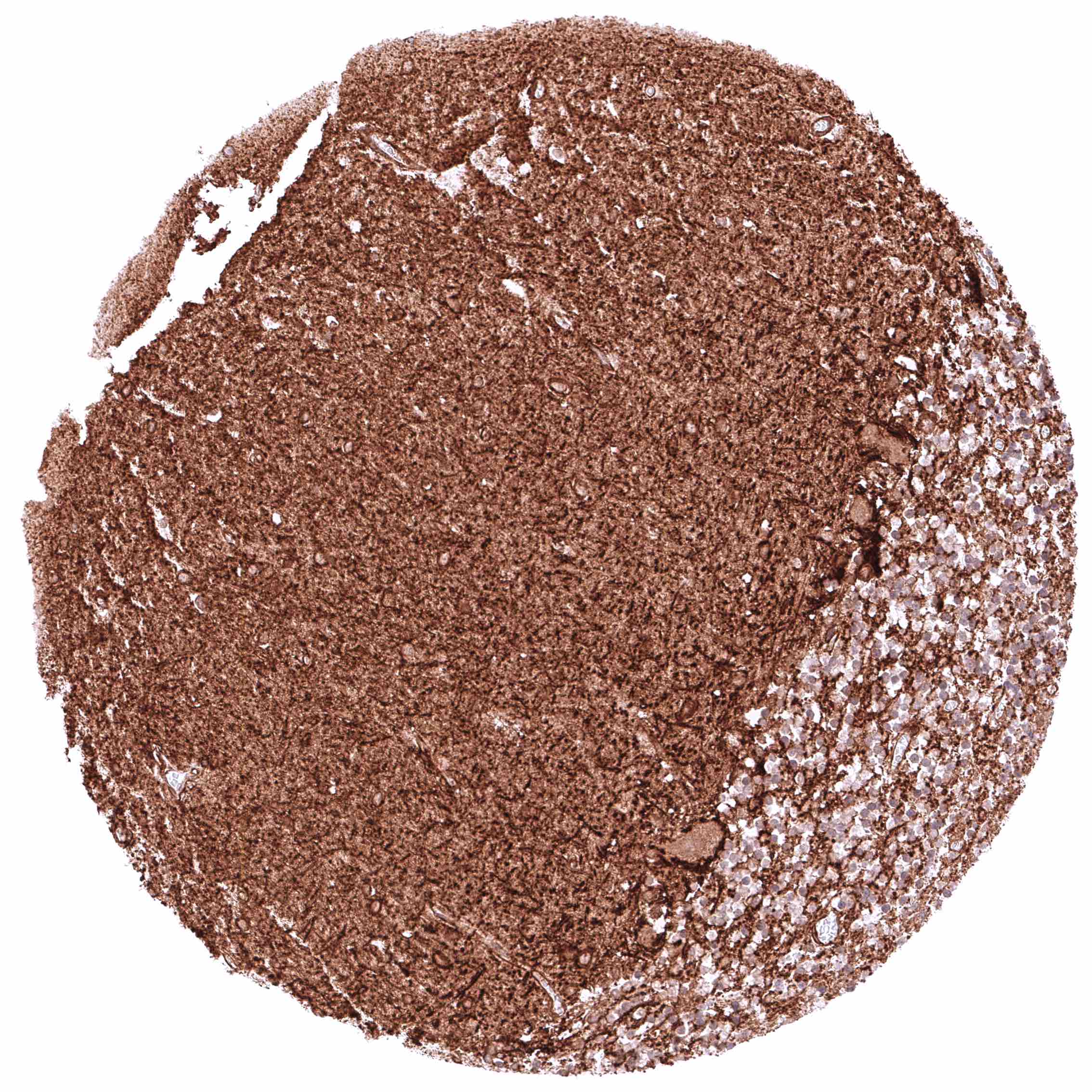
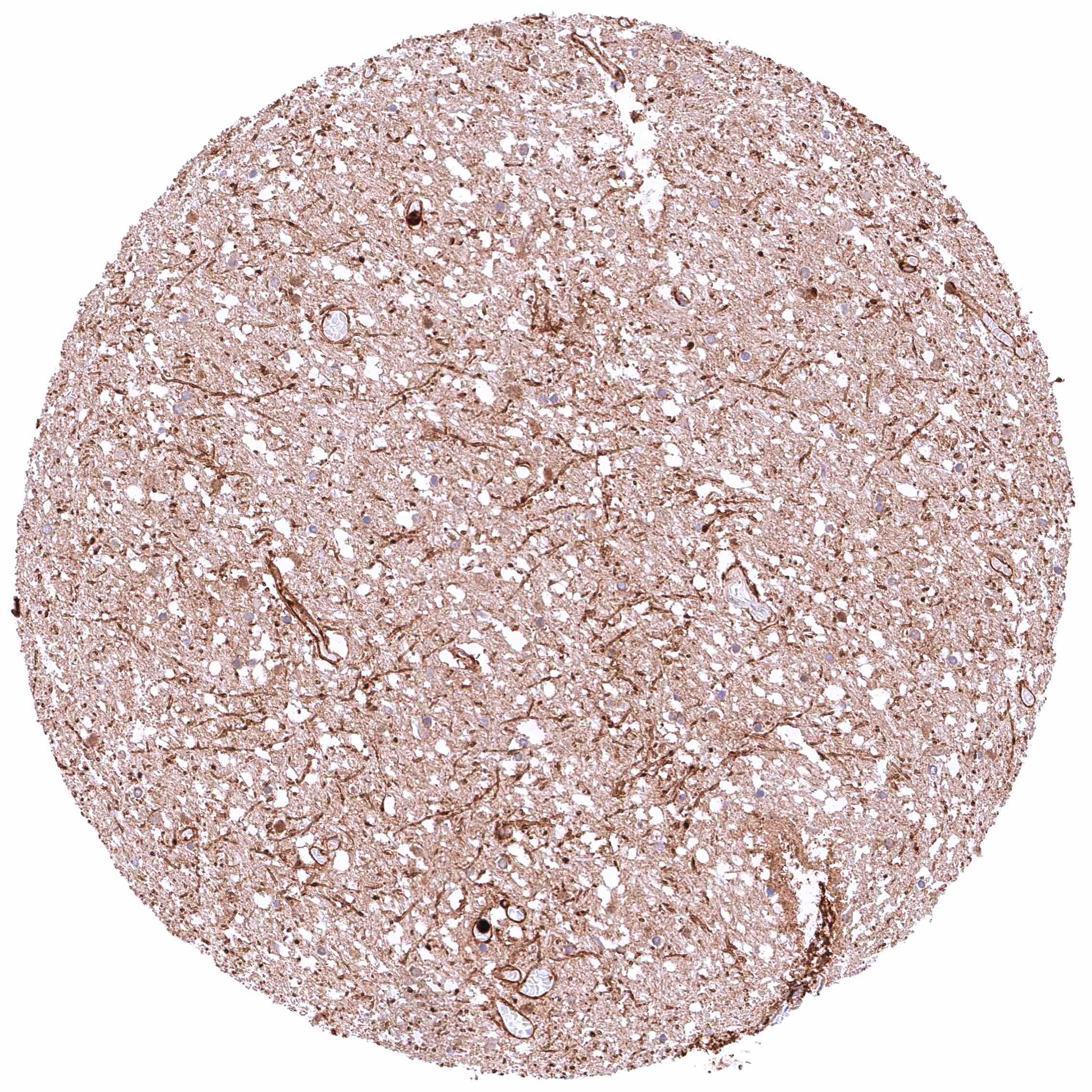
| Brain | Cerebrum | Strong SLC6A1 staining in neurons and in axons. |
| Cerebellum | Strong SLC6A1 staining in neurons (including Purkinje cells) and in axons. | |
| Endocrine Tissues | Thyroid | Negative. |
| Parathyroid | Moderate to strong membranous staining of a subset of epithelial cells. | |
| Adrenal gland | Negative. | |
| Pituitary gland | Negative. | |
| Respiratory system | Bronchus, Respiratory epithelium | Negative. |
| Lung | Negative. | |
| Gastrointestinal Tract | Salivary glands | Negative. |
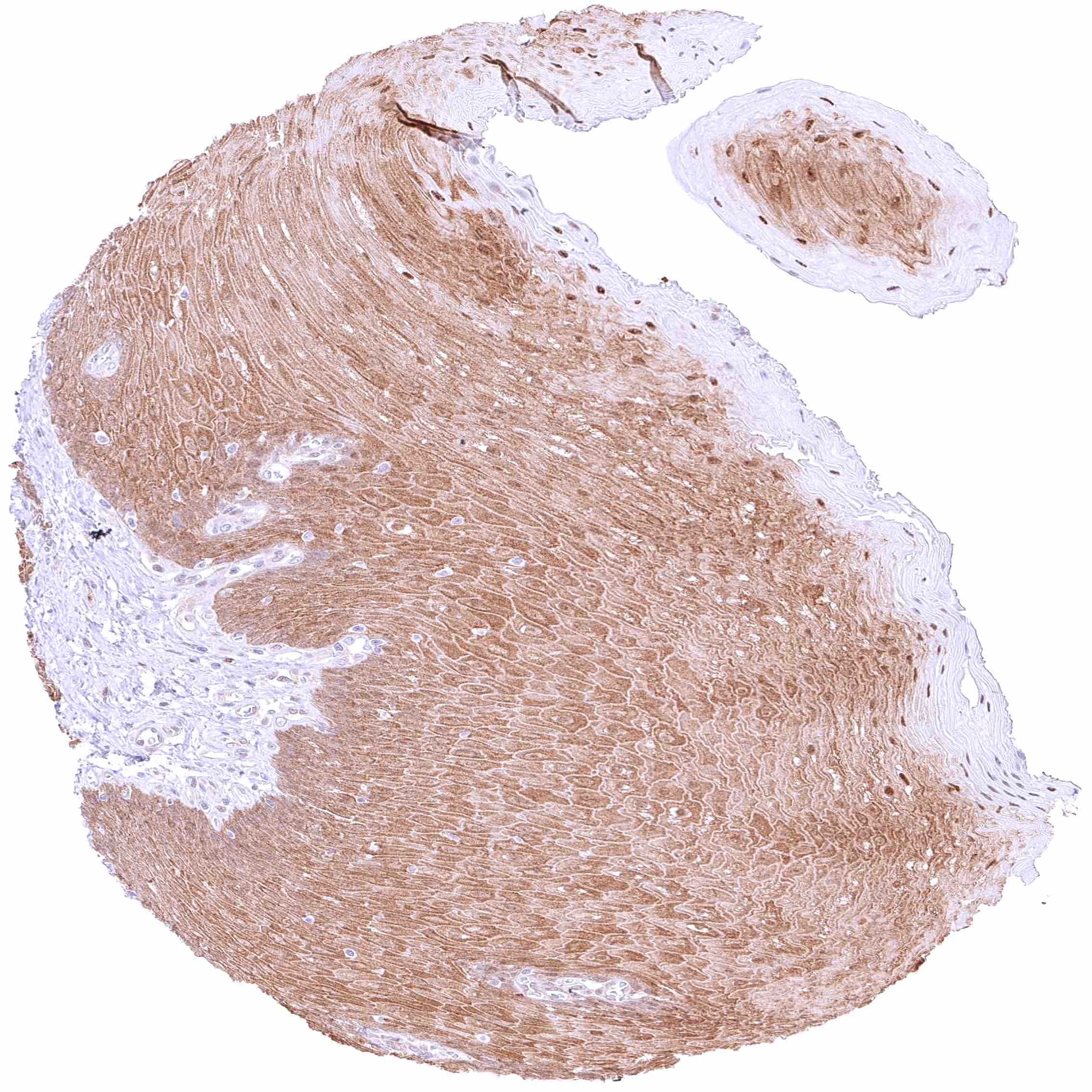
| Esophagus | Weak to moderate membranous SLC6A1 staining of some basal and suprabasal squamous epithelial cells (not in all samples). | |
| Stomach | Negative. | |
| Duodenum | Negative. | |
| Small intestine | Negative. | |
| Appendix | Negative. | |
| Colon | Negative. | |
| Rectum | Negative. | |
| Liver | Membranous SLC6A1 staining of variable intensity ranging from absent/weak to strong of hepatocytes. | |
| Gallbladder | Negative. | |
| Pancreas | Negative. | |
| Genitourinary | Kidney | Weak to moderate granular cytoplasmic staining in tubuli (not in all samples). Perhaps caused by cross-reactivity. |
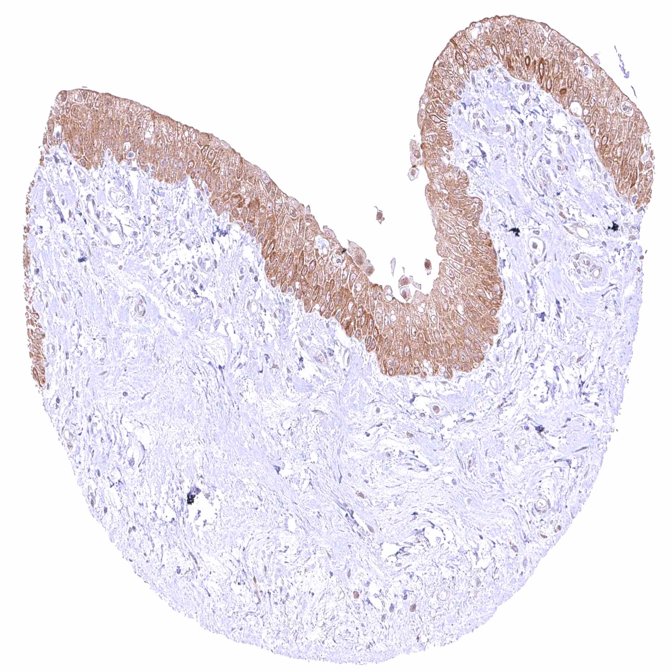
| Urothelium | Weak to moderate membranous SLC6A1 staining of a subset of basal urothelial cells (only in one sample). | |
| Male genital | Prostate | Negative. |
| Seminal vesicles | Negative. | |
| Testis | Negative. | |
| Epididymis | Weak (apical) membranous SLC6A1 staining of a subset of columnar cells in the cauda. | |
| Female genital | Breast | Negative. |
| Uterus, myometrium | Negative. | |
| Uterus, ectocervix | Negative. | |
| Uterus endocervix | Negative. | |
| Uterus, endometrium | Negative. | |
| Fallopian Tube | Negative. | |
| Ovary | Negative. | |
| Placenta early | Negative. | |
| Placenta mature | Negative. | |
| Amnion | Negative. | |
| Chorion | Negative. | |
| Skin | Epidermis | Negative. |
| Sebaceous glands | Negative. | |
| Muscle/connective tissue | Heart muscle | Negative. |
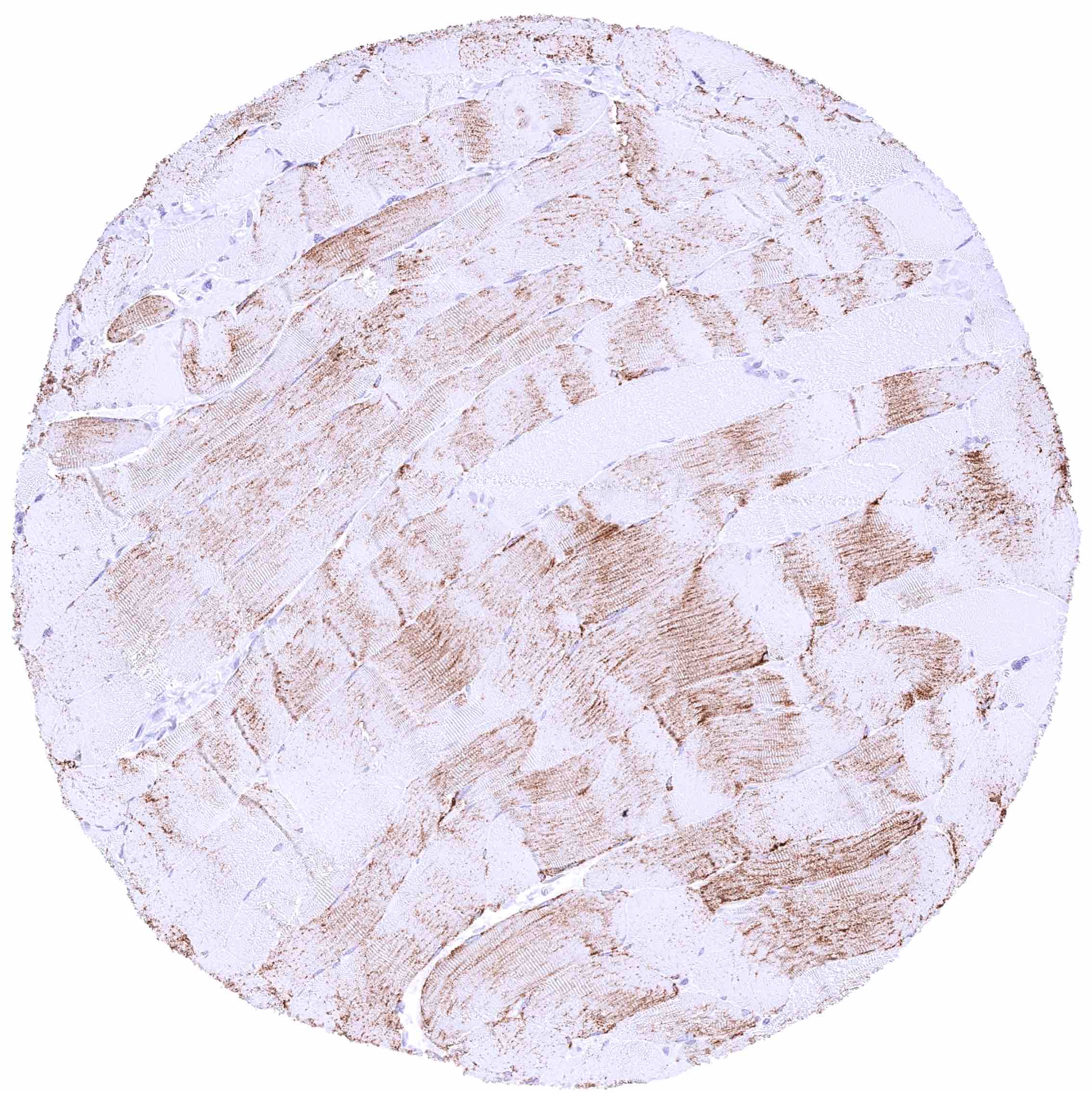
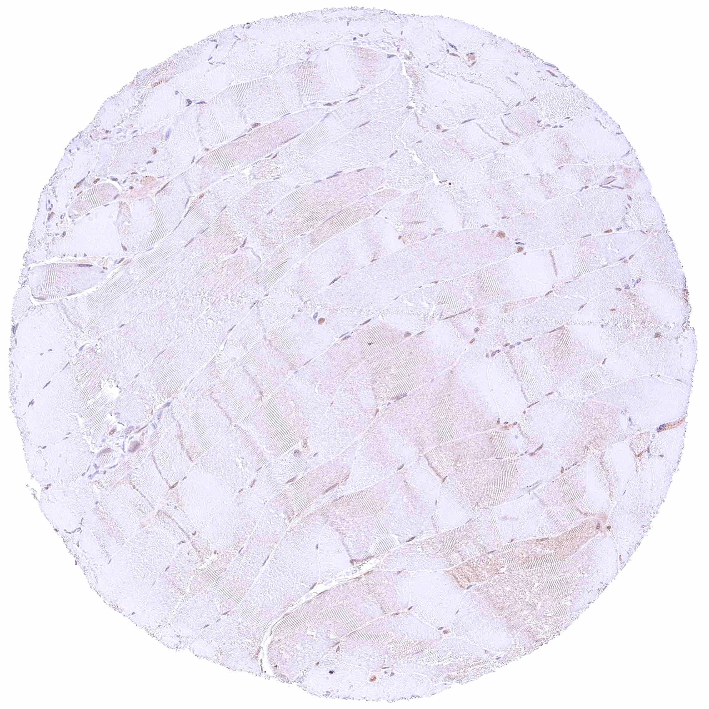
| Skeletal muscle | Cytoplasmic SLC6A1 staining of a subset of skeletal muscle fibers (potential cross-reactivity). | |
| Smooth muscle | Negative. | |
| Vessel walls | Negative. | |
| Fat | Negative. | |
| Stroma | Negative. | |
| Endothelium | Negative. | |
| Bone marrow/ lymphoid tissue | Bone marrow | Negative. |
| Lymph node | Negative. | |
| Spleen | Negative. | |
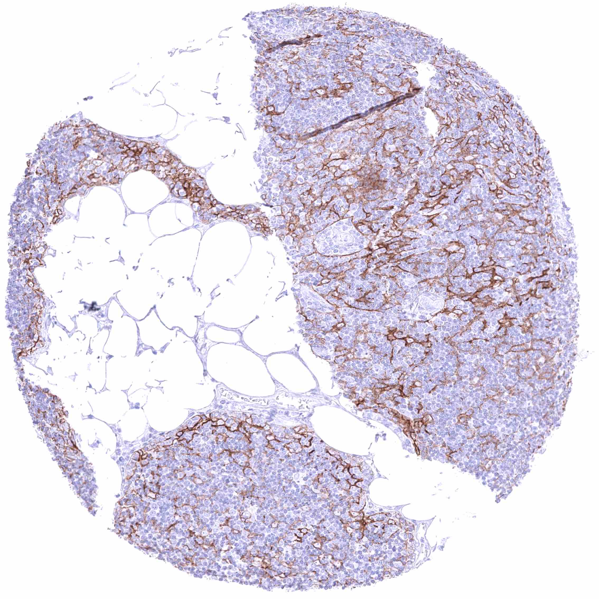
| Thymus | Strong membranous SLC6A1 staining of most if not all thymic epithelial cells. | |
| Tonsil | Negative. | |
| Remarks |
These findings are largely consistent with the RNA data described in the Human Protein Atlas (Tissue expression SLC6A1).
Positive control = Liver: A variable, weak to strong, membranous SLC6A1 staining must be seen in hepatocytes.
Negative control = Lymph node: SLC6A1 staining must be absent in all cell types.
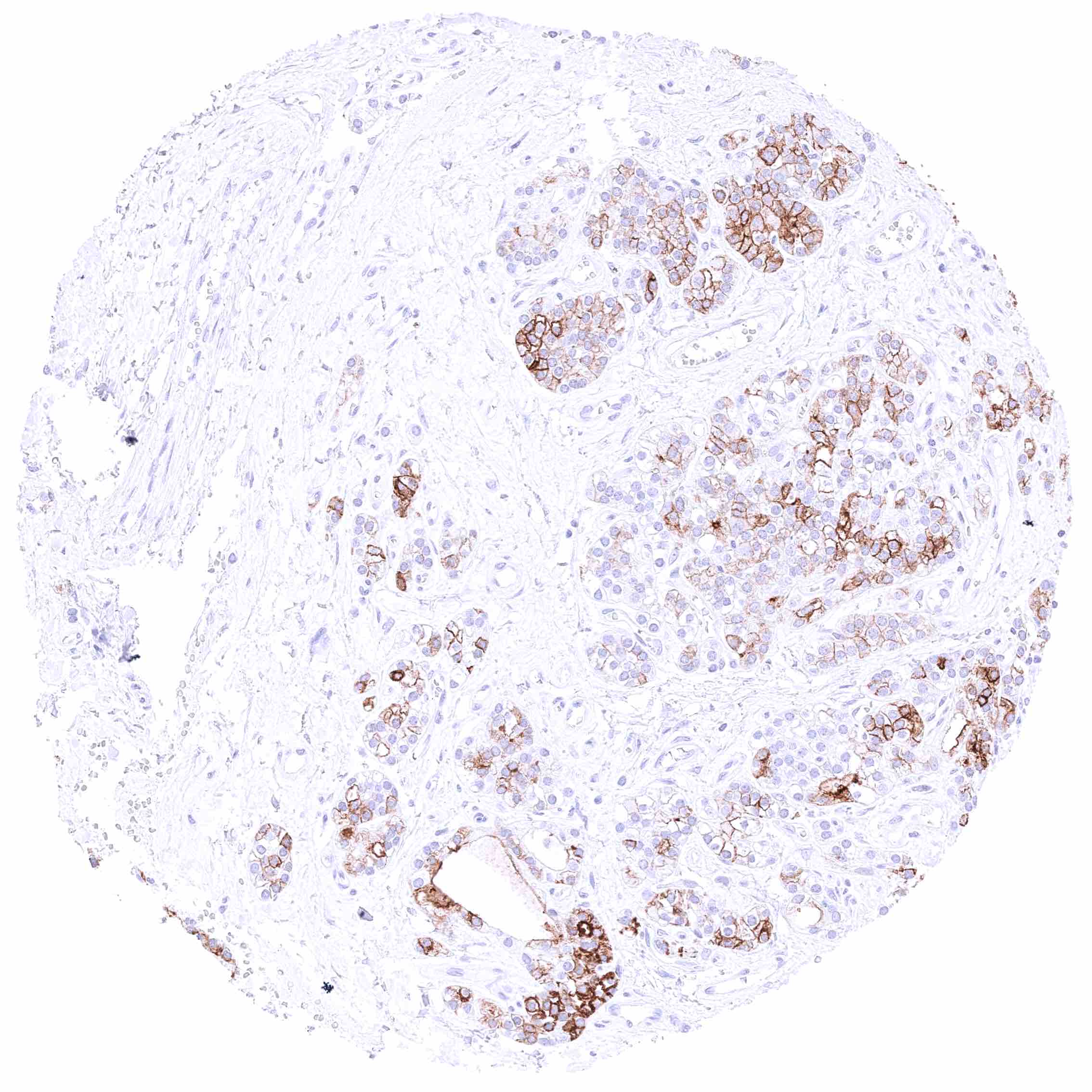
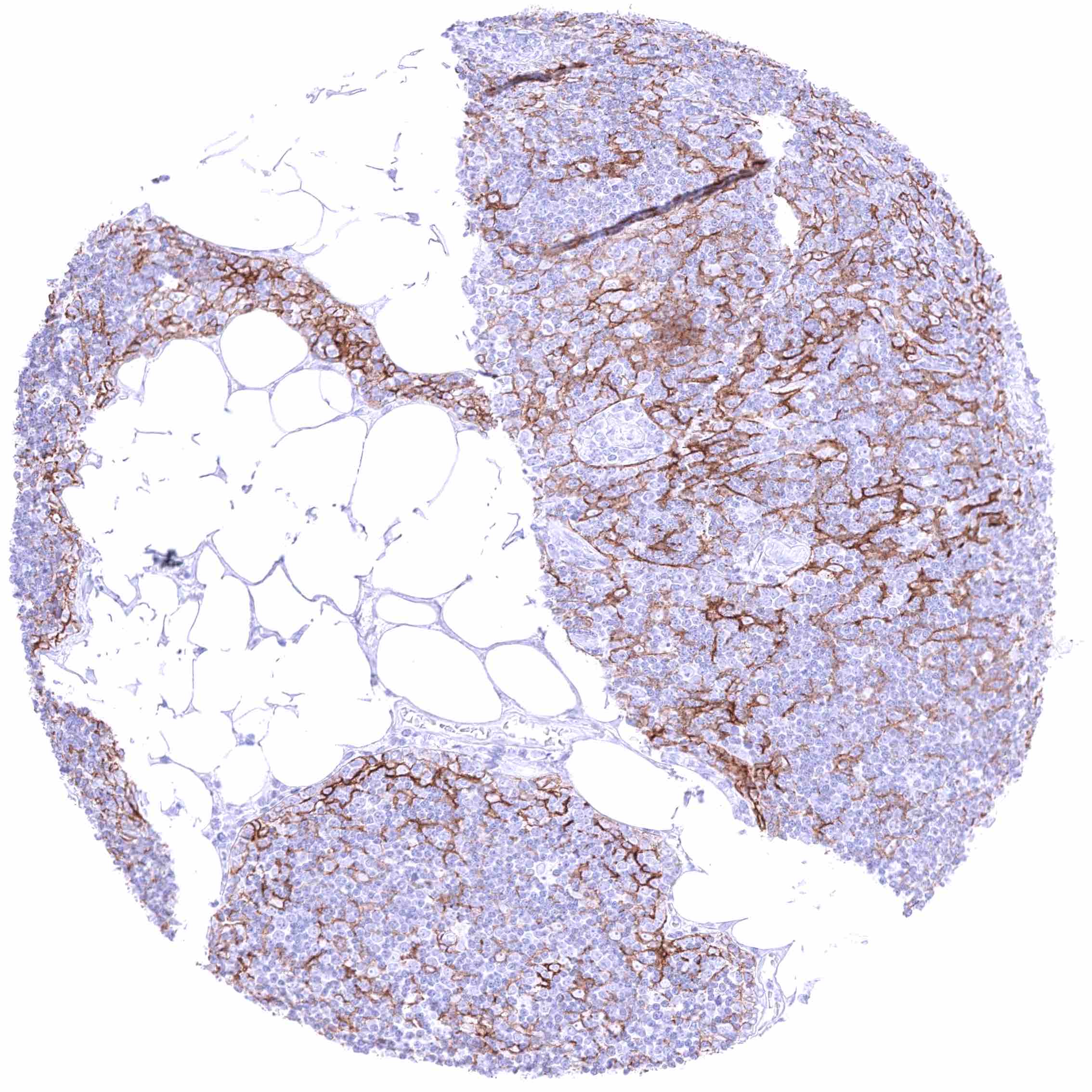
Staining Pattern in Relevant Tumor Types
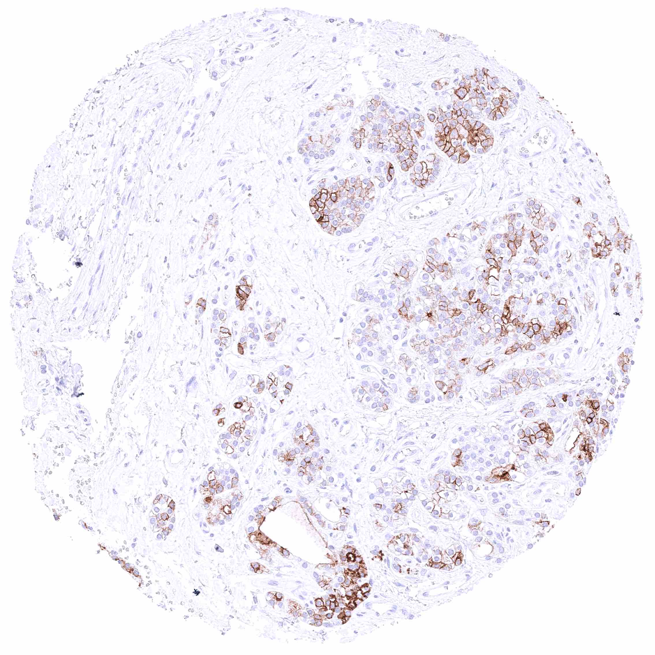
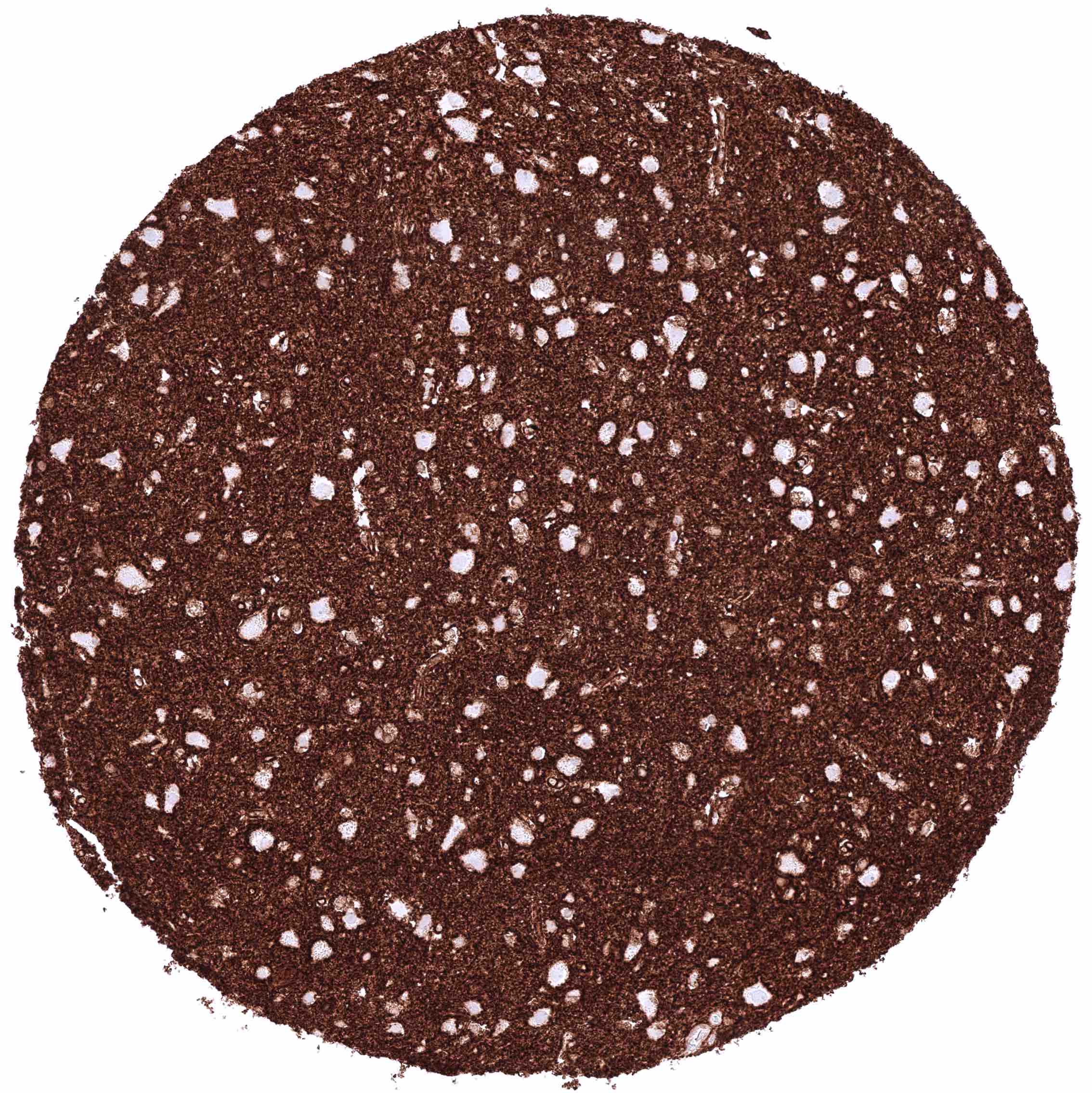
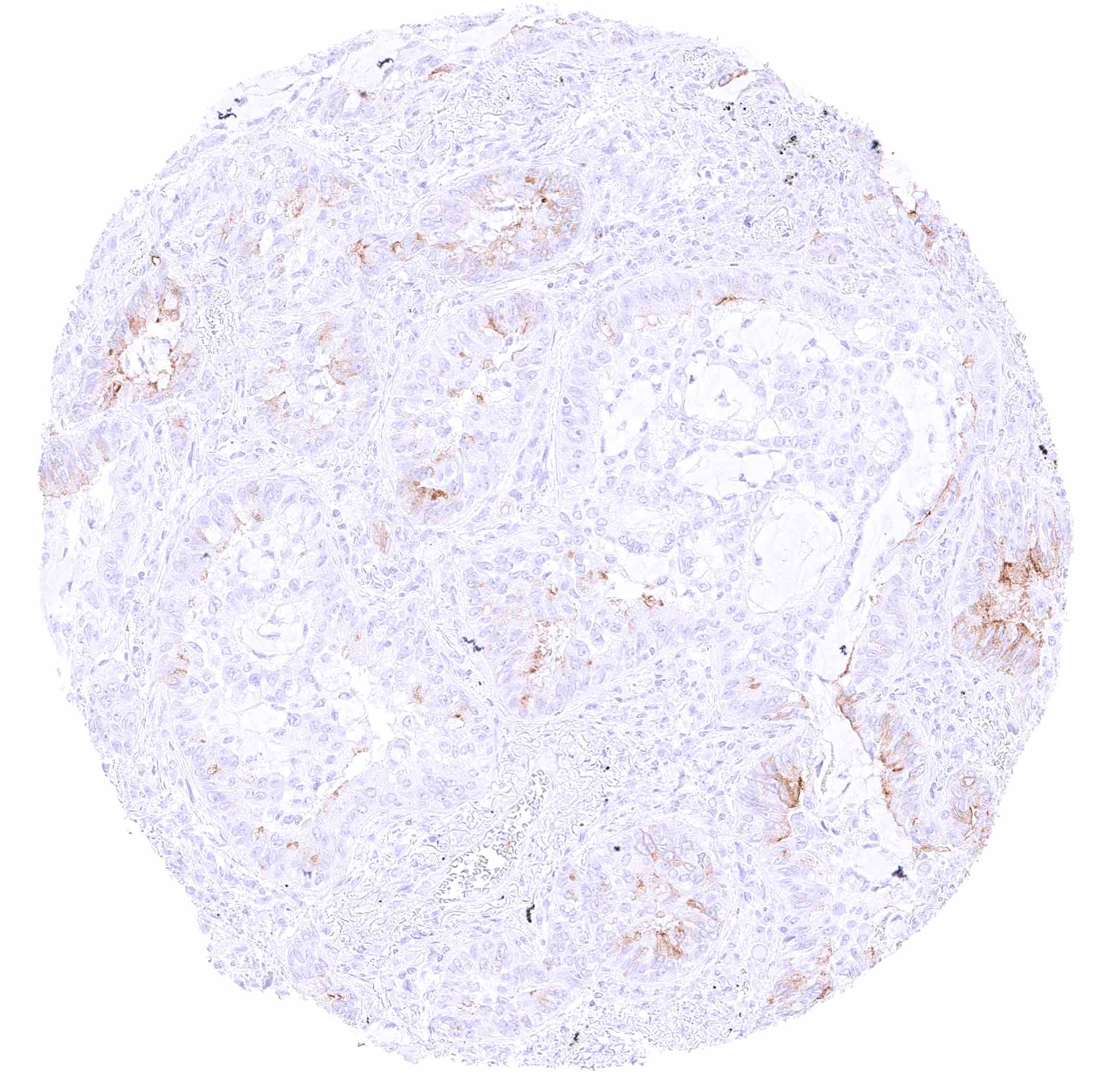
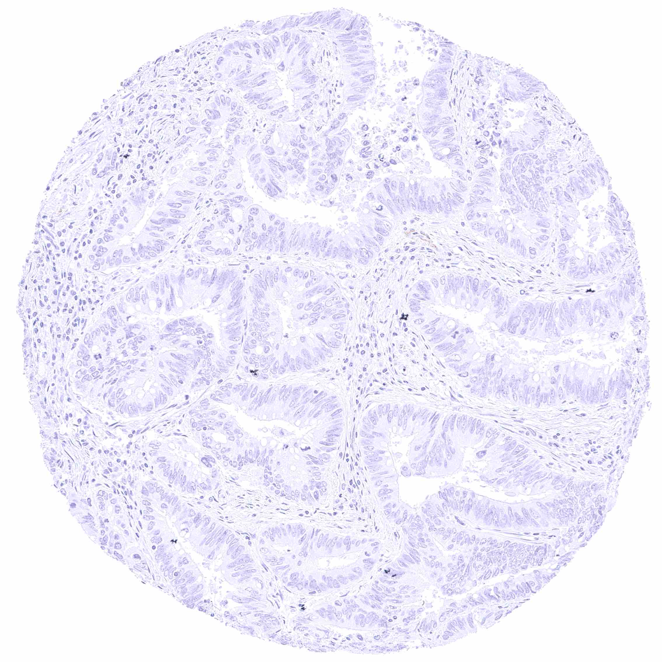
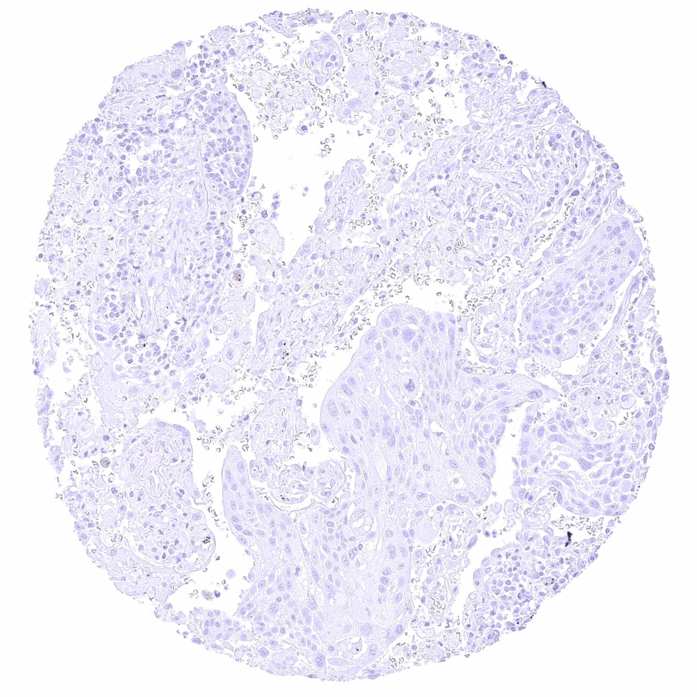
SLC6A1 expression is particularly frequent in hepatocellular carcinomas and it may also occur in astrocytic tumors.
The TCGA findings on SLC6A1 RNA expression in different tumor categories have been summarized in the Human Protein Atlas.
Compatibility of Antibodies
No data available at the moment
Protocol Recommendations
IHC users have different preferences on how the stains should look like. Some prefer high staining intensity of the target stain and even accept some background. Others favor absolute specificity and lighter target stains. Factors that invariably lead to more intense staining include higher concentration of the antibody and visualization tools, longer incubation time, higher temperature during incubation, higher temperature and longer duration of the heat induced epitope retrieval (slide pretreatment). The impact of the pH during slide pretreatment has variable effects and depends on the antibody and the target protein.
All images and data shown here and in our image galleries are obtained by the manual protocol described below. Other protocols resulting in equivalent staining are described as well.
Manual protocol
Freshly cut sections should be used (less than 10 days between cutting and staining). Heat-induced antigen retrieval for 5 minutes in an autoclave at 121°C in pH 7,8 Target Retrieval Solution buffer. Apply HMV4721 at a dilution of 1:150 at 37°C for 60 minutes. Visualization of bound antibody by the EnVision Kit (Dako, Agilent) according to the manufacturer’s directions.
Potential Research Applications
- How do different pathogenic SLC6A1 variants alter SLC6A1 trafficking, localization, and functional expression in human neurons?
- What are the cell-type specific SLC6A1 expression patterns and developmental trajectories of SLC6A1 in human brain regions implicated in epilepsy and neurodevelopmental disorders?
- How does altered SLC6A1 function affect GABAergic synapse physiology, network excitability, and seizure susceptibility in human-relevant models?
- Can pharmacological chaperones or gene therapy restore mutant SLC6A1 function, and what are optimal delivery strategies and windows for therapeutic rescue?
- What is the role of SLC6A1 in the normal liver and in liver cancer?
Evidence for Antibody Specificity in IHC
There are two ways how the specificity of antibodies can be documented for immunohistochemistry on formalin fixed tissues. These are: 1. Comparison with a second independent method for target expression measurement across a large number of different tissue types (orthogonal strategy), and 2. Comparison with one or several independent antibodies for the same target and showing that all positive staining results are also seen with other antibodies for the same target (independent antibody strategy).
Orthogonal validation: For the antibody HMV4721 specificity is in line with RNA expression data in normal tissues which were collected in three independent RNA screening studies, including the Human Protein Atlas (HPA) RNA-seq tissue dataset, the FANTOM5 project, and the Genotype-Tissue Expression (GTEx) project, and which are summarized in the Human Protein Atlas (Tissue expression SLC6A1). The highest levels of SLC6A1 immunostaining by HMV4721 was observed in the tissues with the highest levels of RNA expression such as the brain, the liver, the parathyroid, and the thymus. Also in agreement with SLC6A1 RNA data, immunostaining by HMV4721 was lacking in skin, pancreas, endometrium, breast, gastrointestinal epithelium, salivary glands, thyroid, adrenal gland, respiratory epithelium, or the lung.
Comparison of antibodies: True expression of SLC6A1 in all cell types with SLC6A1 positivity by HMV4721 is corroborated by an identical staining obtained by a commercially available independent second antibody (termed “validation antibody”). The additional cytoplasmic staining in some skeletal muscle fibers and renal tubuli by HMV4721 is considered an antibody specific cross-reactivity of HMV4721. An additional staining of squamous epithelium, urothelium, smooth muscle cells, sebaceous and salivary glands by the validation antibody which was not seen by HMV4721 was considered an antibody specific cross-reactivity of the validation antibody.