295,00 € – 995,00 €
Product details
Synonyms = ATP binding cassette subfamily B member 11, ABC16, BRIC2, BSEP, PFIC-2, PFIC2, PGY4, SPGP
Antibody type = Recombinant Rabbit monoclonal / IgG
Clone = HMV4715
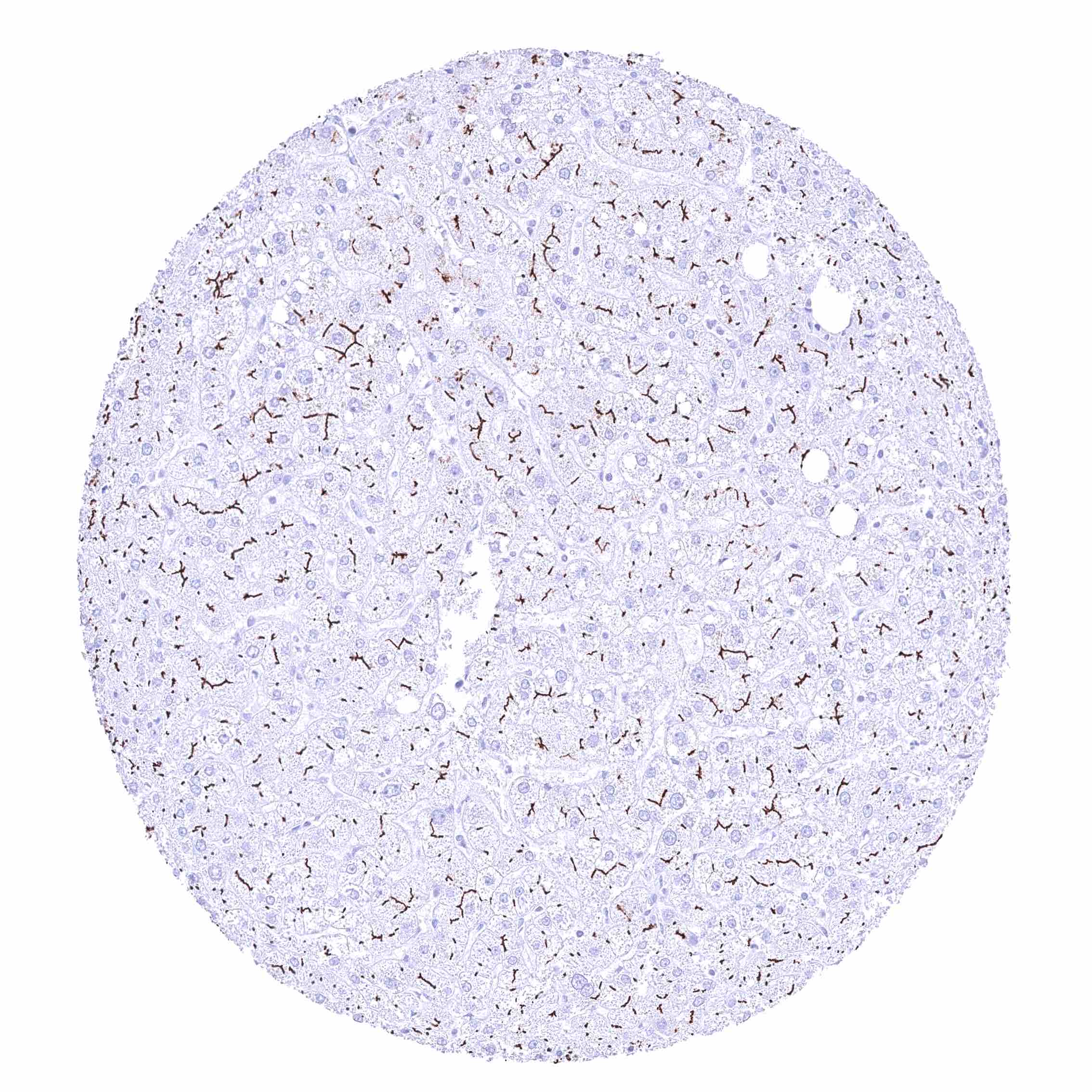
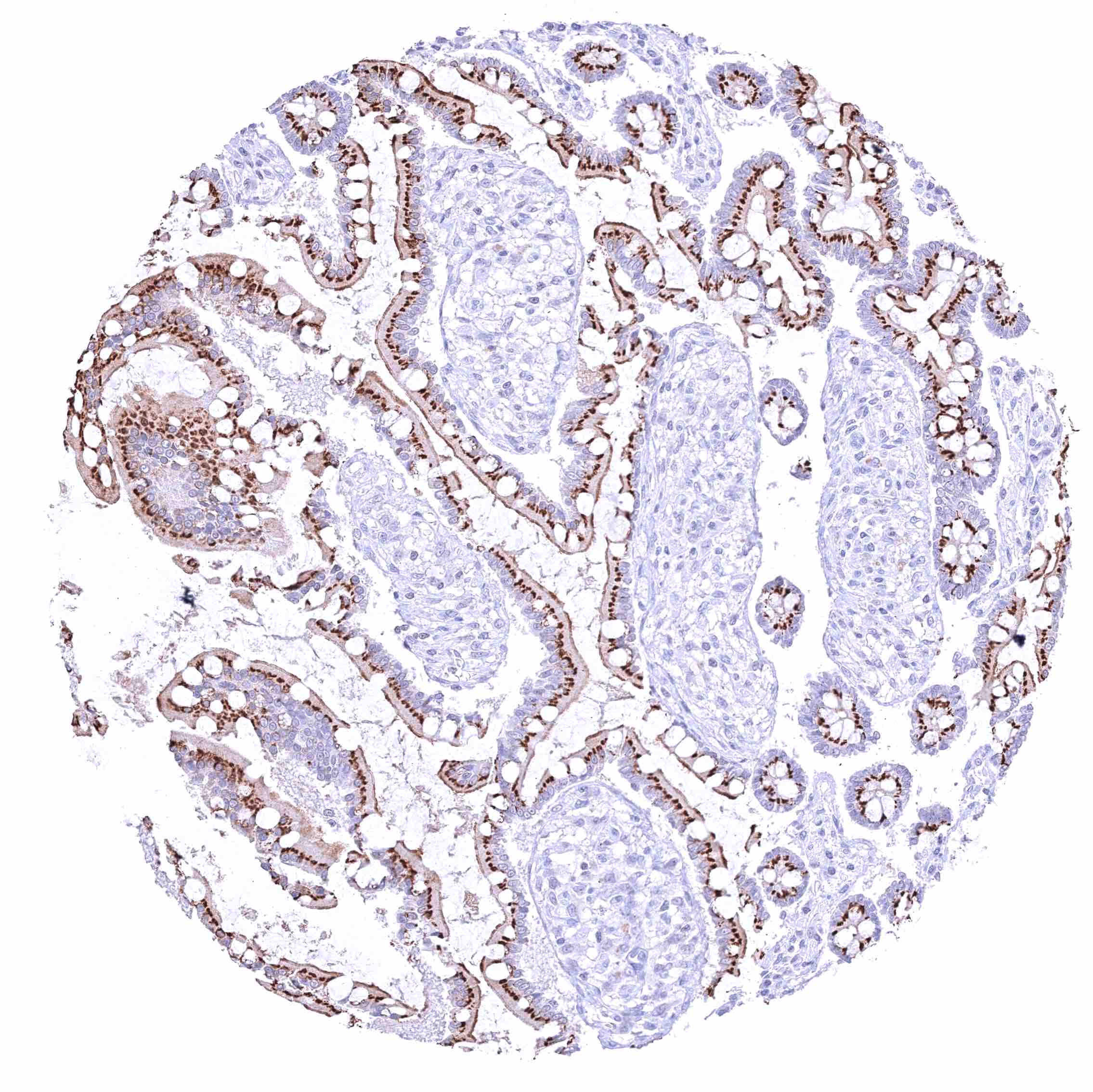
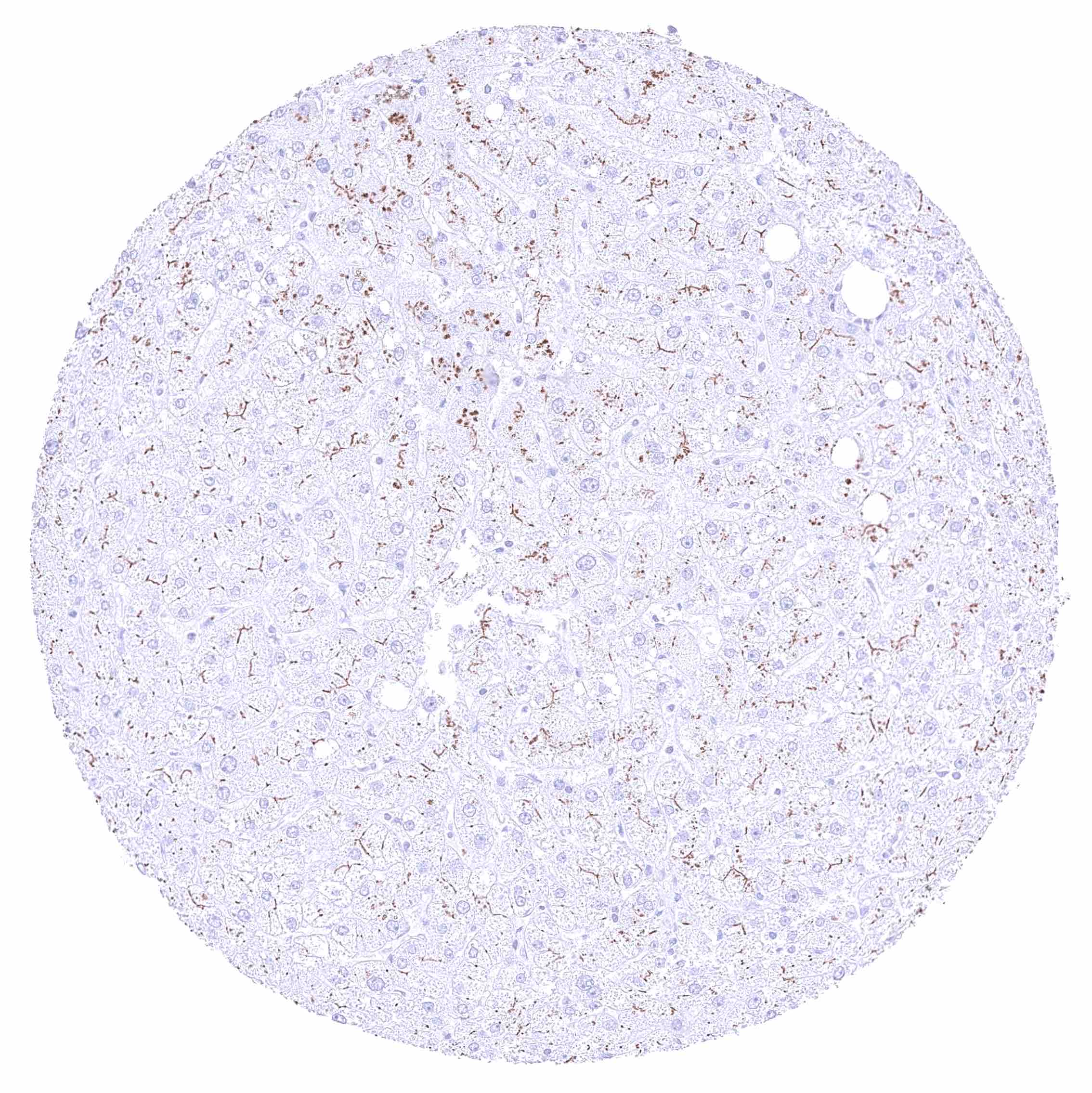
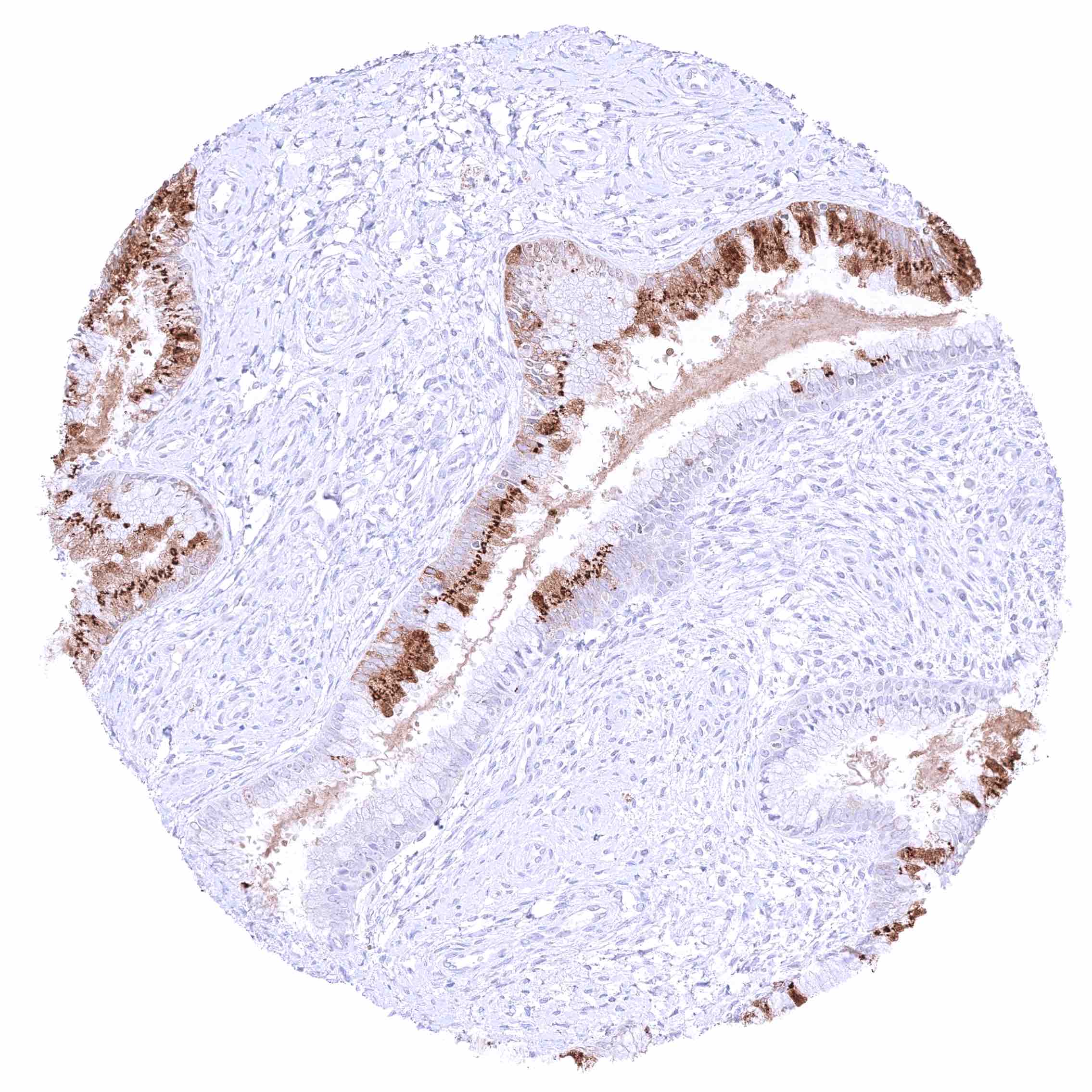
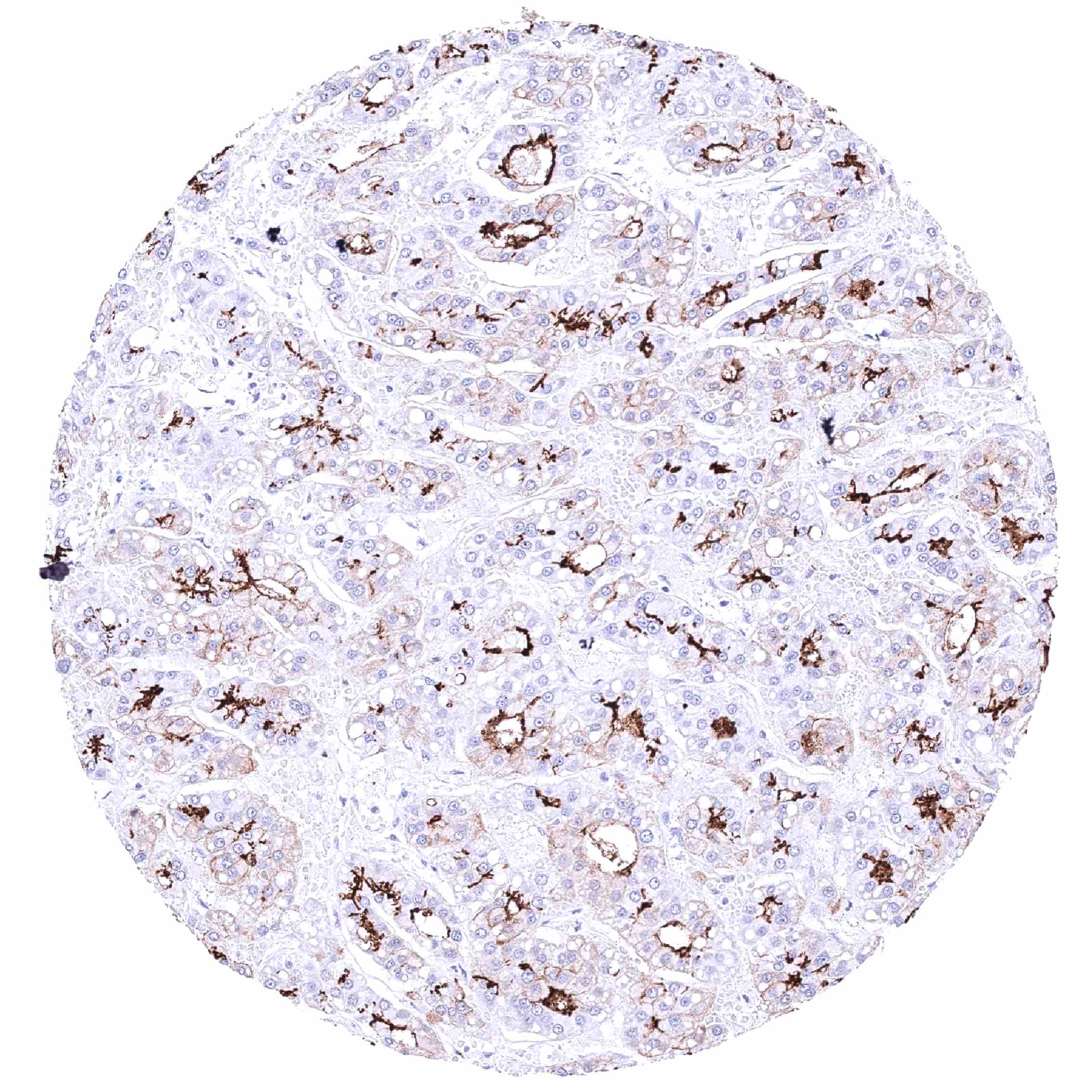
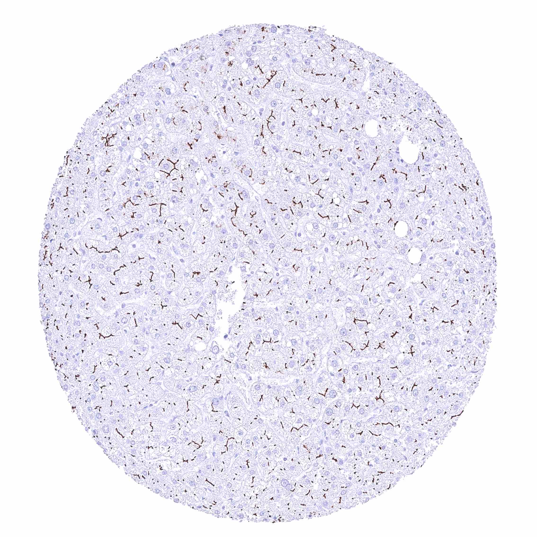
Positive control = Liver: A strong BSEP staining must be seen at the apical “bile secreting” pole of hepatocytes”.
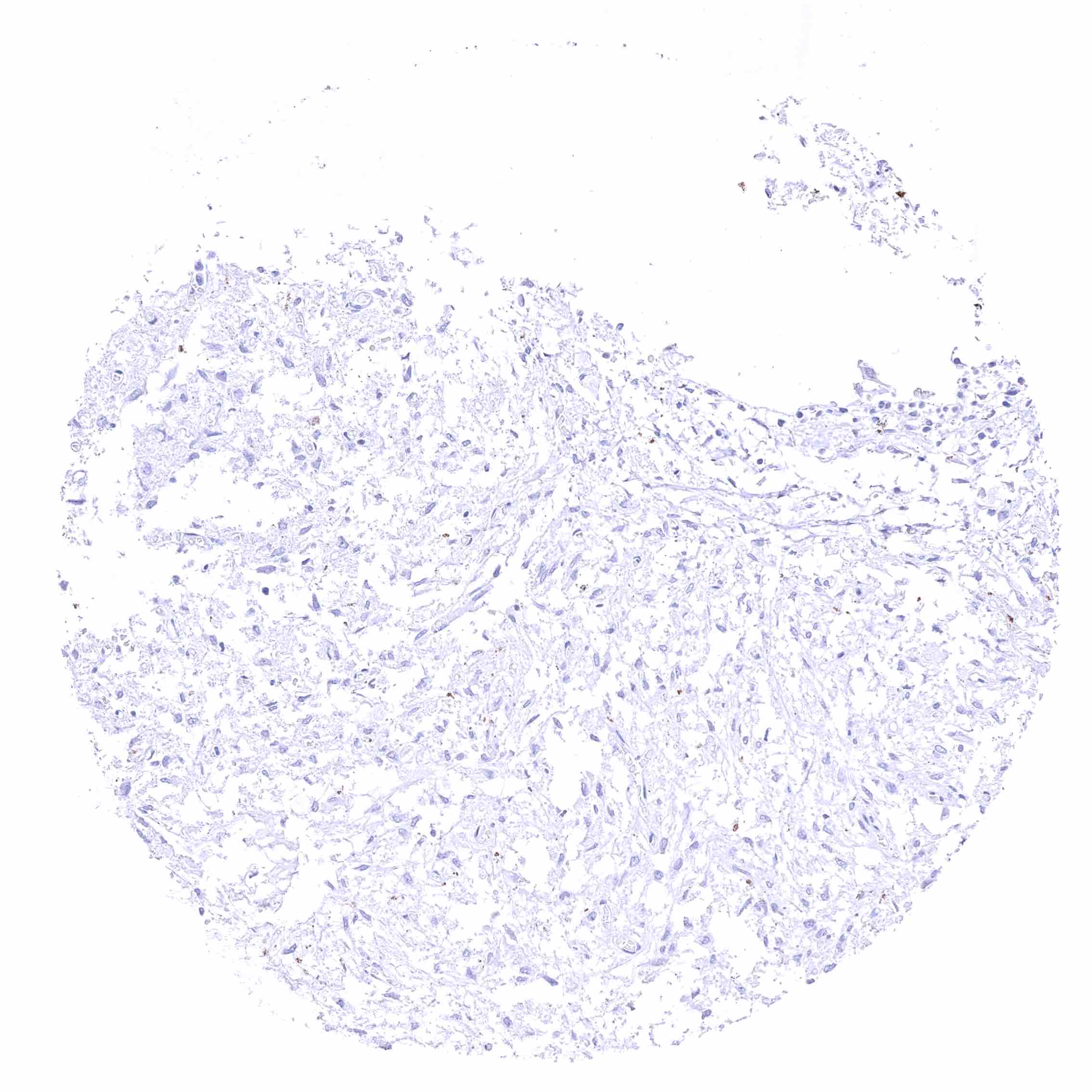
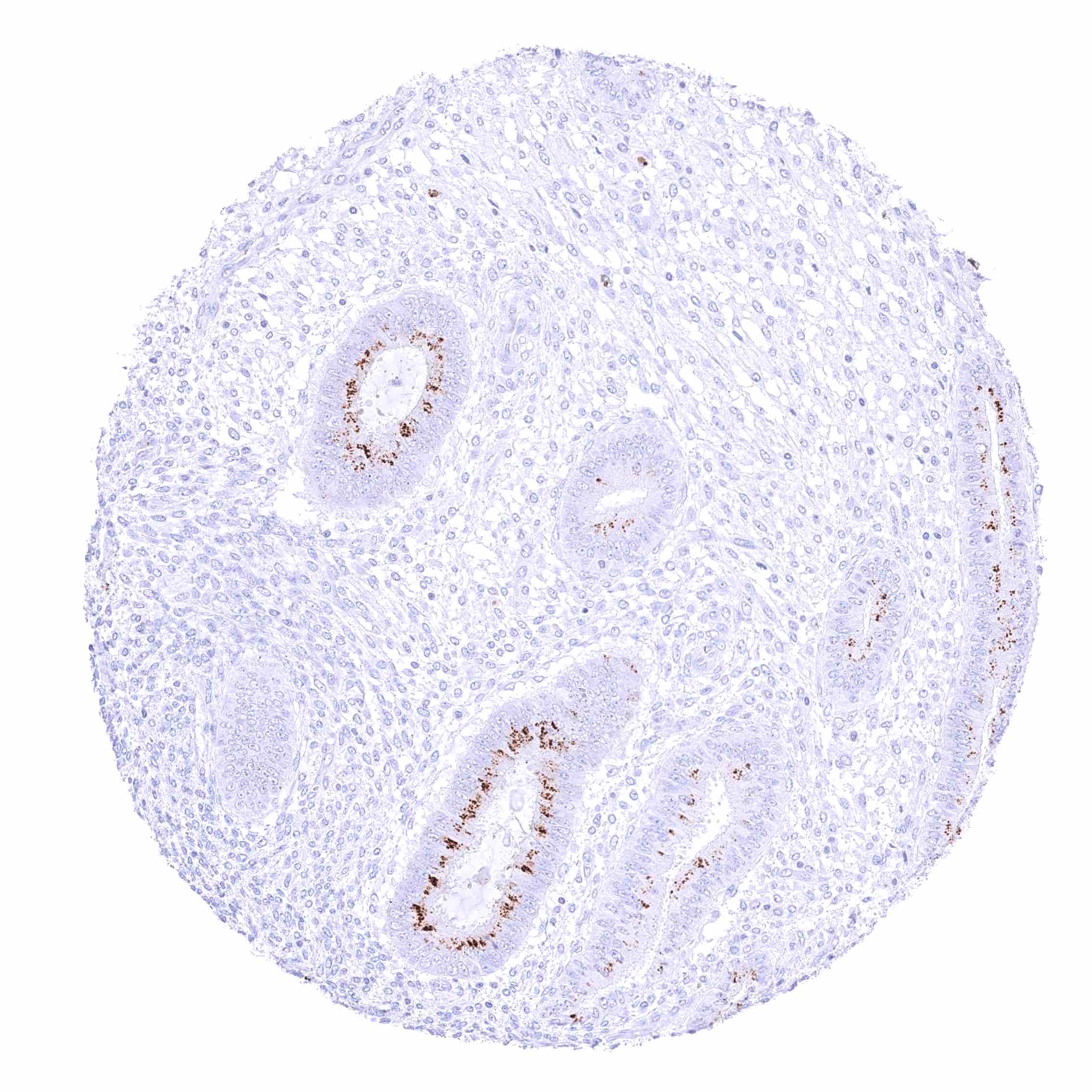
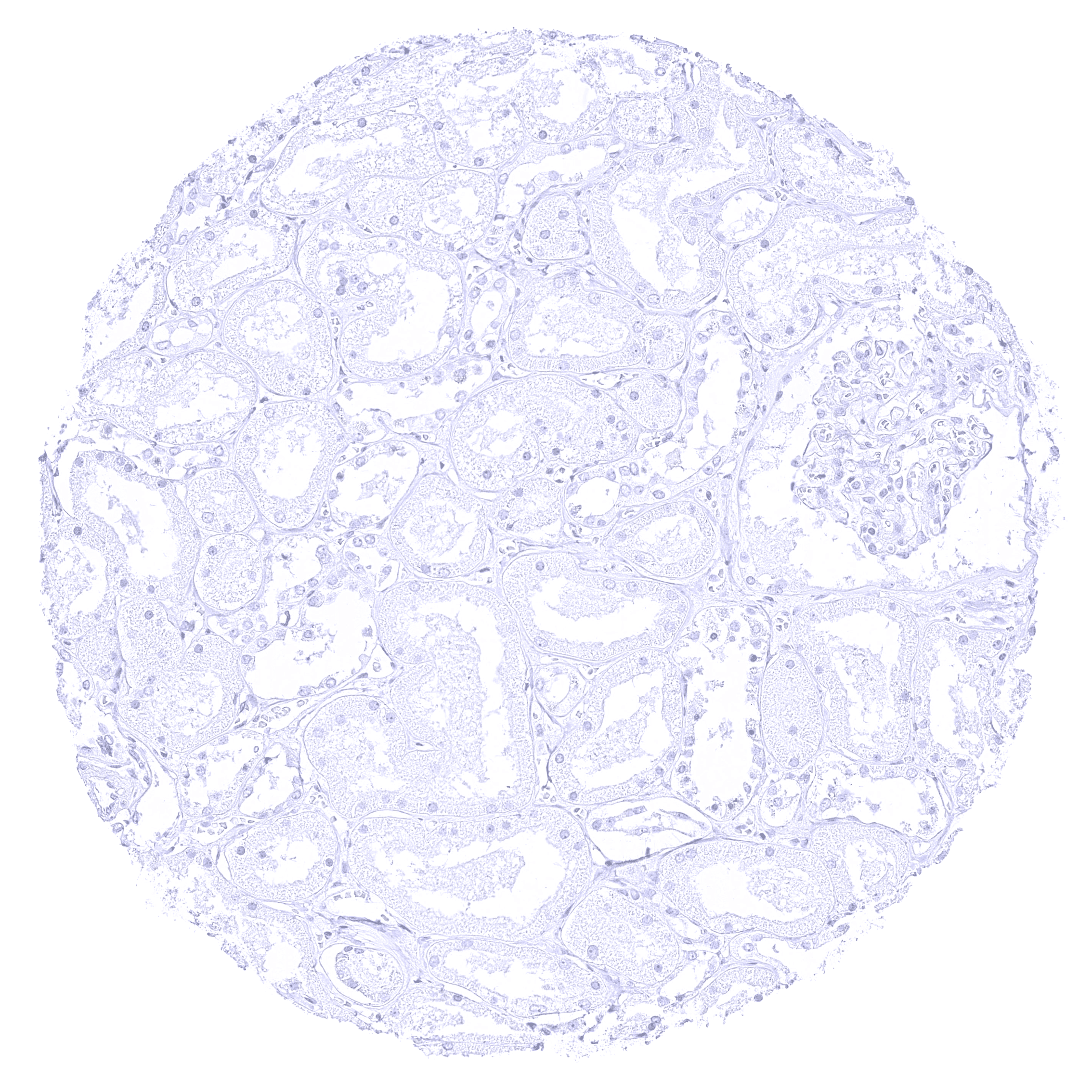
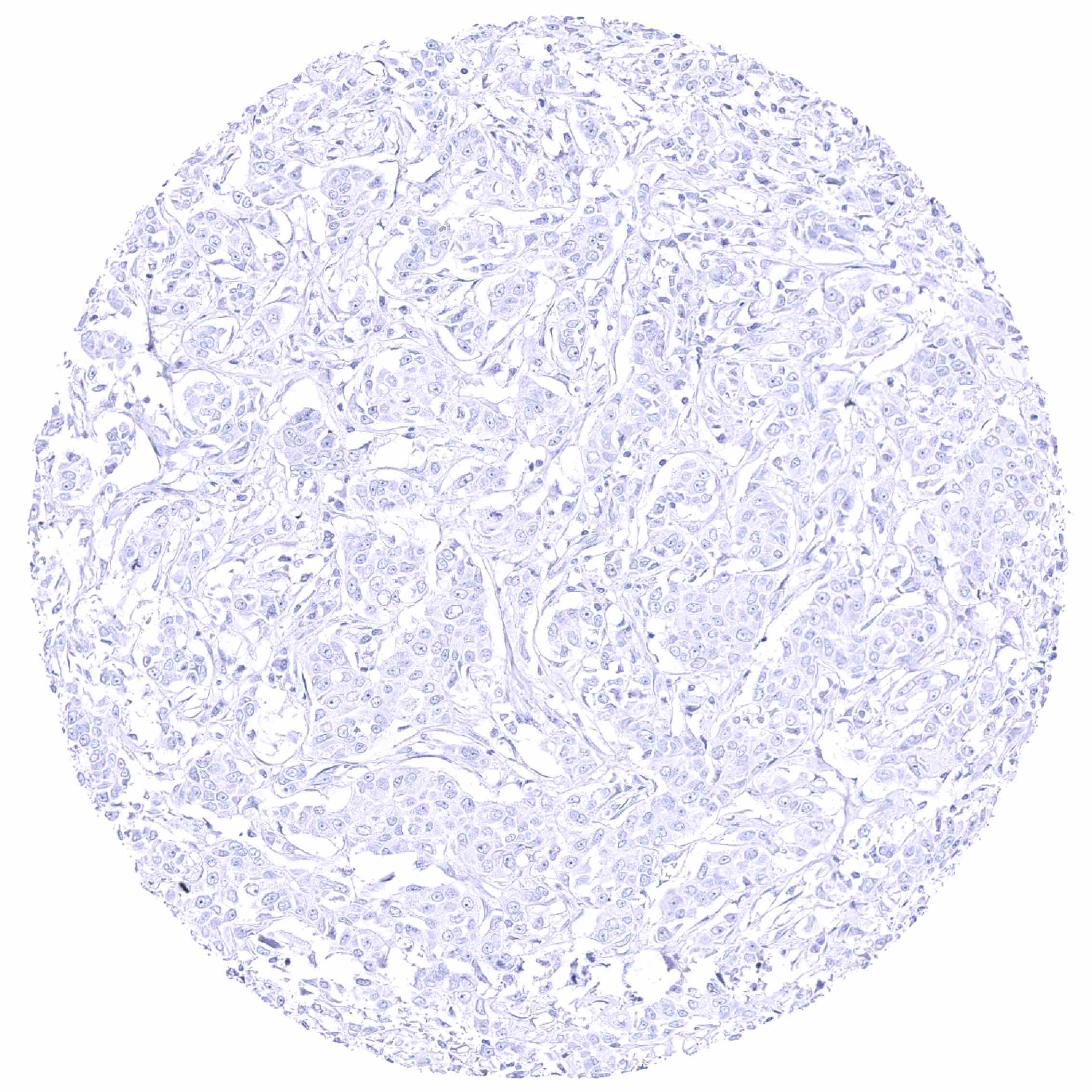
Negative control = Kidney: BSEP staining should be completely absent in all cells (note: other tissues can also be used as negative controls in case of BSEP).
Cellular localization = Membranous
Reactivity = Human
Application = Immunohistochemistry
Dilution = 1:100 – 1:200
Intended Use = Research Use Only
Relevance of Antibody
BSEP is a critical protein for exporting bile acids from hepatocytes.
Biology Behind
The Bile Salt Export Pump (BSEP) is a crucial transporter protein coded by the ABCB11 gene, located at chromosome 2q24.1, which plays an essential role in exporting bile acids from hepatocytes into the bile canaliculi. The BSEP protein features multiple transmembrane domains that span the hepatocyte membrane, facilitating substrate movement using ATP hydrolysis as an energy source. Mutations or dysfunction of BSEP are directly implicated in several cholestatic liver diseases. For example, Progressive Familial Intrahepatic Cholestasis type 2 (PFIC2) is an inherited disorder caused by biallelic mutations in ABCB11, leading to defective BSEP function, cholestasis, and progressive liver damage. Other inherited conditions include Benign Recurrent Intrahepatic Cholestasis (BRIC) and certain drug-induced cholestasis cases, where BSEP impairment results in intracellular bile acid accumulation, hepatocellular injury, and fibrosis. Given its liver-specific expression, BSEP is considered a potential diagnostic marker for the distinction of liver cancer from other tumor entities.
Staining Pattern in Normal Tissues
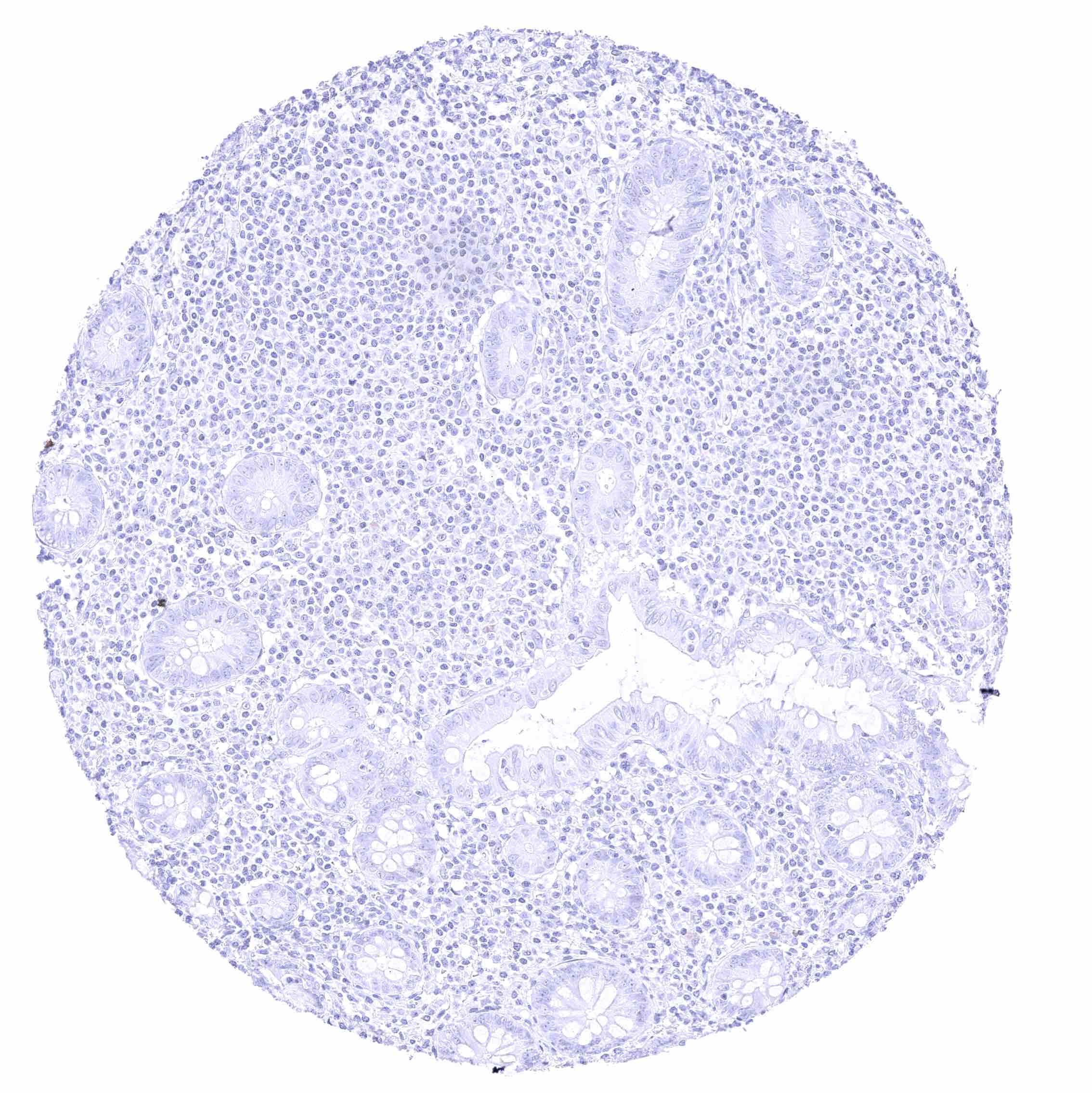
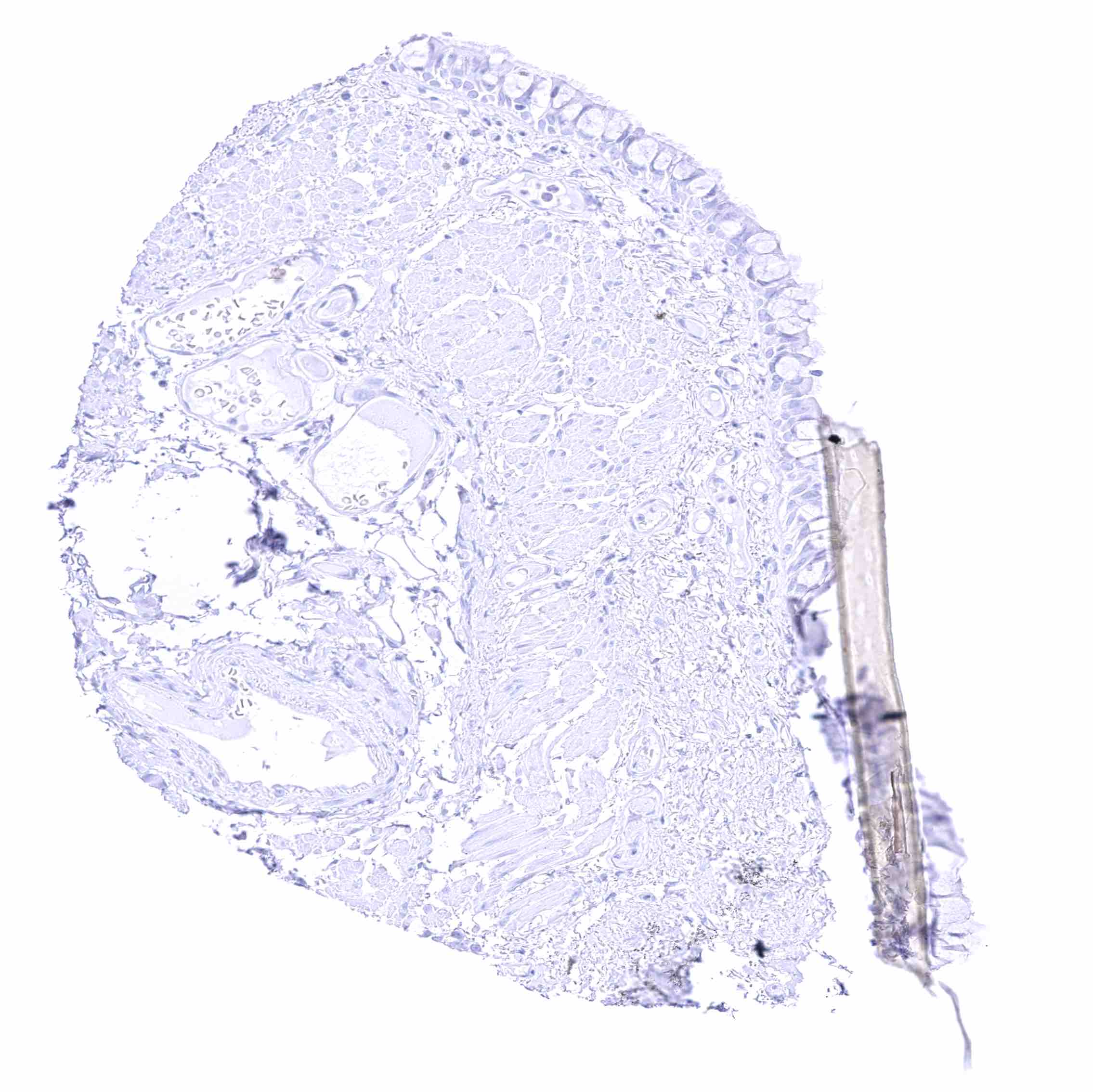
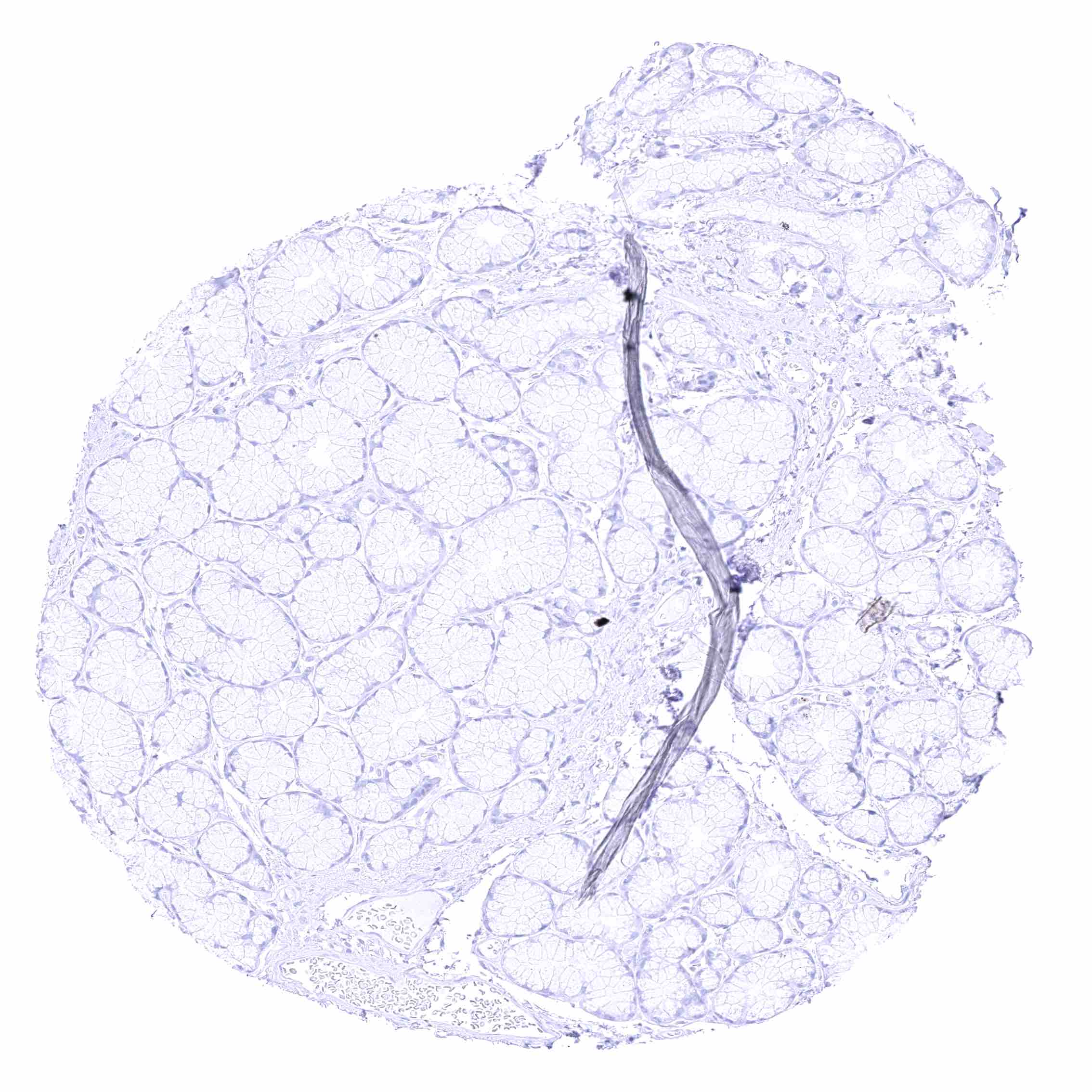
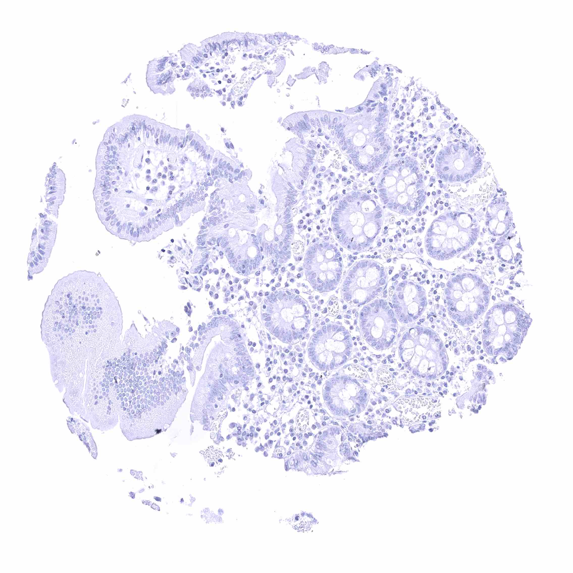
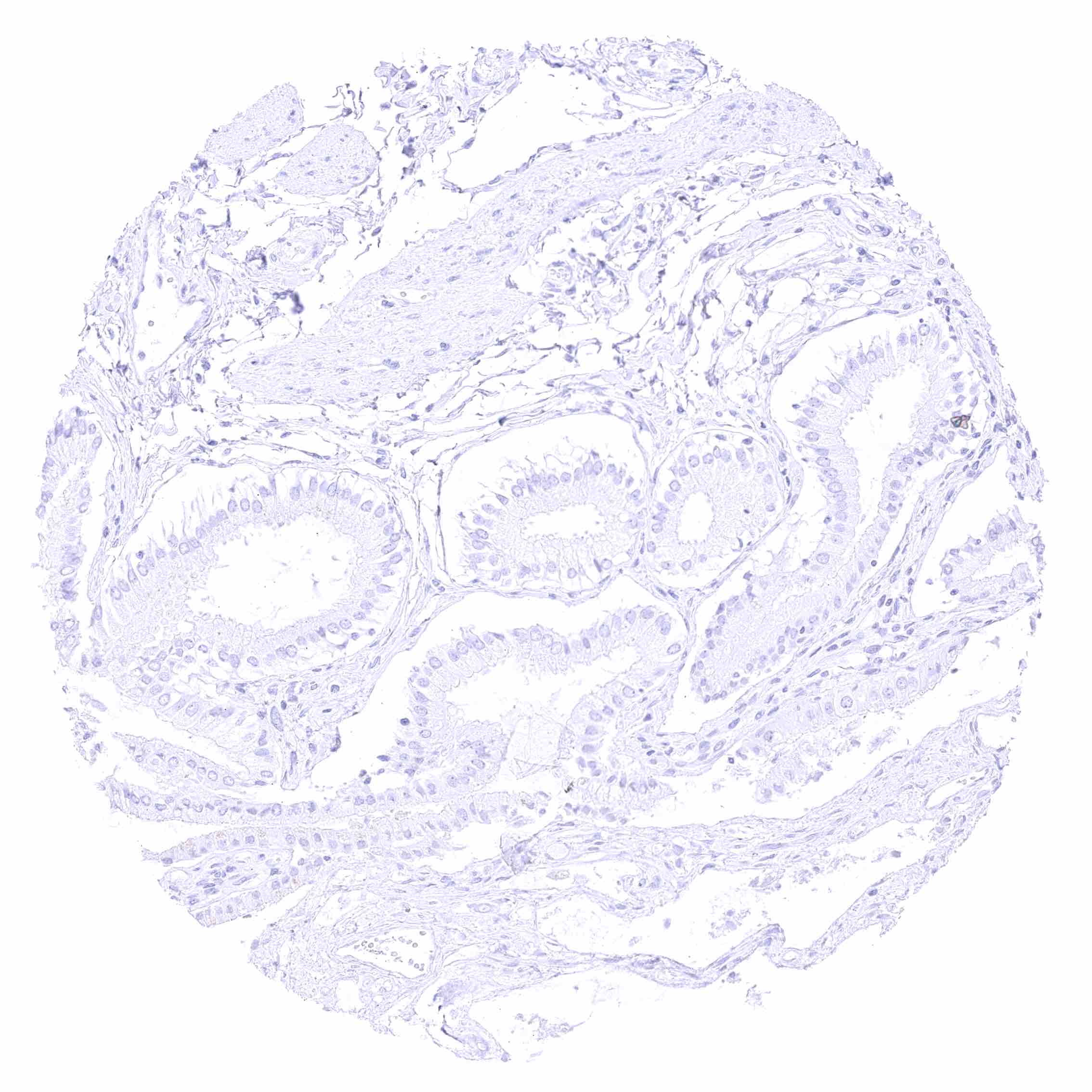
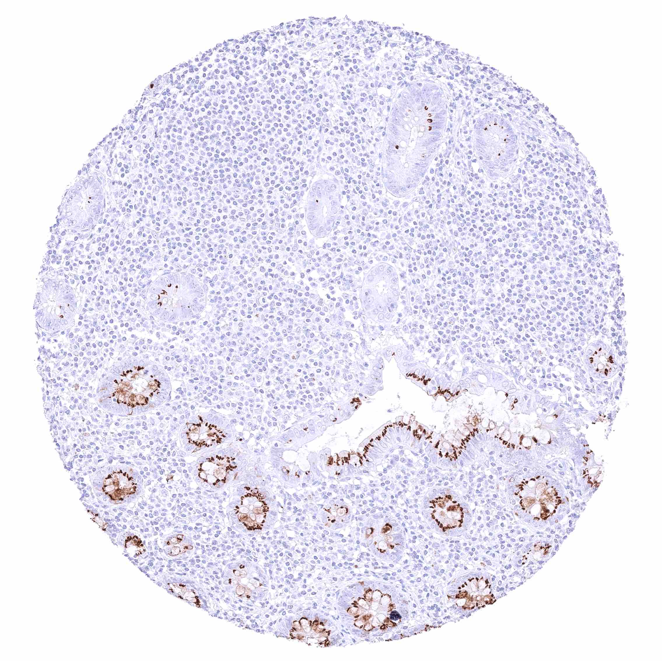
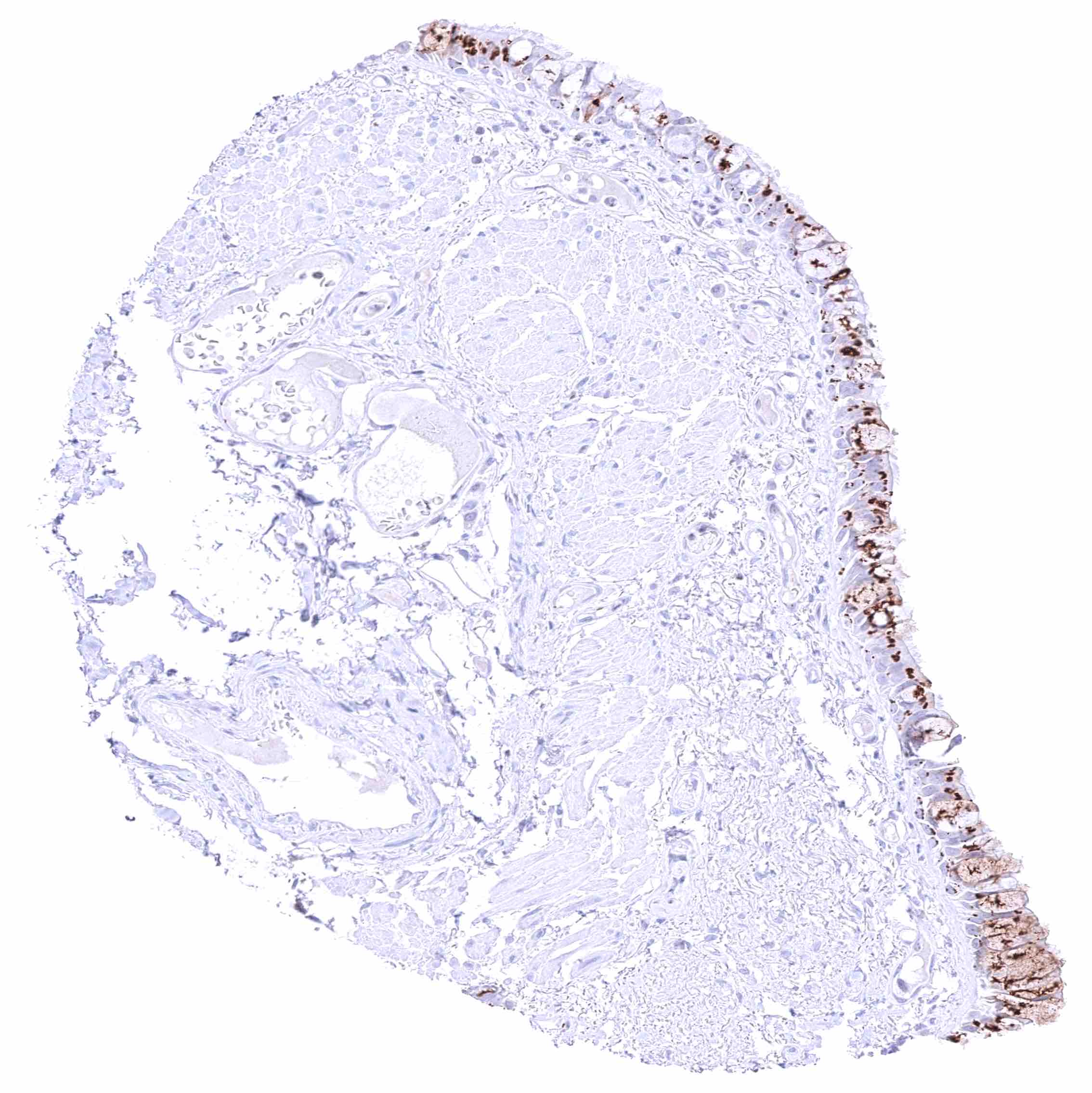
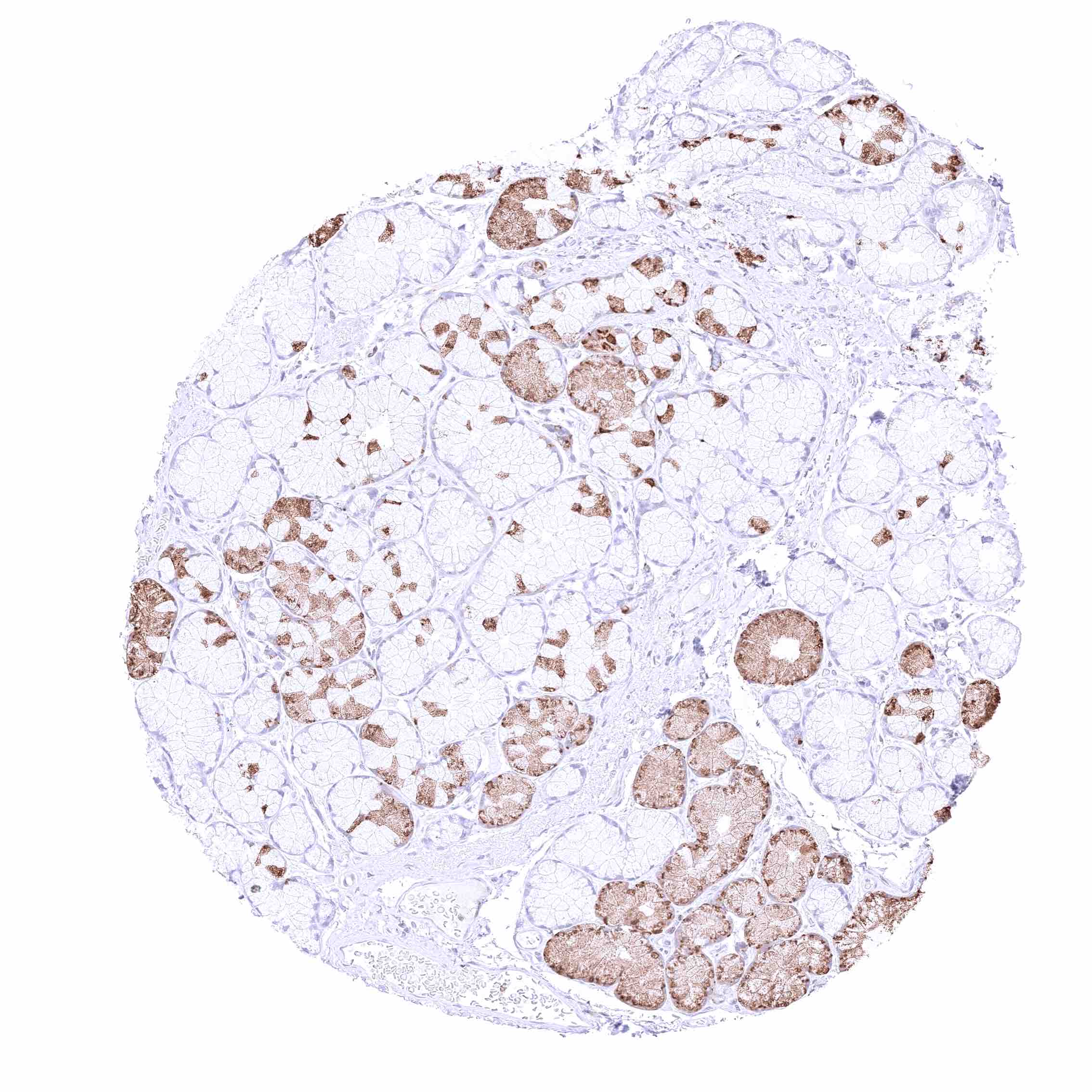
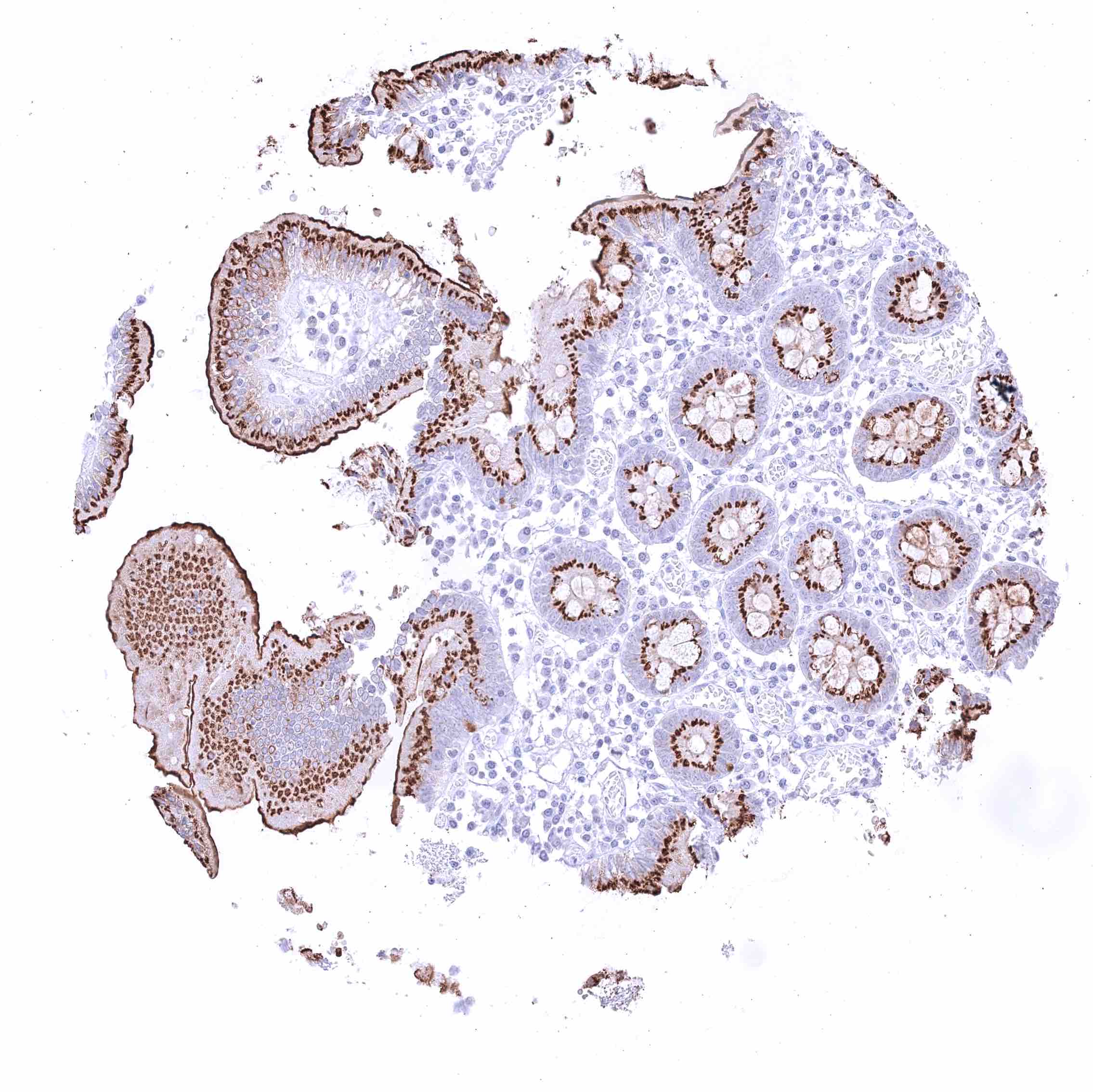
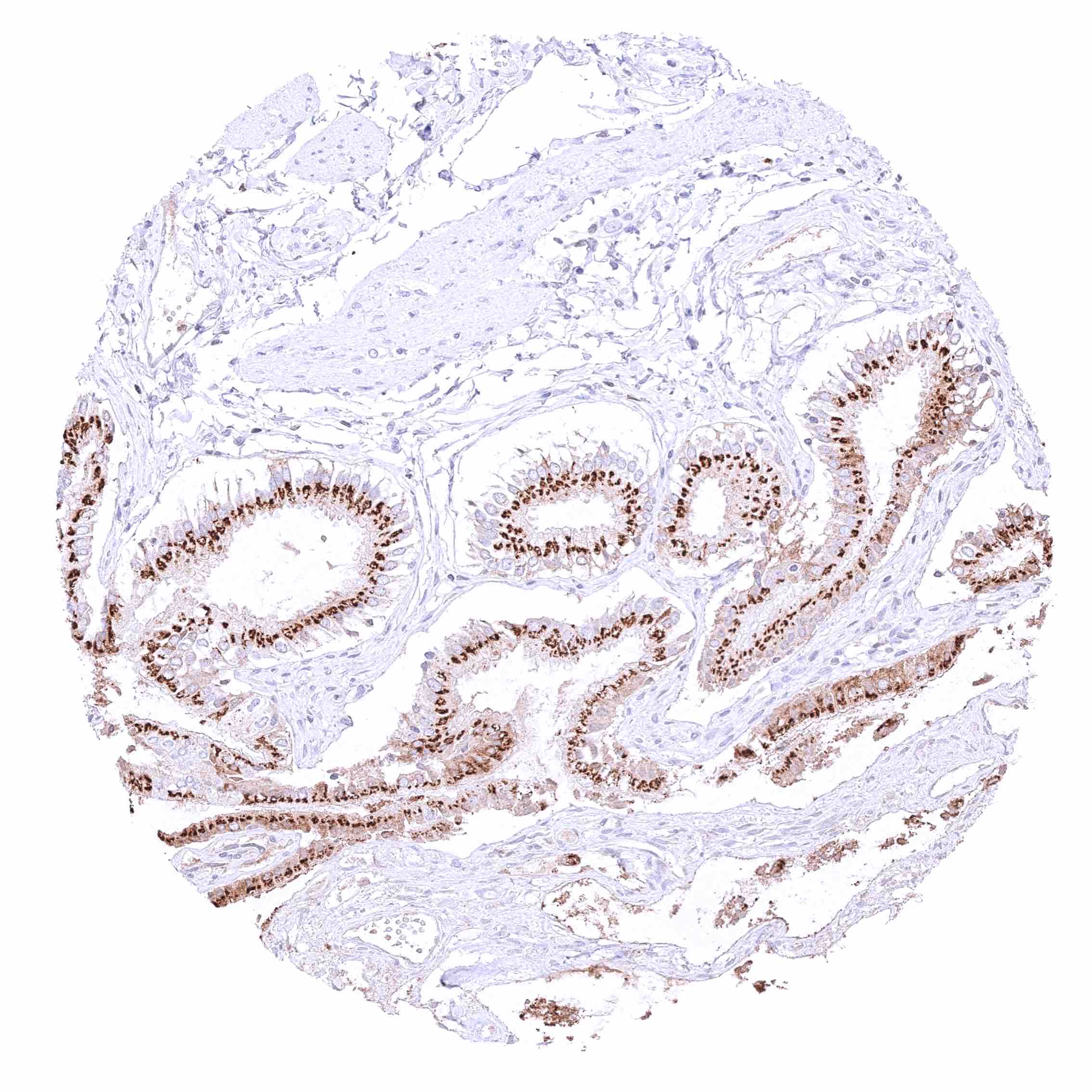
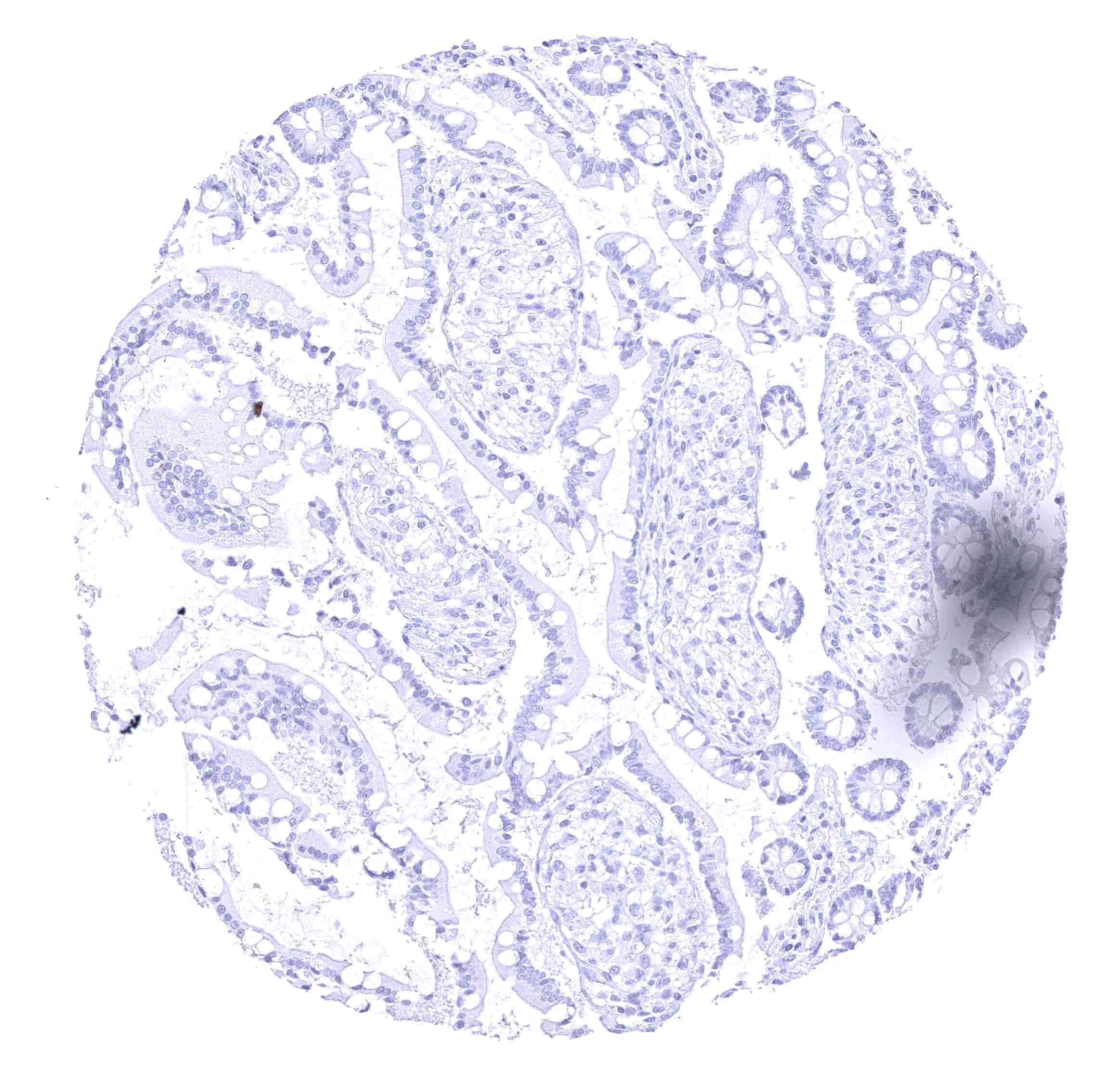
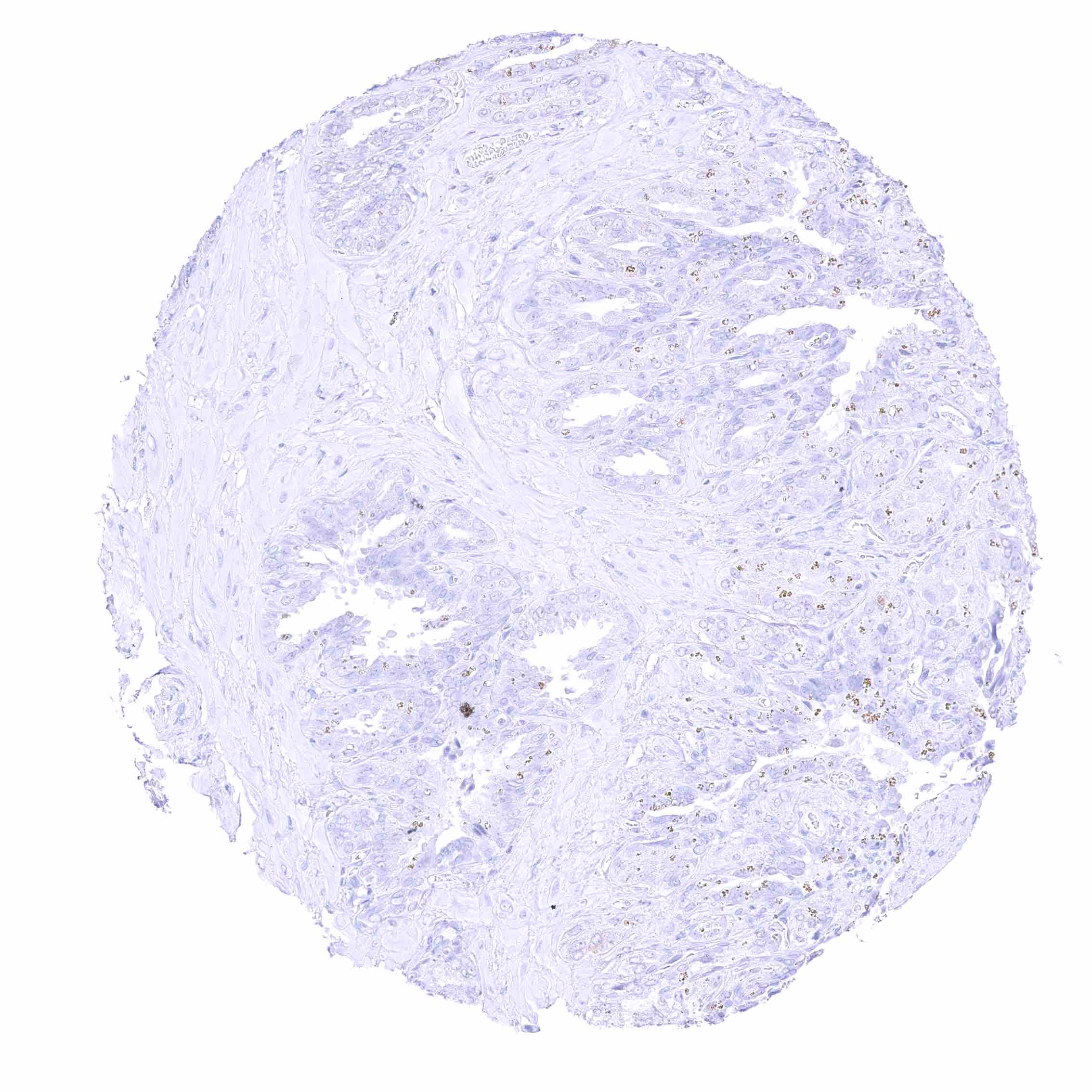
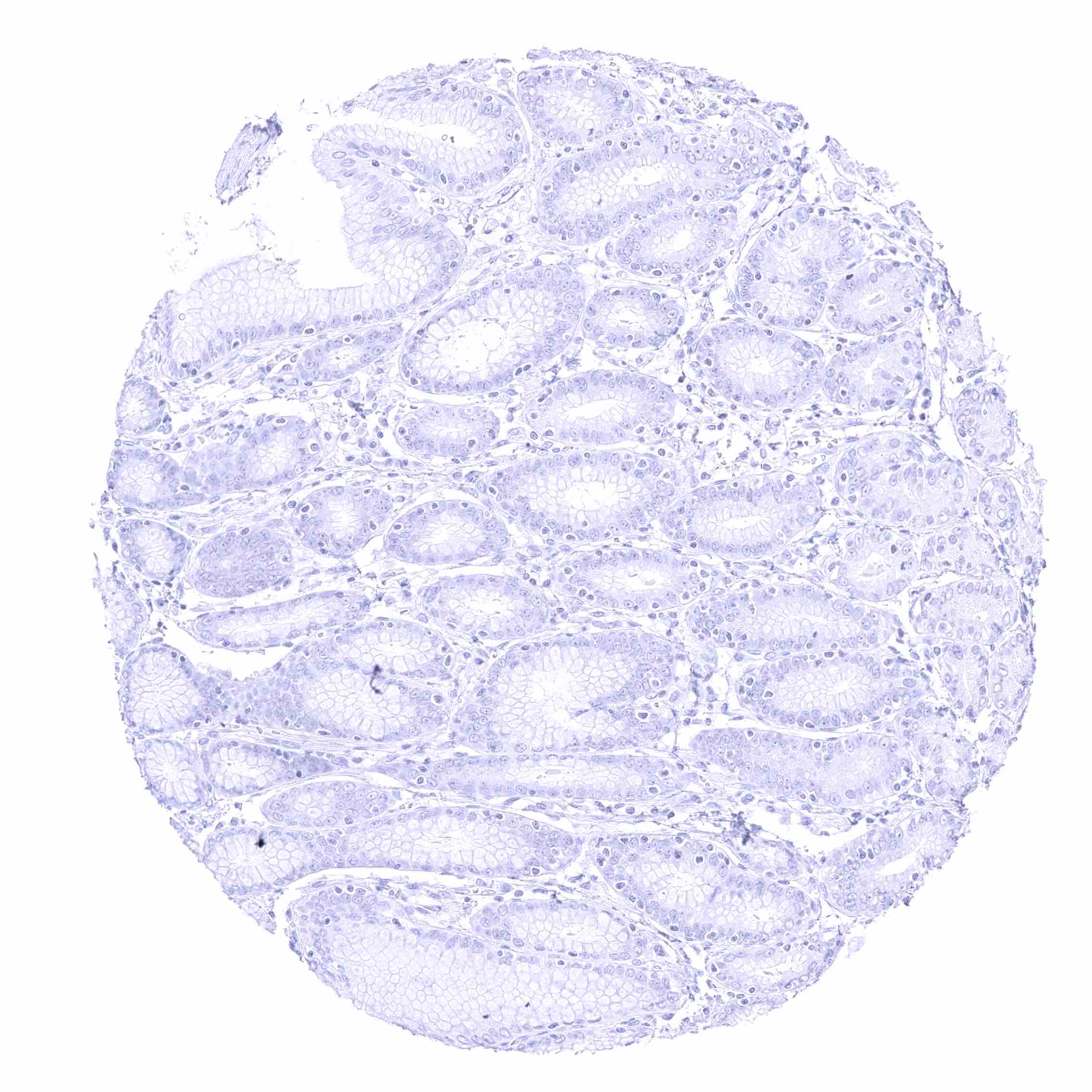
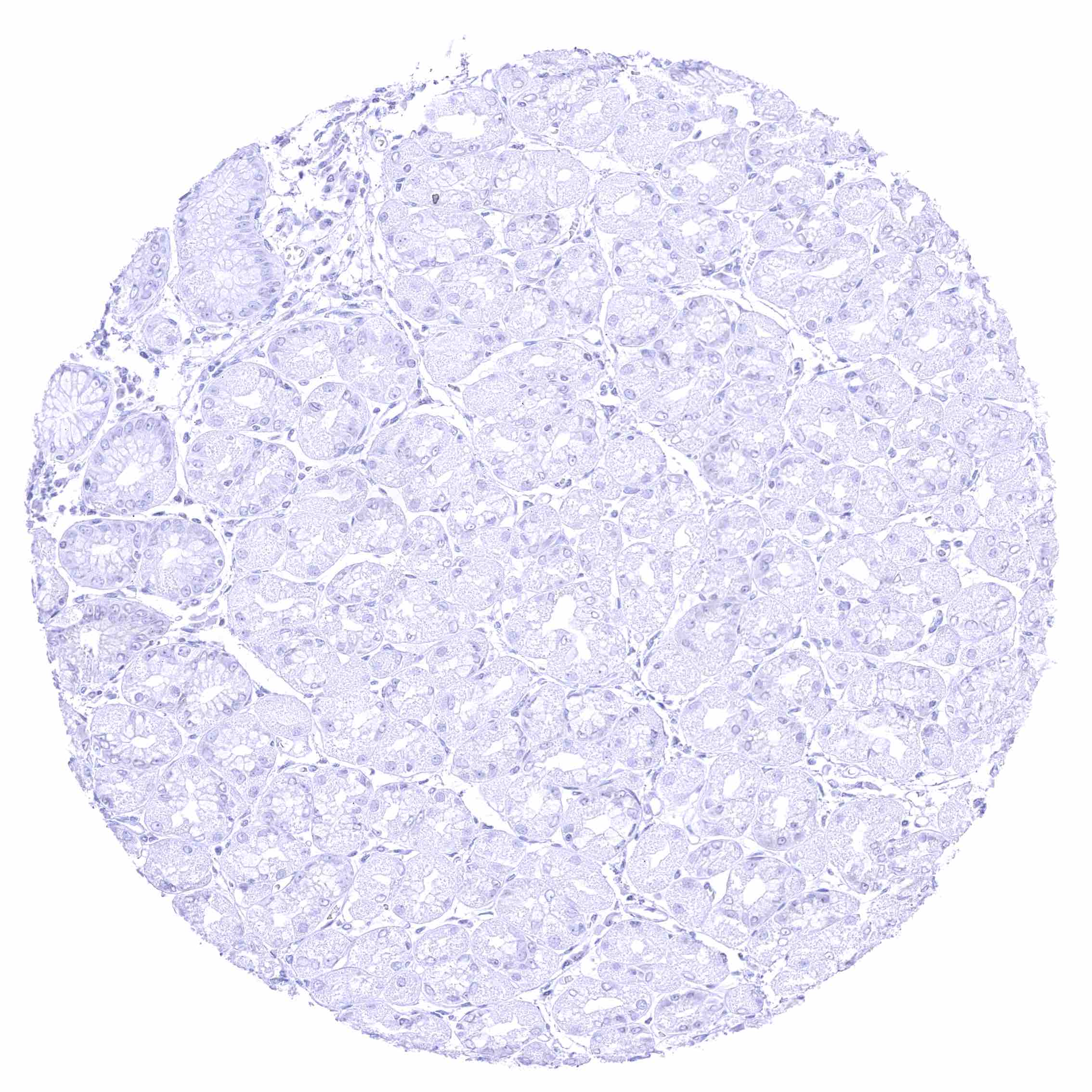
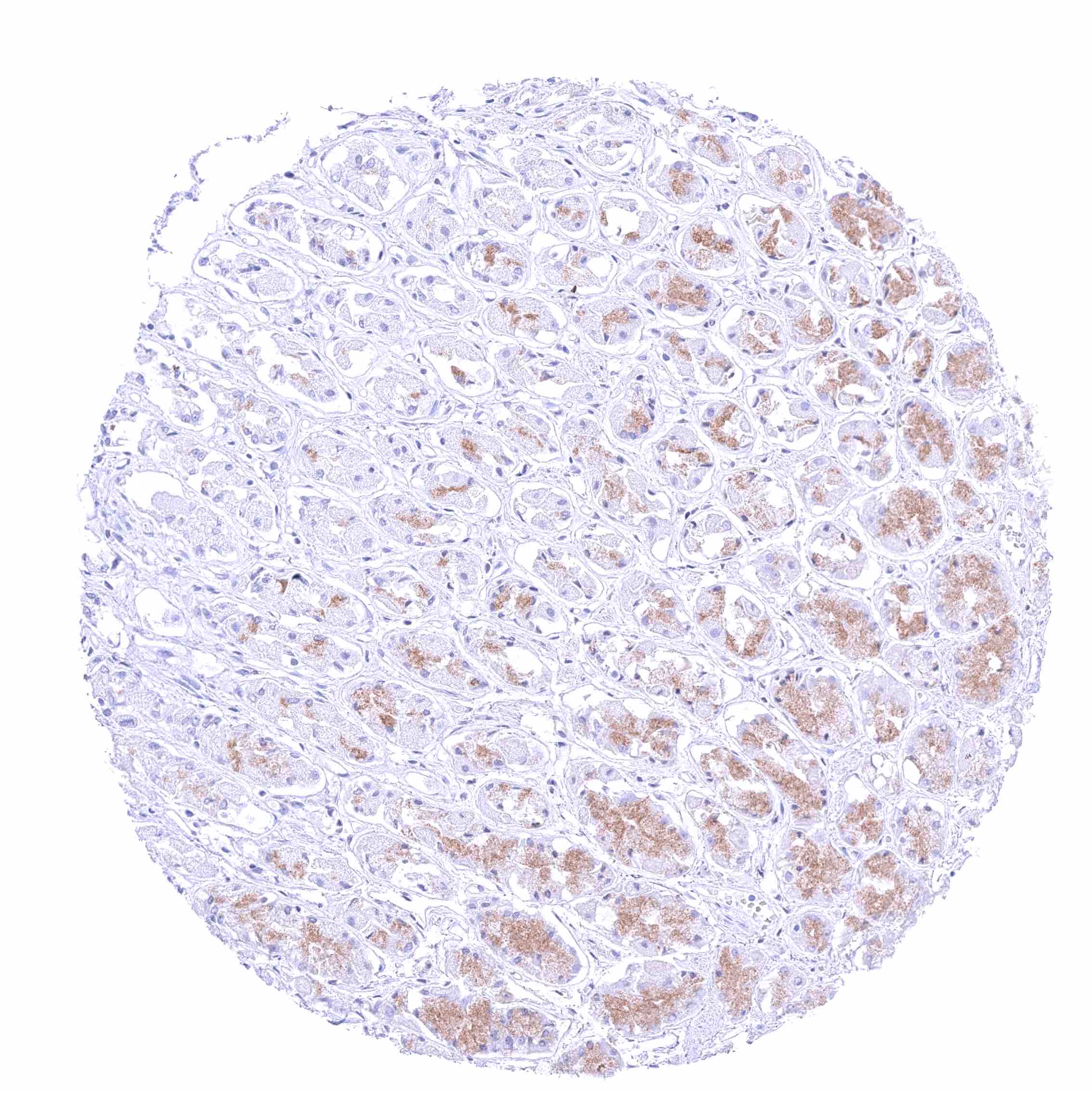
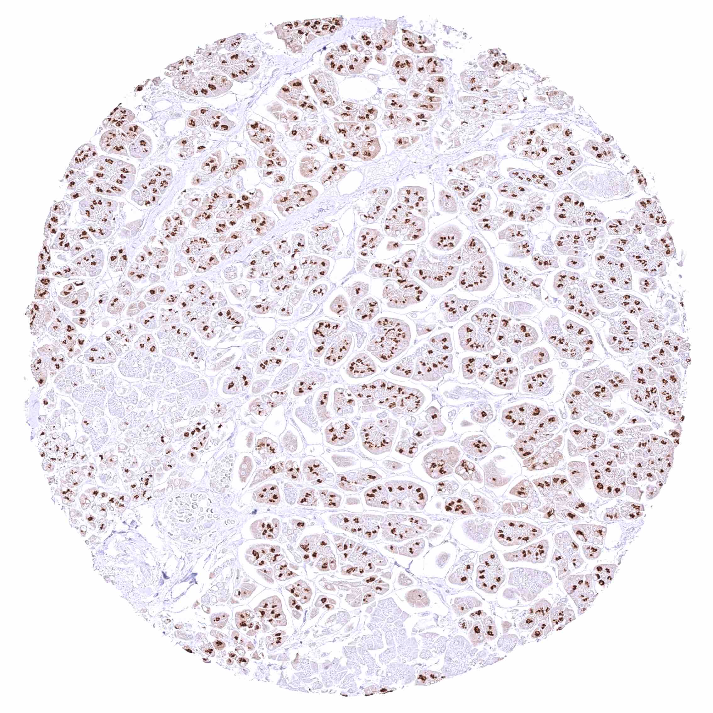
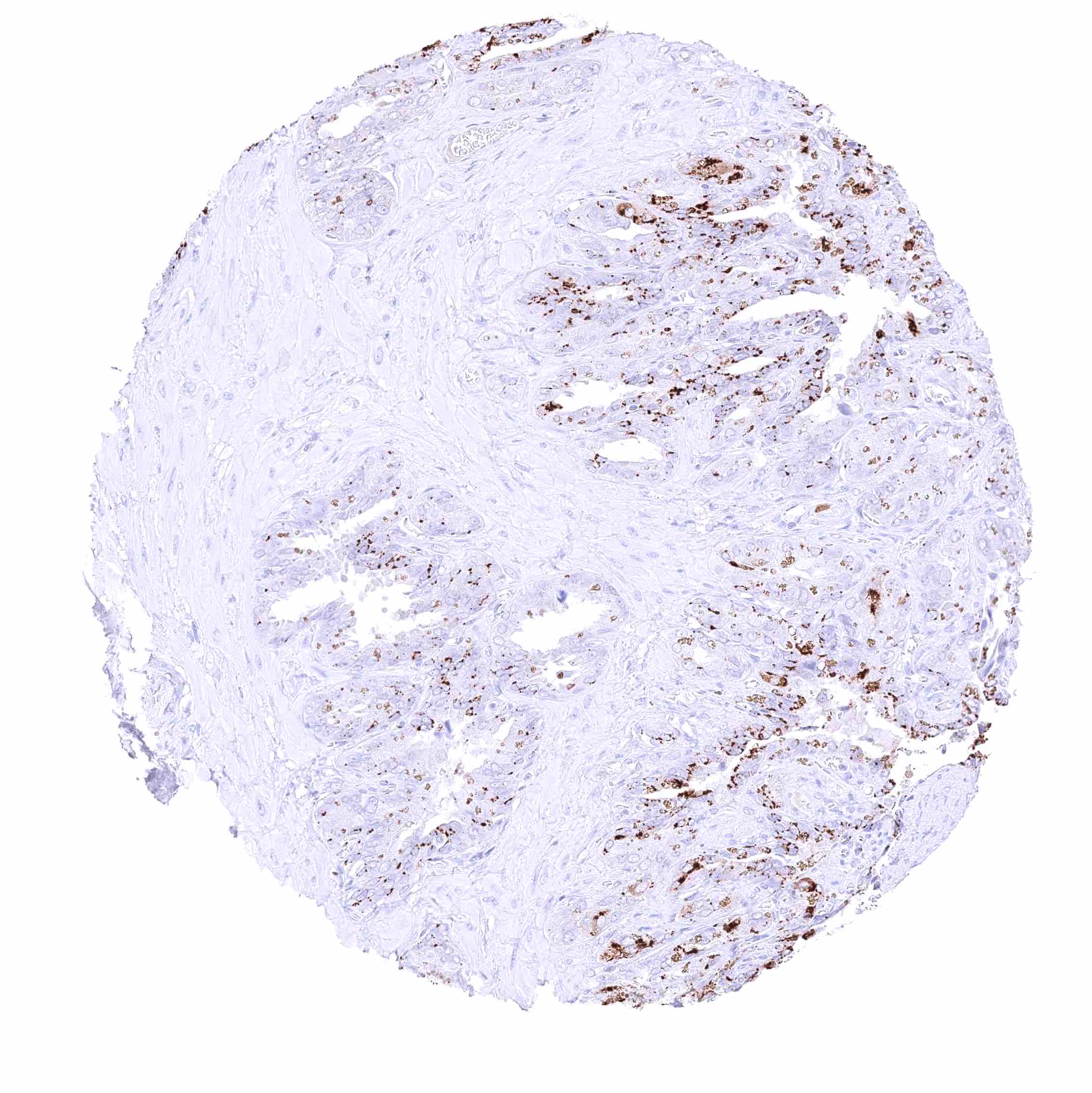
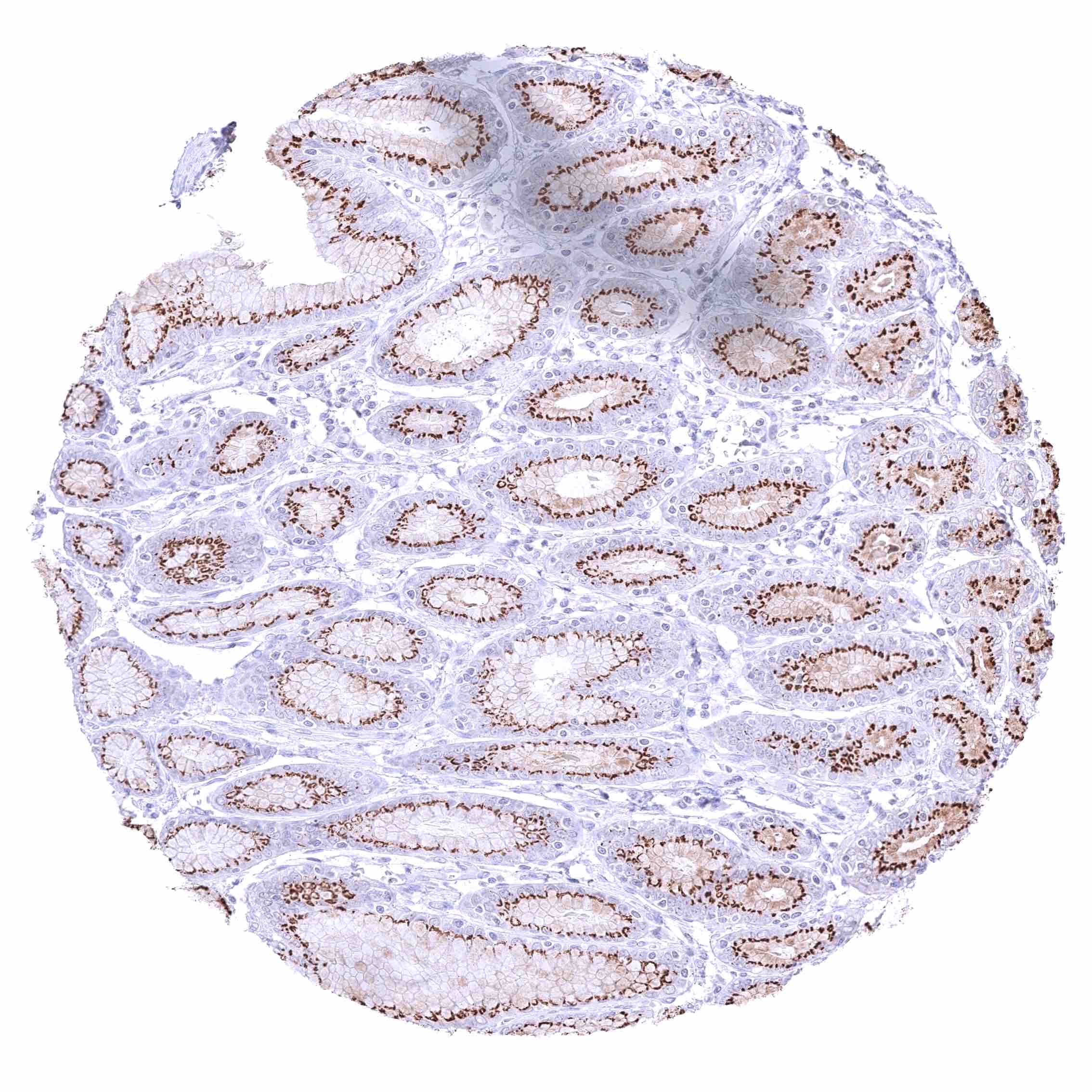
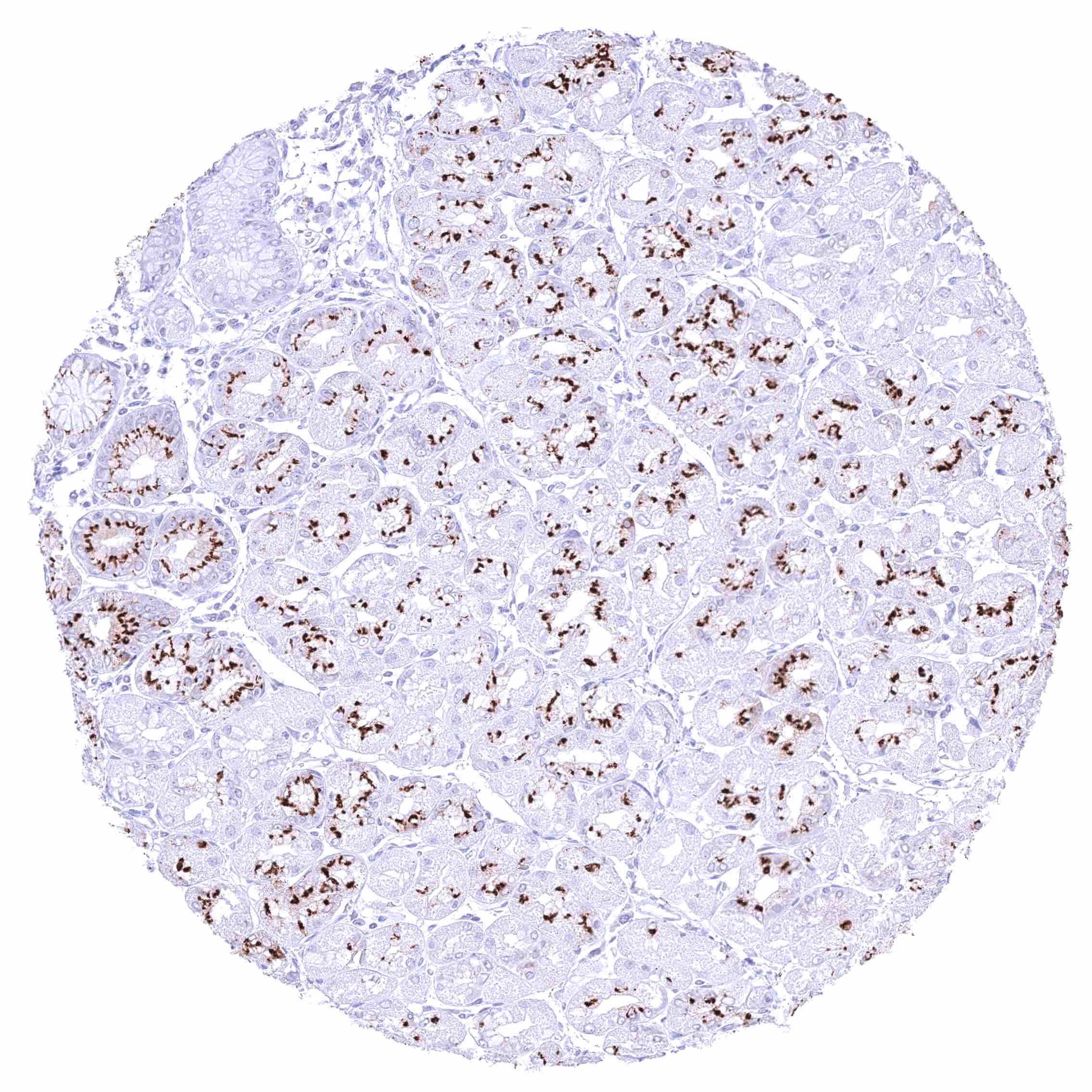
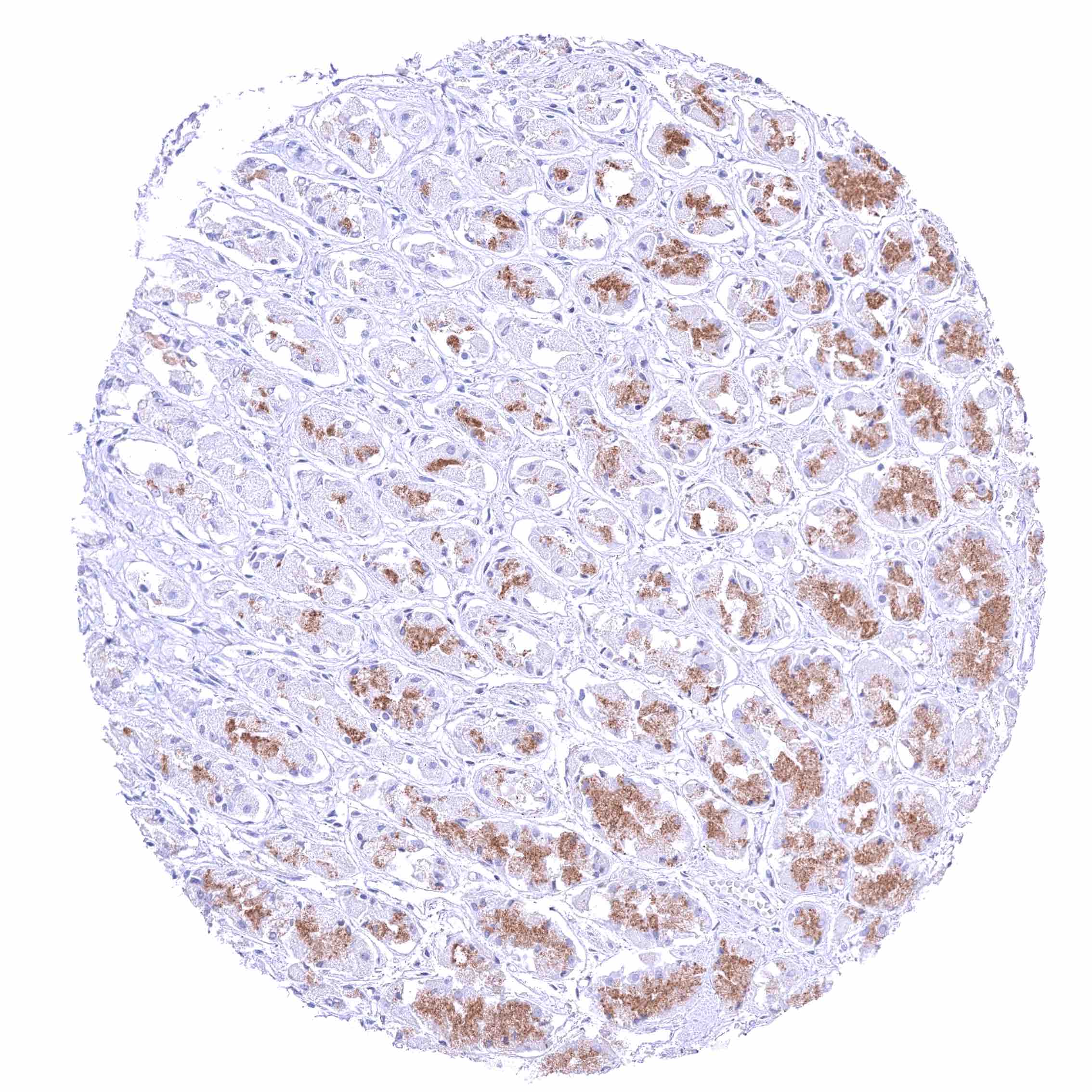
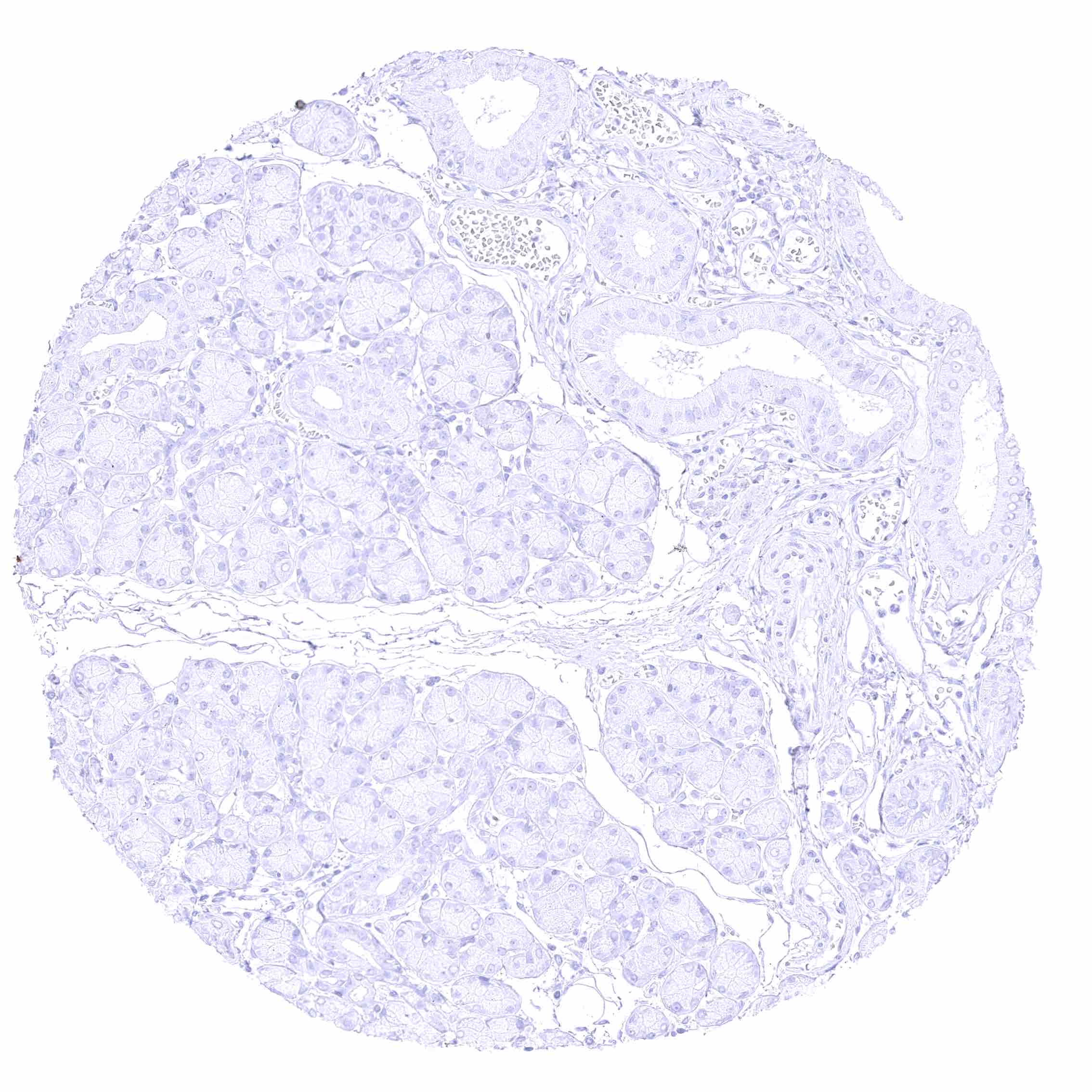
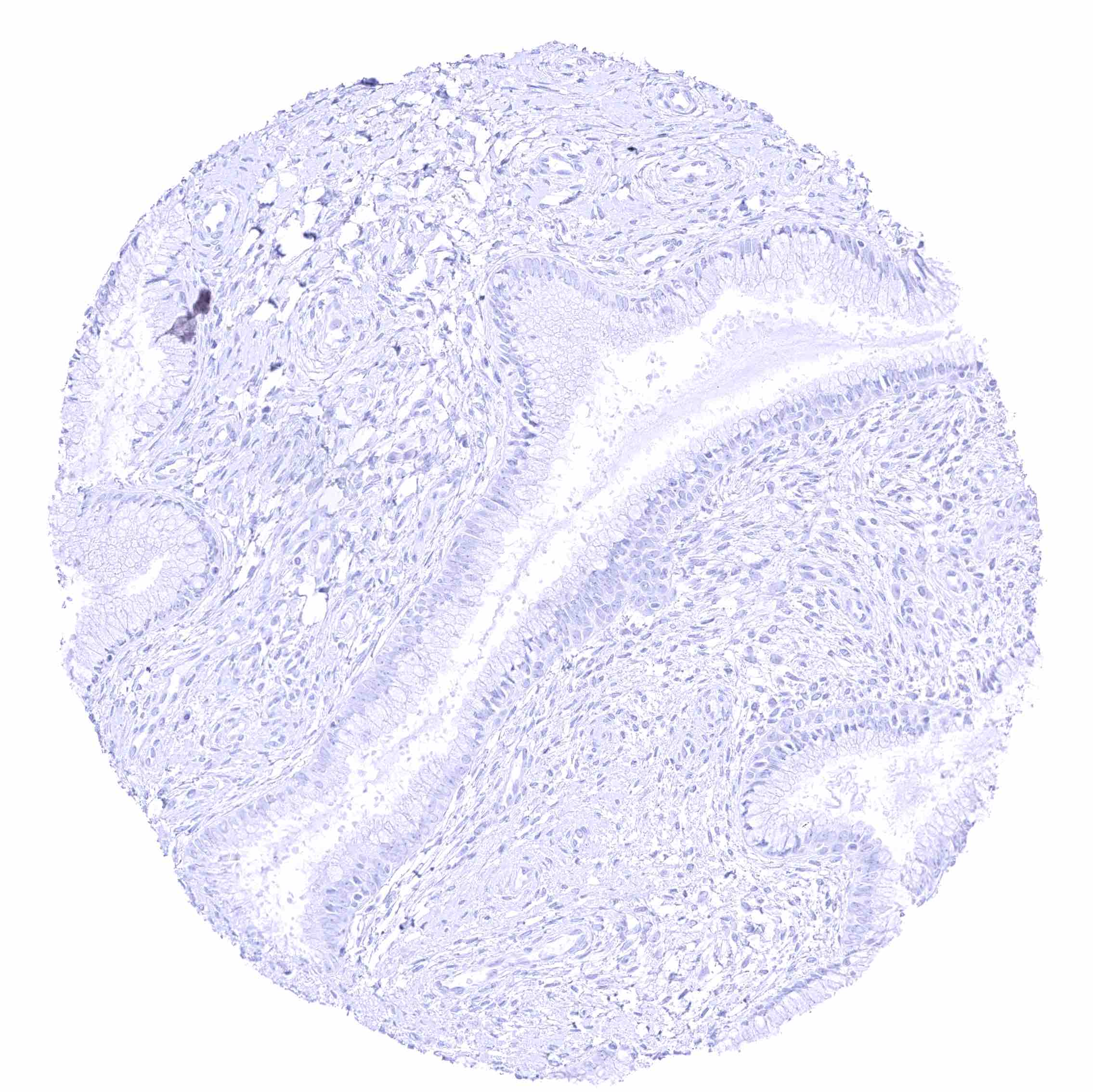
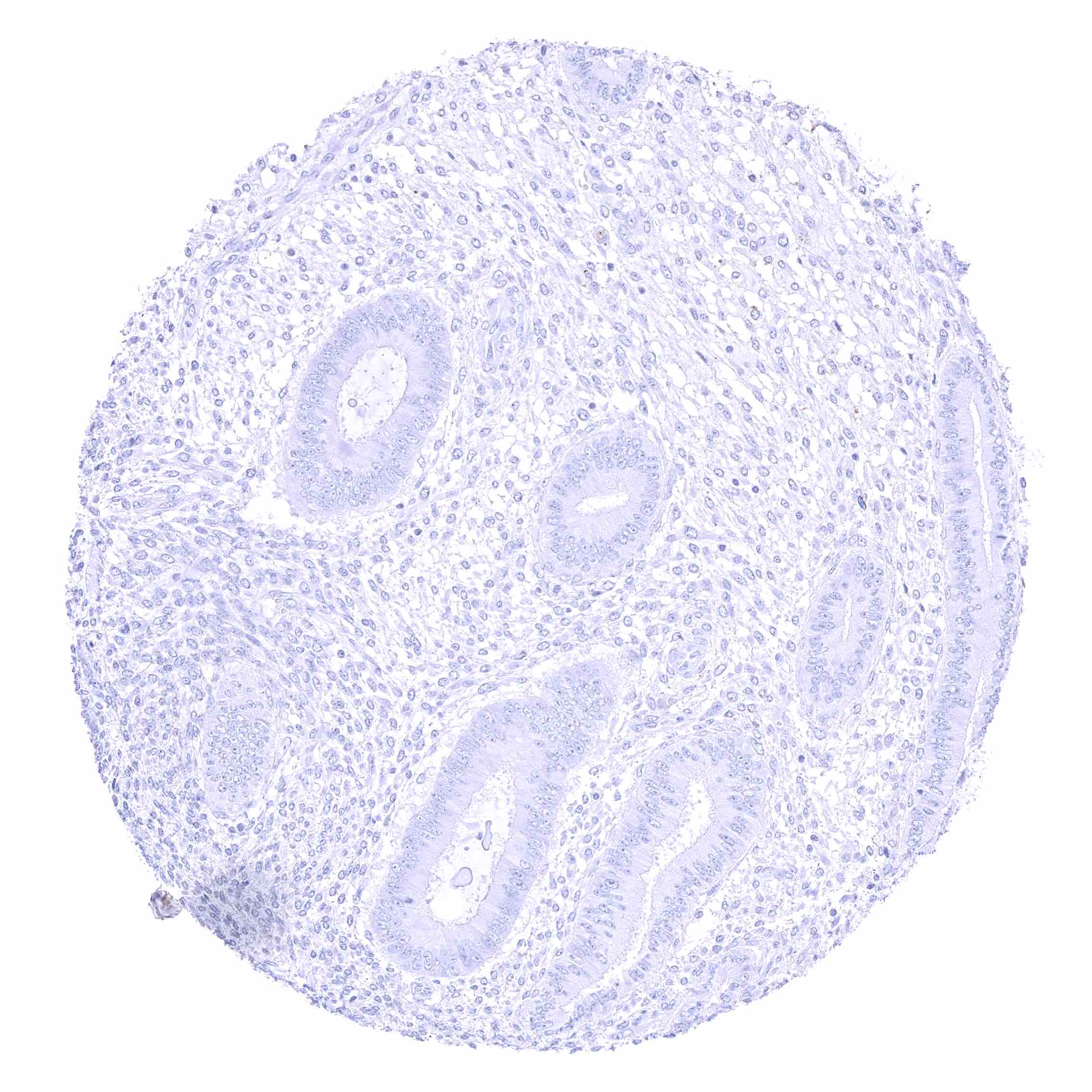
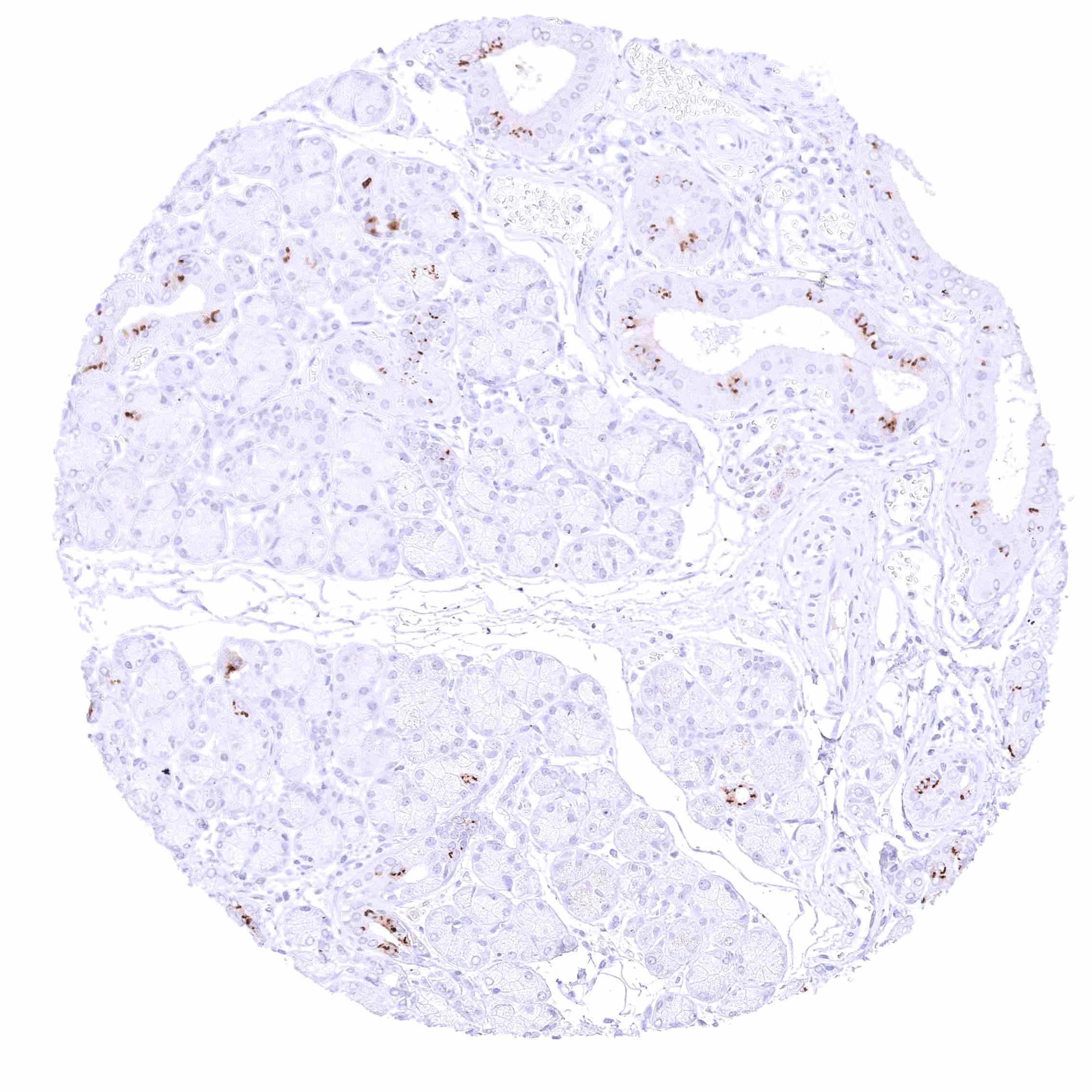
BSEP expression is limited to hepatocytes in the liver. Images describing the BSEP staining pattern in normal tissues obtained by the antibody HMV4715 are shown in our “Normal Tissue Gallery”.
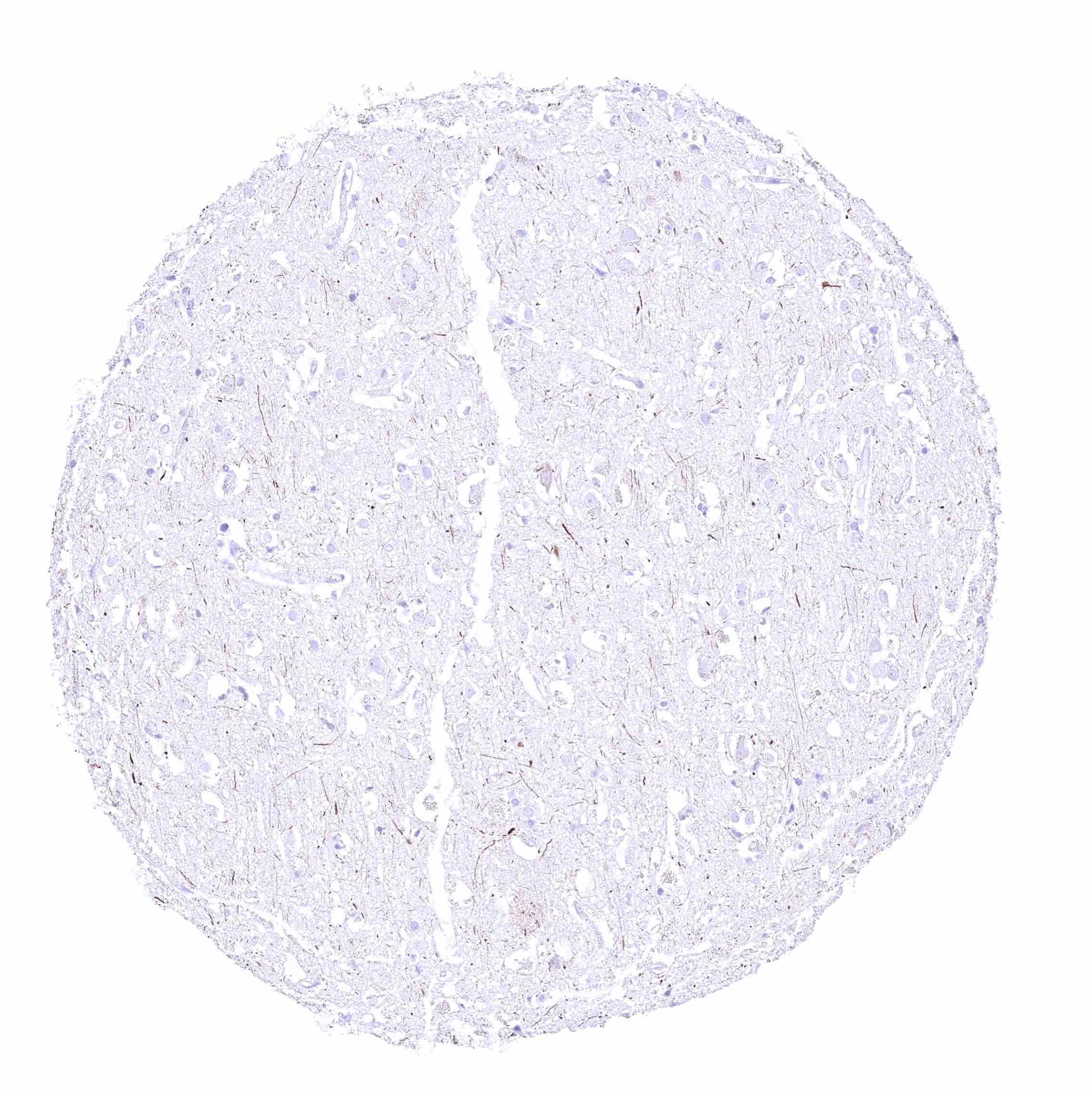
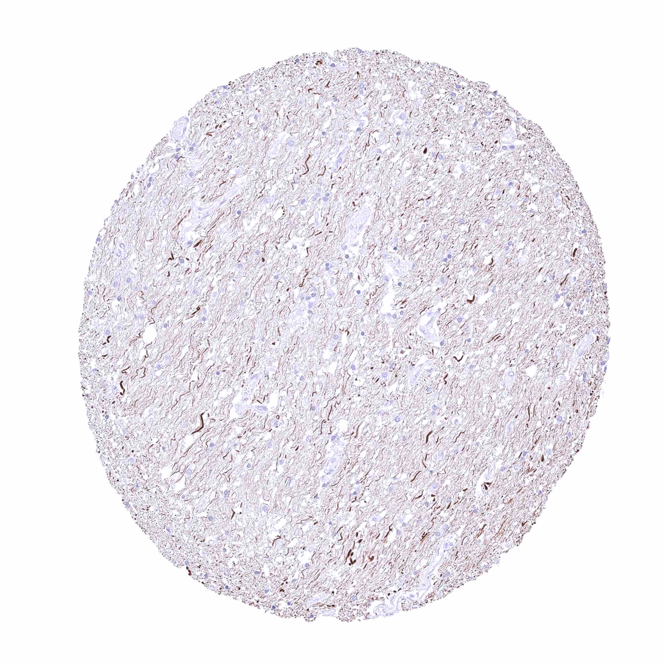
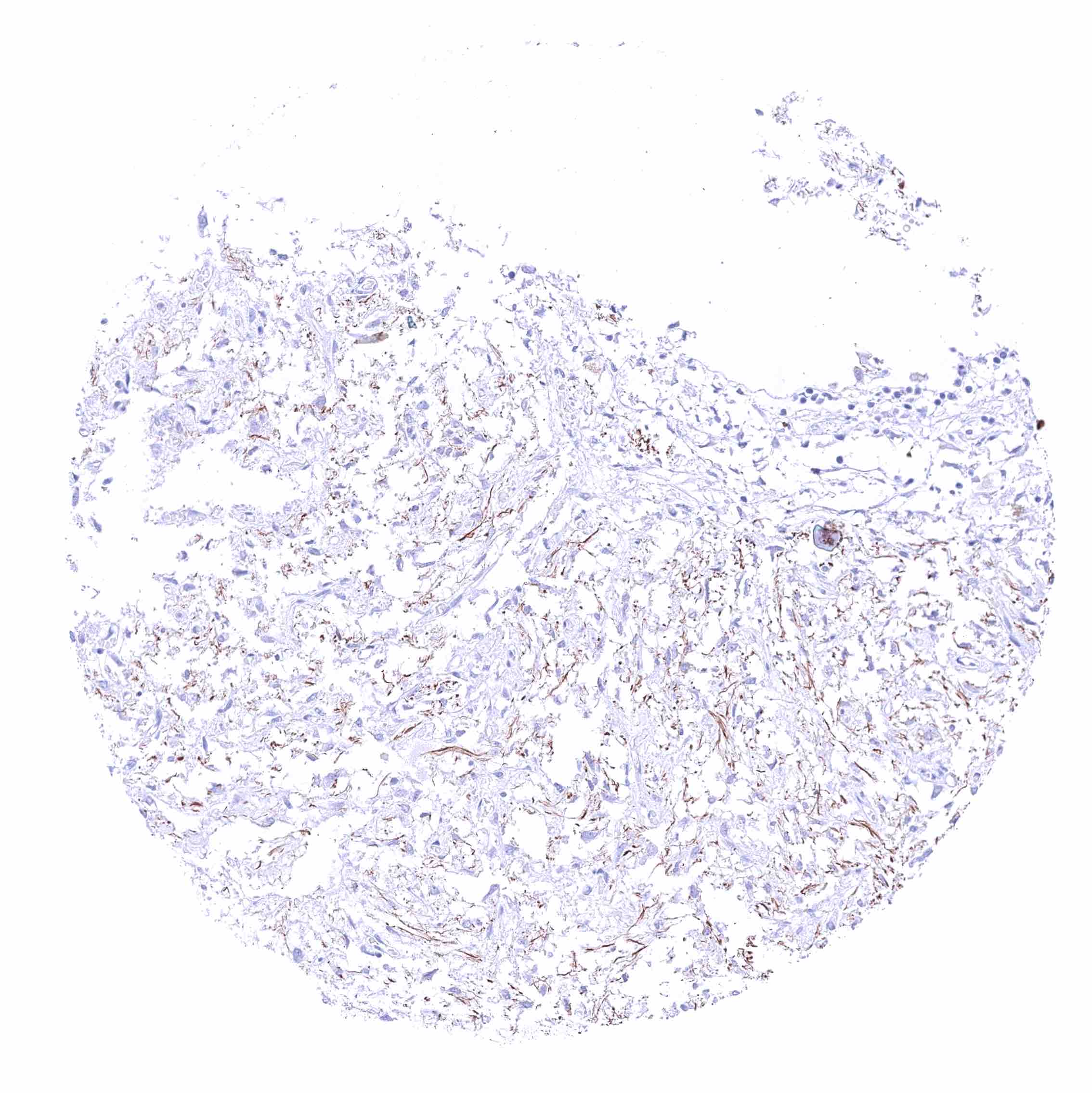
| Brain | Cerebrum | A fibrillar staining occurs in the white matter. This represents a tolerable cross-reactivity of the antibody. |
| Cerebellum | A fibrillar staining occurs in the white matter. This represents a tolerable cross-reactivity of the antibody. | |
| Endocrine Tissues | Thyroid | Negative. |
| Parathyroid | Negative. | |
| Adrenal gland | Negative. | |
| Pituitary gland | A fibrillar staining occurs in the neurohypophysis. This represents a tolerable cross-reactivity of the antibody. | |
| Respiratory system | Respiratory epithelium | Negative. |
| Lung | Negative. | |
| Gastrointestinal Tract | Salivary glands | Negative. |
| Esophagus | Negative. | |
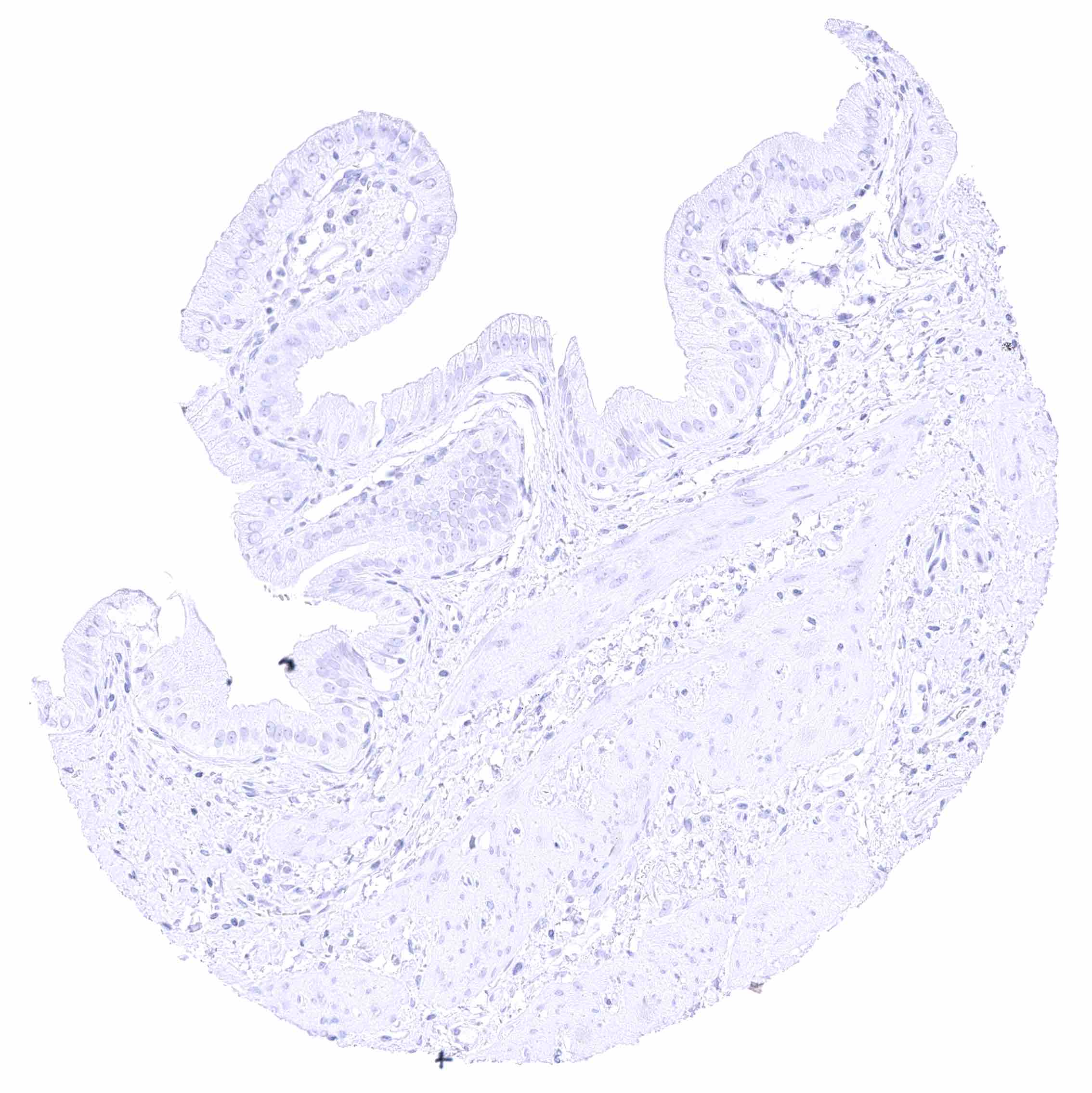
| Stomach | Negative. | |
| Duodenum | Negative. | |
| Small intestine | Negative. | |
| Appendix | Negative. | |
| Colon | Negative. | |
| Rectum | Negative. | |
| Liver | Strong membranous staining at the apical/canalicular („bile-secreting“) pole of hepatocytes. There is a slight zonal variability of the staining intensity. | |
| Gallbladder | Negative. | |
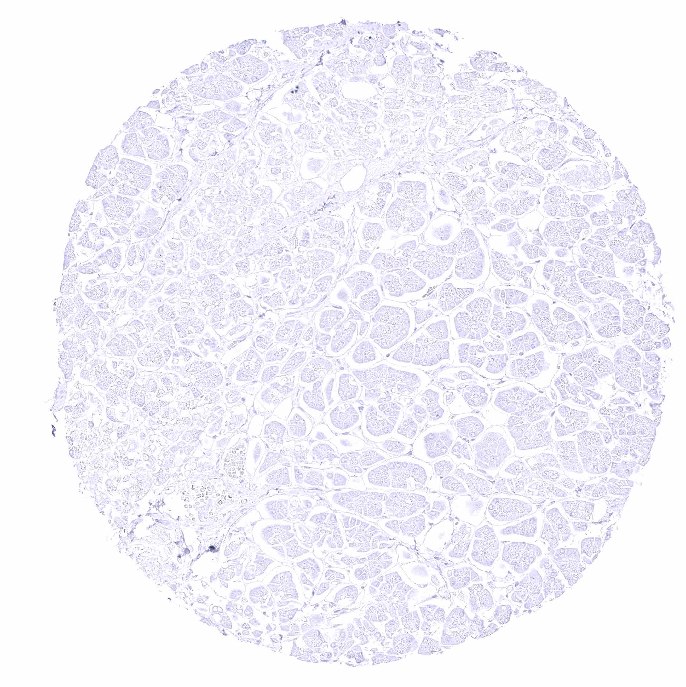
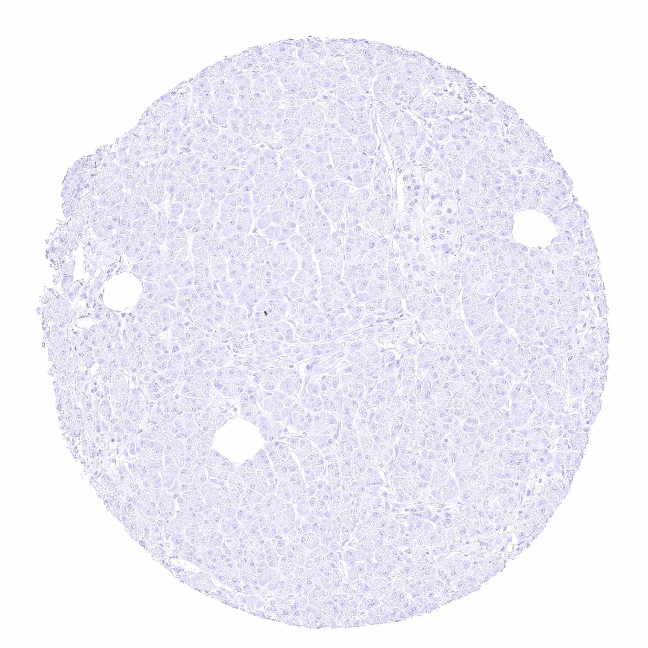
| Pancreas | Negative. | |
| Genitourinary | Kidney | Negative. |
| Urothelium | Negative. | |
| Male genital | Prostate | Negative. |
| Seminal vesicles | Negative. | |
| Testis | Negative. | |
| Epididymis | Negative. | |
| Female genital | Breast | Negative. |
| Uterus, myometrium | Negative. | |
| Uterus, ectocervix | Negative. | |
| Uterus endocervix | Negative. | |
| Uterus, endometrium | Negative. | |
| Fallopian Tube | Negative. | |
| Ovary | Negative. | |
| Placenta early | Negative. | |
| Placenta mature | Negative. | |
| Amnion | Negative. | |
| Chorion | Negative. | |
| Skin | Epidermis | Negative. |
| Sebaceous glands | Negative. | |
| Muscle/connective tissue | Heart muscle | Negative. |
| Skeletal muscle | Negative. | |
| Smooth muscle | Negative. | |
| Vessel walls | Negative. | |
| Fat | Negative. | |
| Stroma | Negative. | |
| Endothelium | Negative. | |
| Bone marrow/ lymphoid tissue | Bone marrow | Negative. |
| Lymph node | Negative. | |
| Spleen | Negative. | |
| Thymus | Negative. | |
| Tonsil | Negative. | |
| Remarks |
These findings are largely consistent with the the Human Protein Atlas (Tissue expression BSEP).
Positive control = Liver: A strong BSEP staining must be seen at the apical “bile secreting” pole of hepatocytes”.
Negative control = Kidney: BSEP staining should be completely absent in all cells (note: other tissues can also be used as negative controls in case of BSEP).
Staining Pattern in Relevant Tumor Types
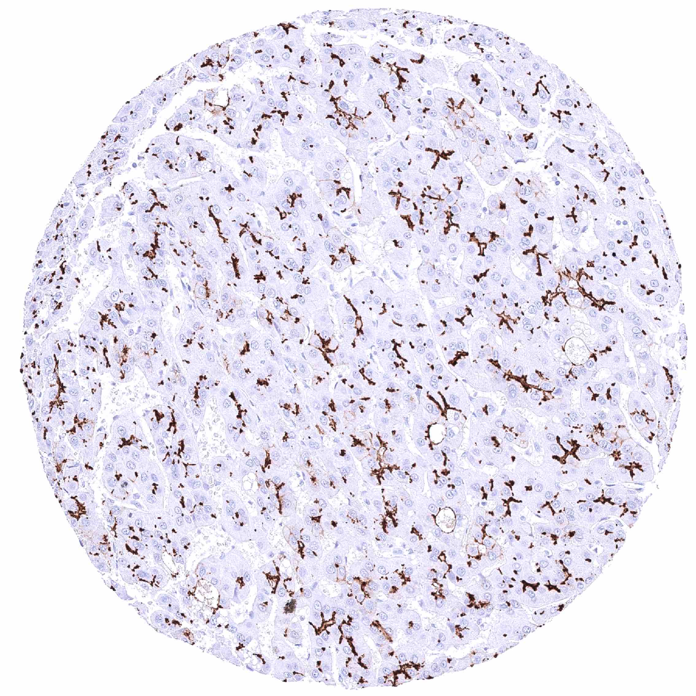
BSEP expression is frequent in hepatocellular carcinoma, but systematic data evaluating its potential expression in other cancers are lacking.
The TCGA findings on BSEP RNA expression in different tumor categories have been summarized in the Human Protein Atlas.
Compatibility of Antibodies
No data available at the moment
Protocol Recommendations
IHC users have different preferences on how the stains should look like. Some prefer high staining intensity of the target stain and even accept some background. Others favor absolute specificity and lighter target stains. Factors that invariably lead to more intense staining include higher concentration of the antibody and visualization tools, longer incubation time, higher temperature during incubation, higher temperature and longer duration of the heat induced epitope retrieval (slide pretreatment). The impact of the pH during slide pretreatment has variable effects and depends on the antibody and the target protein.
All images and data shown here and in our image galleries are obtained by the manual protocol described below. Other protocols resulting in equivalent staining are described as well.
Manual protocol
Freshly cut sections should be used (less than 10 days between cutting and staining). Heat-induced antigen retrieval for 5 minutes in an autoclave at 121°C in pH 7,8 Target Retrieval Solution buffer. Apply HMV4715 at a dilution of 1:150 at 37°C for 60 minutes. Visualization of bound antibody by the EnVision Kit (Dako, Agilent) according to the manufacturer’s directions.
Potential Research Applications
- The sensitivity and specificity of BSEP IHC for the distinction of hepatocellular carcinomas from other tumors needs to be investigated.
- The role of BSEP expression levels in hepatocellular carcinoma are unclear. This includes its potential contribution to chemoresistance and tumor aggressiveness.
- The diverse mutations in ABCB11 that cause different forms of cholestasis (e.g., PFIC2, BRIC) and their functional impacts on BSEP activity need to be further explored.
- The molecular pathways and factors that regulate BSEP expression and activity in the liver need to be investigated.
- Strategies are needed to predict and prevent drug-induced liver injury through impaired BSEP function.
Evidence for Antibody Specificity in IHC
There are two ways how the specificity of antibodies can be documented for immunohistochemistry on formalin fixed tissues. These are: 1. Comparison with a second independent method for target expression measurement across a large number of different tissue types (orthogonal strategy), and 2. Comparison with one or several independent antibodies for the same target and showing that all positive staining results are also seen with other antibodies for the same target (independent antibody strategy).
Orthogonal validation: For the antibody HMV4715, specificity is supported by the near complete concordance of its immunostaining patterm with data from three independent RNA screening studies, including the Human Protein Atlas (HPA) RNA-seq tissue dataset, the FANTOM5 project, and the Genotype-Tissue Expression (GTEx) project, which are all summarized in the Human Protein Atlas (Tissue expression BSEP). Immunostaining by using HMV4715 was especially seen at the apical pole of hepatocytes the liver. The liver is the only organ with documented RNA expression. An additional fibrillary immunostaining by HMV4715 in the white matter of the cerebrum and the cerebellum as well as in the neurohyophysis is to be considered a (tolerable) cross-reactivity of HMV4715.
Comparison of antibodies: True expression of BSEP at the apical (bile-secreting) pole of hepatocytes seen by HMV4715 is confirmed by an identical staining obtained by a second, independent commercially available BSEP antibody, termed “validation antibody”. Independence of the validation antibody is confirmed by its strong staining (partly near the apical membranes) in several tissues including gastric glands, salivary glands, gallbladder, pancreas acinar cells, and small intestine mucosa while validation antibody did not stain fibres in the brain.