295,00 € – 1.045,00 €
Product details
Synonyms = DNA-binding protein SATB2; GLSS; SATB homeobox 2; Special AT-rich sequence-binding protein 2
Antibody type = Recombinant Rabbit monoclonal / IgG
Clone = MSVA-702R
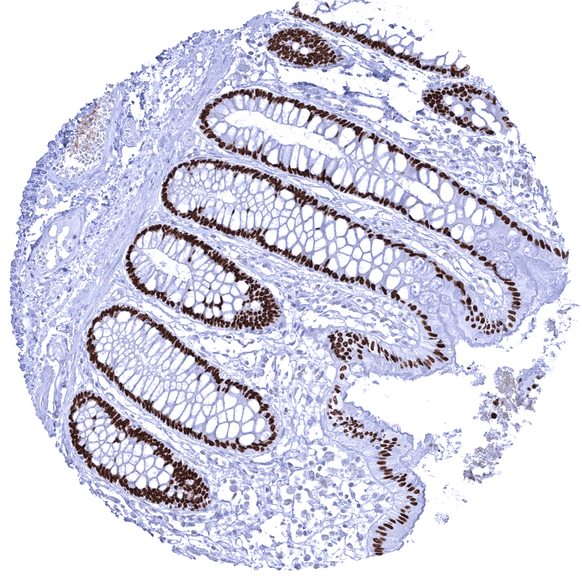
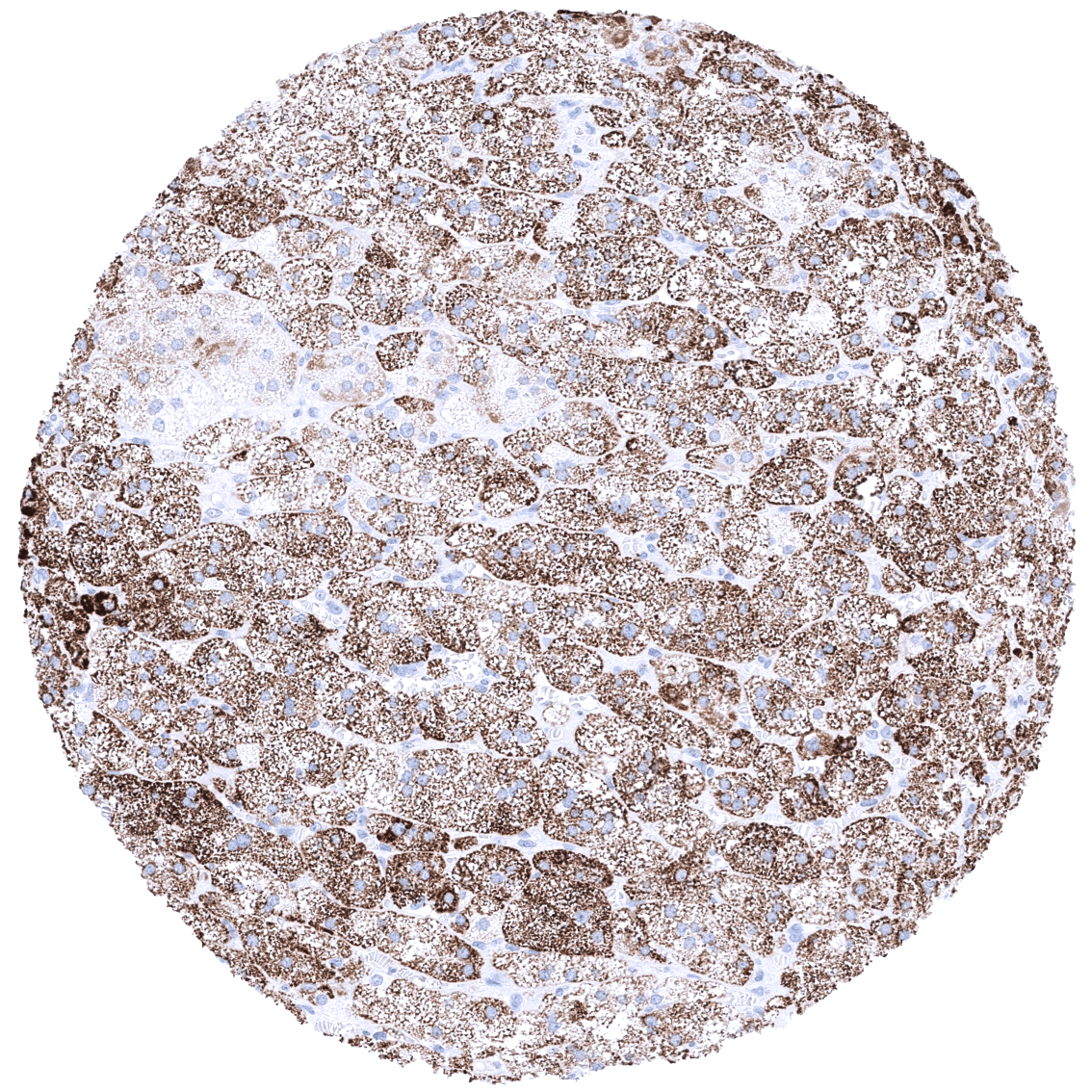
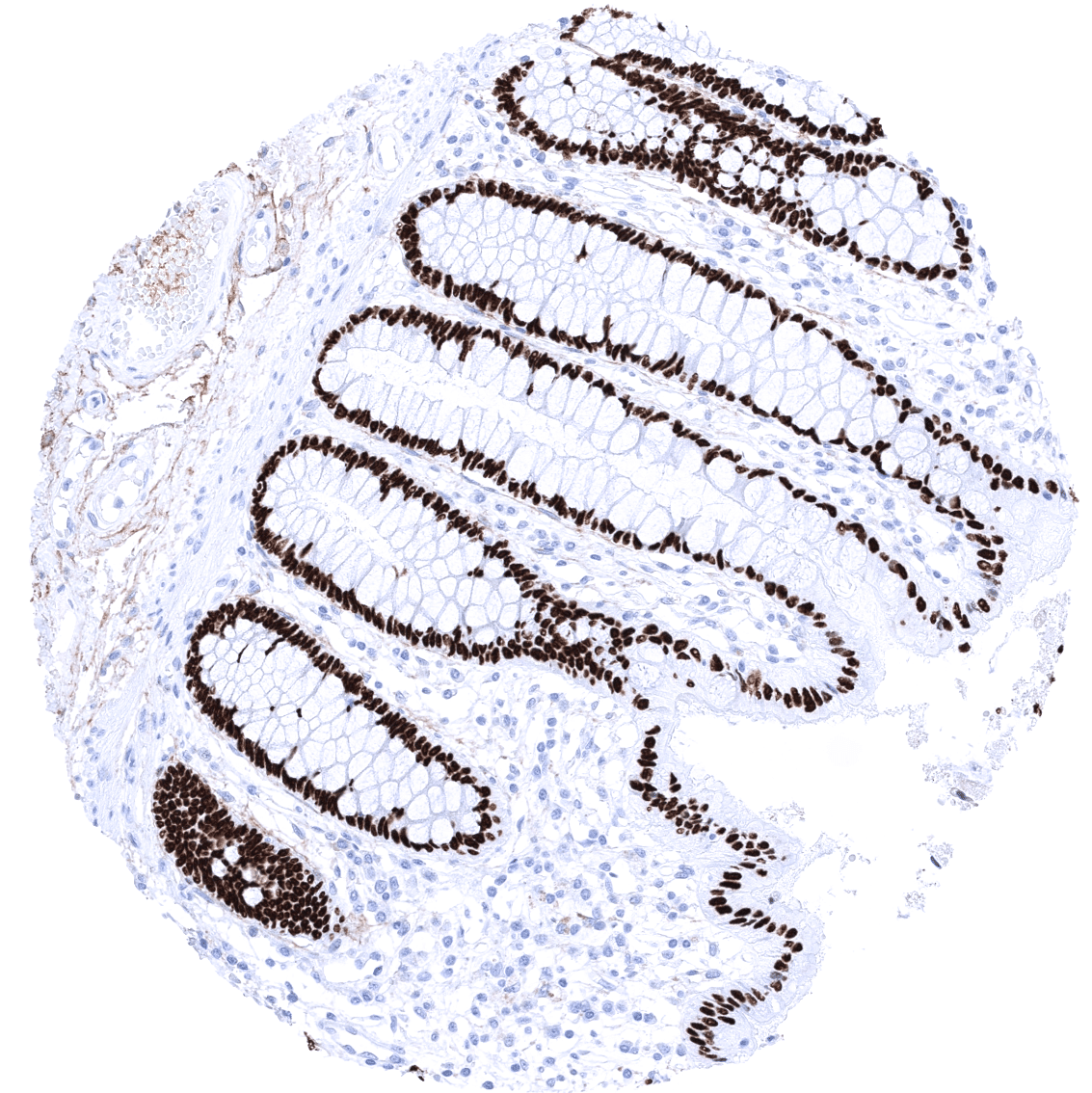
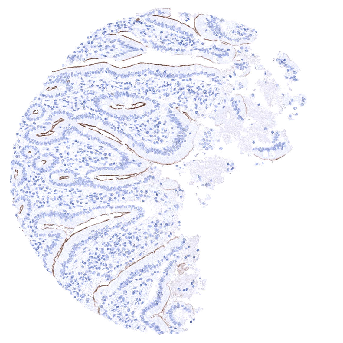
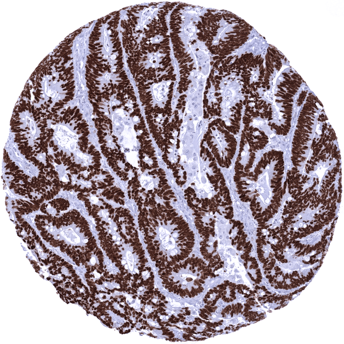
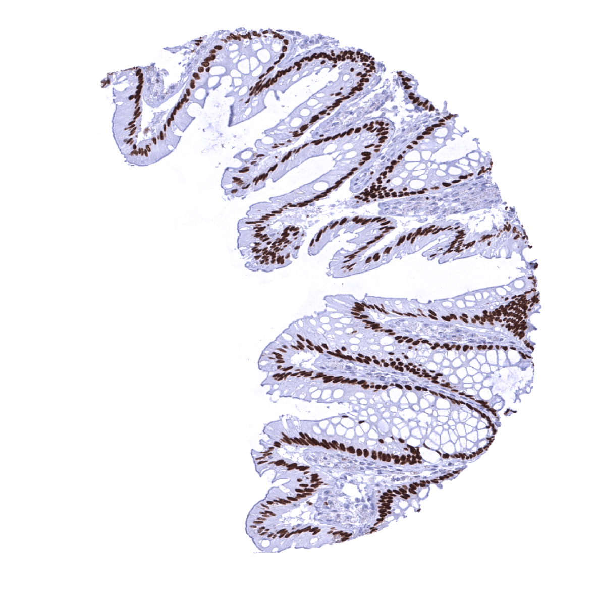
Positive control = Colon: A strong nuclear staining should be seen in virtually all columnar epithelial cells.
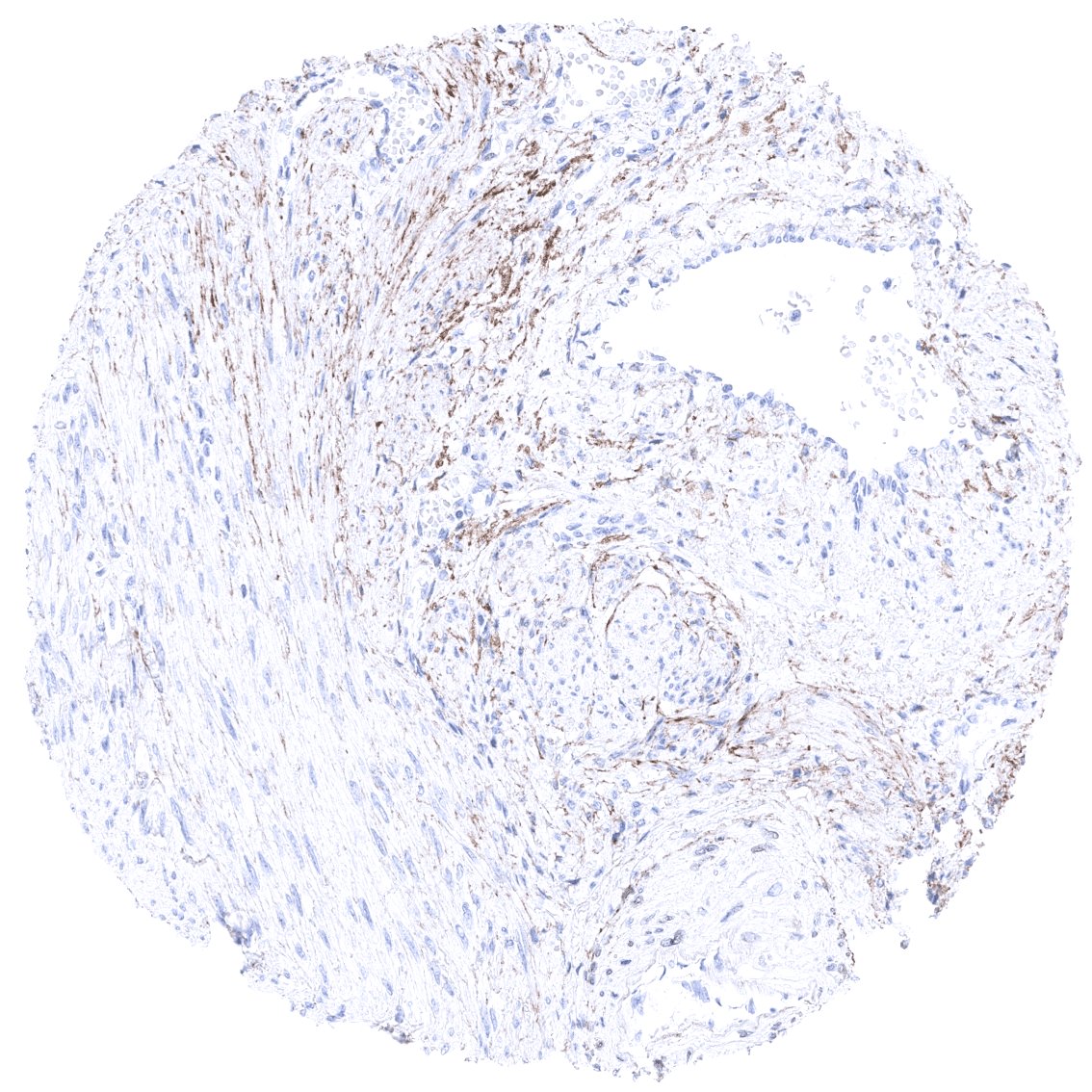
Negative control = Colon: SATB2 staining should be absent in stromal and smooth muscle cells.
Cellular localization = Nuclear
Reactivity = Human
Application = Immunohistochemistry
Dilution = 1:100 – 1:200
Intended Use = Research Use Only
Relevance of Antibody
SATB2 is expressed in colorectal epithelial cells, osteoclasts, and the brain.
Biology Behind
Special AT-rich sequence-binding protein 2 (SATB2) is a nuclear protein with a molecular weight of 82.5 kDa encoded by the SATB2 gene at chromosome 2q33. SATB2 specifically binds to so-called matrix attachment regions (MARs) of the DNA, where it induces local chromatin-loop remodeling that impacts the transcriptional activity of the affected genes. Attached SATB2 recruits additional chromatin remodeling enzymes including histone deacetylases (HDACs) and histone acetyltransferases (HATs). Genes regulated by SATB2 include programs required for neuronal, skeletal, and osteoblast development.
Staining Pattern in Normal Tissues
SATB2 staining pattern in Normal Tissues with antibody MSVA-702R (images are shown in our “Normal Tissue Gallery”)
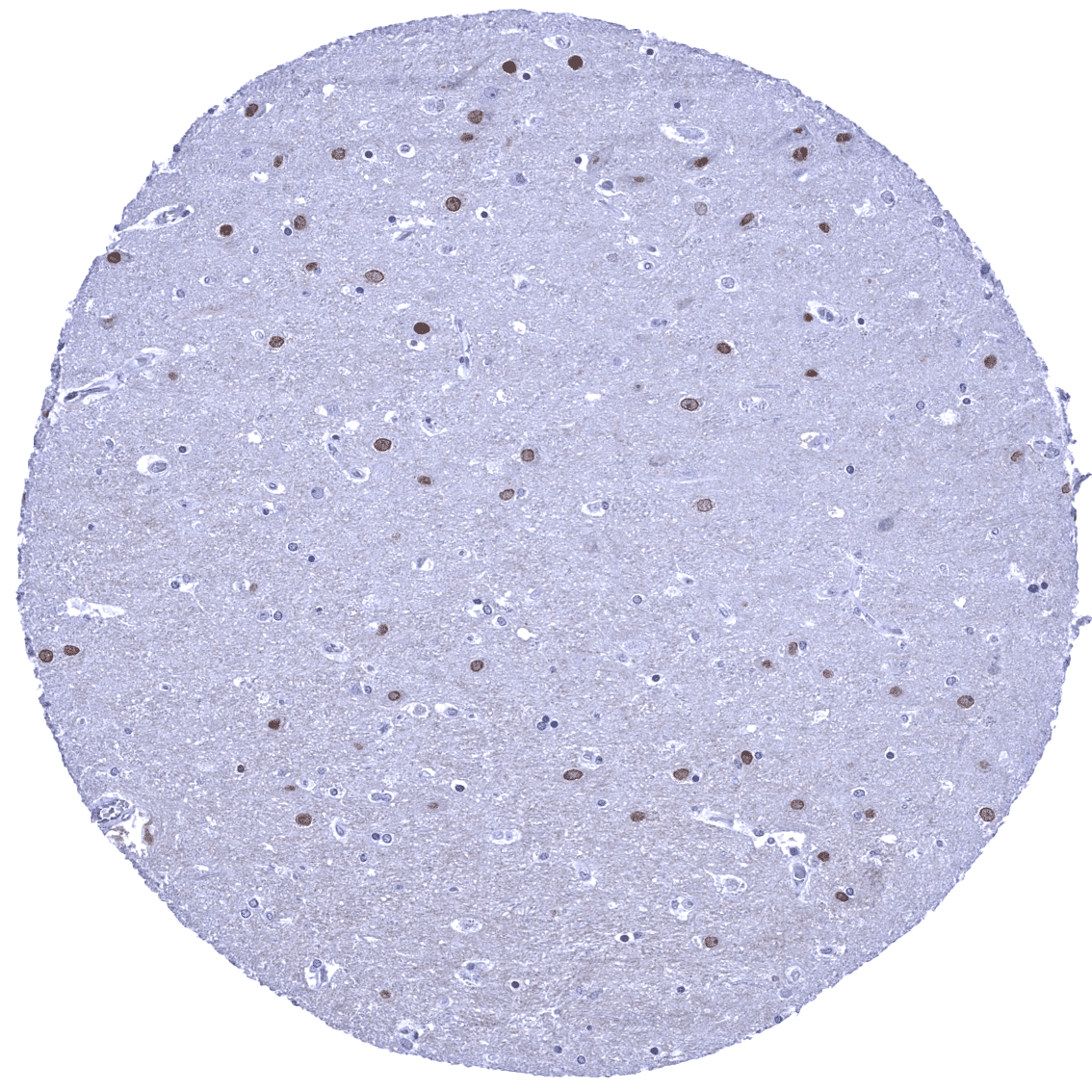
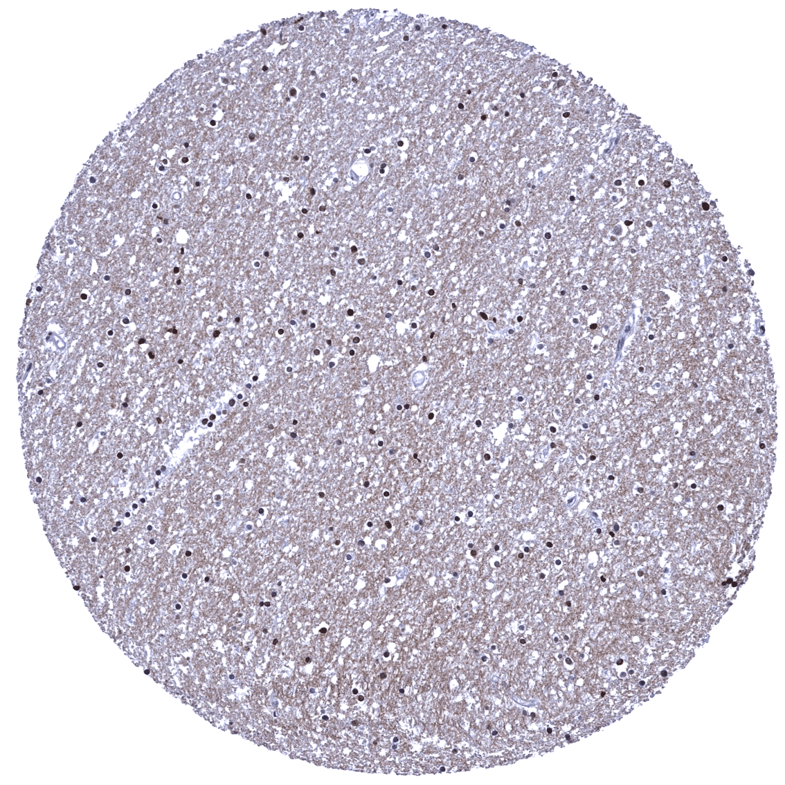
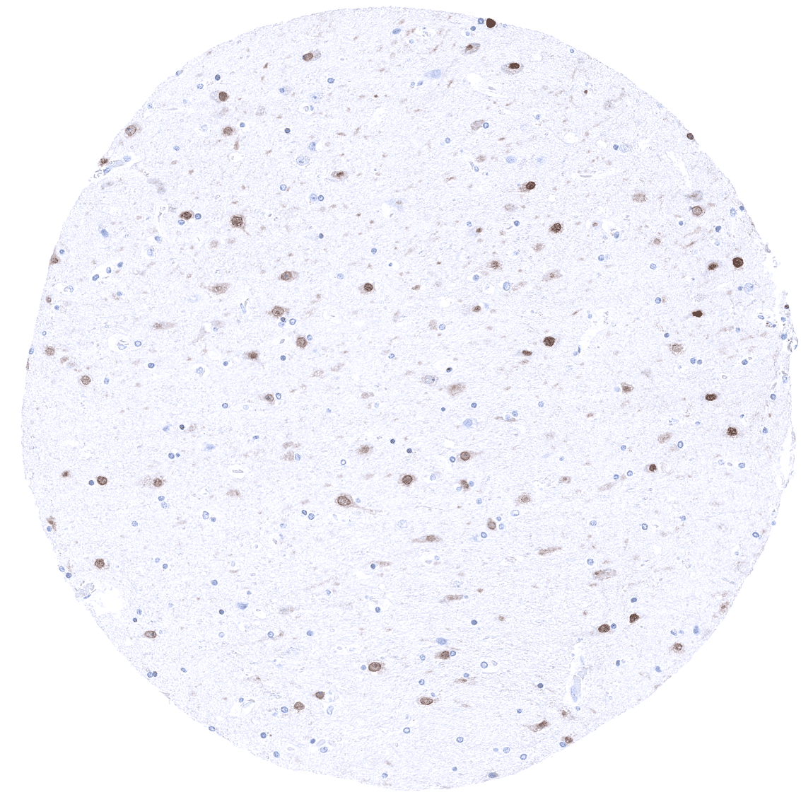
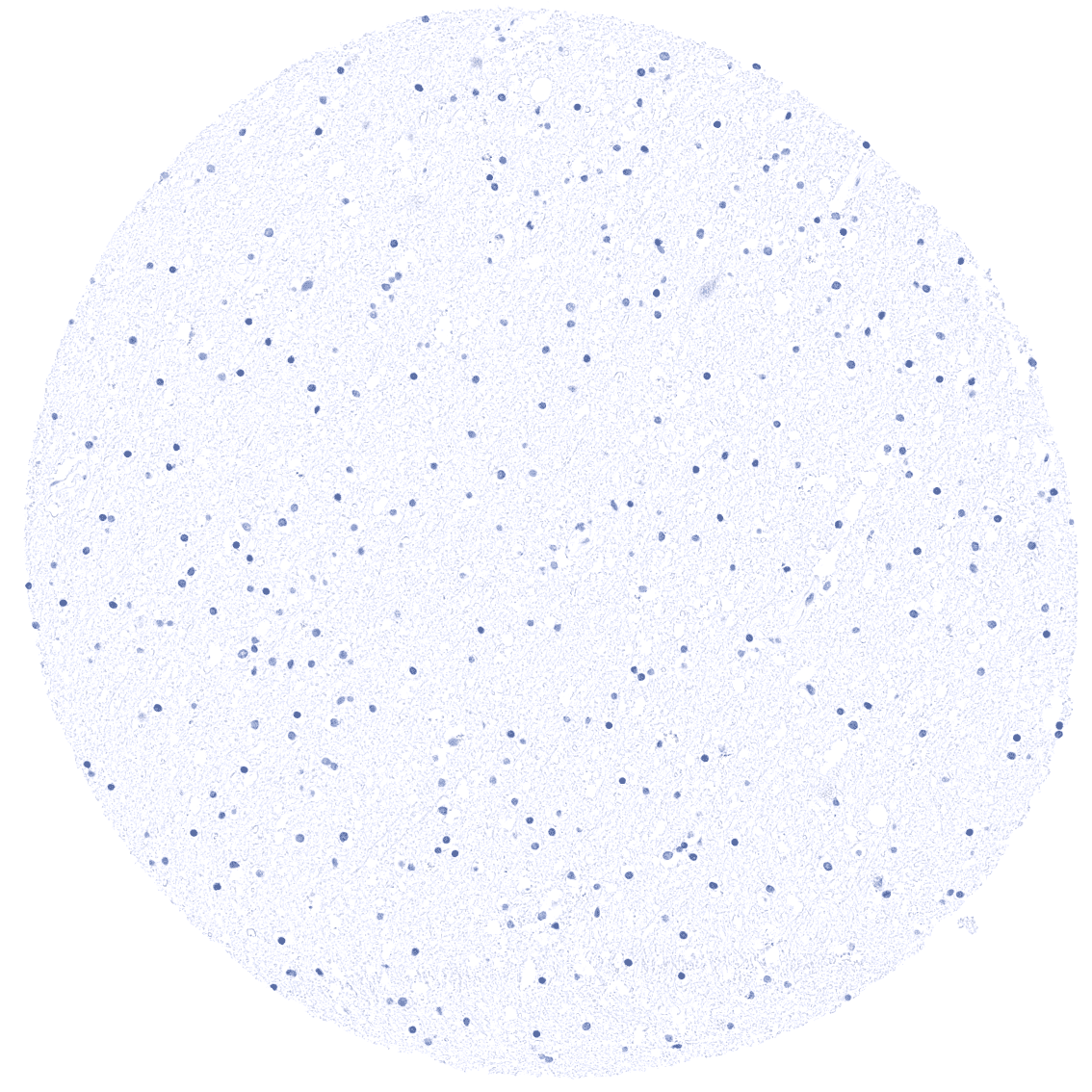
| Brain | Cerebrum | Weak to moderate nuclear SATB2 staining of neurons. An additional fibrillar staining may represent a (tolerable) cross-reactivity. |
| Cerebellum | Weak to moderate nuclear SATB2 staining of neurons. | |
| Endocrine Tissues | Thyroid | Negative. |
| Parathyroid | Negative. | |
| Adrenal gland | Negative. | |
| Pituitary gland | Negative. | |
| Respiratory system | Respiratory epithelium | Negative. |
| Lung | Negative. | |
| Gastrointestinal Tract | Salivary glands | Negative. |
| Esophagus | Negative. | |
| Stomach | Negative. | |
| Duodenum | Negative. | |
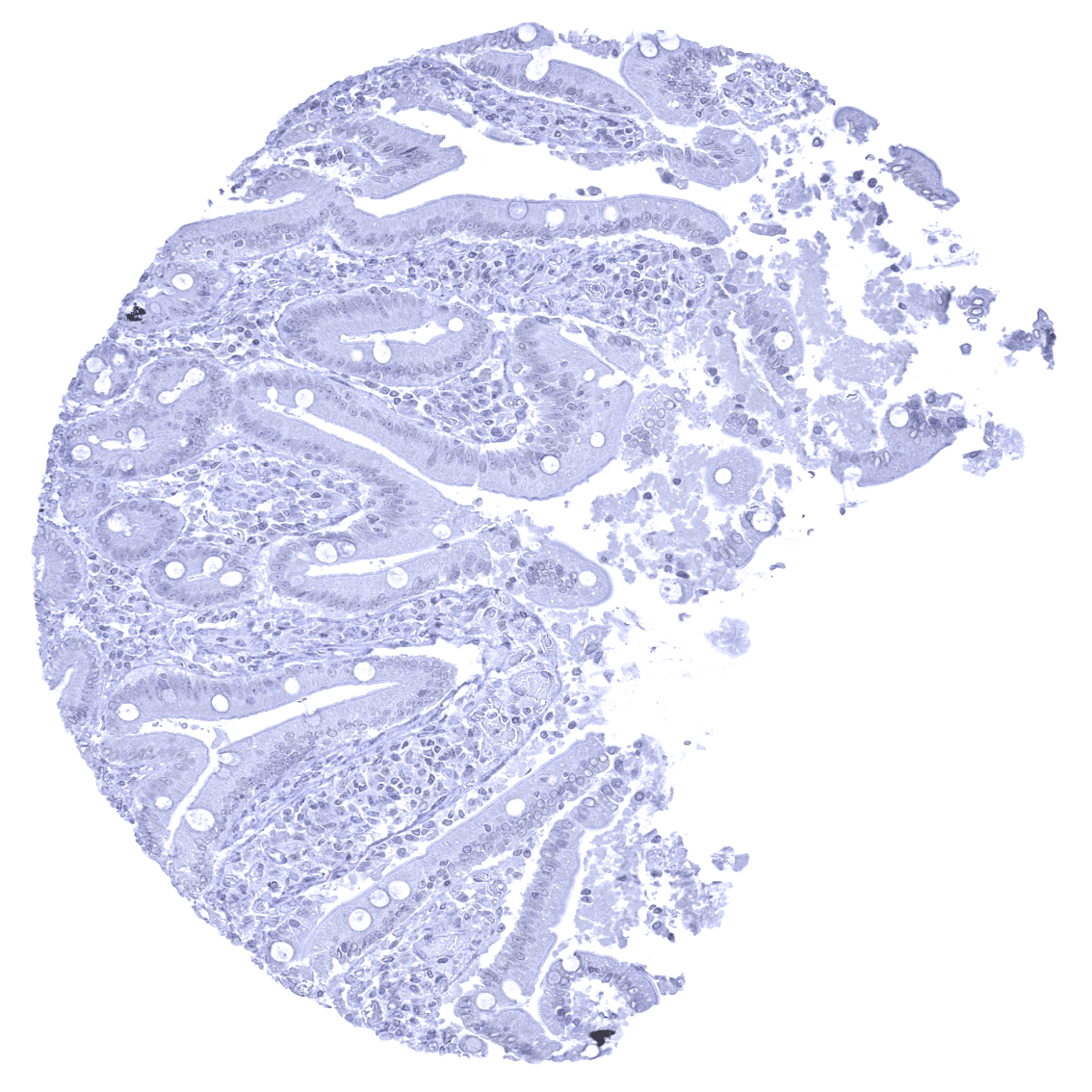
| Small intestine | Weak to moderate nuclear SATB2 staining of all epithelial cells. | |
| Appendix | Strong nuclear SATB2 staining of all epithelial cells. | |
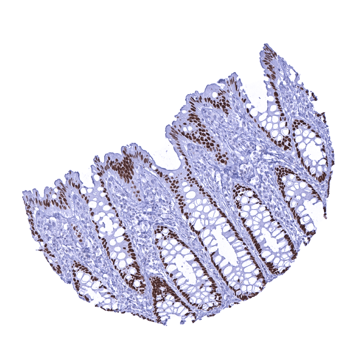
| Colon | Strong nuclear SATB2 staining of all epithelial cells. | |
| Rectum | Strong nuclear SATB2 staining of all epithelial cells. | |
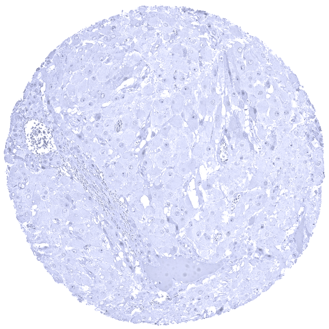
| Liver | Negative. | |
| Gallbladder | Negative. | |
| Pancreas | Negative. | |
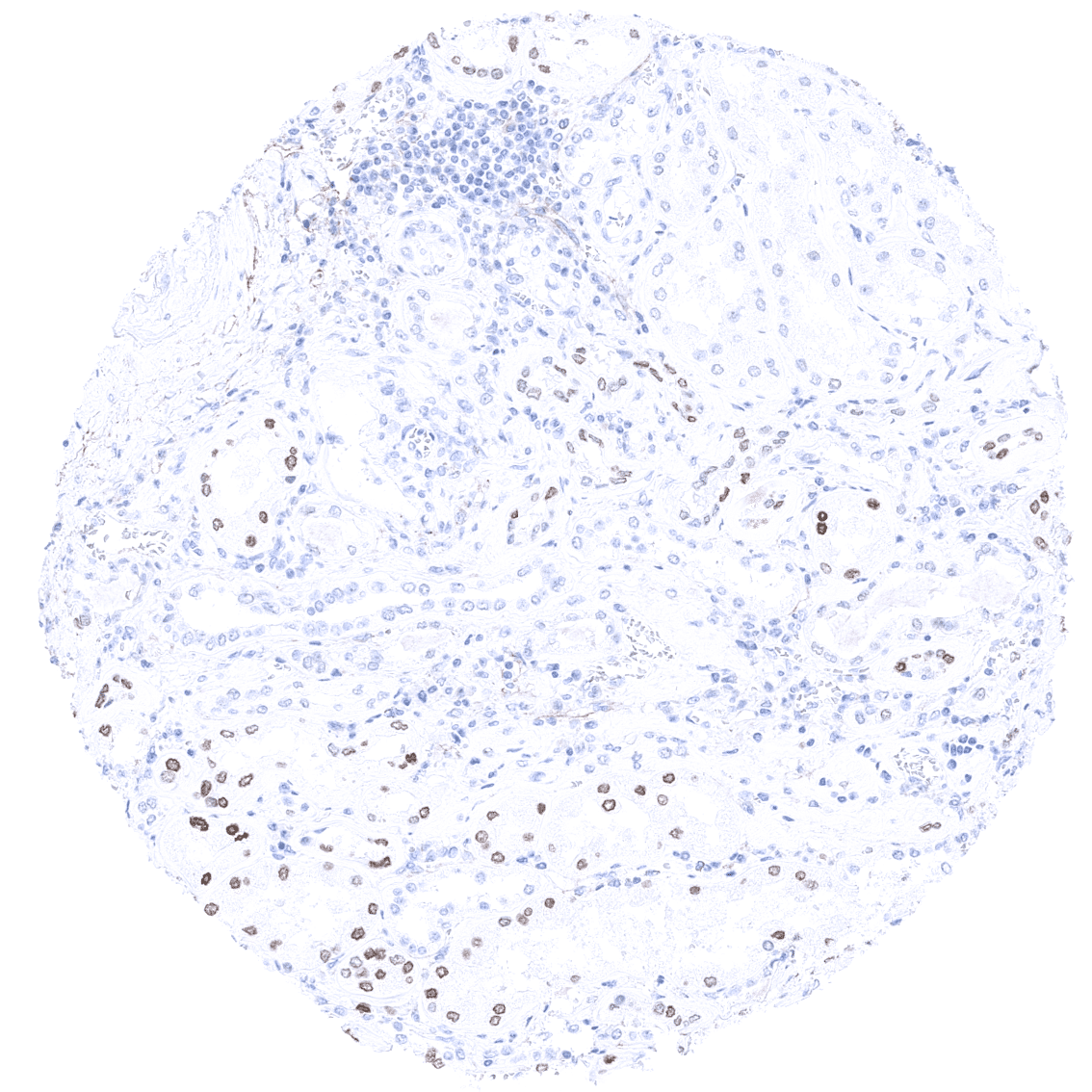
| Genitourinary | Kidney | Weak to moderate nuclear SATB2 staining of a fraction of epithelial cells of distal tubuli and collecting ducts. |
| Urothelium | Negative. | |
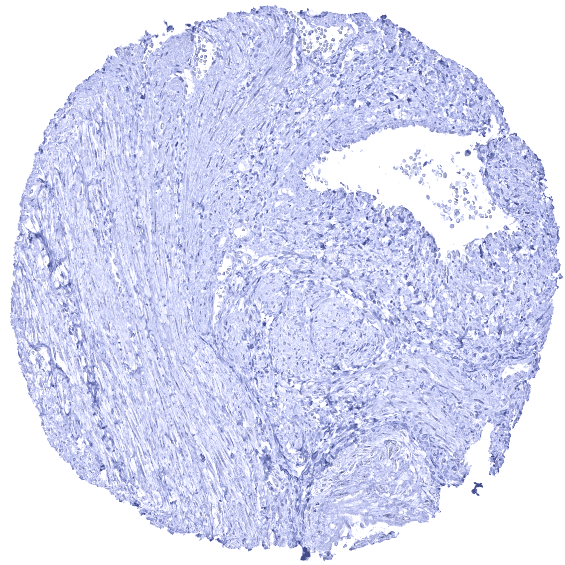
| Male genital | Prostate | Negative. |
| Seminal vesicles | Negative. | |
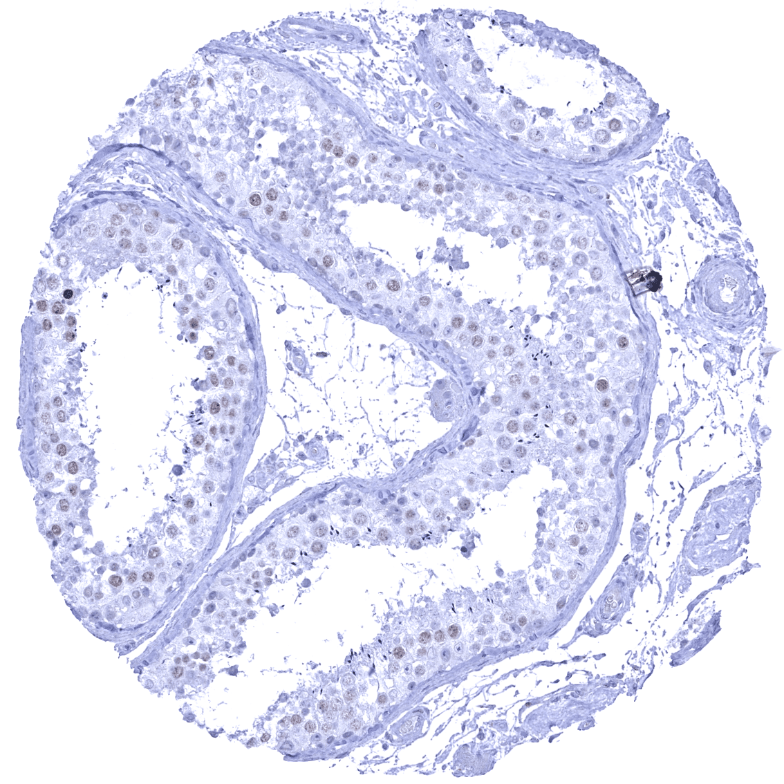
| Testis | Weak to moderate nuclear SATB2 staining of a fraction of spermatocytes. | |
| Epididymis | Negative. | |
| Female genital | Breast | Negative. |
| Uterus, myometrium | Negative. | |
| Uterus, ectocervix | Negative. | |
| Uterus endocervix | Negative. | |
| Uterus, endometrium | Negative. | |
| Fallopian Tube | Negative. | |
| Ovary | Weak to moderate nuclear SATB2 staining of a fraction of oocytes. | |
| Placenta early | Negative. | |
| Placenta mature | Negative. | |
| Amnion | Negative. | |
| Chorion | Negative. | |
| Skin | Epidermis | Negative. |
| Sebaceous glands | Negative. | |
| Muscle/connective tissue | Heart muscle | Negative. |
| Skeletal muscle | Negative. | |
| Smooth muscle | Negative. | |
| Vessel walls | Negative. | |
| Fat | Negative. | |
| Stroma | Negative. | |
| Endothelium | Negative. | |
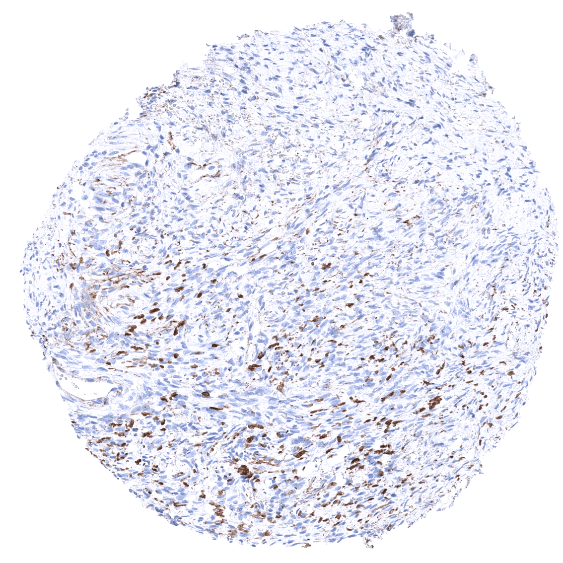
| Bone marrow/ lymphoid tissue | Bone marrow | Strong nuclear SATB2 staining of osteoclasts. |
| Lymph node | Negative. | |
| Spleen | Negative. | |
| Thymus | Negative. | |
| Tonsil | Negative. | |
| Remarks |
These findings are largely consistent with the RNA and protein data described in the Human Protein Atlas (Tissue expression SATB2)
Positive control:
-Colon: A strong nuclear SATB2 staining should be seen in virtually all columnar epithelial cells.
-Testis: dispersed spermatocytes should show at least a weak nuclear staining.
Negative control:
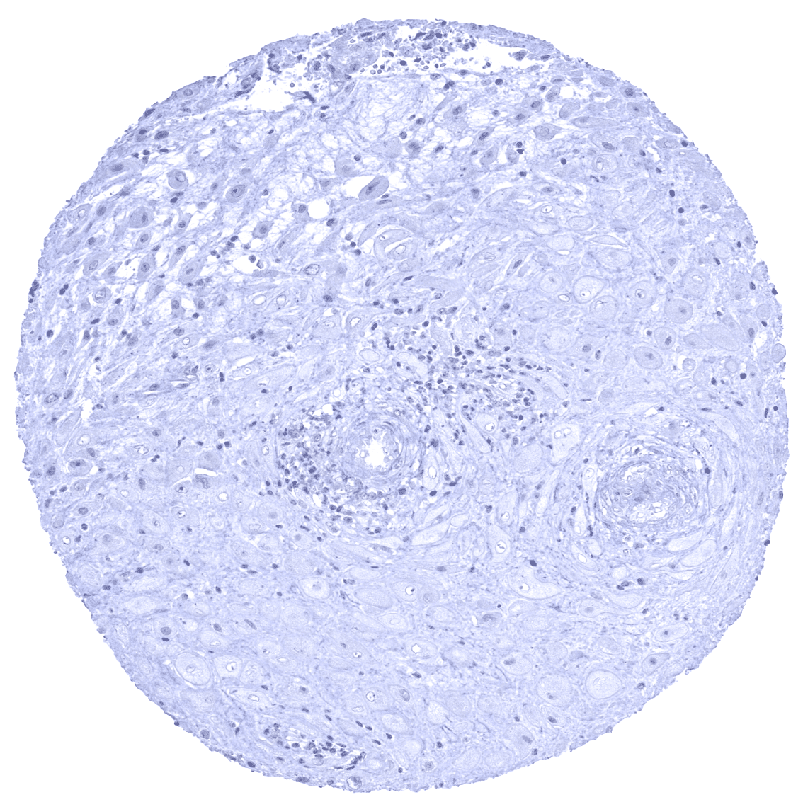
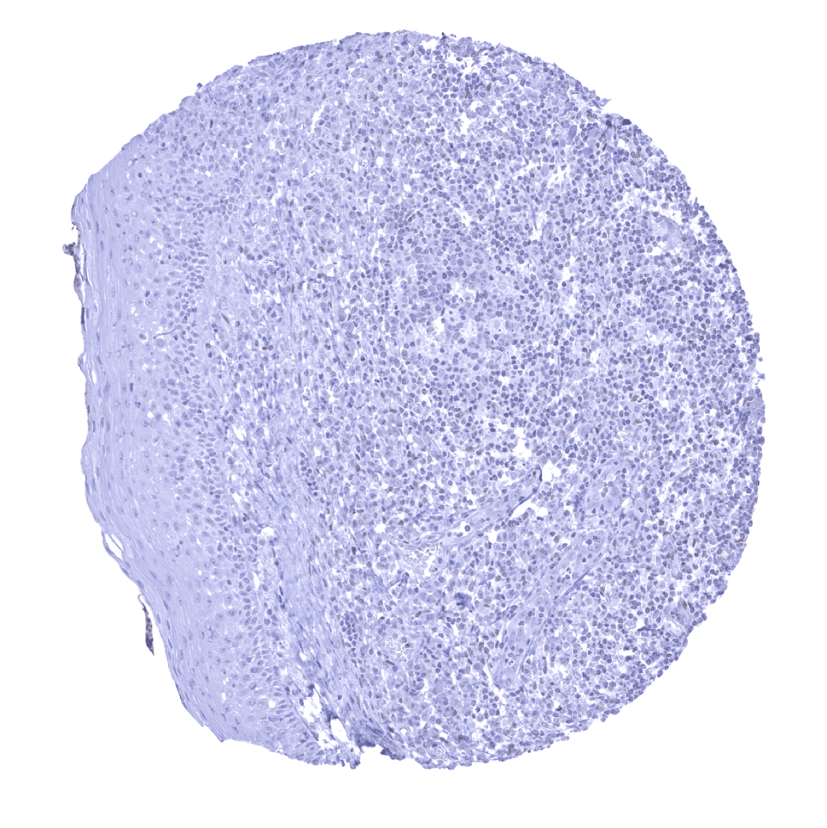
-Colon: SATB2 staining should be absent in stromal and smooth muscle cells.
-Tonsil: SATB2 staining should be absent in epithelial and lymphatic cells.

Staining Pattern in Relevant Tumor Types
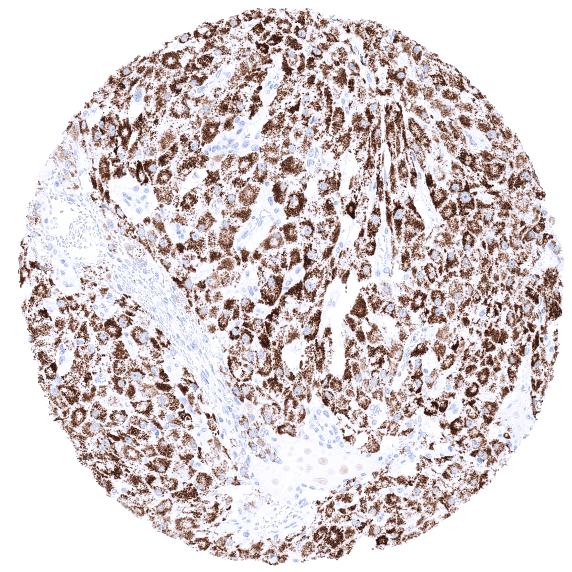
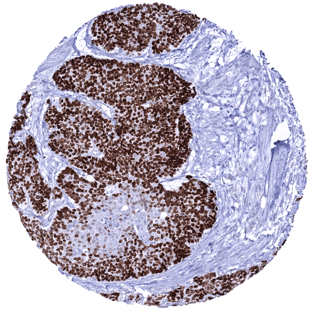
A positive SATB2 immunostaining is most commonly seen in adenocarcinomas and neuroendocrine tumors derived from the colorectum or appendix, osteosarcomas, and in Merkel cell cancer. A positive SATB2 immunostaining, often at lower intensity, has also been described to occur in various other tumor entities.
The TCGA findings on SATB2 RNA expression in different tumor categories have been summarized in the Human Protein Atlas.

Compatibility of Antibodies
SATB2 (MSVA-702R) publication summary
Relevant publication: Dum et al. “SATB2 Expression in Human Tumors: A Tissue Microarray Study on More Than 15 000 Tumors.”
A total of 11’678 tumors were successfully analyzed from 120 different tumor categories by using the following protocol: Heat-induced antigen retrieval for 5 minutes in an autoclave at 121°C in pH7,8 Target Retrieval Solution buffer. MSVA-702R at a dilution of 1:100 at 37°C for 60 minutes. Visualization of bound antibody by the EnVision Kit (Dako, Agilent). This protocol was also used for all stainings depicted in our tumor and normal tissue galleries.
In this study, at least one positive case was seen in 89 (74%) of 120 tumor categories and 38 (32%) tumor categories included at least one case with strong positivity. The highest rates of positive staining and the highest levels of expression were found in adenomas and adenocarcinomas of the colorectum, various subtypes of neuroendocrine tumors of the colorectum and the appendix, Merkel cell carcinomas, osteosarcomas, and papillary renal cell carcinomas. The distribution of positive staining results is shown in an “organ-systematic” and in a “ranking order” figure below (images based on data from Dum et al.). Results on possible associations with histopathological and clinical parameters of tumor aggressiveness are also summarized below (table based on data from Dum et al.).
Authors conclusions on diagnostic utility of SATB2 immunohistochemistry with respect to the distinction of different tumor entities (Dum et al.):
- Distinction of adenocarcinoma of the colorectum (strong SATB2 positivity in about 70%) from other adenocarcinomas (strong positivity in less than 5%).
- Distinction of neuroendocrine tumors derived from of the colorectum/appendix (SATB2 positivity in 28-50%) other neuroendocrine tumors (very rarely positive).
- A strong SATB2 positivity can support the diagnosis of an osteosarcoma in the appropriate morphological context.
Authors conclusions on the prognostic role of SATB2 expression (Dum et al.):
- A loss of SATB2 expression is linked to advanced tumor stage, nodal metastasis, tumor location in the right colon, microsatellite instability, and BRAF V600E mutations in colorectal cancer.
- A low level of SATB2 expression is linked to prognostically unfavorable tumor features in both clear cell and papillary renal cell carcinoma.
Figure 1. SATB2 staining in cancer (“organ-systematic”; according to Dum et al.)

Figure 2. SATB2 staining in cancer (“ranking list””; according to Dum et al.)

Figure 3. Clinico-pathological associations described by Dum et al. (p-value)

Protocol Recommendations
IHC users have different preferences on how the stains should look like. Some prefer high staining intensity of the target stain and even accept some background. Others favor absolute specificity and lighter target stains. Factors that invariably lead to more intense staining include higher concentration of the antibody and visualization tools, longer incubation time, higher temperature during incubation, higher temperature and longer duration of the heat induced epitope retrieval (slide pretreatment). The impact of the pH during slide pretreatment has variable effects and depends on the antibody and the target protein.
All images and data shown here and in our image galleries are obtained by the manual protocol described below. Other protocols resulting in equivalent staining are described as well.
-Manual protocol
Freshly cut sections should be used (less than 10 days between cutting and staining). Heat-induced antigen retrieval for 5 minutes in an autoclave at 121°C in pH 7,8 Target Retrieval Solution buffer. Apply MSVA-702R at a dilution of 1:100 at 37°C for 60 minutes. Visualization of bound antibody by the EnVision Kit (Dako, Agilent) according to the manufacturer’s directions.
-Signs of overstaining
If a very sensitive protocol is applied, some cytoplasmic staining may be seen in squamous epithelium and some tumors.
Potential Research Applications
- The diagnostic utility of SATB2 expression analysis should be further investigated in a large cohort of tumors from different entities
- The prognostic role of SATB2 expression in gastrointestinal adenocarcinomas is unclear.
Evidence for Antibody Specificity in IHC
There are two ways how the specificity of antibodies can be documented for immunohistochemistry on formalin fixed tissues. These are: 1. Comparison with a second independent method for target expression measurement across a large number of different tissue types (orthogonal strategy), and 2. Comparison with one or several independent antibodies for the same target and showing that all positive staining results are also seen with other antibodies for the same target (independent antibody strategy).
Orthogonal validation: For the antibody MSVA-702R specificity is suggested by the strong concordance of its staining results with RNA expression data derived from three independent RNA screening studies, including the Human Protein Atlas (HPA) RNA-seq tissue dataset, the FANTOM5 project, and the Genotype-Tissue Expression (GTEx) project, which are all summarized in the Human Protein Atlas (Tissue expression SATB2). Immunostaining by using MSVA-702R is only detected in organs with documented RNA expression such as the brain, colorectum, appendix, small intestine and the kidney. SATB2 immunostaining also occurs in osteoclasts, a cell type not analyzed in the protein atlas.
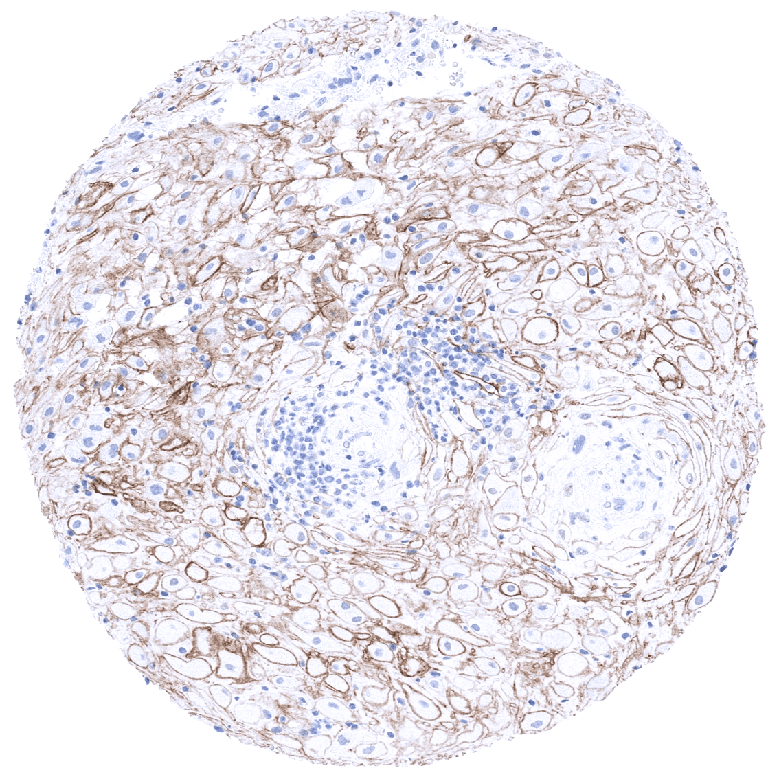
Comparison of antibodies: True expression of SATB2 in the cell types described above is also confirmed by using a second independent commercial antibody termed “validation antibody”. Images comparing MSVA-702R and the independent “validation antibody” are shown below. Both antibodies stain colorectal epithelium, spermatocytes, neurons of the brain, and renal tubular cells. These stainings are thus considered true specific SATB2 staining. Independence of MSVA-702R and “validation antibody” is documented by antibody specific cross-reactivities. Only “validation antibody” shows fibrillar staining in testis, different types of soft tissues as well as a cytoplasmic staining in the adrenal gland, decidua cells and in corpus luteum. These stainings are therefore considered antibody specific cross-reactivity of “validation antibody”. Only MSVA-702R showed a fibrillar staining of intracerebral nerve structures. This staining is therefore considered a (tolerable) antibody specific cross-reactivity of MSVA-702R.
Antibody Comparison: MSVA-702R vs another commercially available SATB2 antibody called “Validation Antibody”