295,00 € – 995,00 €
Product details
Synonyms = Coagulation factor XIII A chain; Coagulation factor XIII A1 polypeptide; F13A; F13a1; Fibrin stabilizing factor, A subunit; Fibrinoligase; FSF, A subunit; Protein-glutamine gamma-glutamyltransferase A chain; TGase; Transglutaminase. plasma
Antibody type = Recombinant Rabbit monoclonal / IgG
Clone = MSVA-813R
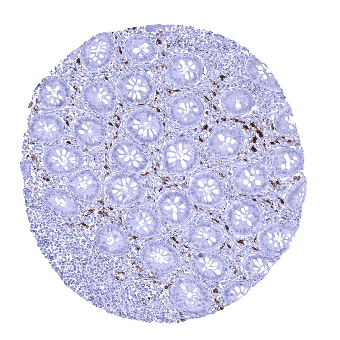
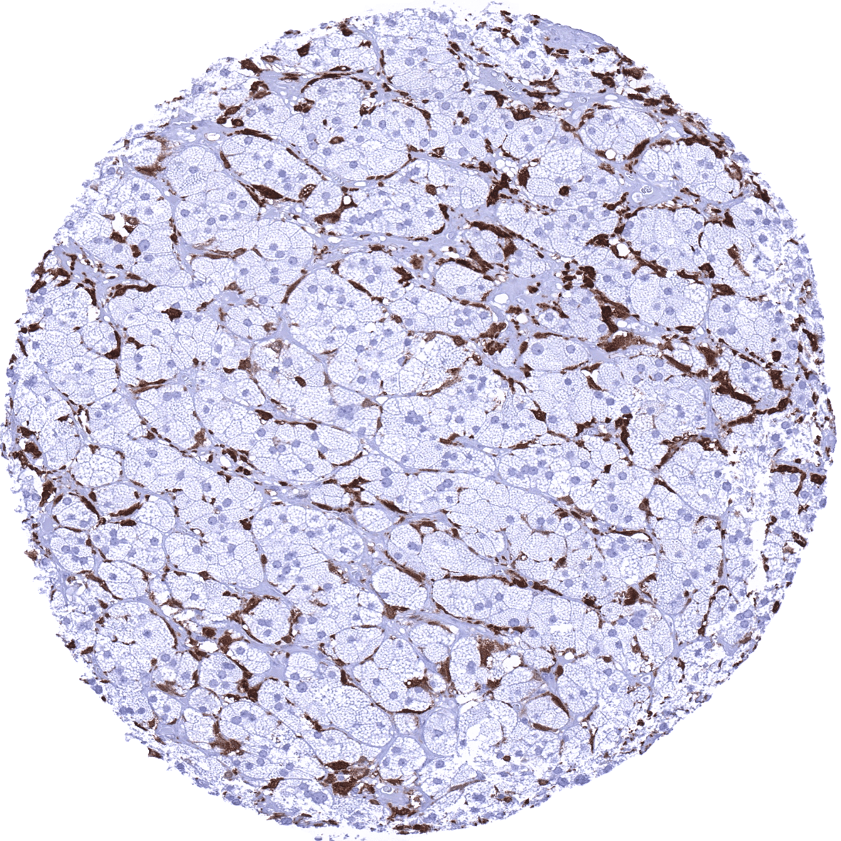
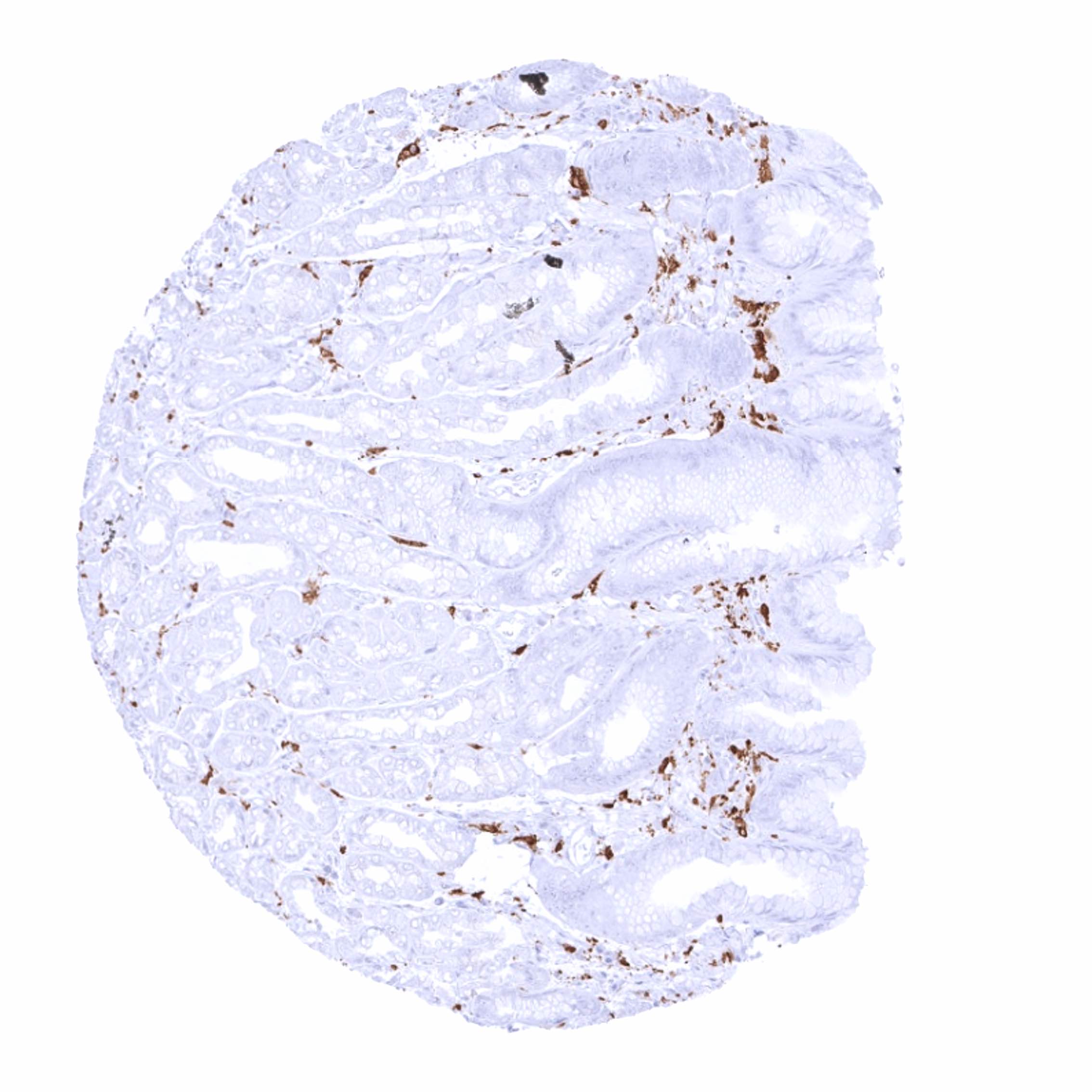
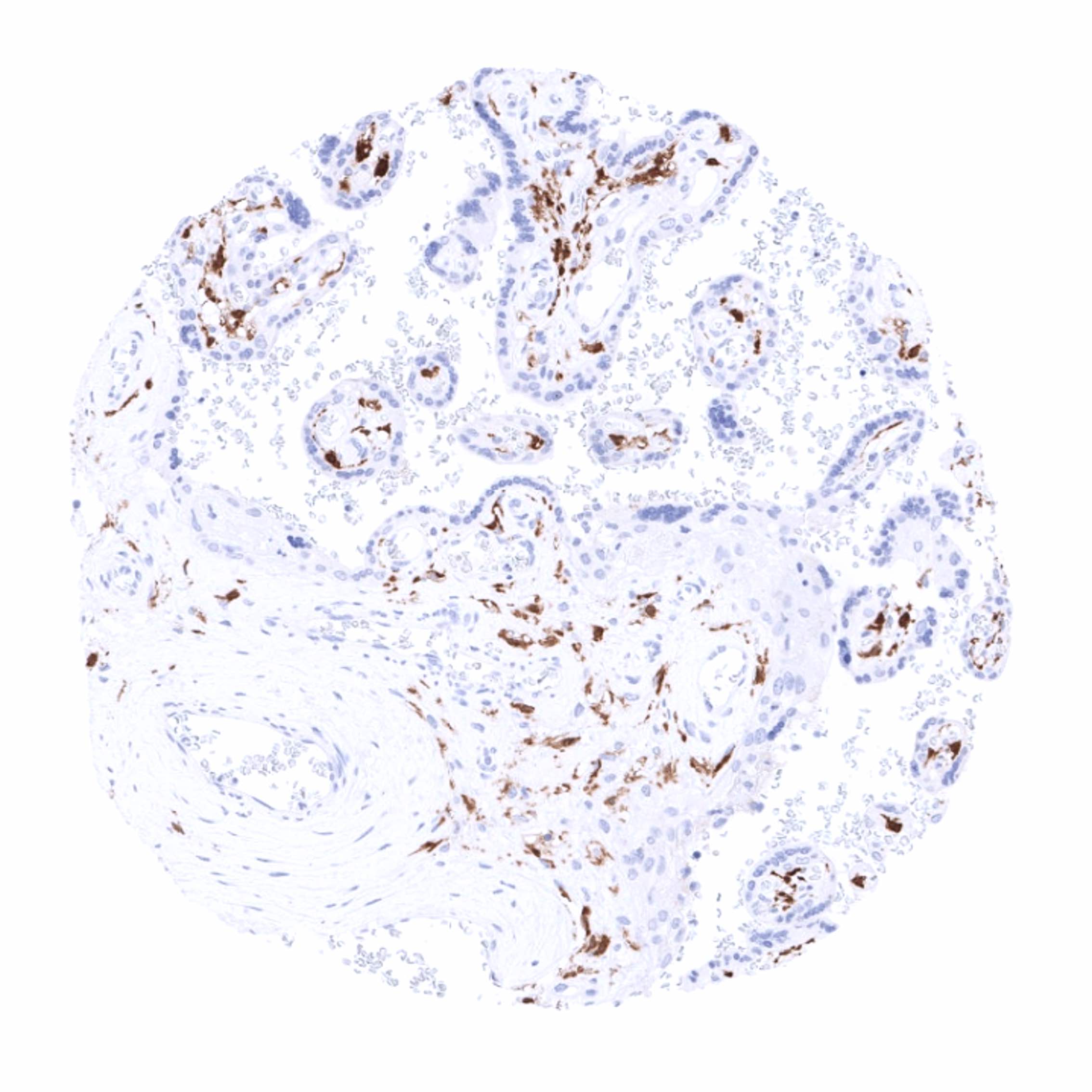
Positive control = Colon: A fraction of monocytic cells in the mucosa should show a strong cytoplasmic factor XIIIa immunostaining.
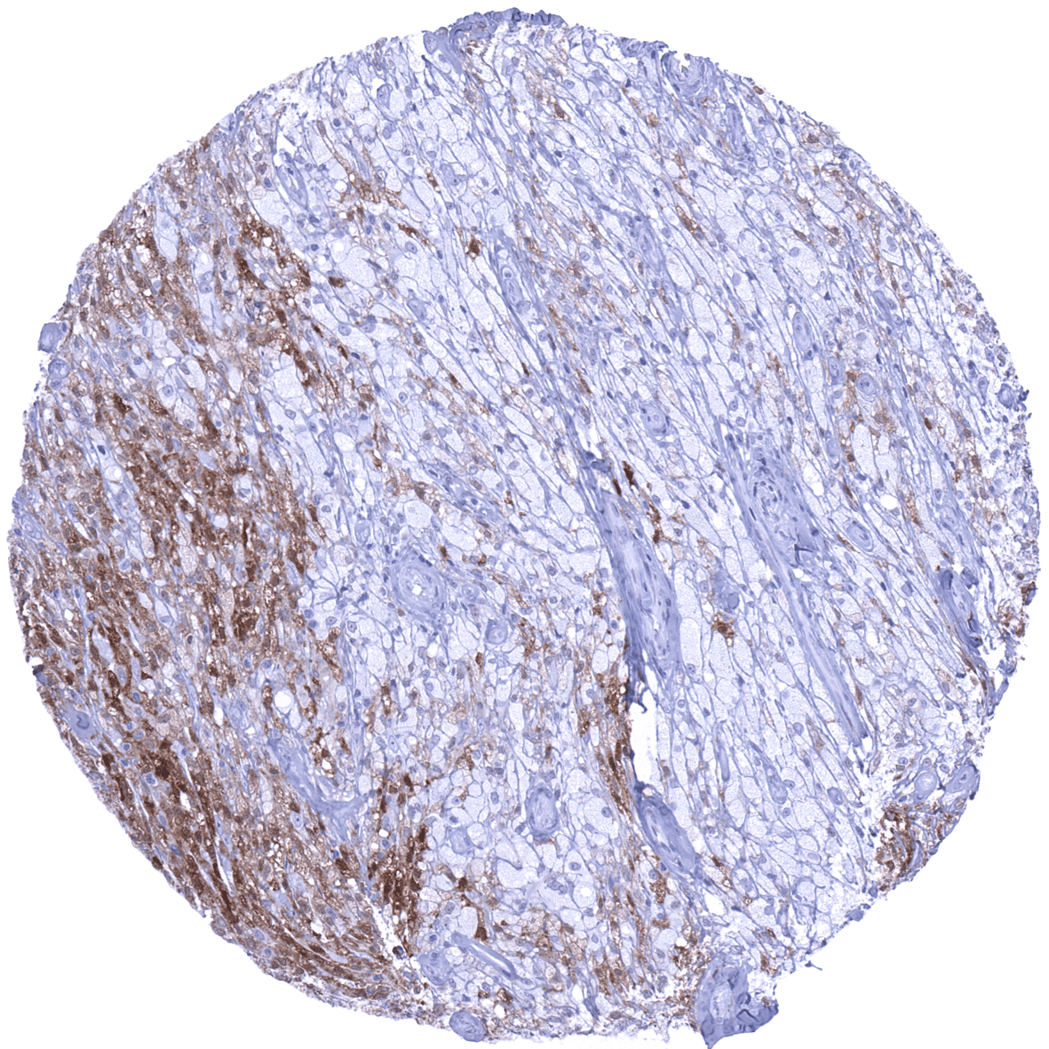
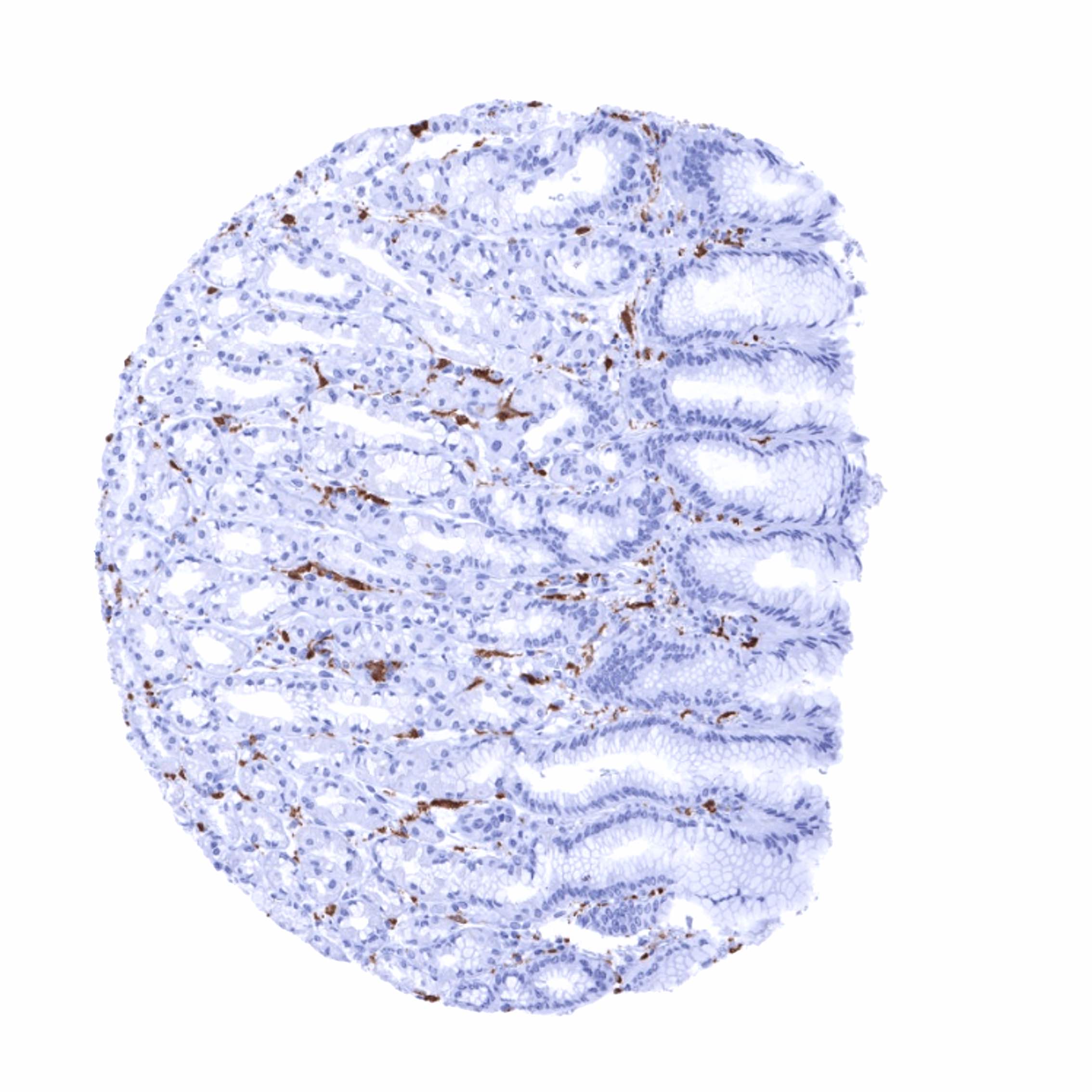
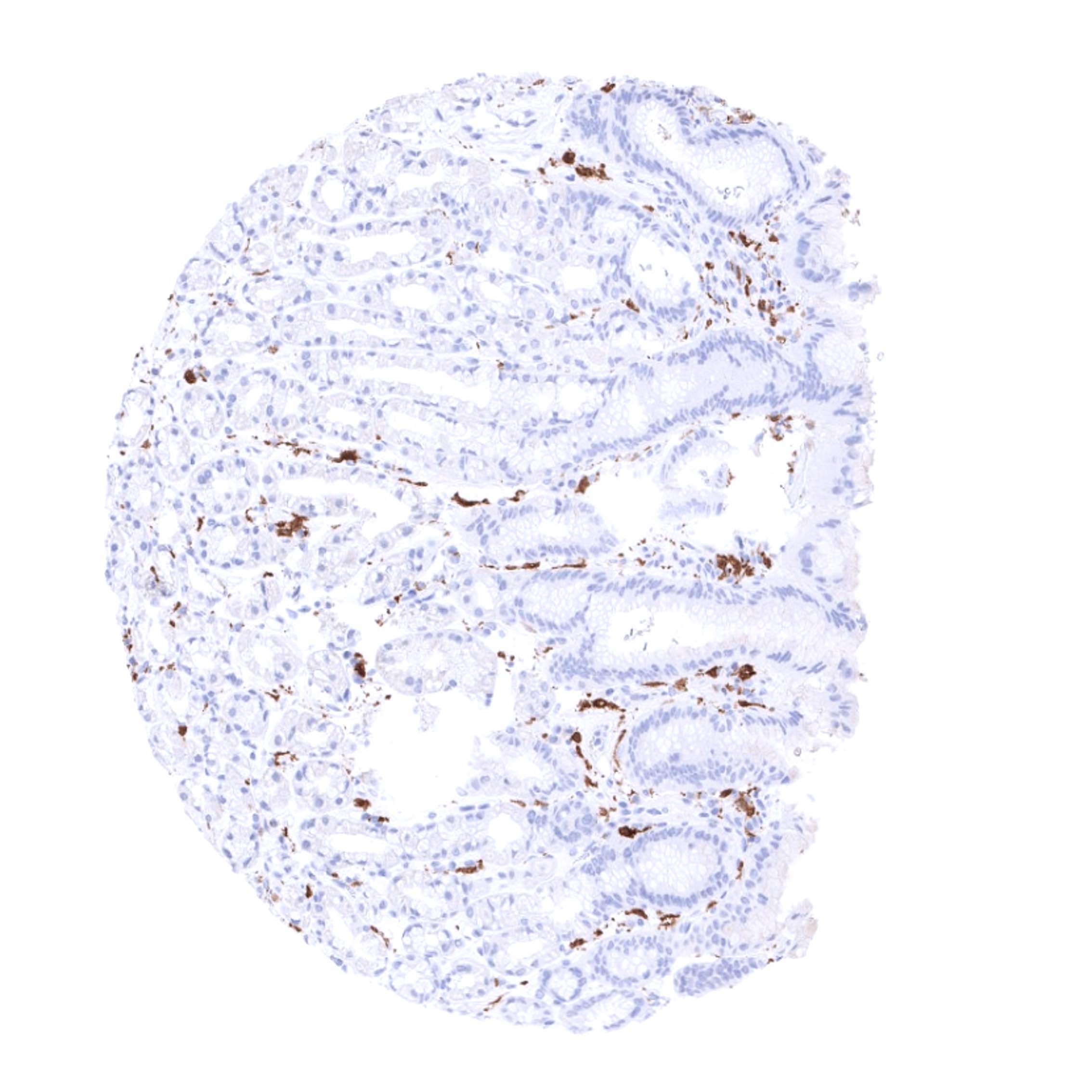
Negative control = Colon: Factor XIIIa immunostaining should be absent in epithelial, endothelial and smooth muscle cells.
Cellular localization = Cytoplasmic and Secreted
Reactivity = Human
Application = Immunohistochemistry
Dilution = 1:100 – 1:200
Intended Use = Research Use Only
Relevance of Antibody
Biology Behind
Factor XIIIa is a part of the blood coagulation system. Factor XIII is a transglutaminase that circulates in the human blood as a heterotetramer of two A and two B subunits. Factor XIIIa is a dimer of activated A peptides. In the process of blood coagulation, factor XIIIa has the function to crosslink fibrin. Factor XIIIs bind to the clot via their B units. In the presence of fibrins, thrombin efficiently cleaves a specific peptide bond of each A unit and activates them. Apart from coagulation and thrombosis, Factor XIII also plays a role in wound healing, bone metabolism, pregnancy and tumor biology.
Staining Pattern in Normal Tissues
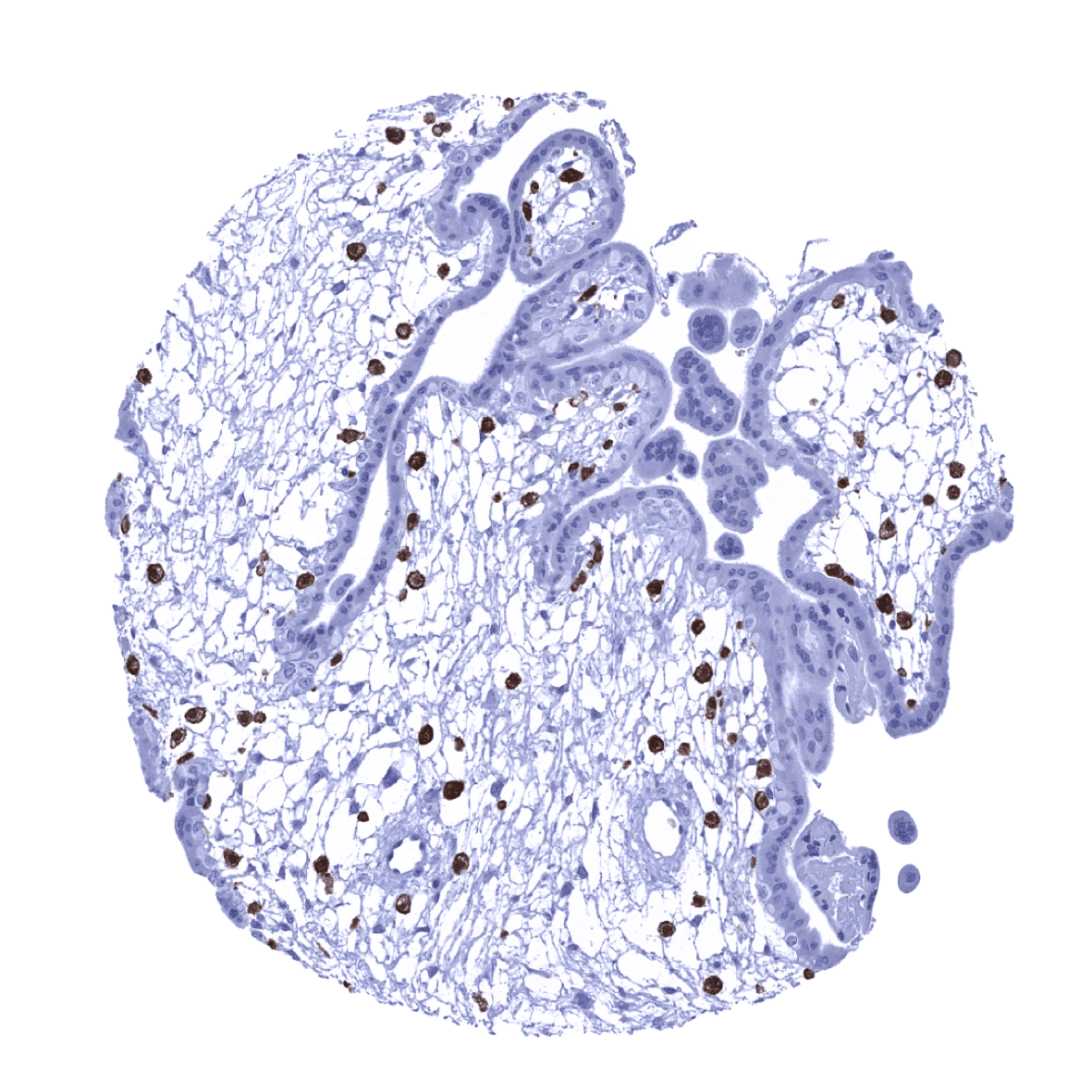
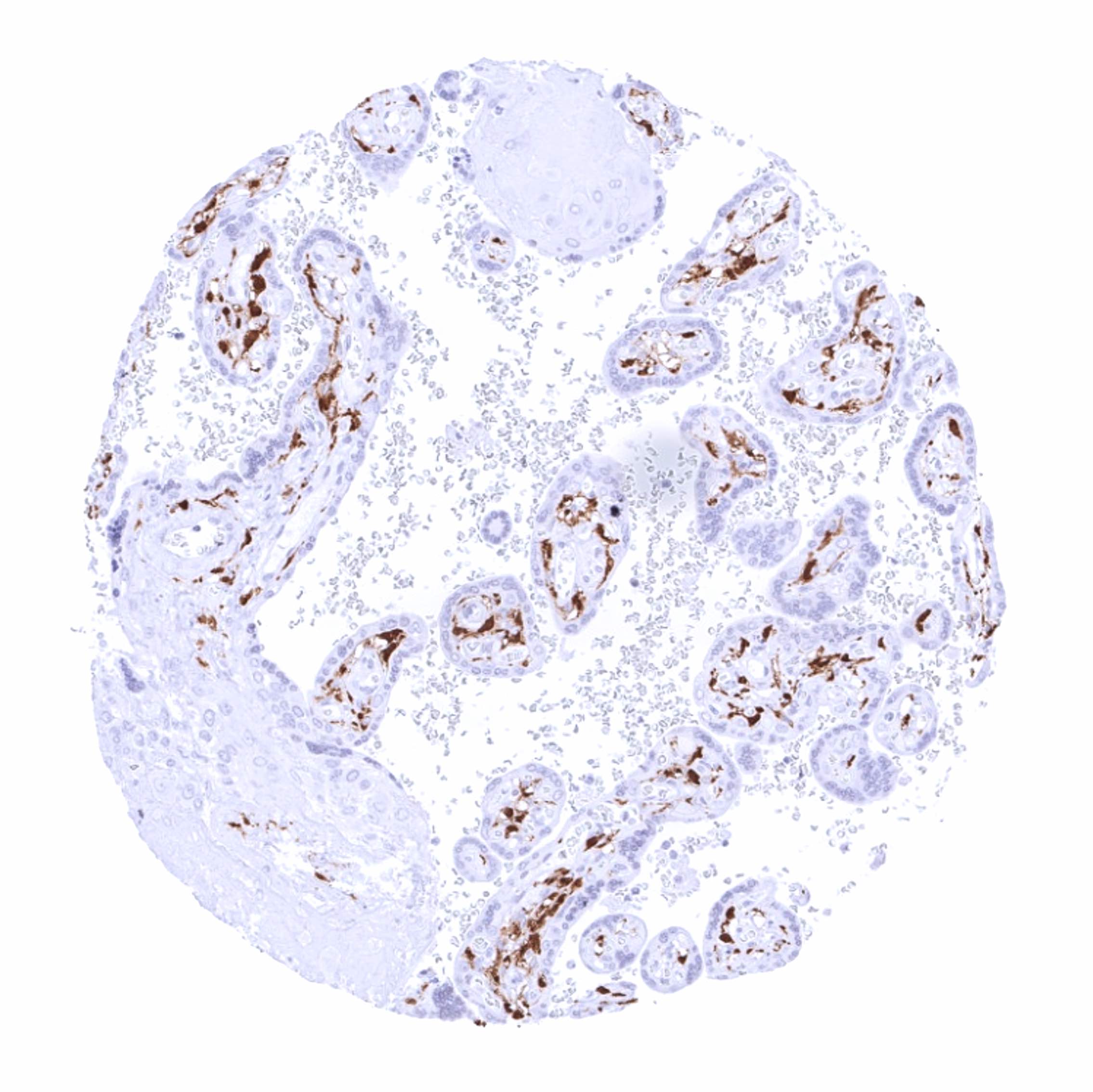
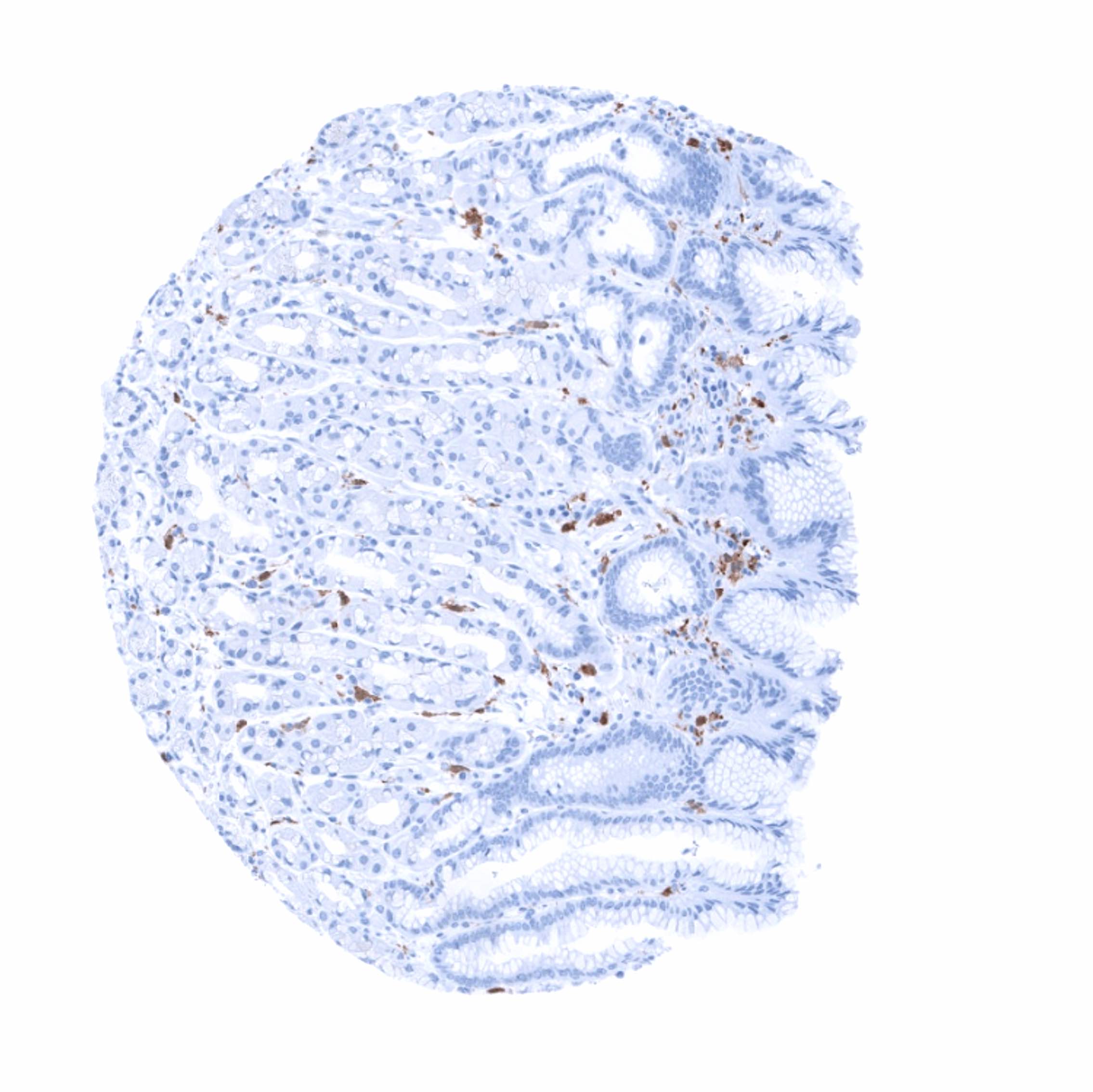
Factor XIIIa occurs in monocytes and macrophages of all tissues including dermal dendrocytes and placenta macrophages, megakaryocytes, platelets, chondrocytes, osteoblasts, and osteocytes.
These findings are largely consistent with the RNA and protein data described in the Human Protein Atlas (Tissue expression Factor XIIIa)
Positive control: Colon: A fraction of monocytic cells in the mucosa should show a strong cytoplasmic factor XIIIa immunostaining.
Negative control: Colon: Factor XIIIa immunostaining should be absent in epithelial, endothelial and smooth muscle cells.


Staining Pattern in Relevant Tumor Types
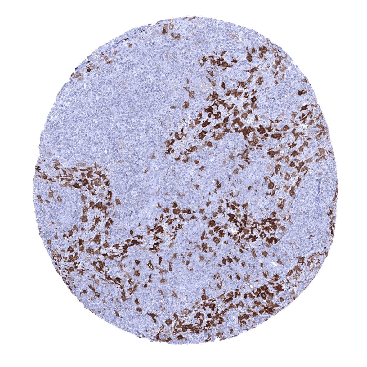
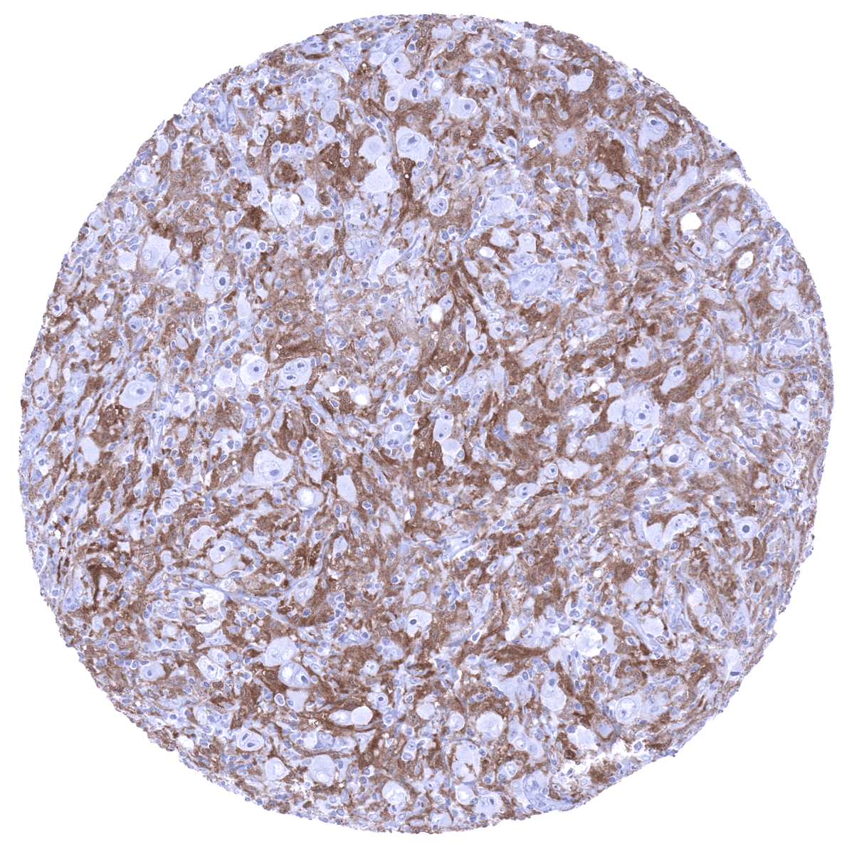
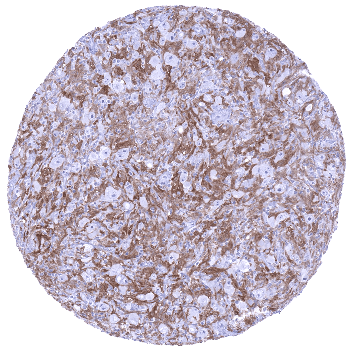
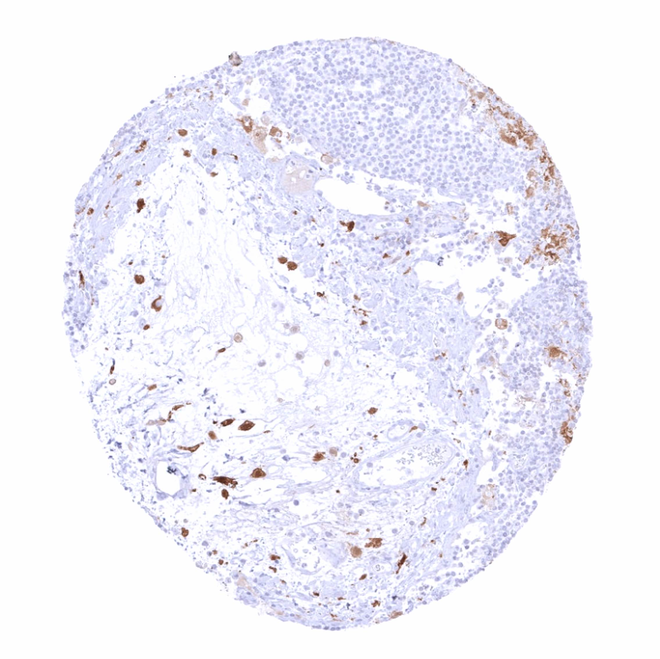
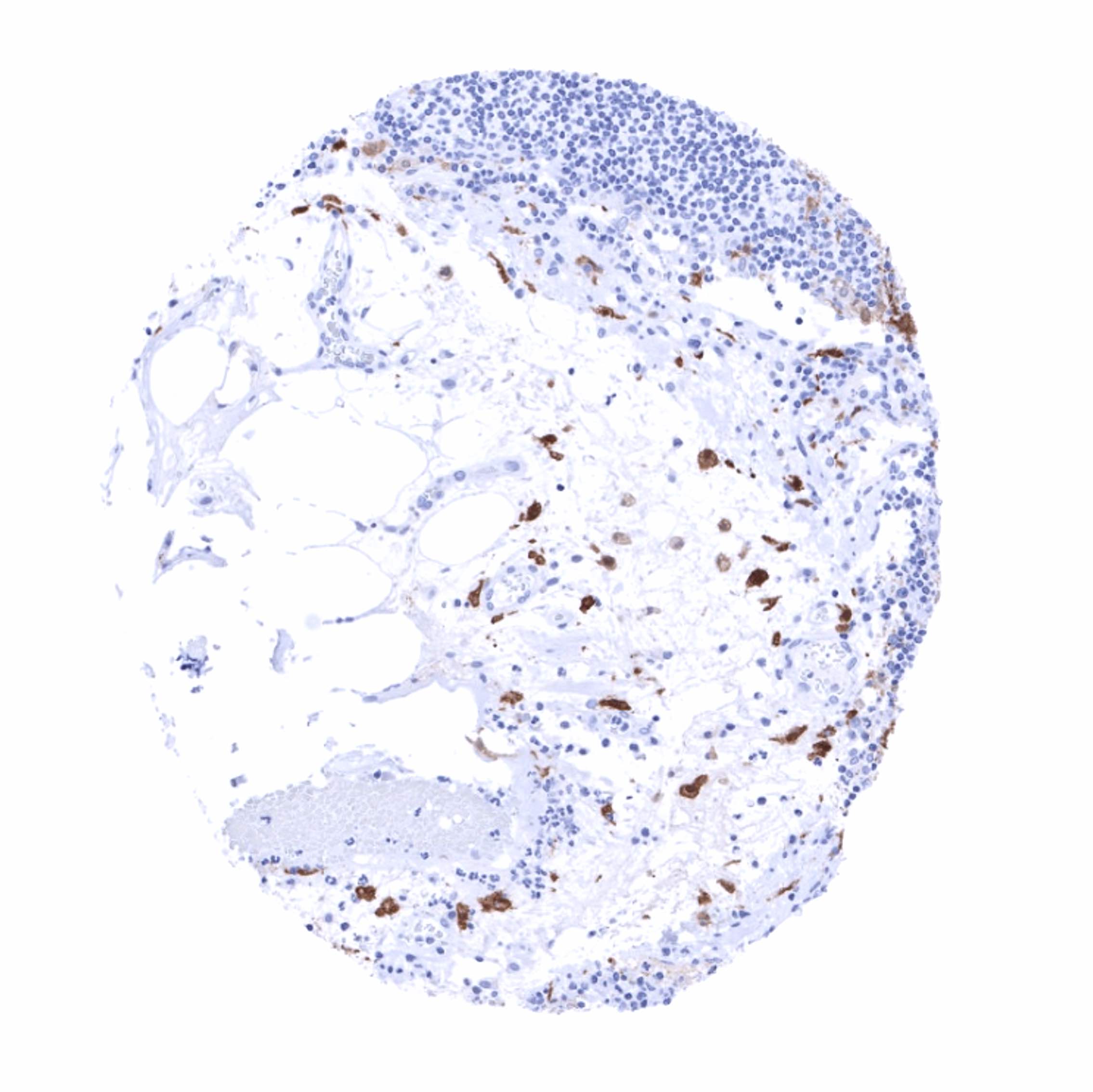
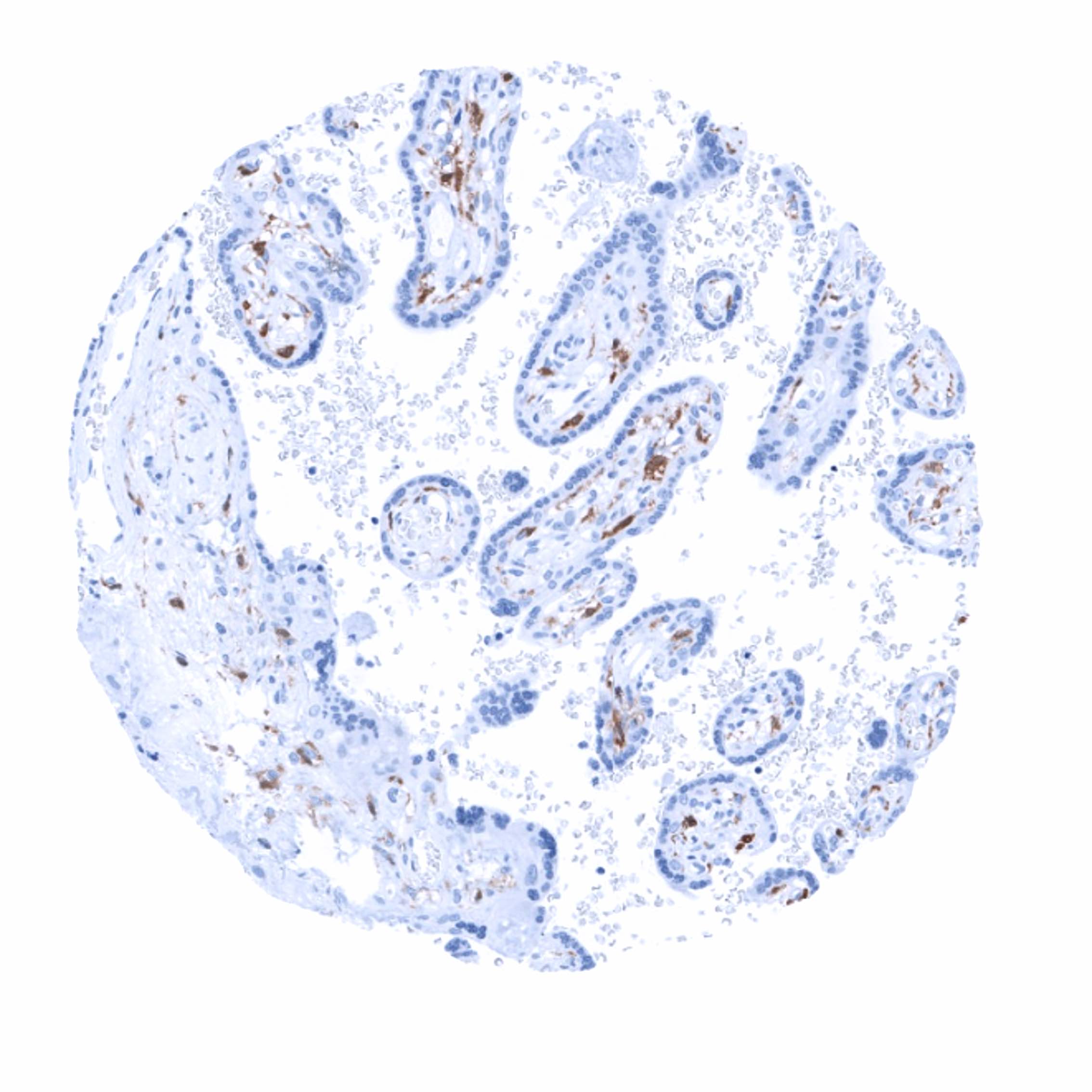
Factor XIIIa is expressed in tumors of monocytic/histiocytic origin. Factor XIIIa immunostaining is often seen in a fraction of tumor associated macrophages.
The TCGA findings on Factor XIIIa RNA expression in different tumor categories have been summarized in the Human Protein Atlas.
Compatibility of Antibodies
No data available at the moment
Protocol Recommendations
IHC users have different preferences on how the stains should look like. Some prefer high staining intensity of the target stain and even accept some background. Others favor absolute specificity and lighter target stains. Factors that invariably lead to more intense staining include higher concentration of the antibody and visualization tools, longer incubation time, higher temperature during incubation, higher temperature and longer duration of the heat induced epitope retrieval (slide pretreatment). The impact of the pH during slide pretreatment has variable effects and depends on the antibody and the target protein.
All images and data shown here and in our image galleries are obtained by the manual protocol described below. Other protocols resulting in equivalent staining are described as well.
Manual protocol
Freshly cut sections should be used (less than 10 days between cutting and staining). Heat-induced antigen retrieval for 5 minutes in an autoclave at 121°C in pH 7,8 Target Retrieval Solution buffer. Apply MSVA-813R at a dilution of 1:150 at 37°C for 60 minutes. Visualization of bound antibody by the EnVision Kit (Dako, Agilent) according to the manufacturer’s directions.
Agilent / Dako – Autostainer Link 48
Pretreatment in PT-Link for 30 minutes at 95°C (pH high); FLEX peroxidase blocking for 5 minutes (room temperature), MSVA-813R 1:150 for 20 minutes (room temperature), FLEX+ mouse/rabbit (LINKER) for 15 minutes (room temperature), horseradish peroxidase (HRP) for 20 minutes (room temperature), FLEX DAB+Sub-Chromo for 10 minutes (room temperature), FLEX hematoxylin for 5 minutes (room temperature).

These images reflect stainings by the protocol described above. It is of note that a comparable staining result can also be obtained by different protocols. In general, a longer pretreatment, a longer incubation time of the primary antibody, a higher antibody concentration, and a longer incubation time of FLEX+LINKER result in stronger staining, potentially at the cost of more background staining. Modifications of the protocol with a strengthening effect on staining intensity in combination with changes of other parameters that result in lower staining intensity can result in a comparable result as shown above.
Leica – BOND RX
Dewax at 72°C for 30 seconds; Pretreatment in Bond Epitope Retrieval Solution (ER2 – EDTA pH9) for 20 minutes at 100°C; Peroxidase blocking for 5 minutes (room temperature), MSVA-813R 1:150 for 15 minutes (room temperature), Post primary (rabbit anti mouse) for 8 minutes (room temperature), Polymer (goat anti rabbit) for 8 minutes (room temperature), mixed DAB refine for 10 minutes (room temperature), hematoxylin for 5 minutes (room temperature).

These images reflect stainings by the protocol described above. It is of note that a comparable staining result can also be obtained by different protocols. In general, a longer pretreatment, a longer incubation time of the primary antibody, a higher antibody concentration, a higher temperature during incubation, and a longer incubation time of Post primary and or the Polymer result in stronger staining, potentially at the cost of more background staining. Modifications of the protocol with a strengthening effect on staining intensity in combination with changes of other parameters that result in lower staining intensity can result in a comparable result as shown above.
Roche – Ventana Discovery ULTRA
Pretreatment for 64 minutes at 100°C (pH 8,4); CM peroxidase blocking for 12 minutes (room temperature), MSVA-813R 1:150 for 20 minutes at 36°C, secondary antibody (anti-rabbit HQ) for 12 minutes at 36°C, anti-HQ HRP for 12 minutes at room temperature, DAB at room temperature, hematoxylin II at room temperature for 8 minutes, bluing reagent at room temperature for 4 minutes.

These images depict staining results obtained by the protocol described above. It is of note, that the Ventana machines generally require higher antibody concentrations than other commonly used autostainers because the antibodies are automatically diluted during the procedure. Various other protocols can result in an identical result as shown above. A longer pretreatment, a longer incubation time of the primary antibody, a higher antibody concentration, a higher temperature during incubation, and a longer incubation time of secondary antibody and or the anti-HQ HRP result in stronger staining, potentially at the cost of more background staining.
Potential Research Applications
- The role of factor XIIIa and of factor XIIIa expressing tumor associated macrophages in cancer biology should be further evaluated.
Evidence for Antibody Specificity in IHC
There are two ways how the specificity of antibodies can be documented for immunohistochemistry on formalin fixed tissues. These are: 1. comparison with a second independent method for target expression measurement across a large number of different tissue types (orthogonal strategy), and 2. Comparison with one or several independent antibodies for the same target and showing that all positive staining results are also seen with other antibodies for the same target (independent antibody strategy).
Orthogonal validation is not well suited for proteins that are expressed in cells occurring in the majority of all organs such as monocytes and macrophages. RNA expression data derived from the Human Protein Atlas (HPA) RNA-seq tissue dataset, the FANTOM5 project, and the Genotype-Tissue Expression (GTEx) project, which are all summarized in the Human Protein Atlas (Tissue expression Factor XIIIa) are consistent, however, with the staining properties of MSVA-813R. Highest RNA expression levels were described for lymph node and placenta, which are organs with a particularly high rate of factor XIIIa positive monocytic cells.
Evidence for specificity of MSVA-813R comes from the strict limitation of immunostaining to cells of the monocyte/macrophage system which are known to express factor XIIIa. In addition, there was a complete lack of staining in all epithelial cell types including those that are prone to non-specific staining such as kidney and colon mucosa.
A comparison with a second independent antibody (Factor XIIIa candidate “MSVA 2b4”) showed an absolutely identical staining pattern across all tissues analyzed.
