
295,00 € – 995,00 €
Product details
Synonyms = Antigen, prostate-specific (APS), Gamma-seminoprotein, hK3, Kallikrein related peptidase 3, Kallikrein-3 (KLK3), KLK2A1, P-30 antigen, Semenogelase, Seminin
Antibody type = Recombinant Rabbit monoclonal / IgG
Clone = MSVA-603R
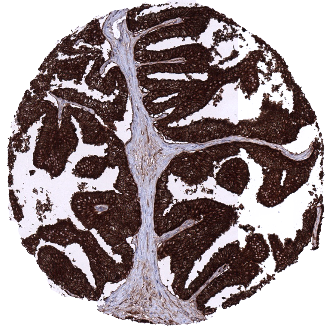
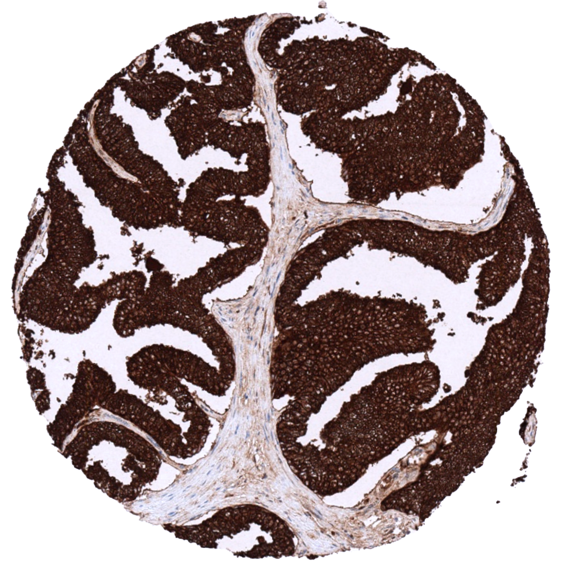
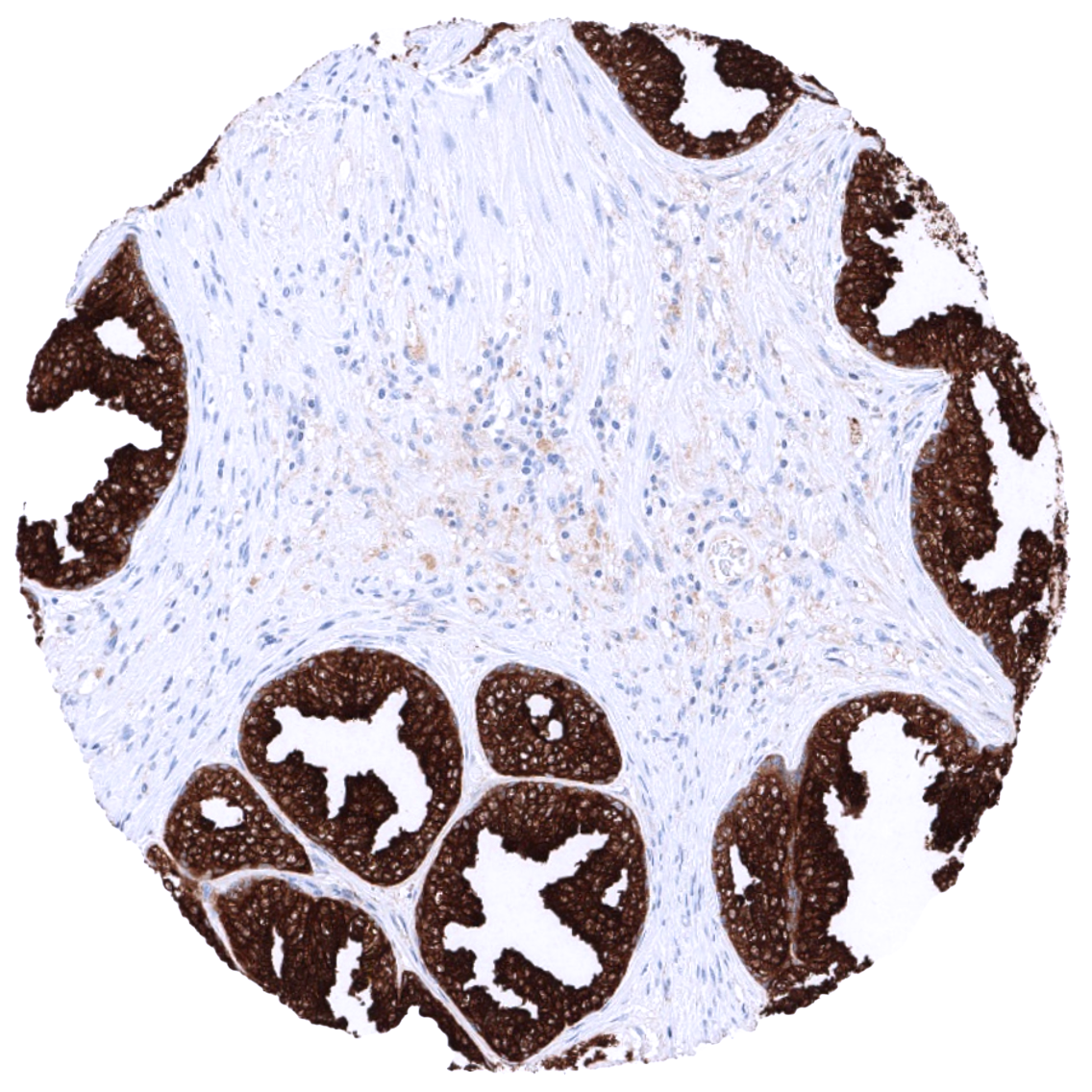
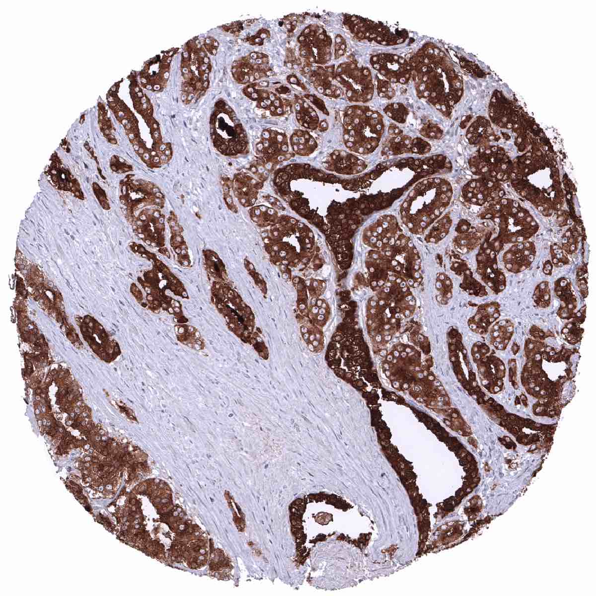
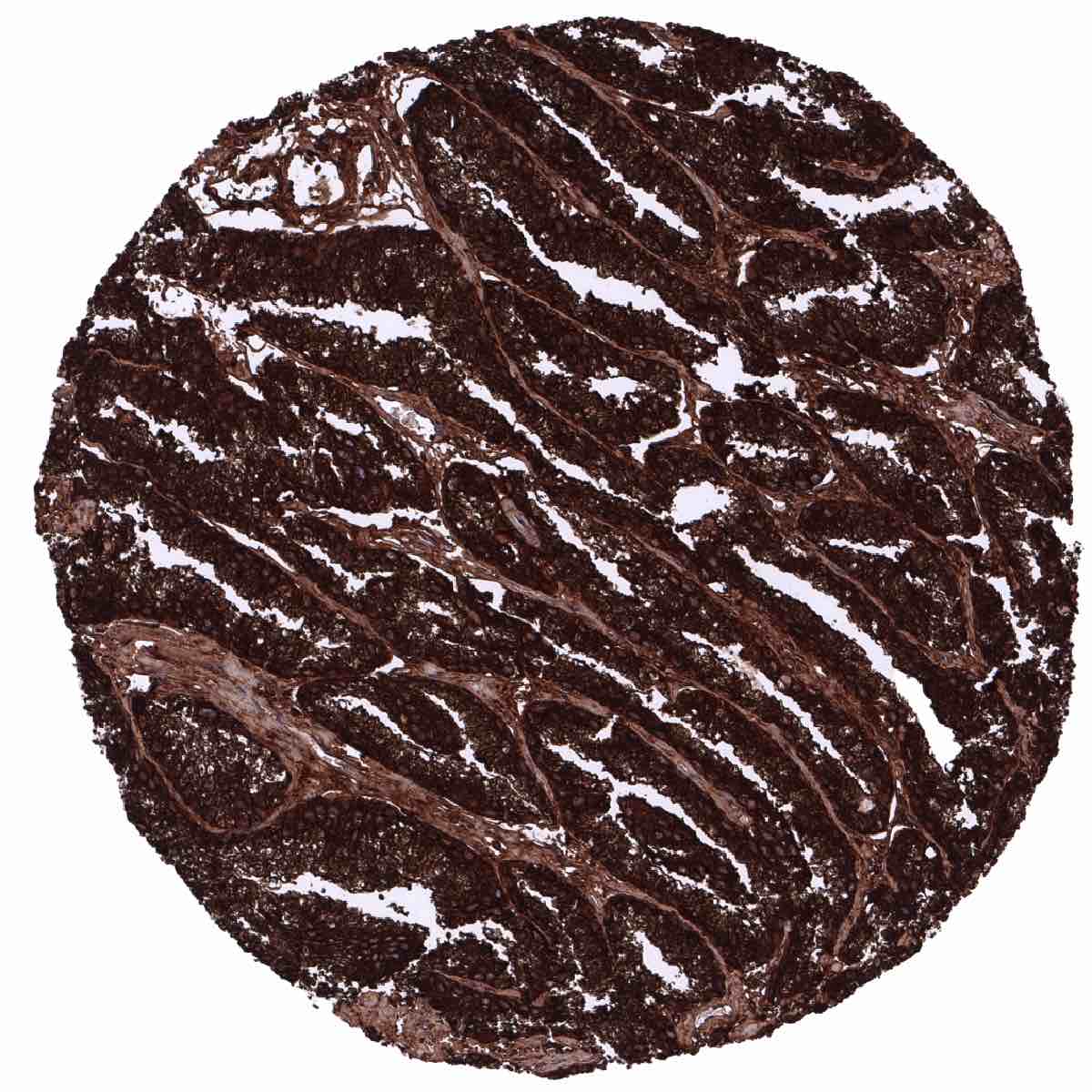
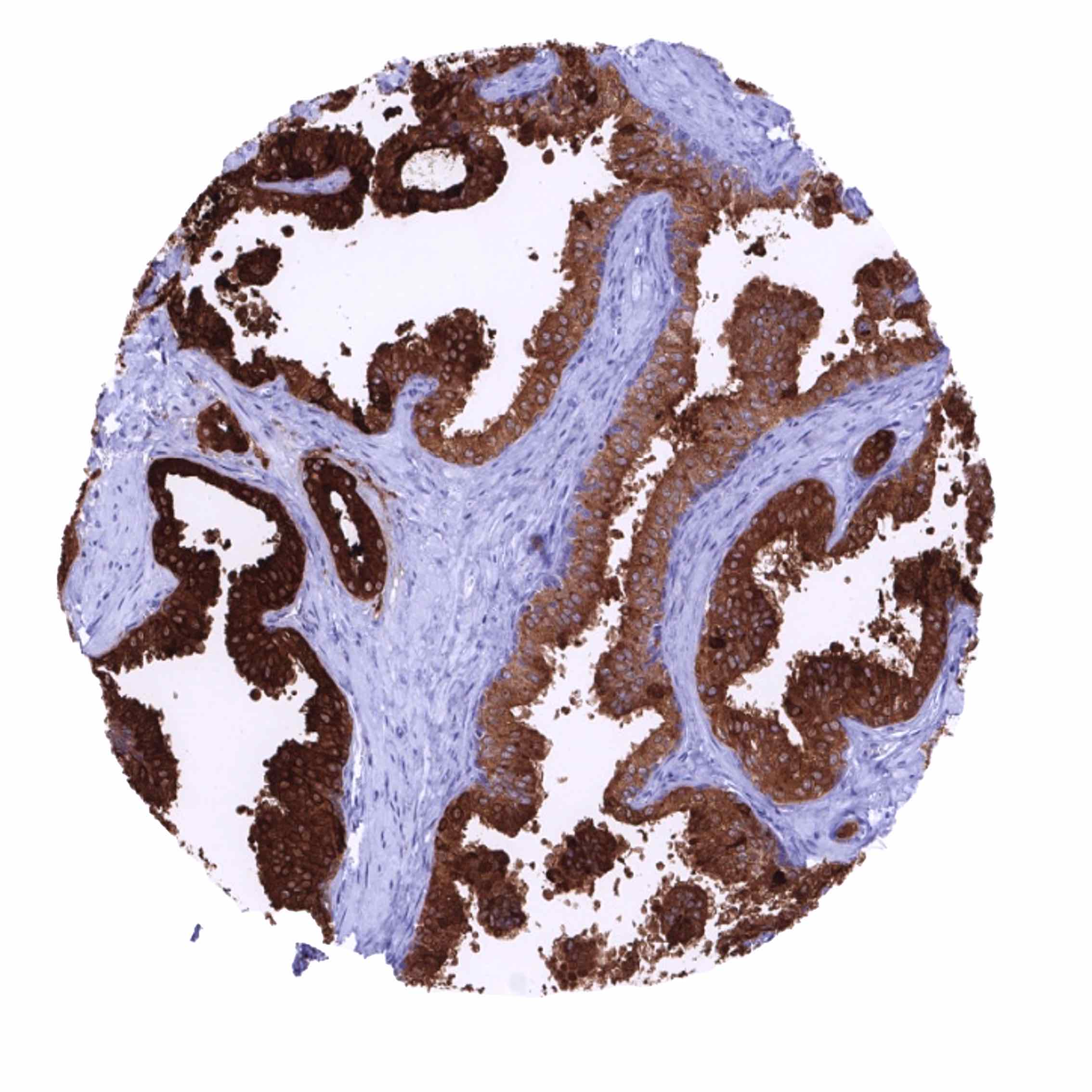
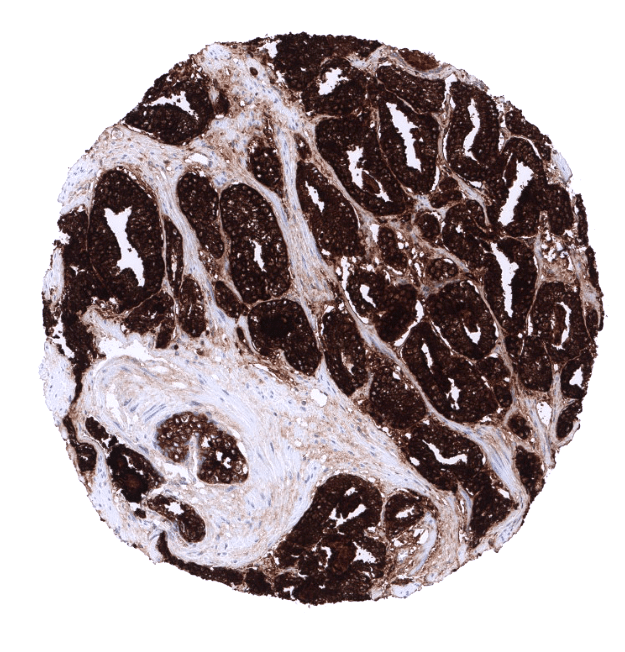
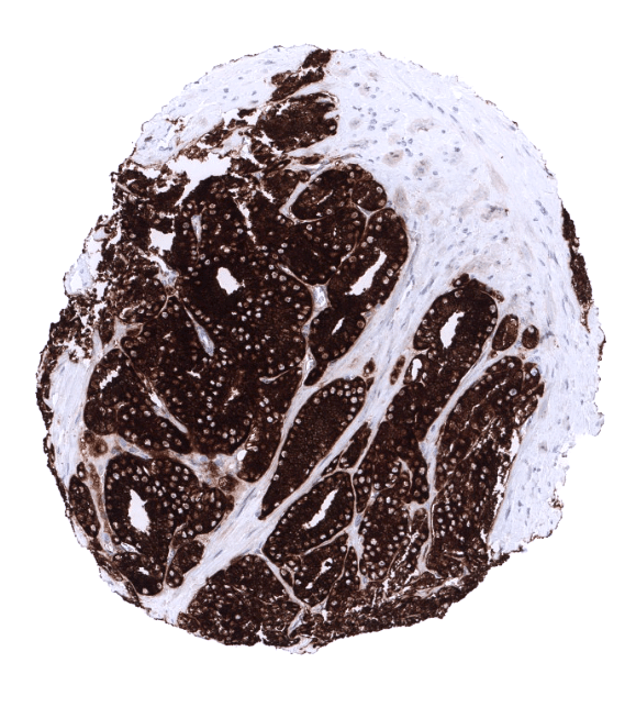
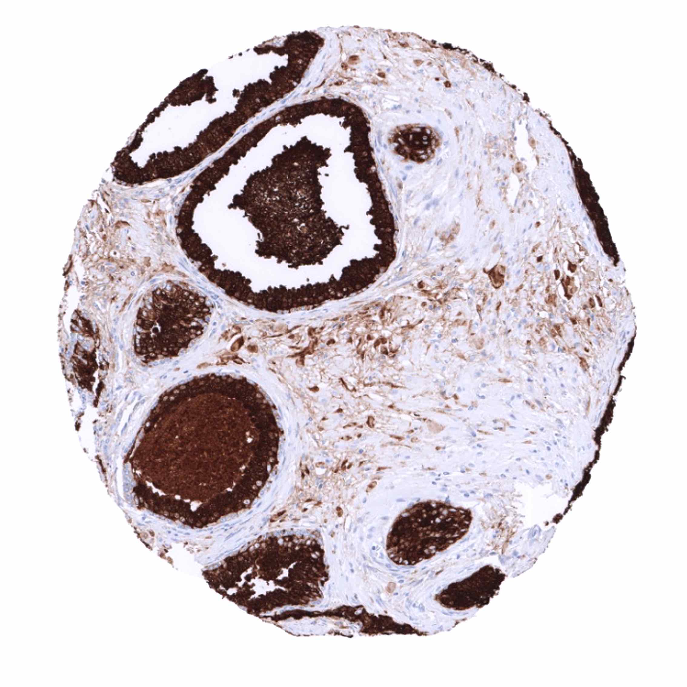
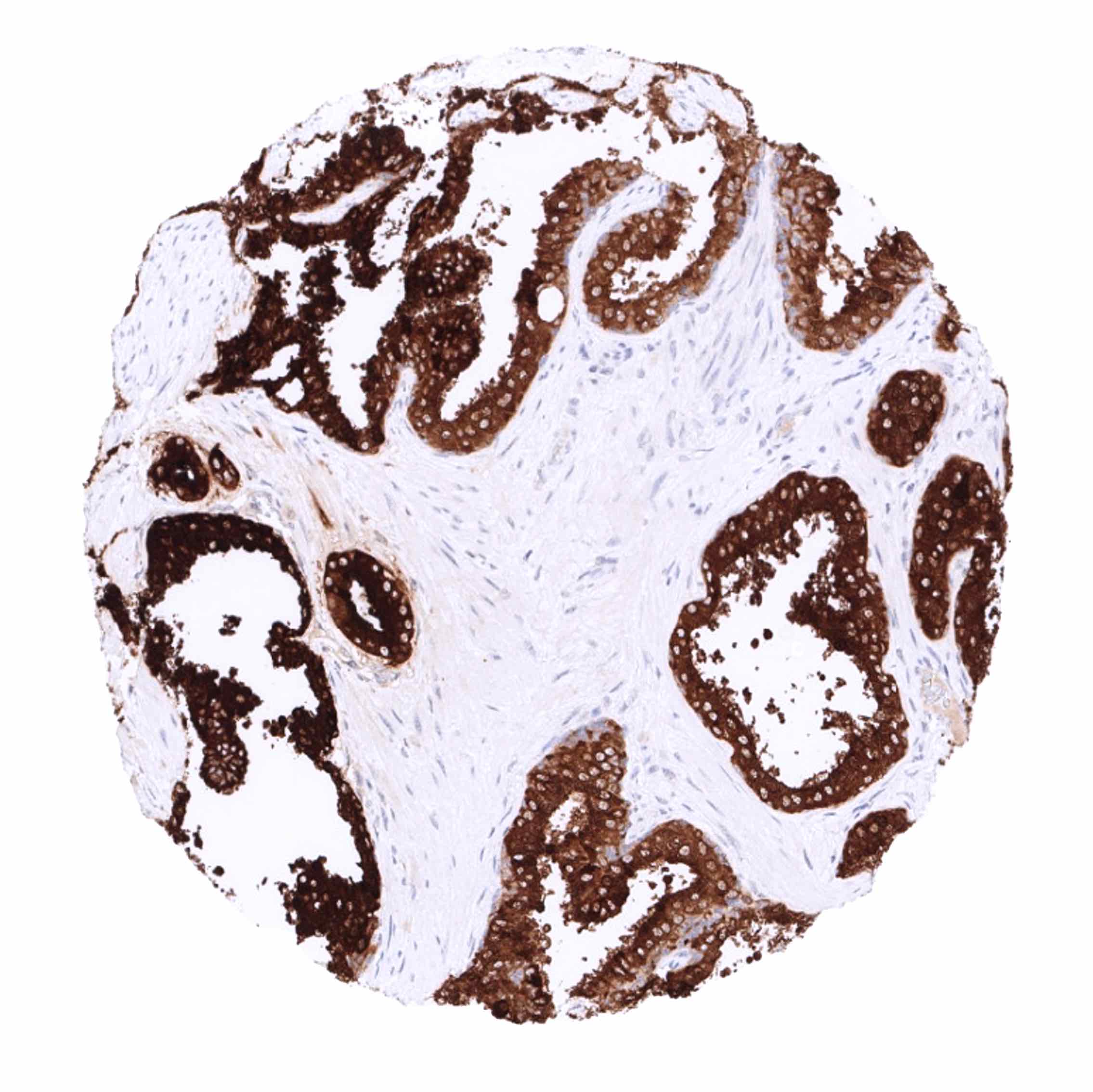
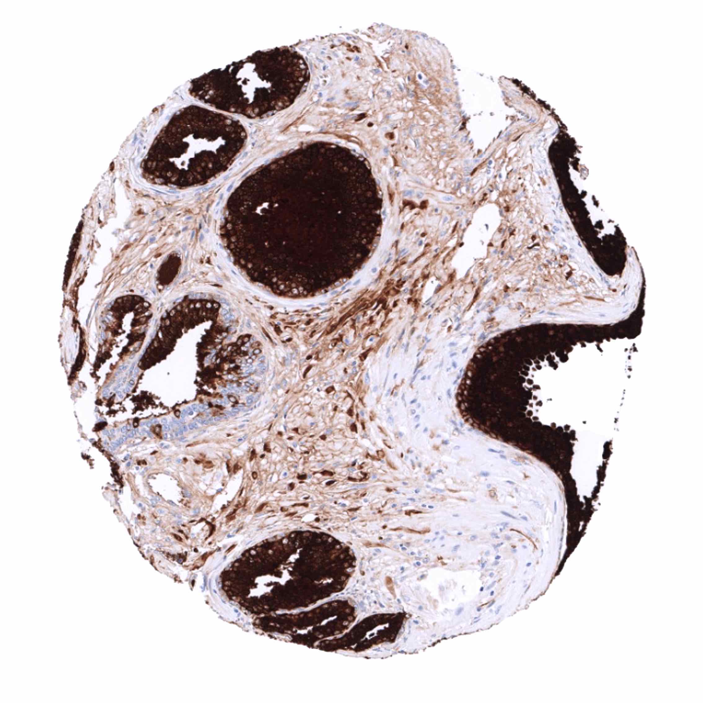
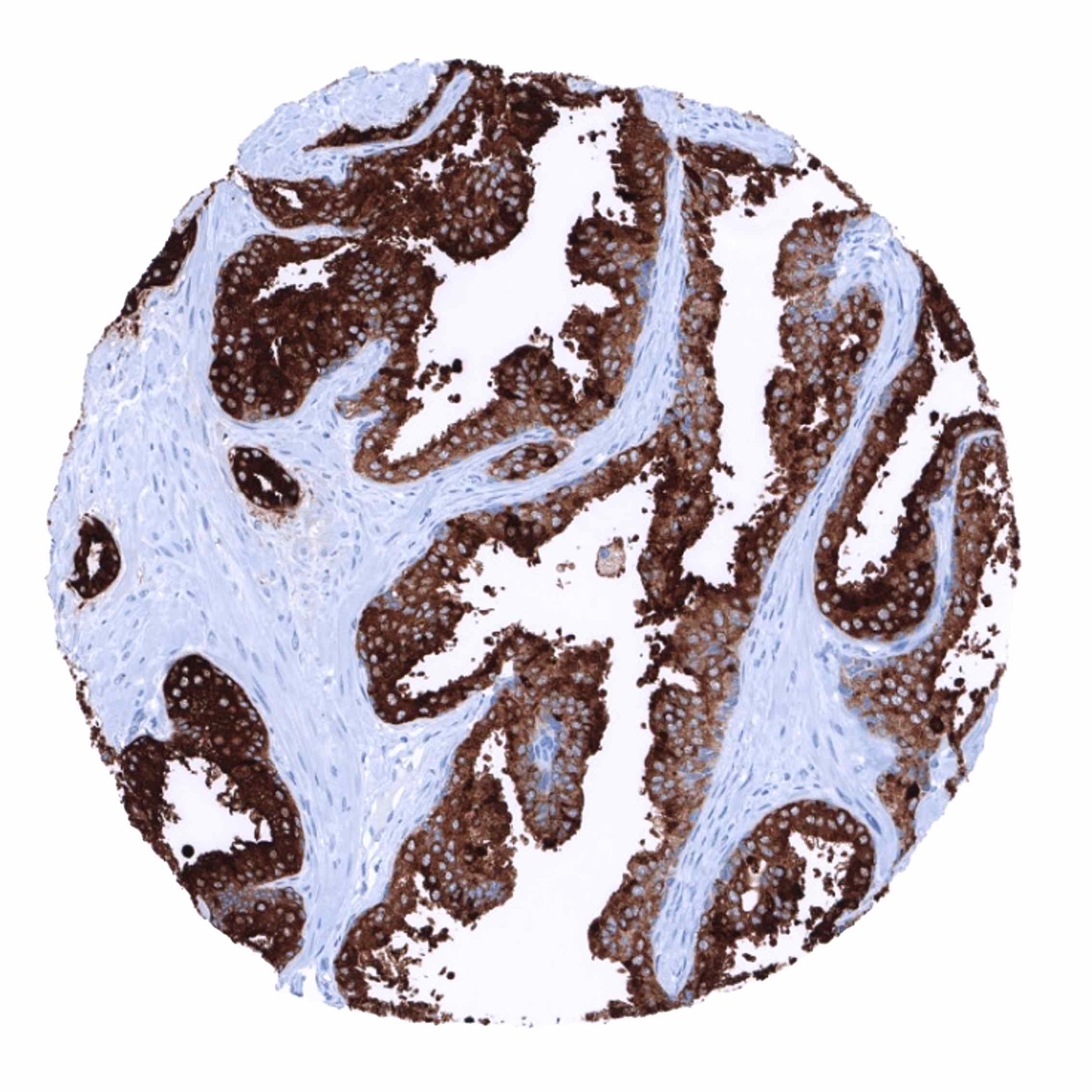
Positive control = Prostate: The epithelial cells of the prostate glands must show a strong cytoplasmic staining. Due to leakage of the antigen in the vicinity of the prostate glands, adjacent stroma cells may display a weak to moderate staining reaction.
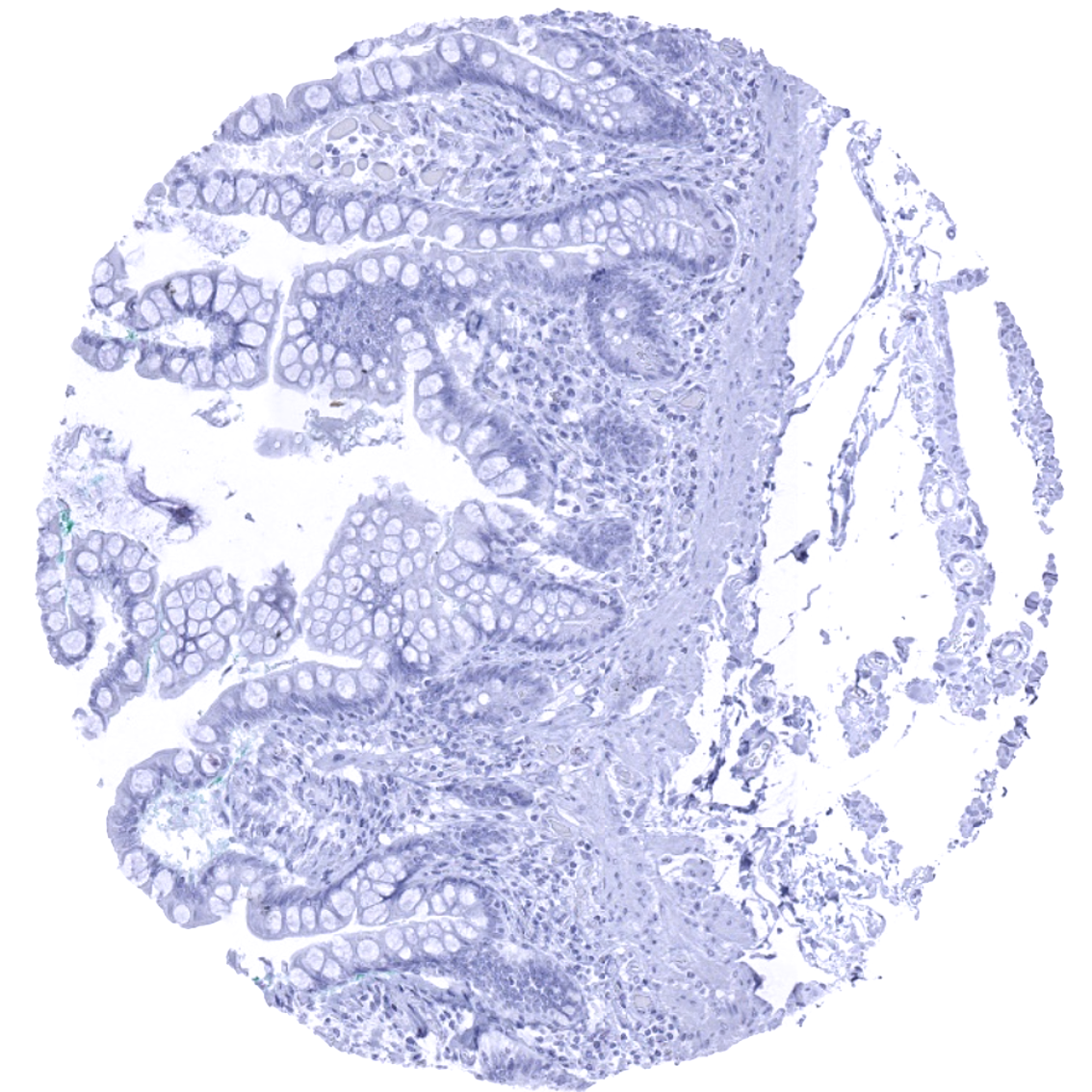
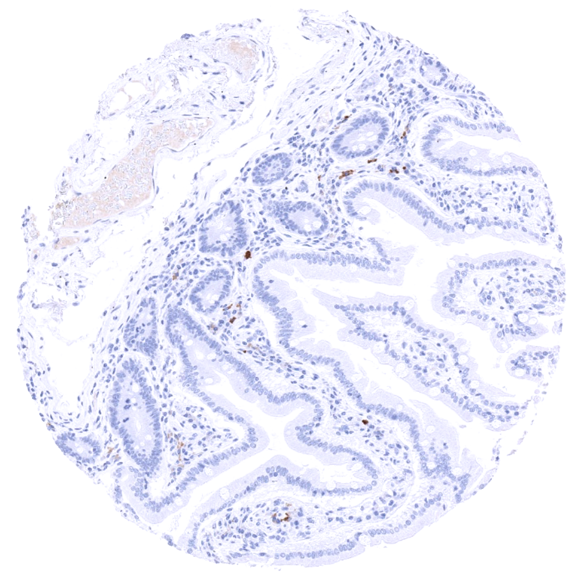
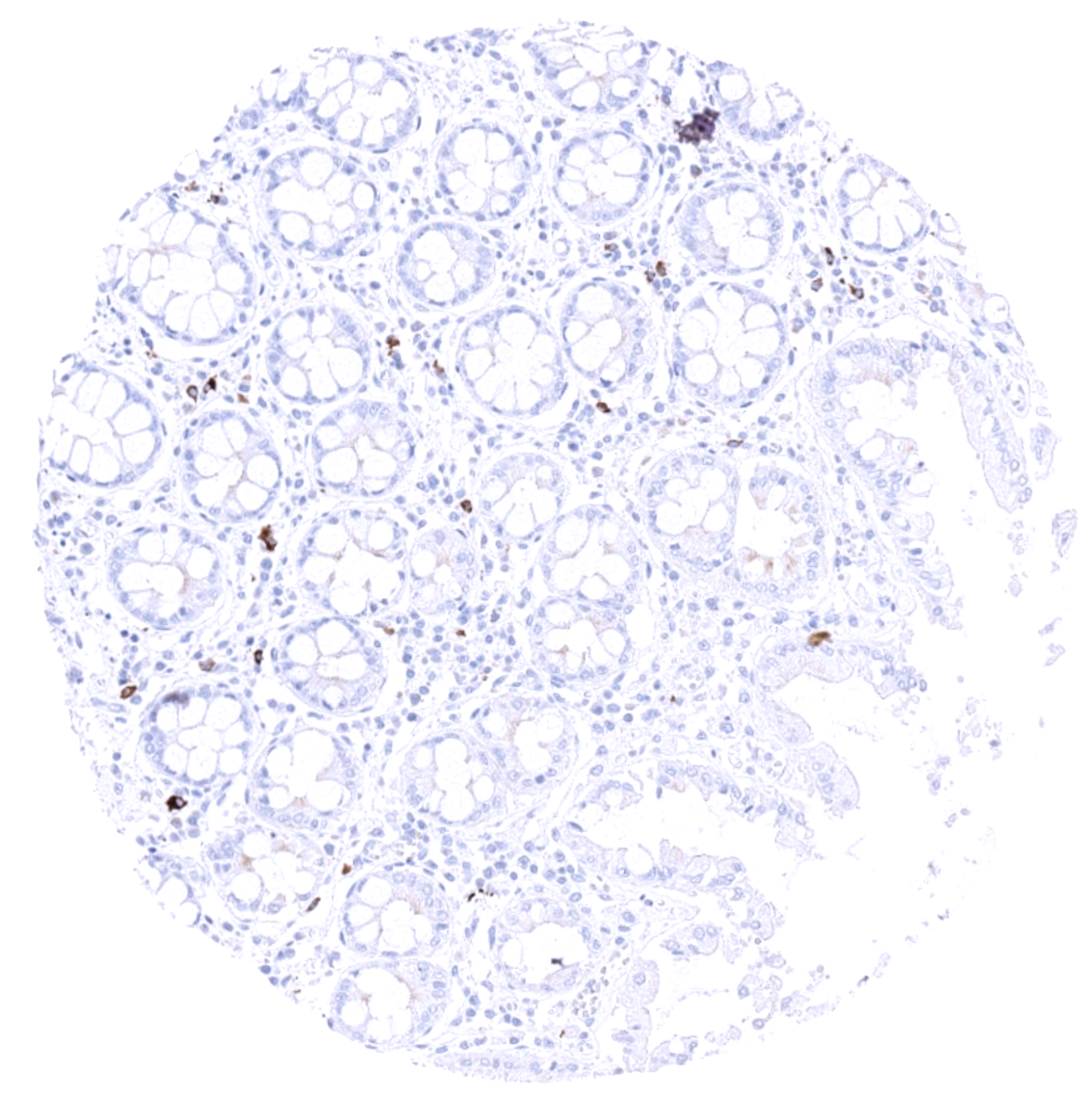
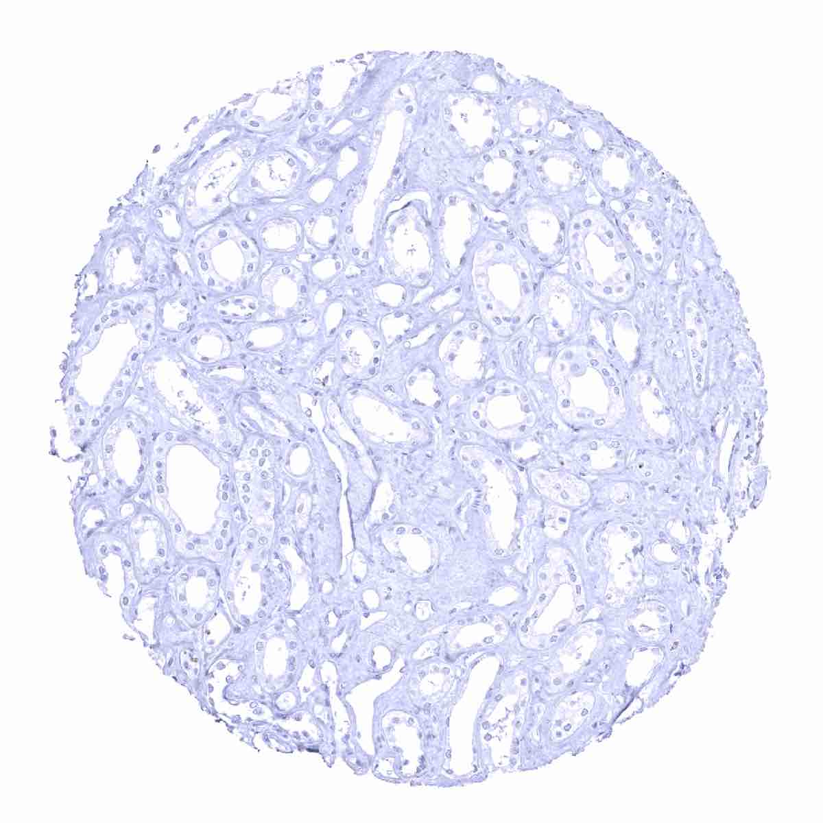
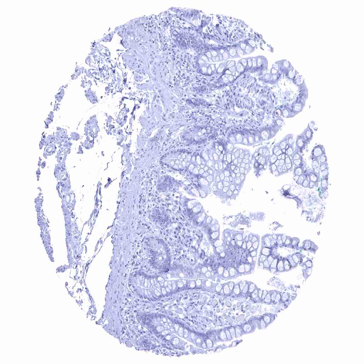
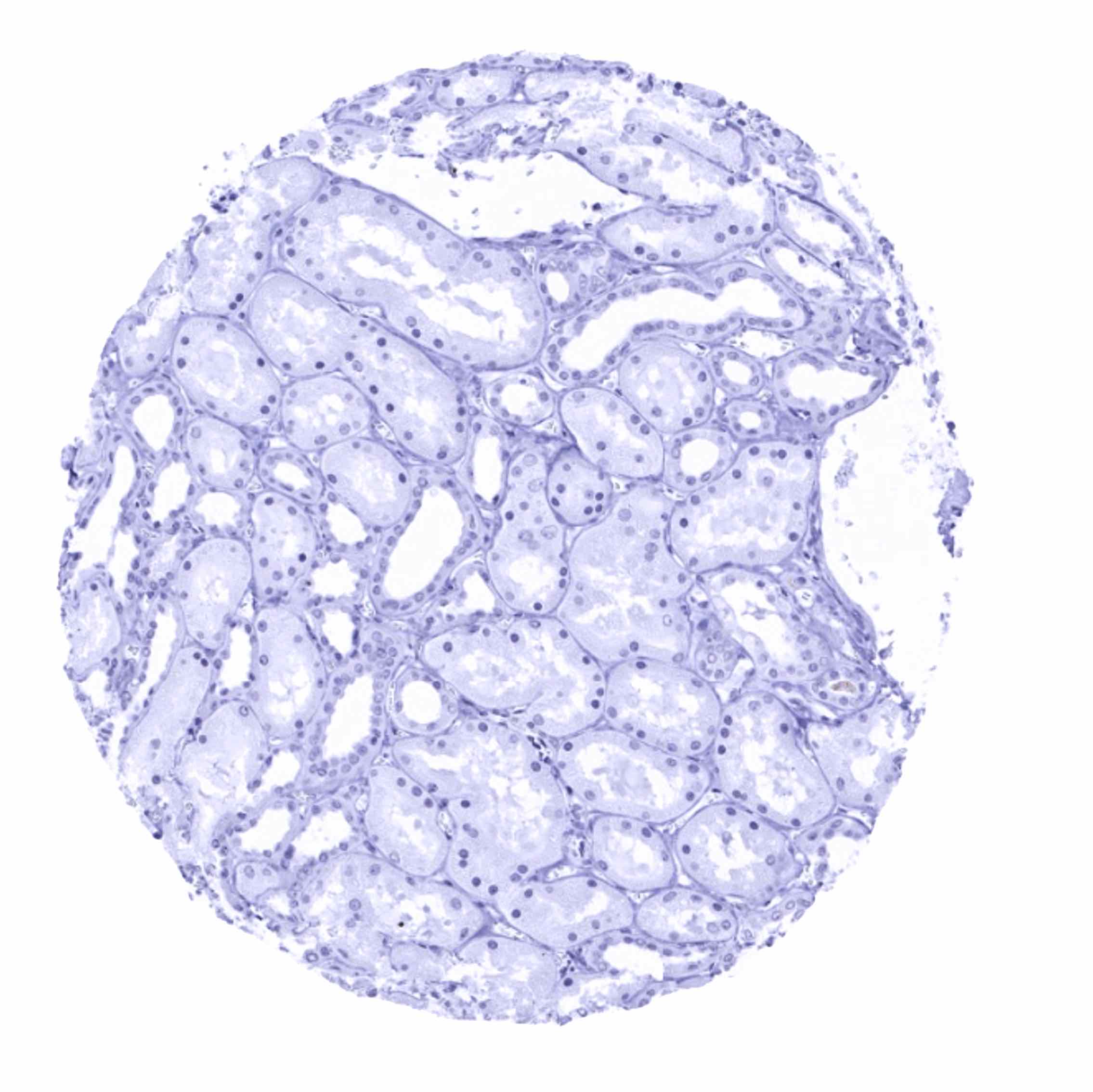
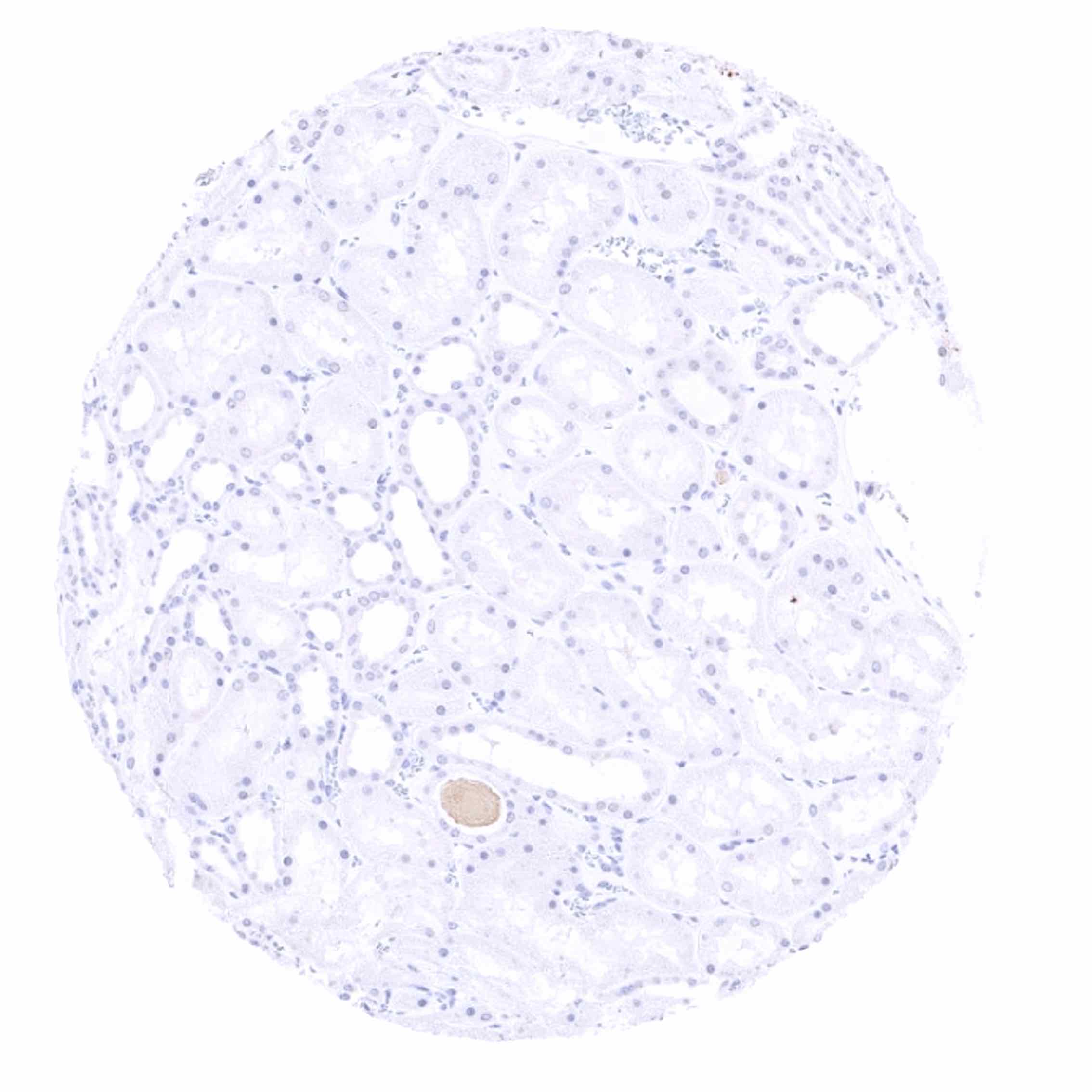
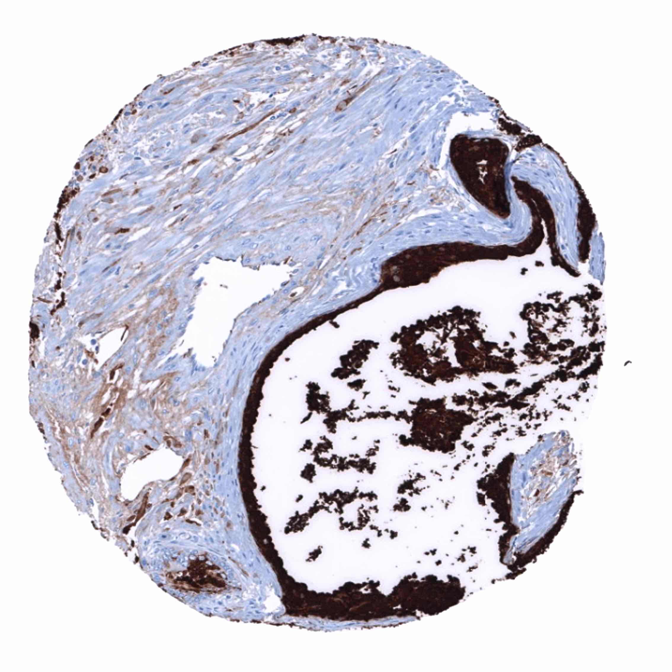
Negative control = Colon: PSA immunostaining should be absent in all cells.
Cellular localization = Cytoplasmic
Reactivity = Human
Application = Immunohistochemistry
Dilution = 1:100 – 1:200
Intended Use = Research Use Only
Relevance of Antibody
PSA is a marker for prostatic epithelial tissue and prostate cancer.
Biology Behind
Prostate specific antigen (PSA) is a proteinase coded by the KLK3 gene at 19q13.33. PSA is the most relevant protein for the management of men with suspected or diagnosed and treated prostate cancer. PSA is exclusively produced in prostate epithelial cells. It is secreted to the seminal fluid and plays a role for its liquefaction. Only minor quantities of PSA reach the bloodstream. The serum PSA level is largely proportionate to the quantity of prostate epithelial cells in the body. An increased serum PSA level is the most common cause for prostate cancer suspicion and subsequent prostate biopsy. In men with diagnosed prostate cancer, serum PSA analysis is the most commonly used parameter to monitor disease progression, disease recurrence, and response to therapy.
Staining Pattern in Normal Tissues
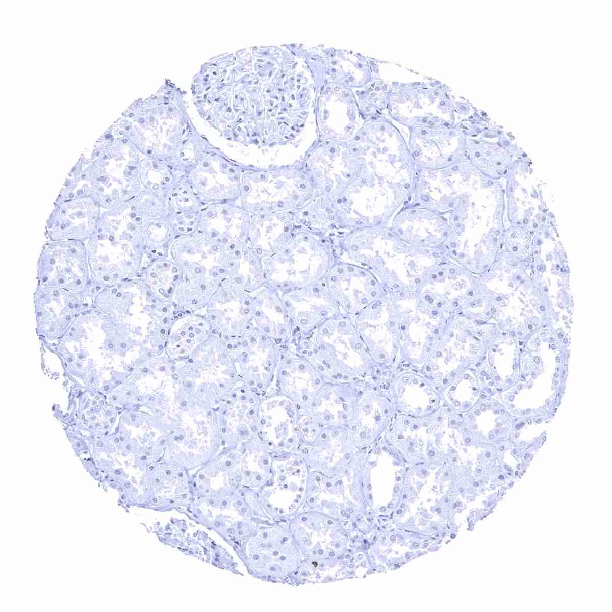
Among normal tissues PSA immunostaining is only seen in prostatic epithelial cells. PSA staining is absent in other normal tissues including aorta, heart, skeletal muscle, smooth muscle, myometrium, ovary (including corpus luteum and follicular cysts), fat, squamous epithelia irrespective of their origin, anal canal transitional epithelium, urothelium, stomach, duodenum, small intestine, appendix, colon, rectum, gallbladder, bronchus, paranasal sinus, lymphatic tissues, node, spleen, thymus, tonsil, liver, pancreas, parotid gland, submandibular gland, sublingual gland, Brunner gland, kidney, seminal vesicle, epididymis, testis, lung, breast, endocervix, endometrium, fallopian tube, placenta, adrenal gland, parathyroid gland, thyroid, cerebellum, cerebrum, and the pituitary gland.
The findings described above are this consistent with the RNA data described in the Human Protein Atlas (Tissue expression PSA)
Positive control = Prostate: The epithelial cells of the prostate glands must show a strong cytoplasmic staining. Due to leakage of the antigen in the vicinity of the prostate glands, adjacent stroma cells may display a weak to moderate staining reaction.
Negative control = Colon: PSA immunostaining should be absent in all cells.

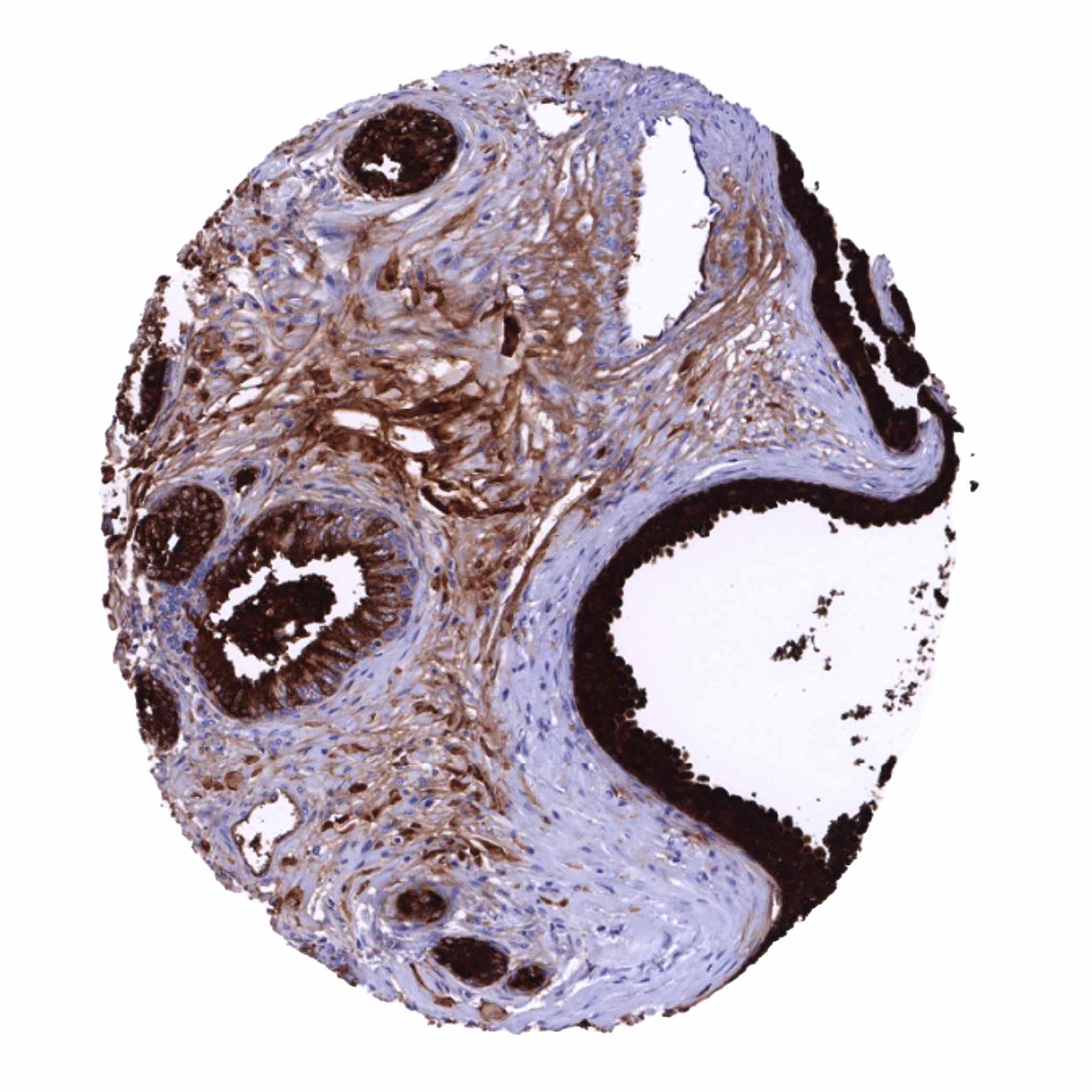
Staining Pattern in Relevant Tumor Types
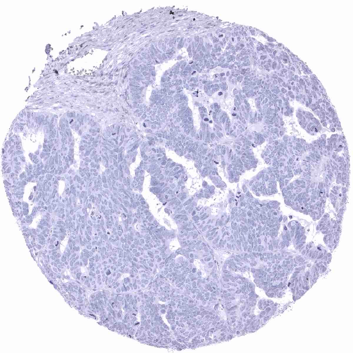
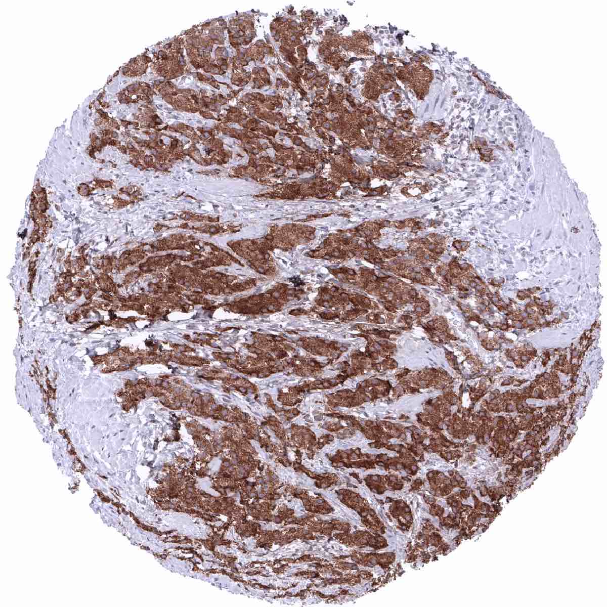
PSA immunostaining is almost completely restricted to prostate cancers. PSA expression is seen in >98% of prostatic adenocarcinomas at the time of initial diagnosis. Advanced high grade prostate cancers can gradually lose PSA expression during tumor progression, however.
The TCGA findings on PSA RNA expression in different tumor categories have been summarized in the Human Protein Atlas.
Compatibility of Antibodies
No data available at the moment
Protocol Recommendations
IHC users have different preferences on how the stains should look like. Some prefer high staining intensity of the target stain and even accept some background. Others favor absolute specificity and lighter target stains. Factors that invariably lead to more intense staining include higher concentration of the antibody and visualization tools, longer incubation time, higher temperature during incubation, higher temperature and longer duration of the heat induced epitope retrieval (slide pretreatment). The impact of the pH during slide pretreatment has variable effects and depends on the antibody and the target protein.
All images and data shown here and in our image galleries are obtained by the manual protocol described below. Other protocols resulting in equivalent staining are described as well.
Manual protocol
Freshly cut sections should be used (less than 10 days between cutting and staining). Heat-induced antigen retrieval for 5 minutes in an autoclave at 121°C in pH 7,8 Target Retrieval Solution buffer. Apply MSVA-603R at a dilution of 1:150 at 37°C for 60 minutes. Visualization of bound antibody by the EnVision Kit (Dako, Agilent) according to the manufacturer’s directions.
Agilent / Dako – Omnis
Unmasking IHC EnV FLEX TRS High pH 97°C ; FLEX peroxidase blocking for 3 minutes (32°C), MSVA-603R 1:150 for 30 minutes at 32°C, FLEX+ mouse/rabbit (LINKER) for 10 minutes at 32°C, horseradish peroxidase (HRP) for 30 minutes (32°C), FLEX DAB+Sub-Chromo for 5 minutes (32°C), FLEX hematoxylin for 5 minutes (room temperature).
These images reflect stainings by the protocol described above. It is of note that a comparable staining result can also be obtained by different protocols. In general, a longer pretreatment, a longer incubation time of the primary antibody, a higher antibody concentration, and a longer incubation time of FLEX+LINKER result in stronger staining, potentially at the cost of more background staining. Modifications of the protocol with a strengthening effect on staining intensity in combination with changes of other parameters that result in lower staining intensity can result in a comparable result as shown above.
Agilent / Dako – Autostainer Link 48
Pretreatment in PT-Link for 30 minutes at 95°C (pH high); FLEX peroxidase blocking for 5 minutes (room temperature), MSVA-603R 1:150 for 20 minutes (room temperature), FLEX+ mouse/rabbit (LINKER) for 15 minutes (room temperature), horseradish peroxidase (HRP) for 20 minutes (room temperature), FLEX DAB+Sub-Chromo for 10 minutes (room temperature), FLEX hematoxylin for 5 minutes (room temperature).


These images reflect stainings by the protocol described above. It is of note that a comparable staining result can also be obtained by different protocols. In general, a longer pretreatment, a longer incubation time of the primary antibody, a higher antibody concentration, and a longer incubation time of FLEX+LINKER result in stronger staining, potentially at the cost of more background staining. Modifications of the protocol with a strengthening effect on staining intensity in combination with changes of other parameters that result in lower staining intensity can result in a comparable result as shown above.
Leica – BOND RX
Dewax at 72°C for 30 seconds; Pretreatment in Bond Epitope Retrieval Solution (ER2 – EDTA pH9) for 20 minutes at 100°C; Peroxidase blocking for 5 minutes (room temperature), MSVA-603R 1:150 for 15 minutes (room temperature), Post primary (rabbit anti mouse) for 8 minutes (room temperature), Polymer (goat anti rabbit) for 8 minutes (room temperature), mixed DAB refine for 10 minutes (room temperature), hematoxylin for 5 minutes (room temperature).
These images reflect stainings by the protocol described above. It is of note that a comparable staining result can also be obtained by different protocols. In general, a longer pretreatment, a longer incubation time of the primary antibody, a higher antibody concentration, a higher temperature during incubation, and a longer incubation time of Post primary and or the Polymer result in stronger staining, potentially at the cost of more background staining. Modifications of the protocol with a strengthening effect on staining intensity in combination with changes of other parameters that result in lower staining intensity can result in a comparable result as shown above.
Roche – Ventana Discovery ULTRA
Pretreatment for 64 minutes at 100°C (pH 8,4); CM peroxidase blocking for 12 minutes (room temperature), MSVA-603R 1:150 for 20 minutes at 36°C, secondary antibody (anti-rabbit HQ) for 12 minutes at 36°C, anti-HQ HRP for 12 minutes at room temperature, DAB at room temperature, hematoxylin II at room temperature for 8 minutes, bluing reagent at room temperature for 4 minutes.

These images depict staining results obtained by the protocol described above. It is of note, that the Ventana machines generally require higher antibody concentrations than other commonly used autostainers because the antibodies are automatically diluted during the procedure. Various other protocols can result in an identical result as shown above. A longer pretreatment, a longer incubation time of the primary antibody, a higher antibody concentration, a higher temperature during incubation, and a longer incubation time of secondary antibody and or the anti-HQ HRP result in stronger staining, potentially at the cost of more background staining.
Potential Research Applications
- The clinical significance of reduced expression of PSA in prostate cancers should be further investigated.
- The level of PSA expression (thought to be related to patient prognosis) might constitute an important parameter for multiparametric prostate cancer prognosis tests
Evidence for Antibody Specificity in IHC
There are two ways how the specificity of antibodies can be documented for immunohistochemistry on formalin fixed tissues. These are: 1. Comparison with a second independent method for target expression measurement across a large number of different tissue types (orthogonal strategy), and 2. Comparison with one or several independent antibodies for the same target and showing that all positive staining results are also seen with other antibodies for the same target (independent antibody strategy).
Orthogonal validation: For the antibody MSVA-603R specificity is suggested by the perfect concordance of the immunostaining data with data from three independent RNA screening studies, including the Human Protein Atlas (HPA) RNA-seq tissue dataset, the FANTOM5 project, and the Genotype-Tissue Expression (GTEx) project, which are all summarized in the Human Protein Atlas (Tissue expression PSA). In these databases, PSA RNA expression was solely expressed in the prostate which also was the only organ where a PSA immunostaining was observed by MSVA-603R.
Comparison of antibodies: Specificity of MSVA-603R was also supported by an identical staining pattern obtained by a second independent commercially available PSA antibody (termed “validation antibody”).
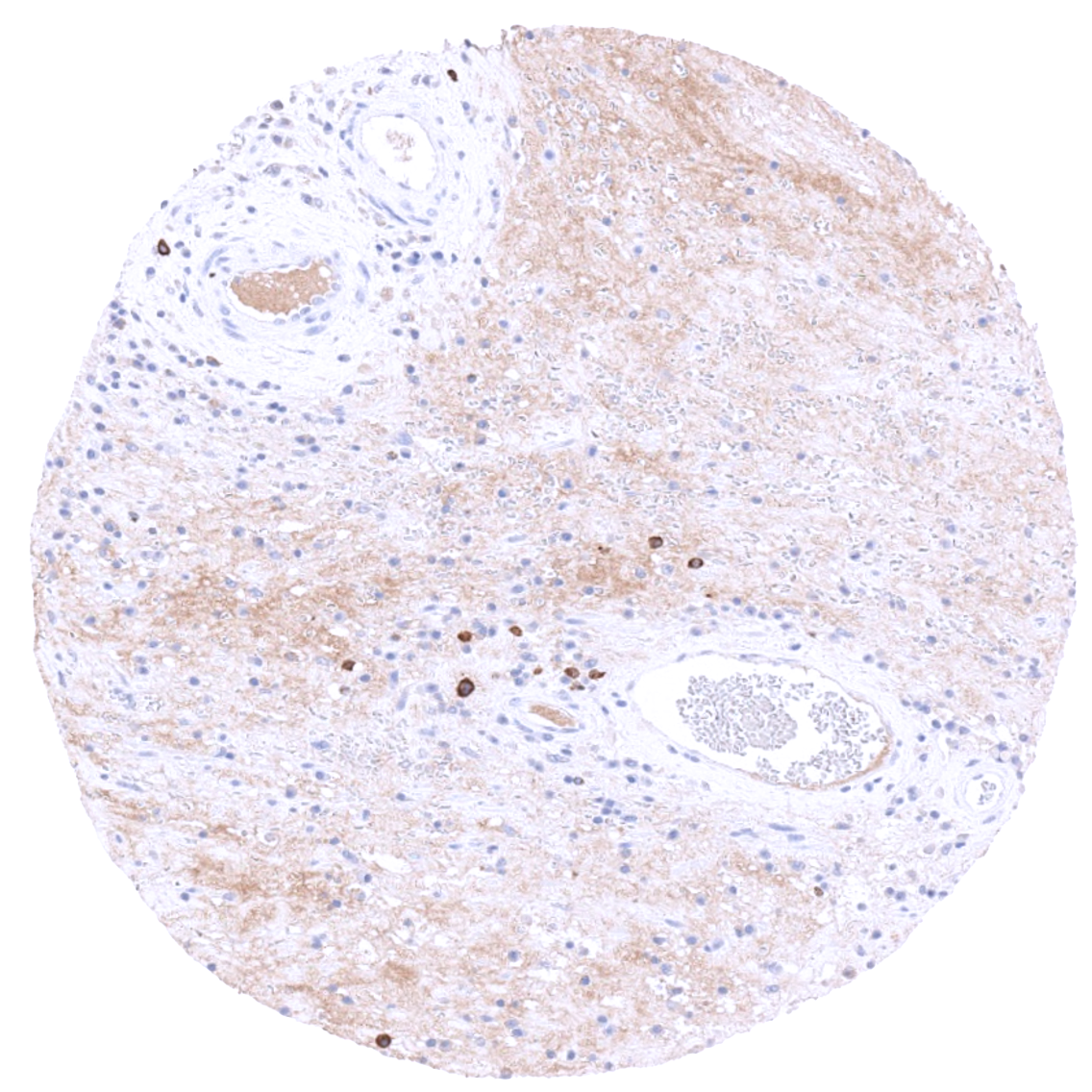
Independence of MSVA-603R and “validation antibody” is demonstrated by a distinct staining of a subset of inflammatory cells (probably plasma cells) by “validation antibody” but not by MSVA-603R. This staining is visible in samples from the small intestine, the stomach and the urinary bladder. It reflects a (tolerable) antibody specific cross-reactivity of “validation antibody”.
Antibody Comparison: MSVA-603R vs another commercially available PSA antibody called “Validation Antibody”